Article

Papain Hydrolysates of Lupin Proteins with Antioxidant, Antimicrobial, and Acetylcholinesterase Inhibitory Activities

1
Department of Analytical Chemistry and Physical Chemistry, University of Food Technologies, 26, Maritza Blvd, 4002 Plovdiv, Bulgaria
2
Department of Biotechnology, University of Food Technologies, 26, Maritza Blvd, 4002 Plovdiv, Bulgaria
3
Department of Tourism and Culinary Technology, Faculty of Economics, University of Food Technologies, 26, Maritza Blvd, 4002 Plovdiv, Bulgaria
4
Department of Mathematics, Physics and Information Technologies, University of Food Technologies, 26, Maritza Blvd, 4002 Plovdiv, Bulgaria
5
Laboratory of Cell Biosystems, Institute of Microbiology, Bulgarian Academy of Sciences, 139, Ruski Blvd, 4000 Plovdiv, Bulgaria
*
Authors to whom correspondence should be addressed.
Appl. Sci. 2022, 12(23), 12370; https://doi.org/10.3390/app122312370
Submission received: 31 October 2022 / Revised: 30 November 2022 / Accepted: 2 December 2022 / Published: 2 December 2022

Abstract

:
Dietary proteins are no longer just nutritional ingredients in our food. During hydrolysis, some of the released peptides may possess properties that favor the health of the human body. In our study enzymatic hydrolysis of lupin proteins was performed using papain. Three enzyme-to-substrate ratios were set for three different duration times. The SDS-PAGE of the samples was performed. Each hydrolysate was studied for the degree of hydrolysis (DH), acetylcholinesterase (AChE) inhibitory, antimicrobial, and antioxidant activities (AOA, according to four spectrophotometric methods). The DH varied from 9.06 ± 0.20 to 27.97 ± 0.37%. According to the results, the best AOA was measured by the ABTS method (from 0.76 ± 0.03 to 1.15 ± 0.46 M TE/100 g protein). All the hydrolysates displayed AChE inhibitory activity (IC50), which varied between 155.58 ± 1.87 and 199.63 ± 0.41 mg/g protein. To the best of our knowledge, this is the first report of the acetylcholinesterase inhibitory activity of lupin protein hydrolysates. In conclusion, lupin proteins prove to have a high potential to serve as a source of bioactive peptides.

1. Introduction

In the twenty-first century, new understandings of food are emerging, which are related to the possible use also as a precursor of functional substances [1]. So far, the research focus has been mainly on non-nutritional components such as polyphenols, pigments, vitamins, etc. with proven potential to affect human health. The research on proteins was mainly on specific amino acid content and their impact on certain functions in the human body. From a recent perspective, it appears that spatial proximity between amino acid residues is more relevant to the presence of functional activity and health benefits [2]. Dietary proteins are major components of the human diet and therefore their sources are important topics [3]. We can consider that meat is no longer a sufficient supplier due to low sustainability and increased resource demand [4]. That is why, in recent years, a lot of alternatives have emerged. Although plant-based proteins are being studied extensively, there is still more to explore [5]. In addition, many efforts are focused on the valorisation of plant protein-rich wastes in the agricultural and food industries [5].
Some of the peptides released during the proteolysis of proteins appear to have a beneficial role in the human body due to their high levels of biospecificity and a wide range of structural features [6]. The role of dietary proteins as a source of bioactive peptides in this regard is constantly emphasized [7,8,9,10]. Bioactive peptides are reported to have properties such as: antihypertensive [11,12], antimicrobial [13,14], antithrombotic [15], antioxidant [16,17], anti-inflammatory [18], osteoprotective [19], immunomodulatory [20,21], and others [22]. There are three approaches to releasing active fractions from the intact protein molecule: microbial fermentation, purified enzyme hydrolysis, and gastrointestinal digestion. While fermentation is associated with a lack of reproducibility, the use of a single, or combination of proteases to digest specific proteins seems to be the better approach. Different proteases, unlike most enzymes, are able to produce different products from the same proteinous substrate when used separately or in combination. This opens the doors for many investigations in different dimensions. Papain (EC 3.4.22.2) is a cysteine endopeptidase with plant origin. When isolated from papaya, it is recognized by the US Food and Drug Administration (FDA) as a GRAS (generally recognized as safe) substance [17]. It has a broad pH range of action (3.0–9.0) and that is why it is used more and more in food processing. In addition, the obtained hydrolysates from different substrates show a high potential for the release of bioactive peptides [23,24,25].
The species Lupinus and family Fabaceae, which include the plant lupin, have more than 4000 years’ history of use in agriculture [26]. The plant has been used primarily as animal feed and for soil development. Lupin’s seeds are gluten-free, high in fibers (30–41%) and proteins [27]. Lupin is considered safe for human consumption; moreover, de-hulled seeds have a low content of anti-nutrients (e.g., phytic acid, lectins and saponins), are free of phytoestrogens, and possess good technological properties [28,29]. The protein content may vary in different species but it is approximately 35–40% db and the ratio of albumins to globulins in the seed protein is roughly 1 to 9 [30]. Two recent review papers summarized the reported activities of bioactive peptides from lupin and revealed the significant potential of this source [31,32]. Compared to established plant protein sources such as soy and pea, lupin proteins are far from being thoroughly investigated. Fermentation and purified proteases (solo or combination) were examined so far. Peptides with antioxidant [33], anti-inflammatory [34], hypocholesterolemic [35], antimicrobial [36], antihypertensive [36,37,38], immunomodulatory [39], as well as multifunctional [40] properties are reported.
Acetylcholinesterase (AChE) (E.C.3.1.1.7) is an enzyme that hydrolyzes the neurotransmitter acetylcholine into choline and acetic acid [41]. Higher levels of AChE expression in the human brain results in disorders of acetylcholine metabolism and are a characteristic condition of Alzheimer’s disease (AD) patients [42]. In these cases, inhibition of the enzyme leads to an increase in acetylcholine levels in the brain thus improving cholinergic synapses. AChE inhibitors are considered as a simple symptomatic short-term intervention [43]. A limited number of drugs are currently used for the treatment of the dementia phase, galantamine being the only naturally occurring compound [44]. Currently, alkaloids, coumarins, terpenes, and polyphenols are extensively studied as highly promising inhibitors [44,45]. The research of peptides with such a function has gained popularity in recent years [46,47], but still, the data is scarce. Hydrolysates form hemp [48,49] and pea [50] were studied. Lupin-derived peptides with AChE inhibitory activity were not reported in the accessible scientific literature.
In this work, we aimed to test the potential of lupin-derived proteins to exhibit antioxidant, antimicrobial, and acetylcholinesterase inhibitory activity after hydrolysis with papain. Various enzyme-to-substrate ratios were tested in combination with three different hydrolysis duration times. To the best of our knowledge, none of the aforementioned activities of lupin protein hydrolysates by papain have been systematically investigated and reported so far.

2. Materials and Methods

2.1. Material

Lupin organic flour (Lupinus angustifolius L.) was purchased from the commercial chain. The chemicals included-O-Phthaldialdehyde (OPA), sodium tetraborate, sodium dodecyl sulfate, 2-mercaptoethanol, 3-(2-Pyridyl)-5,6-diphenyl-1,2,4-triazine-p, p′-disulfonic acid monosodium salt hydrate (ferrozine), 1,1-diphenyl-2-picrylhydrazyl (DPPH), 2,2′-Azino-bis (3-ethylbenzothiazoline-6-sulfonic acid) (ABTS), 2,4,6-tris(2-pyridyl)-S-triazine, neocuproine, 6-hydroxy-2,5,7,8-tetramethylchroman-2-carboxylic acid (Trolox), papain (A3824), AChE (Acetylcholinesterase, C3389-500U, Sigma Aldrich, Merck, Darmstadt, Germany), DTNB (5,5′-dithio-bis(2-nitrobenzoic acid), ATChI (Acetylthiocholine Iodide), PBS (phosphate buffered saline), hydrochloric acid (HCl), 1,4-dithiothreitol (DTT), Bovine serum albumin (BSA), Coomassie Blue G-250, Sodium bicarbonate, Sodium hydroxide (NaOH), etc.

2.2. Sample Preparation

The flour was defatted using 2-propanol in a ratio of 1:4 w/v for 4 h at room temperature followed by centrifugation at 6000× g for 20 min. The residue was collected and the procedure was repeated twice.
Alkaline extraction of proteins was applied. The defatted lupin flour was suspended in distilled water (1:10 w/v) and the pH was adjusted to 9 using 1 M NaOH. The suspension was centrifuged at 4250× g for 30 min after being agitated for an hour at room temperature. The extraction procedure was repeated twice on the residue. The protein samples were lyophilized and prepared for subsequent analyses.

2.3. Enzymatic Hydrolysis of Lupin Protein Isolates (LPI)

For the preparation of protein hydrolysates (LPH), commercially available papain was used. The lupin protein extracts (0.05 g/mL) were dissolved in PBS buffer, 100 mM, pH 6. Three enzyme-to-substrate ratios were set up: 1:50; 1:100; and 1:200, each tested for 30 min, 1 h, and 2 h duration of hydrolysis. The pH and the temperature were optimal for the enzyme’s activity at 6 and 65 °C, respectively. The digested samples were heated at 95 °C for 20 min and cooled down in ice to stop the enzymatic reaction. The pH of the samples was corrected to 4.5 followed by a centrifugation at 4 °C, 15 min, and 4250× g. Under the same conditions, negative control, lupin protein dispersion without enzyme, was used. Subsequently, the LPH was lyophilized for further use. The abbreviations of the resulting samples are presented in Table 1.

2.4. Protein Assay

The protein content was measured by the Lowry method with some modifications [51]. Bovine Serum Albumin (BSA) was used as a standard, concentration range 250–1000 µg/mL.

2.5. Evaluation of the Degree of Hydrolysis (DH)

The degree of hydrolysis of each hydrolysate was calculated using the methodology previously reported by Nielsen [52] in duplicate using serine as a standard. The OPA working solution was prepared as follows: 3.81 g of di-Na- tetraborate decahydrate and 0.1 g of Na-dodecyl-sulfate (SDS) were dissolved in 75 mL distilled water. After that, 0.08 g of o-phthaldialdehyde (OPA) was dissolved in 2 mL 95% ethanol and added quantitatively to the solution above. By washing with distilled water, 0.088 g of dithiothreitol (DTT) was added to the solution. Consequently, a serine standard solution (0.4758 meqv/L) was prepared. To obtain samples with various concentrations, the lupin hydrolysates solutions were diluted with water. Briefly, 100 μL of each sample was mixed with 750 μL OPA reagent, vortexed for 10 s, and after 2 min of incubation, the absorbance was measured at 340 nm using spectrophotometer SPECTROstar Nano Microplate Reader (BMG LABTECH, Ortenberg, Germany). The percentage of the degree of hydrolysis is calculated according to the method of Nielsen [52].

2.6. Sodium Dodecyl Sulfate-Polyacrylamide Gel Electrophoresis (SDS-PAGE)

According to Laemmli’s protocol [53], SDS-PAGE was carried out using 6% stacking gel (w/v) and 15% polyacrylamide gel (w/v) to evaluate the molecular weight distribution of the lupin protein hydrolysates. To break up noncovalent bonds, lupin protein hydrolysates at a concentration of 2 mg/mL was dissolved in distilled water and then heated for five minutes at 95 °C. In a vertical electrophoresis cell (Hoefer SE250 Mighty Small II Mini Vertical Protein Electrophoresis Unit) the protein samples were separated at 130 V. Sigma ultra low molecular weight markers were used in the range of 1060–26,600 Da (Sigma Aldrich, Merck, Darmstadt, Germany).

2.7. Determination of the Antioxidant Activity

2.7.1. DPPH Radical Scavenging Assay

The donation of an electron and scavenge DPPH radicals of hydrolysed lupin fractions/samples have been determined using the methodology of Brand-Williams et al. [54] with some modifications.
Each hydrolysate (250 µL) was added to 1 mL 12.10−5 M DPPH methanol solution. After 30 min of incubation at room temperature, the DPPH radical scavenging activity was analysed [55] and the light absorption was measured at 517 nm using SPECTROstar Nano Microplate Reader (BMG LABTECH, Ortenberg, Germany). The results were expressed as Trolox equivalent (TEAC, mM TE/g DW) to discuss the results. Instead of lupin hydrolysates, distilled water was used as the blank. The following equation was used to determine the scavenging activity.
Radical scavenging activity, % = (Acontrol − Asample)/Acontrol × 100
Acontrol represents the absorbance of the reaction mixture without the sample.
Asample represents the absorbance of the sample and the reaction mixture.
The Trolox standard curve was plotted to determine TEAC and the results are presented as mM TE/100 g protein.

2.7.2. ABTS•+ Radical Scavenging Assay

The scavenging activity of lupin protein hydrolysates has been evaluated against 2,2′ -azino-bis (3-ethylbenzothiazoline-6-sulfonic acid) radical action (ABTS•+) using the methodology of Re et al. [56]. For the ABTS•+ radical scavenging assay, to produce (ABTS•+) radical cation, a mixture of ABTS stock solution (7 mM) with 2.45 mM potassium persulfate (final concentration) is left at room temperature in the dark for at least 12 h. The ABTS•+ solution was then diluted with 96% ethanol until reaching an absorbance of 0.70 (±0.02) at 734 nm and a temperature of 30 °C. Then, 10 µL of the hydrolysate was added to 1 mL ABTS solution and the light absorption was measured at 734 nm after 6 min incubation and temperature 30 °C using SPECTROstar Nano Microplate Reader (BMG LABTECH, Ortenberg, Germany). The control sample was a solution made in the same way as the experimental sample, but with distilled water in its place.
The radical scavenging activity was calculated as follows:
Radical scavenging activity, % = (Acontrol − Asample)/Acontrol × 100
The percentage of radical scavenging activity was calculated as a function of the concentration of Trolox standard (TEAC, M TE/100 g protein).

2.7.3. Cupric Ion Reducing Antioxidant Capacity (CUPRAC) Assay

The CUPRAC assay was carried out according to the procedure of Apak et al. [57]. One mL of CuCl2 solution (1.0 × 10⁻² M) was mixed with 1 mL of neo-cuproine methanolic solution (7.5 × 10⁻³ M), 1 mL of ammonium-acetate buffer solution (1 M, pH 7.0), and 0.1 mL of the sample followed by the addition of 1 mL distilled water (total volume = 4.1 mL). The mixture was then vortexed for 10 sec and incubated for 30 min at 22 °C. The absorbance was measured at 450 nm against a blank solution prepared in the same manner but with distilled water instead of a sample using SPECTROstar Nano Microplate Reader (BMG LABTECH, Ortenberg, Germany). Trolox was used as a standard (TEAC, mM TE/100 g protein).

2.7.4. Ferric-Reducing Antioxidant Power

Determination of the antioxidant potential was performed according to the method described by Benzie and Strain [58] with some modifications. The oxidant in the FRAP assay consisted of an acetate buffer (pH 3.6), a ferric chloride solution (20 mM) and a 2,4,6- tripyridyl-s-triazine solution (10 mM TPTZ in 40 mM HCl) at 10:1:1 (v/v/v), respectively, and was freshly prepared on the day of analysis. A volume of 150 μL of hydrolysate solution (10 mg/mL) was added to 2.85 mL of the FRAP solution and vortexed. The tubes were incubated at 37 °C for 4 min, and after that, absorbance was measured at 593 nm using SPECTROstar Nano Microplate Reader (BMG LABTECH, Ortenberg, Germany). The results were calculated as TEAC—mM TE/100 g protein.

2.8. Acetylcholinesterase Inhibitory Activity Assay (AChE)

The experimental conditions of the in vitro AChE-inhibitory assay were based on the method described by Lobens et al. and modified by [59]. The acetylcholinesterase inhibitory assay was carried out in a 96-well microplate. Each well contained 30 µL AChE (final concentration of 0.05 U/mL), 125 µL 1.5 mM 5,5′ -dithiobis(2-nitrobenzoic acid) (DTNB, D 218200, Sigma Aldrich, Merck, Darmstadt, Germany) dissolved in phosphate-buffered saline (PBS) pH 7.5, 45 µL PBS pH 7.5, and 25 µL test solution or 25 µL negative control (water). A blank sample was prepared by adding buffer instead of the enzyme. The microplate was shaken for 10 s and left at 30 °C for 5 min. Subsequently, 30 µL of 7.5 mM acetylthiocholine (ATCI, 01480, Sigma Aldrich, Merck, Darmstadt, Germany) dissolved in water was added to each well and the absorbance was measured at 412 nm every 30 s for 1 min. The blank corrected data were plotted against time and the reaction rate (the slope of the plot) was calculated. Finally, the inhibition was calculated by comparing the reaction rate in the test solution compared to the negative control. The experiment was performed in triplicate.
The inhibition was expressed as a percentage as follows:
%inhibition = 100 − (Slopesample/Slopenegative control) × 100
The results are expressed as the concentration of extract (IC50) in µg/mL that inhibited 50% of acetylcholinesterase.

2.9. In Vitro Antimicrobial Assay

Test-Microorganisms and Nutrient Media

Gram-positive bacteria—Listeria monocytogenes NCTC 11994, Staphylococcus aureus ATCC 25093, Gram-negative bacteria—Escherichia coli ATCC 8739, and Salmonella enterica subsp. Enterica serovar abony NCTC 6017 were used in this study. The National Bank supplied the strains for Industrial Microorganisms and Cell Cultures. The following selective bacteriological media were used: Listeria Oxford Agar Base with an additive containing cycloheximide (Biolife, Heidelberg, Germany); ENDO agar (Sigma-Aldrich, Heidelberg, Germany); LEIFSON Agar (Merck, Darmstadt, Germany); Baird Parker Agar Base (Biolife) with yolk-tellurite additive and Plate Count Agar (Merck), respectively.
Antibacterial activity was determined by modifying the agar diffusion method by measuring the inhibition zones of pathogen growth around metal rings into which a certain amount of test material was introduced. Selective media for test cultures were inoculated with pathogen suspensions prepared from a 24-hour culture on PCA. From a suitable ten-fold dilution of the suspension, the melted and cooled to 45–50 °C selective media was inoculated. After solidifying the media, sterilized metal rings with a diameter of Ø = 6 mm were placed on their surface, in which 0.10 and 0.15 μL of the extract were imported, respectively. Test cultures were incubated at 37 °C. The diameter (mm) of the growth inhibition zones of the test cultures was measured at 24 and 48 h, and a comparative assessment of their antibacterial activity was made.

2.10. Statistical Analysis

All experiments and analyses were done in triplicate and mean values are presented. The experimental data were analysed statistically by correlation and regression analysis. Different regression models (linear, logistic, exponential, polynomial, and power-law) were tested to determine the most accurate one according to the coefficient of determination R2. A two-factor variance analysis was implemented in order to determine the influence of the factor’s enzyme-to-substrate ratio and hydrolysis duration, on degree of hydrolysis and on different biological activities (FRAP, CUPRAC, DPPH, ABTS). The research was done with the help of specialized statistical software IBM SPSS Statistics 26. Part of the statistical calculations were performed using the MS Excel 2019 software package (Microsoft Corp., Redmond, WA, USA).

3. Results and Discussion

3.1. Enzyme Hydrolysis

Discovering functional, nutraceutical, and therapeutic agents requires systematic scientific research of the biological effects of dietary elements. One of the crucial factors for this is food protein. It has been demonstrated in the literature that the nutritional value of proteins generated through enzymatic hydrolysis can lead to improved functional and therapeutic properties [60].
In this study, enzymatic hydrolysis was carried out to produce lupin protein hydrolysates. The percentage of cleaved peptide bonds in a protein hydrolysate is referred to as the degree of hydrolysis (DH). The results are presented in Table 2. All the results were statistically different (p < 0.05) between samples as analysed by one-way ANOVA ad Tukey test. Accordingly, the DH increased with the E:S ratio and the duration of hydrolysis. The highest degree of hydrolysis was calculated for sample P9—27.97 ± 0.37%. The solo and combined effects of E:S ratio and duration of hydrolysis were analyzed by two-way ANOVA (results in Supplementary file). Both factors and combination influenced the DH (p < 0.05). Schlegel et al. [61] also studied the degree of hydrolysis of lupin proteins by papain. In their study, only a 0.2% E:S ratio for 2 h was used and the temperature was set to 80°C instead of 65 °C that was used in this study. The authors reported a DH of 2.61 ± 0.66%, which is much lower than the one we obtained.
Papain, being a cysteine endopeptidase, possesses broad specificity for peptide bonds; however, it prefers an amino acid bearing a large hydrophobic side chain at the P2 position. The resulting lupin hydrolysates were subjected to SDS-PAGE analysis. The electrophoretic pattern of protein molecular weight distribution of each sample is given in Supplementary file. Presented in Table 3 are the corresponding molecular weights (in Daltons) of each identified fraction. Overall, in all the samples, the resulting polypeptide fragments possessed molecular weight below 16 kDa. Four to five fractions are identified in all the samples. We might also consider that the bands’ intensity decreased as the hydrolysis progressed. Similar electrophoretic profile was reported by Schlegel et al. [61] for papain lupin hydrolysates after 120 min of reaction. In a study by Goggin, it was demonstrated that lupin protein fractions possess IgE-reactivity in the range 40–26 kDa [62]. In this regard, the treatment with papain most likely resulted in breaking down the allergen fractions. Papain was demonstrated to remarkably hydrolyze all soy protein proteins into small peptides and amino acids not visible in the electrophoretic pattern [63]. LPH were demonstrated to crosses the human intestinal Caco-2 monolayer, as well as to exert remarkable reduction in nitric oxide and ROS in the cell-based system [33].
Considering our results, we may conclude that enzymatic hydrolysis with papain led to the release of low molecular weight peptides which exhibit certain biological activities described below.

3.2. Antioxidant Activity (AOA)

One of the most popular types of biologically active compounds that intrigues researchers is antioxidants. The importance of oxidative stress and the protective functions of antioxidants encourage researchers to create different techniques to evaluate the antioxidant potential of various functional components in food. Recent studies have focused on proteins that can be enzymatically hydrolyzed to produce peptide antioxidants. As there is no universal method for its measuring, several methods are normally used to calculate the number of antioxidant molecules present in diverse food products and their potential to prevent oxidative food deterioration [64], including chemical techniques, in vitro cell biology techniques, and in vivo animal investigations.
In our study, the AOA of the released peptides was measured by four generally recognized spectrophotometric methods, and the corresponding results are presented in Figure 1. Two-way ANOVA was performed in order to study the effect of the E:S ratio and the duration of hydrolysis on the AOA. For CUPRAC, FRAP, and ABTS, the single effect of both factors had impact (p < 0.05, results in Supplementary file). For these methods, the combined effect of both factors did not influence the AOA activity. For the DPPH method, only the effect of the E:S ratio mattered.
According to the results, presented in Figure 1, FRAP and ABTS displayed the most pronounced data. However, depending on the type of antioxidant molecule, each assay employed to evaluate antioxidant capacity should be considered as well [65]. When tested with ABTS•+ scavenging assay, a thousand times higher AOA was measured for all the hydrolysates, compared to the other three techniques. P2, corresponding to an E:S ratio of 0.5% showed the highest antioxidant activity (1.15 MTE/100 g protein). Until the first hour, we see a slow increase in the activity, and the last three protein hydrolysates (P7; P8 and P9) show a reduction in the free radical scavenging potential. The measurement with FRAP methodology appeared to exhibit significant reducing power. The activity varied between 5.25 and 6.45 mMTE/100 g protein, samples P2 and P6 being the most active. Similar results were obtained when CUPRAC and DPPH methods were applied. Values below 1.0 mMTE/100 g protein were measured. According to earlier research, protein hydrolysates or peptides with significant DPPH or other radical scavenging capabilities typically have high hydrophobic amino acid content or hydrophobicity [66]. The sample preparation in our study included only aqueous solution, which could be the reason for the lower values of activities.
Food-derived antioxidant peptides typically range in length from 2 to 15 amino acids [67]. It is important to point out that antioxidant peptides usually contain hydrophobic amino acids including leucine, isoleucine, and proline [68], as well as aromatic amino residues such as tyrosine [69]. The major factor of the size, content, and amino acid sequence of the peptides is the type of peptidase, which has an impact on the antioxidant activity of protein hydrolysates [70]. The optimization of the enzymatic conditions, such as temperature, pH, E:S, and time, as well as the molecular weight, are also important determining factors in obtaining antioxidant peptides. The molecular weight of peptides of 500–1500 Daltons is considered to have higher antioxidant activity than peptides with a molecular weight of 1500 Daltons or less than 500 Daltons [71]. This type of peptide can be isolated and identified from different sources: beans [72], nuts [73], seeds [74], and other food products [67].
A study from 2020 [70] focused on the production, separation, and purification of different antioxidant peptides generated from plant proteins over the last 20 years and it summarizes them along with possible mechanisms and evaluation approaches. However, there is not necessarily a linear relationship between the antioxidant activity and the degree of hydrolysis of protein hydrolysates. On one side, favorable pH can have a considerable impact on the efficiency of enzymatic hydrolysis; on the other side, caution should be taken because a percentage of peptide fragments with antioxidant properties could be converted into passive amino acids or peptides by excessive hydrolysis. Numerous studies have demonstrated the possible use of antioxidant peptides produced from plant proteins in the food systems [67,70].

3.3. Antimicrobial Activity of LPH

Infectious diseases are the primary cause of death worldwide and are becoming a global issue. Antibiotics are frequently used to treat bacterial infections, due to their active characteristics [75]. However, this caused the establishment and spread of resistant types of bacteria which have consequences on human health [76]. Even low concentrations of antibiotics put into the environment can “enrich” the population of resistant bacterial strains. In this regard, there is some evidence that peptides may be able to satisfy this need because of their ability to suppress the activity of several types of pathogenic microorganisms: Gram-positive (Listeria monocytogenes, Bacillus cereus, Staphylococcus aureus, Micrococcus luteus) and Gram-negative (Escherichia coli, Yersinia enterocolitica) [77]. The antibacterial activity of peptides is primarily caused by the presence of sequences rich in positive amino acids and the amphiphilic nature of the peptide sequence [78]. The structural characteristics of these physiologically active peptides, such as peptide size, amino acid content, or charge, influence their efficacy as antibacterial compounds [79]. It is established/demonstrated that lysine and arginine are the essential amino acids with the most substantial antibacterial effects [80]. These antimicrobial peptides’ characteristics ultimately enable them to break through bacterial membranes, produce pores, and remove undesirable bacteria by modifying homeostasis. It was also demonstrated that peptide hydrolysates with molecular weights between 400 and 1400 Da had the strongest antibacterial activity [77]. Antimicrobial peptides were considered a new horizon in science because of the simplicity of their structure compared to that of proteins, which is related to the faster knowledge of the function–structure relationship. Peptides may satisfy this need because studies suggest that they can suppress the activity of several types of pathogenic bacteria.
In our study, the antibacterial activity of nine lupin hydrolysates was tested by plate growth inhibition (Table 4). Strains of pathogenic bacteria reported causing infections, toxic infections, and toxicoses were used as test microorganisms. Antimicrobial assays were carried out against the following bacterial strains: Escherichia coli, Salmonella enterica, S. aureus and L. monocytogenes. As positive control, tetracycline was used, and sterile water with the bacterial suspension as a negative control.
From the obtained results, it could be concluded that LPH has enhanced inhibitory potential against Gram (-) bacterium (Escherichia coli). The strongest inhibition is demonstrated by sample P7, with an inhibition zone of 1.3 cm. Regarding Gram (+) bacteria (S. aureus), only one sample (P4) showed inhibition. None of the other hydrolysates tested have demonstrated significant activity against Gram (+) bacteria. The only published study on the topic was from Osman et al. (2016) [81] who reported similar results for alcalase hydrolysates of lupin protein on E. coli. In addition, they reported inhibition of S. aureus that was not detected in our study. The findings of this study demonstrated that lupin protein could demonstrate antibacterial activity against Gram (-) bacteria by enzymatic hydrolysis by papain at pH 6 and 65 °C for 120 min, and this method could be used as a bio-preservation technique in food systems. Such application of bioactive peptides of pea and red kidney on enhancing the quality and safety of raw buffalo meat was reported [82].

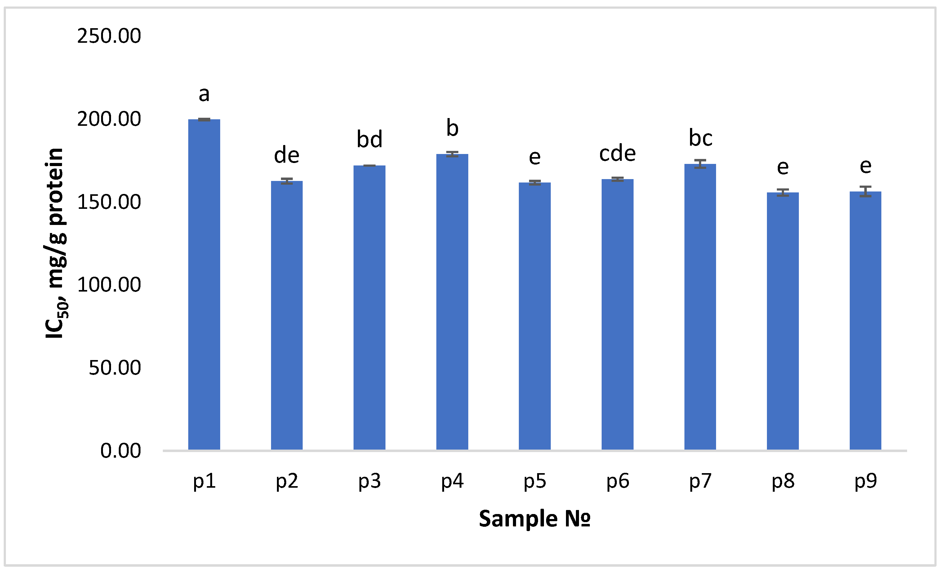
3.4. Acetylcholinesterase Inhibitory Actzivity of LPH

The importance of acetylcholinesterase currently represents the major clue pointing to the cause of Alzheimer’s disease and comprises much of the scientific attention and research. The protein structure of the enzymes suggests their blocking by peptides of suitable structure; however, it was so far neglected by the scientific community until now. Nowadays, only few studies have examined whether enzymatic hydrolysates of dietary proteins can block the enzyme [83,84]. More investigations are needed to clarify the relationships between particular amino acids and their order on a peptide chain with AChE-inhibitory effectiveness both in vitro and in vivo. The findings of our study have shown that lupin proteins could be considered as a highly prominent precursor of peptides with anti-AChE activity. Presented in Figure 2 are the results from the analysis of LPH against acetylcholinesterase inhibitory activity. According to the results in Figure 2, all of the papain protein hydrolysates exhibited acetylcholinesterase inhibitory activity. The effect of E:S ratio and duration of hydrolysis was tested by two-way ANOVA and Tukey HSD multiple comparisons test (Results in Supplementary file). Both factors are considered as the combination of them has significant impact (p-value ≤ 0.05, results in Supplementary file) on the manifestation of the activity. Statistically significant difference was calculated between the three different E:S ratios. The tests between the hydrolysis duration levels showed p-value ≤ 0.05 only between 30–60 and 30–120. Sample P1 exhibited the highest AChE inhibition (IC50 = 199.63 mg/g protein), which is suggestive of the presence of peptides with significant affinity for the enzyme. We may assume that the AChE-inhibitory effects of papain protein hydrolysates could be also the result of the synergistic interactions between various peptides. The study on hempseed protein hydrolysates derived by the action of alcalase, papain, and pepsin reported active peptides in the range 6–11 µg/mL (IC50) [49]. The analysis of molecular weight distribution suggested that the active peptides possess ultra-low molecular weight (<1000 Da). Some papers have documented specific proteins and amino acids as relevant to the inhibition of AChE [47]. The same authors assumed that the presence of high concentration of lysine (39.62–40.18%) in the active hydrolysates could be also a contribution. Nevertheless, further studies are needed to clarify the kinetics of inhibition. Future research on Alzheimer’s disease should take the inhibitory peptides into perspective to demonstrate their potential efficacy as therapeutic agents and/or functional food additives. While in vivo studies are required to definitively determine the positive impact of adding hydrolysates to functional meals, it is crucial to do previous in vitro research to identify lupin hydrolysates with AChE inhibitory activity.

3.5. Correlation Analysis

The dependence of various biological activities (FRAP, CUPRAC, DPPH, ABTS, and AChE inh.) on the degree of hydrolysis (DH) of papain enzyme was investigated using correlation analysis. The results are summarized in the following Table 5.
In the first column of the table, the investigated dependencies are described; in the second column the values of the correlation coefficients are given; in the third column are the values of the coefficient of determination for different types of regression (linear, exponential, logarithmic, etc.); and in the last column are the p-values. According to Cohen’s scale [85], the correlation between DH-Ache inh is very high, DH-CUPRAC is high, between DH-DPPH and DH-ABTS is medium, and between DH-FRAP is weak. The data in the last column show that the DH-CUPRAC and DH-AChE inh. Relationships are statistically significant as p-value ≤ 0.05. Therefore, these two dependencies are subjected to regression analysis and their coefficients of determination are determined in different types of regression.
The relationship between DH-CUPRAC has the highest coefficient of determination in a fourth-order polynomial regression and is therefore best described by the following equation:
y = 4.10−6x4 − 0.0005x3 + 0.0221x2 − 0.4239x + 3.1945
The relationship between DH-AchE inh. Has the highest coefficient of determination in a fourth-order polynomial regression and is therefore best described by the following equation:
y = 0.0002x4 − 0.0352x3 + 2.0624x2 − 53.092x + 677.12

4. Conclusions

This study provides valuable information about the potential of papain-generated lupin protein hydrolysates to exert antimicrobial, antioxidant, and acetylcholinesterase inhibitory activities. Three E:S ratios were studied with three hydrolysis duration times. A total of nine different hydrolysates were generated. Both factors and combination influenced the DH and AchE inhibition (p < 0.05). All the hydrolysates possessed antioxidant activity, acetylcholinesterase inhibitory activity, and inhibited the growth of E. coli ATCC 8739. When tested with ABTS•+ scavenging assay, a thousand times higher AOA was measured for all the hydrolysates, compared to the other three techniques (DPPH, CUPRAC, FRAP). While antioxidant peptides from various sources have been more or less studied, research on protein acetylcholinesterase inhibitors is rather scarce. In this regard, our findings are of significant importance. We measured IC50 from 155.56 to 199.63 mg/g protein between samples that, in our opinion, highlights the need for more detailed studies on that topic. Further studies of the peptide structure–activity relationship are needed.

Supplementary Materials

The following supporting information can be downloaded at: https://www.mdpi.com/article/10.3390/app122312370/s1, Figure S1: The densitometric profile of low molecular weight standard markers (1.060–17.0 kDa); Figure S2: Molecular weight calibration curve (based on the densitometric profile from Figure S1); Figure S3: The densitometric profile of sample P1; Figure S4: The densitometric profile of sample P2; Figure S5: The densitometric profile of sample P3; Figure S6: The densitometric profile of sample P4; Figure S7: The densitometric profile of sample P5; Figure S8: The densitometric profile of sample P6; Figure S9: The densitometric profile of sample P7; Figure S10: The densitometric profile of sample P8; Figure S11: The densitometric profile of sample P9. Table S1. Two-way ANOVA of the impact of E:S ratio and enzyme hydrolysis dura-tion (EHD) on the DH. Table S2. Two-way ANOVA of the impact of E:S ratio and enzyme hydrolysis dura-tion (EHD) on the AChE inhibitory activity. Table S3. Tukey HSD multiple comparisons on the AChE inh. between E:S ratio levels. Table S4. Tukey HSD multiple comparisons on the AChE inh. between EHD. Table S5. Two-way ANOVA of the impact of E:S ratio and enzyme hydrolysis duration (EHD) on AOA (FRAP). Table S6. Two-way ANOVA of the impact of E:S ratio and enzyme hydrolysis dura-tion (EHD) on AOA (CUPRAC). Table S7. Two-way ANOVA of the impact of E:S ratio and enzyme hydrolysis dura-tion (EHD) on AOA (DPPH). Table S8. Two-way ANOVA of the impact of E:S ratio and enzyme hydrolysis dura-tion (EHD) on AOA (ABTS).

Author Contributions

Conceptualization, I.D.; methodology, I.D.; software, I.D. and M.T.; validation, I.D. and M.T.; formal analysis, A.G., I.D., D.M., and H.F.; resources, A.G. and I.D.; writing—original draft preparation, A.G., I.D., D.M., H.F., and M.T.; writing—review and editing, I.D. and D.M.; visualization, I.D. and D.M.; supervision, I.D. and A.P.; project administration, I.D.; funding acquisition, I.D. All authors have read and agreed to the published version of the manuscript.

Funding

This research was funded by the Bulgarian Scientific Fund, grant No. KP-06-M36/2.

Institutional Review Board Statement

Not applicable.

Informed Consent Statement

Not applicable.

Data Availability Statement

Not applicable.

Conflicts of Interest

The authors declare no conflict of interest.

References

  1. Ortea, I. Foodomics in health: Advanced techniques for studying the bioactive role of foods. TrAC Trends Anal. Chem. 2022, 150, 116589. [Google Scholar] [CrossRef]
  2. Tulipano, G. Role of Bioactive Peptide Sequences in the Potential Impact of Dairy Protein Intake on Metabolic Health. Int. J. Mol. Sci. 2020, 21, 8881. [Google Scholar] [CrossRef]
  3. Wu, G. Dietary protein intake and human health. Food Funct. 2016, 7, 1251–1265. [Google Scholar] [CrossRef] [Green Version]
  4. Mehrabi, Z.; Gill, M.; van Wijk, M.; Herrero, M.; Ramankutty, N. Livestock policy for sustainable development. Nat. Food 2020, 1, 160–165. [Google Scholar] [CrossRef]
  5. Aimutis, W.R. Plant-Based Proteins: The Good, Bad, and Ugly. Annu. Rev. Food Sci. Technol. 2022, 13, 1–17. [Google Scholar] [CrossRef]
  6. Cruz-Casas, D.E.; Aguilar, C.N.; Ascacio-Valdés, J.A.; Rodríguez-Herrera, R.; Chávez-González, M.L.; Flores-Gallegos, A.C. Enzymatic hydrolysis and microbial fermentation: The most favorable biotechnological methods for the release of bioactive peptides. Food Chem. Mol. Sci. 2021, 3, 100047. [Google Scholar] [CrossRef]
  7. Rutherfurd-Markwick, K.J. Food proteins as a source of bioactive peptides with diverse functions. Br. J. Nutr. 2012, 108, 149–157. [Google Scholar] [CrossRef] [Green Version]
  8. Chakrabarti, S.; Guha, S.; Majumder, K. Food-Derived Bioactive Peptides in Human Health: Challenges and Opportunities. Nutrients 2018, 10, 1738. [Google Scholar] [CrossRef] [Green Version]
  9. Zaky, A.A.; Simal-Gandara, J.; Eun, J.-B.; Shim, J.-H.; Abd El-Aty, A.M. Bioactivities, Applications, Safety, and Health Benefits of Bioactive Peptides From Food and By-Products: A Review. Front. Nutr. 2022, 8, 815640. [Google Scholar] [CrossRef]
  10. Daroit, D.J.; Brandelli, A. In vivo bioactivities of food protein-derived peptides—A current review. Curr. Opin. Food Sci. 2021, 39, 120–129. [Google Scholar] [CrossRef]
  11. Korhonen, H.; Pihlanto, A. Food-derived Bioactive Peptides—Opportunities for Designing Future Foods. Curr. Pharm. Des. 2005, 9, 1297–1308. [Google Scholar] [CrossRef] [Green Version]
  12. Okagu, I.U.; Ezeorba, T.P.C.; Aham, E.C.; Aguchem, R.N.; Nechi, R.N. Recent findings on the cellular and molecular mechanisms of action of novel food-derived antihypertensive peptides. Food Chem. Mol. Sci. 2022, 4, 100078. [Google Scholar] [CrossRef]
  13. Daliri, E.B.M.; Lee, B.H.; Oh, D.H. Current trends and perspectives of bioactive peptides. Crit. Rev. Food Sci. Nutr. 2018, 58, 2273–2284. [Google Scholar] [CrossRef]
  14. Baindara, P.; Mandal, S.M. Plant-Derived Antimicrobial Peptides: Novel Preservatives for the Food Industry. Foods 2022, 11, 2415. [Google Scholar] [CrossRef]
  15. Cheng, S.; Tu, M.; Liu, H.; Zhao, G.; Du, M. Food-derived antithrombotic peptides: Preparation, identification, and interactions with thrombin. Crit. Rev. Food Sci. Nutr. 2019, 59, S81–S95. [Google Scholar] [CrossRef]
  16. Memarpoor-yazdi, M.; Mahaki, H.; Zare-zardini, H. Antioxidant activity of protein hydrolysates and purified peptides from Zizyphus jujuba fruits. J. Funct. Foods 2012, 5, 62–70. [Google Scholar] [CrossRef]
  17. Xiong, Y.L. Antioxidant Peptides. In Bioactive Proteins and Peptides as Functional Foods and Nutraceuticals; Wiley-Blackwell: Oxford, UK, 2010; pp. 29–42. ISBN 9780813811048. [Google Scholar]
  18. Liu, W.; Chen, X.; Li, H.; Zhang, J.; An, J.; Liu, X. Anti-Inflammatory Function of Plant-Derived Bioactive Peptides: A Review. Foods 2022, 11, 2361. [Google Scholar] [CrossRef]
  19. Pandey, M.; Kapila, S.; Kapila, R.; Trivedi, R.; Karvande, A. Evaluation of the osteoprotective potential of whey derived-antioxidative (YVEEL) and angiotensin-converting enzyme inhibitory (YLLF) bioactive peptides in ovariectomised rats. Food Funct. 2018, 9, 4791–4801. [Google Scholar] [CrossRef]
  20. Chalamaiah, M.; Yu, W.; Wu, J. Immunomodulatory and anticancer protein hydrolysates (peptides) from food proteins: A review. Food Chem. 2018, 245, 205–222. [Google Scholar] [CrossRef]
  21. Martínez-Medina, G.A.; Chávez-González, M.L.; Méndez-Carmona, J.Y.; de la Rosa, O.; Carranza-Méndez, R.; Cruz-Casas, D.E.; Espitia-Hernández, P.; Amaya-Chantaca, D.P.; Aguilar, C.N. Immunomodulatory Properties of Proteins and Peptides: Food Derivatives Approach. In Immunomodulators and Human Health; Kesharwani, R.K., Sharma, A.K., Eds.; Springer Nature: Singapore, 2022; pp. 415–438. ISBN 978-981-16-6379-6. [Google Scholar]
  22. Liu, L.; Li, S.; Zheng, J.; Bu, T.; He, G.; Wu, J. Safety considerations on food protein-derived bioactive peptides. Trends Food Sci. Technol. 2020, 96, 199–207. [Google Scholar] [CrossRef]
  23. Tacias-Pascacio, V.G.; Castañeda-Valbuena, D.; Morellon-Sterling, R.; Tavano, O.; Berenguer-Murcia, Á.; Vela-Gutiérrez, G.; Rather, I.A.; Fernandez-Lafuente, R. Bioactive peptides from fisheries residues: A review of use of papain in proteolysis reactions. Int. J. Biol. Macromol. 2021, 184, 415–428. [Google Scholar] [CrossRef]
  24. Chandrasekaran, S.; Gonzalez de Mejia, E. Optimization, identification, and comparison of peptides from germinated chickpea (Cicer arietinum) protein hydrolysates using either papain or ficin and their relationship with markers of type 2 diabetes. Food Chem. 2022, 374, 131717. [Google Scholar] [CrossRef]
  25. Cotabarren, J.; Rosso, A.M.; Tellechea, M.; García-Pardo, J.; Rivera, J.L.; Obregón, W.D.; Parisi, M.G. Adding value to the chia (Salvia hispanica L.) expeller: Production of bioactive peptides with antioxidant properties by enzymatic hydrolysis with Papain. Food Chem. 2019, 274, 848–856. [Google Scholar] [CrossRef]
  26. Austrelian Government of the Gene Technology Regulator The Biology of Lupinus L. (lupin or lupine). 2013. Available online: https://www.ogtr.gov.au/resources/publications/biology-lupinus-l-lupin-or-lupine (accessed on 1 December 2022).
  27. Lo, B.; Kasapis, S.; Farahnaky, A. Lupin protein: Isolation and techno-functional properties, a review. Food Hydrocoll. 2021, 112, 106318. [Google Scholar] [CrossRef]
  28. Mazumder, K.; Biswas, B.; Kerr, P.G.; Blanchard, C.; Nabila, A.; Golder, M.; Aziz, M.G.; Farahnaky, A. Comparative assessment of nutritional, thermal, rheological and functional properties of nine Australian lupin cultivars. Sci. Rep. 2021, 11, 21515. [Google Scholar] [CrossRef]
  29. Visvanathan, R.; Madhujith, T.; Gamage, A.; Zhang, N. Lupin. In Pulses Processing and Product Development; Manickavasagan, A., Thirunathan, P., Eds.; Springer Nature: Cham, Switzerland, 2020; pp. 169–204. ISBN 978-3-030-41375-0. [Google Scholar]
  30. Arnoldi, A.; Boschin, G.; Zanoni, C.; Lammi, C. The health benefits of sweet lupin seed flours and isolated proteins. J. Funct. Foods 2015, 18, 550–563. [Google Scholar] [CrossRef]
  31. Garmidolova, A.; Desseva, I.; Mihaylova, D.; Lante, A. Bioactive Peptides from Lupinus spp. Seed Proteins-State-of-the-Art and Perspectives. Appl. Sci. 2022, 12, 3766. [Google Scholar] [CrossRef]
  32. Okagu, I.U.; Ndefo, J.C.; Aham, E.C.; Obeme-Nmom, J.I.; Agboinghale, P.E.; Aguchem, R.N.; Nechi, R.N.; Lammi, C. Lupin-Derived Bioactive Peptides: Intestinal Transport, Bioavailability and Health Benefits. Nutrients 2021, 13, 3266. [Google Scholar] [CrossRef]
  33. Montserrat-de la Paz, S.; Villanueva, A.; Pedroche, J.; Millan, F.; Martin, M.E.; Millan-Linares, M.C. Antioxidant and Anti-Inflammatory Properties of Bioavailable Protein Hydrolysates from Lupin-Derived Agri-Waste. Biomolecules 2021, 11, 1458. [Google Scholar] [CrossRef]
  34. Millán-Linares, M.D.C.; Yust, M.D.M.; Alcaide-Hidalgo, J.M.; Millán, F.; Pedroche, J. Lupine protein hydrolysates inhibit enzymes involved in the inflammatory pathway. Food Chem. 2014, 151, 141–147. [Google Scholar] [CrossRef]
  35. Lammi, C.; Bollati, C.; Lecca, D.; Abbracchio, M.P.; Arnoldi, A. Lupin Peptide T9 (GQEQSHQDEGVIVR) Modulates the Mutant PCSK9D374Y Pathway: In vitro Characterization of its Dual Hypocholesterolemic Behavior. Nutrients 2019, 11, 1665. [Google Scholar] [CrossRef] [PubMed] [Green Version]
  36. Kamran, F.; Phillips, M.; Reddy, N. Functional properties of Australian blue lupin (Lupinus angustifolius) protein and biological activities of protein hydrolysates. Legum. Sci. 2021, 3, e65. [Google Scholar] [CrossRef]
  37. Boschin, G.; Scigliuolo, G.M.; Resta, D.; Arnoldi, A. Optimization of the Enzymatic Hydrolysis of Lupin (Lupinus) Proteins for Producing ACE-Inhibitory Peptides. J. Agric. Food Chem. 2014, 62, 1846–1851. [Google Scholar] [CrossRef] [PubMed]
  38. Chin, Y.Y.; Chew, L.Y.; Toh, G.T.; Salampessy, J.; Azlan, A.; Ismail, A.; Yee, Y.; Yee, L.; Theng, G.; Salampessy, J.; et al. Nutritional composition and angiotensin converting enzyme inhibitory activity of blue lupin (Lupinus angustifolius). Food Biosci. 2019, 31, 100401. [Google Scholar] [CrossRef]
  39. Santos-Sánchez, G.; Cruz-Chamorro, I.; Álvarez-Ríos, A.I.; Álvarez-Sánchez, N.; Rodríguez-Ortiz, B.; Álvarez-López, A.I.; Fernández-Pachón, M.-S.; Pedroche, J.; Millán, F.; Millán-Linares, M.D.C.; et al. Bioactive Peptides from Lupin (Lupinus angustifolius) Prevent the Early Stages of Atherosclerosis in Western Diet-Fed ApoE–/– Mice. J. Agric. Food Chem. 2022, 70, 8243–8253. [Google Scholar] [CrossRef]
  40. Lammi, C.; Aiello, G.; Dellafiora, L.; Bollati, C.; Boschin, G.; Ranaldi, G.; Ferruzza, S.; Sambuy, Y.; Galaverna, G.; Arnoldi, A.; et al. Assessment of the Multifunctional Behavior of Lupin Peptide P7 and Its Metabolite Using an Integrated Strategy. J. Agric. Food Chem. 2020, 68, 13179–13188. [Google Scholar] [CrossRef]
  41. Rajaretinam, R.K.; Samuel Gnana, P.V. Rapid neurobehavioural analysis based on the effects of an acetylcholinesterase inhibitor from Tephrosia purpurea in Zebrafish. Ann. Neurosci. 2012, 19, 8–13. [Google Scholar] [CrossRef] [Green Version]
  42. Rees, T.M.; Brimijoin, S. The role of acetylcholinesterase in the pathogenesis of Alzheimer’s disease. Drugs Today 2003, 39, 75–83. [Google Scholar] [CrossRef]
  43. Racchi, M.; Mazzucchelli, M.; Porrello, E.; Lanni, C.; Govoni, S. Acetylcholinesterase inhibitors: Novel activities of old molecules. Pharmacol. Res. 2004, 50, 441–451. [Google Scholar] [CrossRef]
  44. Santos, T.C.d.; Gomes, T.M.; Pinto, B.A.S.; Camara, A.L.; Paes, A.M.D.A. Naturally Occurring Acetylcholinesterase Inhibitors and Their Potential Use for Alzheimer’s Disease Therapy. Front. Pharmacol. 2018, 9, 1192. [Google Scholar] [CrossRef]
  45. Dembitsky, V.M.; Dzhemileva, L.; Gloriozova, T.; D’yakonov, V. Natural and synthetic drugs used for the treatment of the dementia. Biochem. Biophys. Res. Commun. 2020, 524, 772–783. [Google Scholar] [CrossRef] [PubMed]
  46. Prasasty, V.; Radifar, M.; Istyastono, E. Natural Peptides in Drug Discovery Targeting Acetylcholinesterase. Molecules 2018, 23, 2344. [Google Scholar] [CrossRef] [PubMed] [Green Version]
  47. Aluko, R.E. Food-derived Acetylcholinesterase Inhibitors as Potential Agents against Alzheimer’s Disease. eFood 2021, 2, 49–58. [Google Scholar] [CrossRef]
  48. Yan, X.; Tang, J.; dos Santos Passos, C.; Nurisso, A.; Simões-Pires, C.A.; Ji, M.; Lou, H.; Fan, P. Characterization of Lignanamides from Hemp (Cannabis sativa L.) Seed and Their Antioxidant and Acetylcholinesterase Inhibitory Activities. J. Agric. Food Chem. 2015, 63, 10611–10619. [Google Scholar] [CrossRef]
  49. Malomo, S.A.; Aluko, R.E. In Vitro Acetylcholinesterase-Inhibitory Properties of Enzymatic Hemp Seed Protein Hydrolysates. J. Am. Oil Chem. Soc. 2016, 93, 411–420. [Google Scholar] [CrossRef]
  50. Asen, N.D.; Odagu, O.; Udenigwe, C.; Aluko, R.E. In vitro inhibition of acetylcholinesterase activity by yellow field pea (Pisum sativum) protein-derived peptides as revealed by kinetics and molecular docking. Front. Nutr. 2022, 9, 1021893. [Google Scholar] [CrossRef]
  51. Lowry, O.H.; Rosenbrough, N.J.; Farr, A.L.; Randall, R.J. Protein measurement with the Folin Phenol Reagent. J. Biol. Chem. 1951, 193, 265–275. [Google Scholar] [CrossRef]
  52. Nielsen, P.M.; Petersen, D.; Dambmann, C. Improved method for determining food protein degree of hydrolysis. J. Food Sci. 2001, 66, 642–646. [Google Scholar] [CrossRef]
  53. Sun, H.; Yang, J.; Lin, C.; Huang, X.; Xing, R.; Zhang, K.Q. Purification and properties of a β-1,3-glucanase from Chaetomium sp. that is involved in mycoparasitism. Biotechnol. Lett. 2006, 28, 131–135. [Google Scholar] [CrossRef]
  54. Cuvelier, M.E.; Berset, C. Use of a Free Radical Method to Evaluate Antioxidant Activity. LWT Food Sci. Technol. 1995, 30, 25–30. [Google Scholar]
  55. Mihaylova, D.; Lante, A.; Krastanov, A. Total phenolic content, antioxidant and antimicrobial activity of Haberlea rhodopensis extracts obtained by pressurized liquid extraction. Acta Aliment. Acta Aliment. 2015, 44, 326–332. [Google Scholar] [CrossRef]
  56. Re, R.; Pellegrini, N.; Proteggente, A.; Pannala, A.; Yang, M.; Rice-Evans, C. Antioxidant activity applying an improved ABTS radical cation decolorization assay. Free Radic. Biol. Med. 1999, 26, 1231–1237. [Google Scholar] [CrossRef] [PubMed]
  57. Apak, R.; Güçlü, K.; Özyürek, M.; Karademir, S.E. Novel total antioxidant capacity index for dietary polyphenols and vitamins C and E, using their cupric ion reducing capability in the presence of neocuproine: CUPRAC Method. J. Agric. Food Chem. 2004, 52, 7970–7981. [Google Scholar] [CrossRef] [PubMed]
  58. Benzie, I.F.F.; Strain, J.J. Ferric reducing/antioxidant power assay: Direct measure of total antioxidant activity of biological fluids and modified version for simultaneous measurement of total antioxidant power and ascorbic acid concentration. Methods Enzymol. 1999, 299, 15–27. [Google Scholar] [CrossRef]
  59. Mihaylova, D.; Desseva, I.; Popova, A.; Dincheva, I.; Vrancheva, R.; Lante, A.; Krastanov, A. GC-MS metabolic profile and α-glucosidase-, α-amylase-, lipase-, and acetylcholinesterase-inhibitory activities of eight peach varieties. Molecules 2021, 26, 4183. [Google Scholar] [CrossRef]
  60. Vogelsang-O’Dwyer, M.; Sahin, A.W.; Arendt, E.K.; Zannini, E. Enzymatic Hydrolysis of Pulse Proteins as a Tool to Improve Techno-Functional Properties. Foods 2022, 11, 1307. [Google Scholar] [CrossRef]
  61. Schlegel, K.; Sontheimer, K.; Hickisch, A.; Wani, A.A.; Eisner, P.; Schweiggert-Weisz, U. Enzymatic hydrolysis of lupin protein isolates—Changes in the molecular weight distribution, technofunctional characteristics, and sensory attributes. Food Sci. Nutr. 2019, 7, 2747–2759. [Google Scholar] [CrossRef] [Green Version]
  62. Goggin, D.E.; Mir, G.; Smith, W.B.; Stuckey, M.; Smith, P.M.C. Proteomic Analysis of Lupin Seed Proteins To Identify Conglutin β as an Allergen, Lup an 1. J. Agric. Food Chem. 2008, 56, 6370–6377. [Google Scholar] [CrossRef]
  63. Peñta-Ramos, E.A.; Xiong, Y.L. Antioxidant Activity of Soy Protein Hydrolysates in a Liposomal System. J. Food Sci. 2002, 67, 2952–2956. [Google Scholar] [CrossRef]
  64. Sohaib, M.; Anjum, F.M.; Sahar, A.; Arshad, S.; Rahman, U.U.; Imran, A.; Hussain, S. comprehensive review. Int. J. Food Prop. 2016, 2912, 2581–2593. [Google Scholar] [CrossRef]
  65. López-Pedrouso, M.; Lorenzo, J.M.; Borrajo, P.; Franco, D. In search of antioxidant peptides from porcine liver hydrolysates using analytical and peptidomic approach. Antioxidants 2022, 11, 27. [Google Scholar] [CrossRef] [PubMed]
  66. Wang, X.; Tang, C.; Chen, L.; Yang, X. Characterization and Antioxidant Properties of Hemp Protein Hydrolysates Obtained with Neutrase®. Food Technol. Biotechnol. 2009, 47, 428–434. [Google Scholar]
  67. Wong, F.C.; Xiao, J.; Wang, S.; Ee, K.Y.; Chai, T.T. Advances on the antioxidant peptides from edible plant sources. Trends Food Sci. Technol. 2020, 99, 44–57. [Google Scholar] [CrossRef]
  68. Jiang, Y.; Zhang, M.; Lin, S.; Cheng, S. Contribution of specific amino acid and secondary structure to the antioxidant property of corn gluten proteins. Food Res. Int. 2018, 105, 836–844. [Google Scholar] [CrossRef] [PubMed]
  69. Sun, C.; Tang, X.; Ren, Y.; Wang, E.; Shi, L.; Wu, X.; Wu, H. Novel Antioxidant Peptides Purified from Mulberry (Morus atropurpurea Roxb.) Leaf Protein Hydrolysates with Hemolysis Inhibition Ability and Cellular Antioxidant Activity. J. Agric. Food Chem. 2019, 67, 7650–7659. [Google Scholar] [CrossRef] [PubMed]
  70. Wen, C.; Zhang, J.; Zhang, H.; Duan, Y.; Ma, H. Plant protein-derived antioxidant peptides: Isolation, identification, mechanism of action and application in food systems: A review. Trends Food Sci. Technol. 2020, 105, 308–322. [Google Scholar] [CrossRef]
  71. Perez Espitia, P.J.; de Fátima Ferreira Soares, N.; dos Reis Coimbra, J.S.; de Andrade, N.J.; Souza Cruz, R.; Alves Medeiros, E.A. Bioactive Peptides: Synthesis, Properties, and Applications in the Packaging and Preservation of Food. Compr. Rev. Food Sci. Food Saf. 2012, 11, 187–204. [Google Scholar] [CrossRef]
  72. Dong, J.; Wang, S.; Yin, X.; Fang, M.; Gong, Z.; Wu, Y. Food Science and Human Wellness Angiotensin I converting enzyme (ACE) inhibitory activity and antihypertensive effects of rice peptides. Food Sci. Hum. Wellness 2022, 11, 1539–1543. [Google Scholar] [CrossRef]
  73. Liu, M.; Yang, S.; Yang, J.; Lee, Y.; Kou, J.; Wang, C. Neuroprotective and Memory-Enhancing Effects of Antioxidant Peptide from Walnut (Juglans regia L.) Protein Hydrolysates. Nat. Prod. Commun. 2019, 14. [Google Scholar] [CrossRef] [Green Version]
  74. Girgih, A.T.; Alashi, A.; He, R.; Malomo, S.; Aluko, R.E. Preventive and treatment effects of a hemp seed (Cannabis sativa L.) meal protein hydrolysate against high blood pressure in spontaneously hypertensive rats. Eur. J. Nutr. 2014, 53, 1237–1246. [Google Scholar] [CrossRef]
  75. Kolář, M. Bacterial Infections, Antimicrobial Resistance and Antibiotic Therapy. Life 2022, 12, 468. [Google Scholar] [CrossRef] [PubMed]
  76. Ventola, C.L. The Antibiotic Resistance Crisis Part 1: Causes and Threats. Pharm. Ther. 2015, 40, 277–283. [Google Scholar]
  77. Borrajo, P.; Pateiro, M.; Barba, F.J.; Mora, L.; Franco, D.; Toldrá, F.; Lorenzo, J.M. Antioxidant and Antimicrobial Activity of Peptides Extracted from Meat By-products: A Review. Food Anal. Methods 2019, 12, 2401–2415. [Google Scholar] [CrossRef]
  78. Akbarian, M.; Khani, A.; Eghbalpour, S.; Uversky, V.N. Bioactive Peptides: Synthesis, Sources, Applications, and Proposed Mechanisms of Action. Int. J. Mol. Sci. 2022, 23, 1445. [Google Scholar] [CrossRef] [PubMed]
  79. Tincu, J.A.; Taylor, S.W. Antimicrobial peptides from marine invertebrates. Antimicrob. Agents Chemother. 2004, 48, 3645–3654. [Google Scholar] [CrossRef] [Green Version]
  80. Cutrona, K.J.; Kaufman, B.A.; Figueroa, D.M.; Elmore, D.E. Role of arginine and lysine in the antimicrobial mechanism of histone-derived antimicrobial peptides. FEBS Lett. 2015, 589, 3915–3920. [Google Scholar] [CrossRef] [Green Version]
  81. Osman, A.; El-araby, G.M.; Taha, H. Potential use as a bio-preservative from lupin protein hydrolysate generated by alcalase in food system. J. Appl. Biol. Biotechnol. 2016, 4, 76–81. [Google Scholar] [CrossRef]
  82. El-Saadony, M.T.; Abd El-Hack, M.E.; Swelum, A.A.; Al-Sultan, S.I.; El-Ghareeb, W.R.; Hussein, E.O.S.; Ba-Awadh, H.A.; Akl, B.A.; Nader, M.M. Enhancing quality and safety of raw buffalo meat using the bioactive peptides of pea and red kidney bean under refrigeration conditions. Ital. J. Anim. Sci. 2021, 20, 762–776. [Google Scholar] [CrossRef]
  83. Mondal, P.; Gupta, V.; Das, G.; Pradhan, K.; Khan, J.; Gharai, P.K.; Ghosh, S. Peptide-Based Acetylcholinesterase Inhibitor Crosses the Blood-Brain Barrier and Promotes Neuroprotection. ACS Chem. Neurosci. 2018, 9, 2838–2848. [Google Scholar] [CrossRef]
  84. Sanchis, I.; Spinelli, R.; Aschemacher, N.; Humpola, M.V.; Siano, A. Acetylcholinesterase inhibitory activity of a naturally occurring peptide isolated from Boana pulchella (Anura: Hylidae) and its analogs. Amino Acids 2020, 52, 387–396. [Google Scholar] [CrossRef]
  85. Cohen, J. Statistical Power Analysis for the Behavioral Sciences, 2nd ed.; Lawrence Erlbaum Associates: Hillsdale, MI, USA; New York, NY, USA, 1988; ISBN 0-8058-0283-5. [Google Scholar]
Figure 1. Antioxidant activity of lupin protein hydrolysates according to (A) ABTS, and (B) DPPH, FRAP, and CUPRAC. Values are means ± SEM, n = 3 per treatment group. Different letters (a–f) within each assay chart columns indicate significant differences (p < 0.05) between treatments as analyzed by one-way ANOVA and TUKEY test.
Applsci 12 12370 g001
Figure 2. IC50 of lupin protein hydrolysates toward acetylcholinesterase activity. Values are means ± SEM, n = 3 per treatment group. Different letters (a–e) indicate significant differences (p < 0.05) between treatments as analyzed by one-way ANOVA and TUKEY test.
Applsci 12 12370 g002
Table 1. Hydrolysates abbreviation and hydrolysis conditions.
Sample Abbreviation E:S Ratio, %Enzymatic Hydrolysis Duration, min
P10.530
P20.560
P30.5120
P41.030
P51.060
P61.0120
P72.030
P82.060
P92.0120
Table 2. Degree of hydrolysis of lupin protein hydrolysates.
Sample №DH, %
P19.06 ± 0.20 g
P214.20 ± 0.18 e
P318.83 ± 0.11 c
P413.18 ± 0.11 f
P517.01 ± 0.11 d
P623.09 ± 0.48 b
P716.97 ± 0.04 d
P822.80 ± 0.08 b
P927.97 ± 0.37 a
Values are means ± SEM, n = 3 per treatment group. Means in a column without a common superscript letter differ (p < 0.05) as analyzed by one-way ANOVA and the TUKEY test.
Table 3. Molecular weights (in Daltons) of identified fractions by SDS-PAGE.
Band/MWP1P2P3P4P5P6P7P8P9
115,66016,68616,50915,43316,91016,60616,16615,62514,135
2938710,31011,12710,20814,64611,74211,43811,7002301
323082506236524882758275025722531753
4843108885794011091151956909<500
5-835-<500<500<500<500<500-
“-”—no corresponding band detected
Table 4. Antibacterial activity of lupin protein hydrolysate.
MicroorganismP1P2P3P4P5P6P7P8P9
Zone of Inhibition, cm
E. coli ATCC 87390.9 ± 0.01 d1 ± 0.06 c0.9 ± 0.00 d0.8 ± 0.010.6 ± 0.00 g1.1 ± 0.00 b1.3 ± 0.00 a0.7 ± 0.01 f1.1 ± 0.01 b
S. enterica NCTC 6017---------
S. aureus ATCC 25093---0.7-----
L. monocytogenes NCTC 11994---------
“-”—not detected. Values are means ± SEM, n = 3 per treatment group. Means in a row without a common superscript letter differ (p < 0.05) as analyzed by one-way ANOVA and the TUKEY test.
Table 5. Dependence of various biological activities on the degree of hydrolysis (DH) of papain.
CorrelationsrR2p-Value
LinearExponentialLogarithmicPolynomialPower
DH-FRAP−0.22750.05180.05510.130.8740.1380.556
DH-CUPRAC0.58860.34650.37910.290.95520.310.0095
DH-DPPH0.38480.14800.13800.1730.3870.1670.277
DH-ABTS−0.40660.16500.15900.1270.4150.1210.306
DH-AChE inh.−0.79940.63900.65600.730.85330.7470.0097
Publisher’s Note: MDPI stays neutral with regard to jurisdictional claims in published maps and institutional affiliations.

