Article

Geminated Maxillary Incisors: The Success of an Orthodontic Conservative Approach: 15 Years Follow-Up Study

1
Galilee Medical Center, Orthodontic Department, College of Dental Sciences, Nahariya 2210001, Israel
2
Orthodontic and Craniofacial Department, Graduate School of Dentistry, Rambam Health Care Campus, Haifa 3109601, Israel
*
Author to whom correspondence should be addressed.
Appl. Sci. 2022, 12(3), 1389; https://doi.org/10.3390/app12031389
Submission received: 18 December 2021 / Revised: 18 January 2022 / Accepted: 25 January 2022 / Published: 27 January 2022
(This article belongs to the Special Issue Current Advances in Dentistry)

Abstract

:
Tooth gemination is often presented clinically and radiologically as merged crowns or “megadonts” with or without a longitudinal fissure, single root, and a single pulp chamber. The increased mesiodistal width of these teeth results in poor anterior aesthetics, anterior crowding, and incisor rotation. Our aim is to present a conservative non-extraction orthodontic approach that provides both good aesthetics and long-term stability. It aims to achieve extra space to incorporate the megadonts into the dental arch and to exceed “super” class I dental relationships. Accordingly, this study shows that geminated teeth were successfully retained by means of conservative non-extraction orthodontic mechanotherapy and without premolar extractions or enamel reduction. It demonstrated stability during a long-term 15-year follow-up, in addition to high patient satisfaction, good aesthetics, and periodontal health. The solution protocol is definitive and achieved the aesthetic, psychological, and functional objectives at a feasible cost. Thus, this treatment has proven to be preferable to other alternative surgical prosthodontic and endodontic approaches in terms of its long-term stability, and by providing a solution that does not necessitate further multidisciplinary interventions. Dental practitioners in the fields of pediatric dentistry, endodontics and prosthodontics should be well aware of this option.

1. Introduction

Tooth gemination is defined as an incomplete formation of two teeth from a single tooth bud. Clinically, it is often presented in the incisor area of the maxilla as a merged crown or “megadont” with or without a longitudinal crown fissure and a single undivided root [1,2,3]. Radiographically, the single root, which is enlarged in the mesiodistal dimension, incorporates a single, wide pulp chamber that may include two root canals. It appears that gemination is caused by complex genetic and environmental factors [4]. The incidence of gemination is 0.1–0.47% in the general population and 0.07% in orthodontic patients [5], with no gender predilection [6]. It can be diagnosed in the permanent and primary dentitions [7] and may be associated with dental anomalies, such as dens in dente, macrodontia, hypodontia, and supernumerary teeth [2]. The differential diagnosis of gemination is tooth fusion: a complete or incomplete fusion of two adjacent teeth.
As fusion with a supernumerary tooth or missing teeth have also been reported [8], accurate diagnosis cannot rely on teeth counting, but rather on clinical and radiographic assessment. Patients presenting with anterior teeth gemination often report aesthetic concerns due to the crown’s malformation, longitudinal fissure, and the unique enlarged mesiodistal dimension, which may exceed or double the regular incisors’ crown size [9]. The enlarged crown size may also contribute to malocclusion, functional disorders, and compromised oral hygiene. Anterior crowding and rotations may be aggravated by the increased geminated tooth crown’s width [10], which can lead to increased caries. Other than the approach we suggest in this paper, a few treatment modalities are available for patients with geminated central incisors (Table 1). The first option is extraction and later prosthodontic treatment. The second treatment is more complicated and extensive, combining a multidisciplinary approach of endodontic, surgical, and later prosthodontic treatment. The aim of this study was to present an alternative conservative non-surgical orthodontic treatment option, instead of the aforementioned approaches, to provide both good aesthetics and long-term stability.

2. Materials and Methods

(a)
Patients
Five patients with geminated central incisors were studied retrospectively. All of the patients were examined at the mixed dentition stages. Their pretreatment demographic and orthodontic information is presented in Table 2. Four of the patients had maxillary geminated central incisors (3 cases—bilateral, and 1 case—unilateral) and one had a mandibular geminated lateral incisor.
(b)
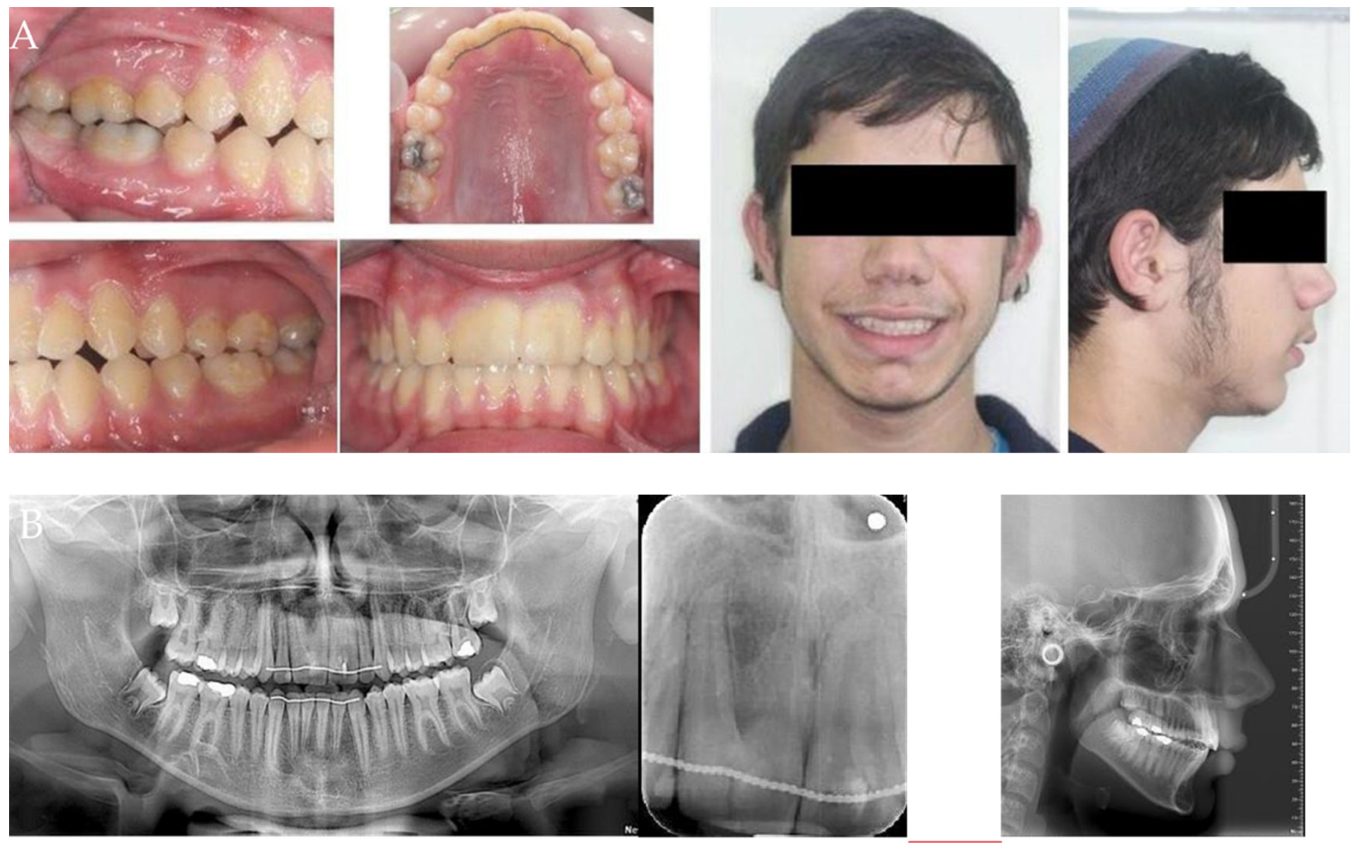
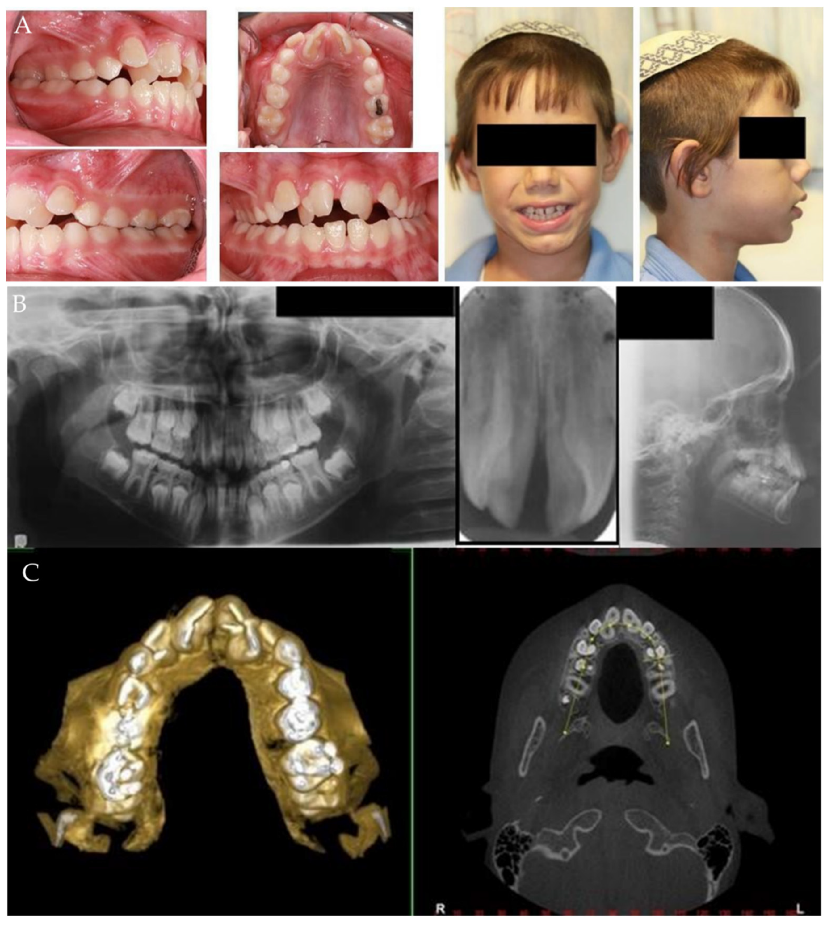
Gemination Characteristics
Gemination in the first three patients occurred in the mixed dentition stage with bilateral double maxillary central incisors. In addition, two cases of gemination of either the upper or lower lateral incisors were present. All cases presented a class II division 1 (Figure 1A) malocclusion associated with an enlarged overjet and overbite tendency. Transversally, the maxilla ranged between normal and narrow, with a bilateral posterior X-bite. Figure 1 presents a 10-year-old patient with maxillary right and left central incisors that demonstrate malalignment and rotation with an abnormal shape. The mesiodistal width of the two central maxillary abnormal incisors was 14 mm on average. The panoramic, periapical, and Cone Beam Computerized Tomography (CBCT) views of the upper incisors revealed fused crowns on both central incisors; a single wide root with a single root canal is clearly demonstrated in the CBCT view (Figure 1B,C). A pretreatment lateral cephalometric radiograph analysis revealed a skeletal class II relationship with a retrognathic mandible with a convex profile, a high angle pattern, and a mandibular clockwise rotation with an enlarged lower facial height (Table 3). The maxillary and mandibular incisors were proclined.
(1)
SNA (Sella, Nasion, A point) indicates the position of the maxilla mandible (whether or not it is normal, prognathic, or retrognathic);
(2)
SNB (Sella, Nasion, B point) Indicates the position of the mandible (whether or not it is normal, prognathic, or retrognathic);
(3)
ANB (A point, Nasion, B point) indicates whether the skeletal relationship between the maxilla and mandible is a normal skeletal class I (+2 degrees), a skeletal class II (+4 degrees or more), or skeletal class III (0 or negative) relationship;
(4)
Lower facial height to indicate facial proportions;
(5)
Angle between Frankfort horizontal line and the line intersecting Gonion–Menton;
(6)
Upper incisors inclination;
(7)
Lower incisors inclination.
(c)
Treatment Protocol and Technique
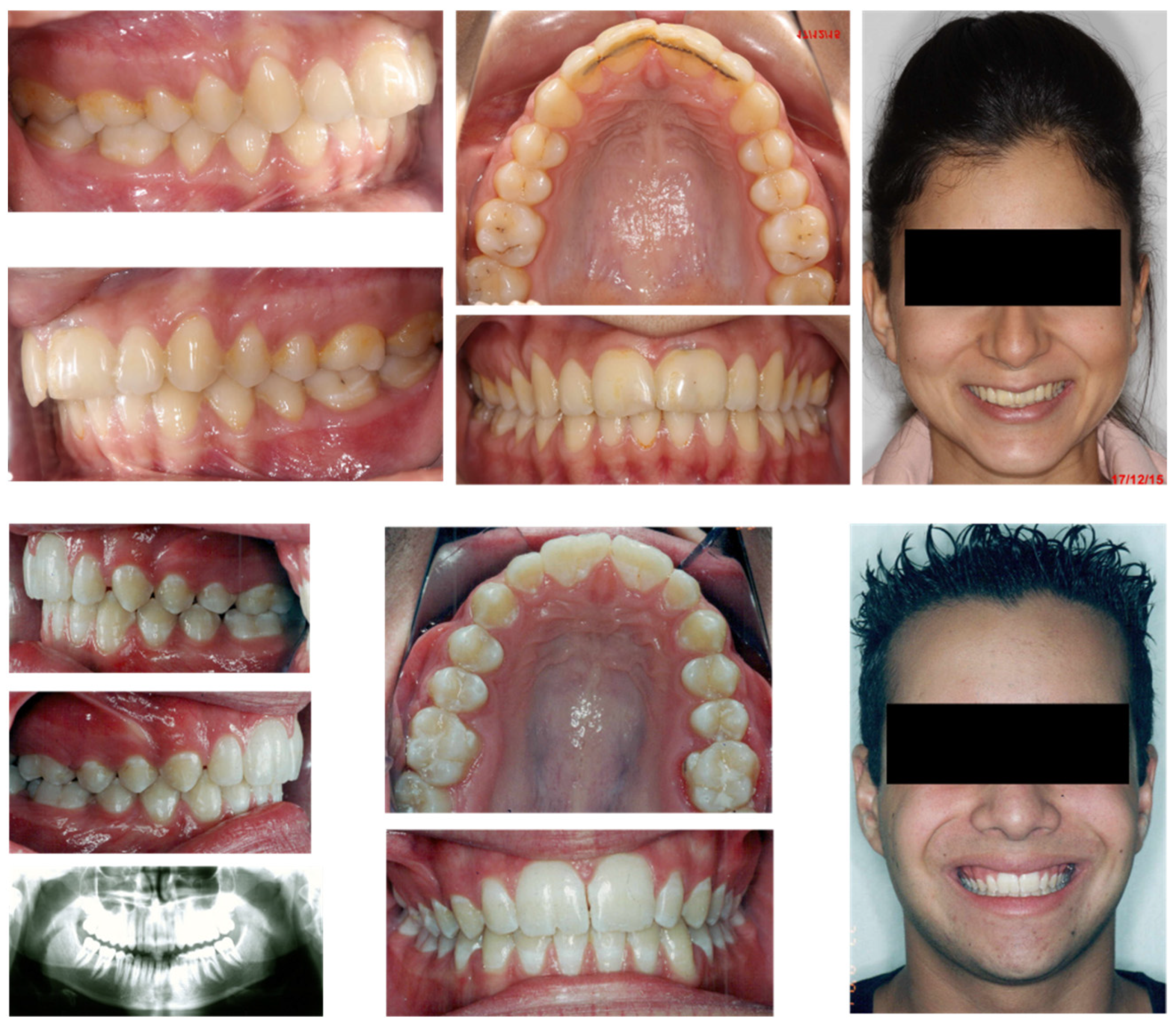
An orthodontic treatment approach that consisted of a distalization and expansion, without premolar extractions was planned. The geminated incisors non-extraction orthodontic conservative technique included 3 components: (1) a maxillary molar teeth distalization of 3–4 mm to exceed the “super” class I dental relationship; (2) a maxillary expansion of 3–4 mm per side; and (3) an incisor teeth protrusive inclination of 7–8 degrees. At first, distalization with a pendulum, a noncompliance orthodontic appliance, was started. Its adverse effect of incisor proclination is also favorable in gemination for its contribution to arch length. Thus, cervical head gear aimed to increase distalization, but which limits the incisor proclination was achieved just 3–4 months later. After the class I relationship was accomplished, a bonded pre-adjusted upper fixed orthodontic appliance was used to expand the upper arch. In the other patients an upper arch expansion was started by Hyrax, enabling an upper median diastema opening. A fixed pre-adjusted appliance was immediately bonded to take advantage of the space created in the upper frontal area to alleviate the upper crowding. The Hyrax was removed after 6 months of retention. In the next step, a lower fixed pre-adjusted appliance was performed to achieve arch coordination and additional lower arch expansion. In addition, a slight upper proclination was considered. The main objectives of the treatment modality were: (1) to obtain healthy periodontal tissue while avoiding bone loss in the median region; (2) to meet aesthetic and functional requirements; (3) to reach upper and lower anterior crowding alleviation; and (4) to establish normal overjet and overbite with full alignment of the geminated teeth.

3. Results

(a) The post-treatment results indicate that the treatment objectives were fully achieved. The facial profile (Figure 2) was improved, with good periodontal health. The angle class III molar relationship and class I canine relationship was achieved with a 4–5 mm overjet and normal overbite. Teeth 11 and 21 were aligned in the arch as well as leveling, and alignment of the maxillary and mandibular dental arches was achieved. The lower incisors were retruded and the upper incisors sustained the same inclination. No change in the skeletal Anteroposterior (AP) discrepancy and vertical dimension was measured (Table 3). A conservative acid-etched restoration concealed the aesthetic defect in the central incisors in two of the cases; the third was not found to require any restoration. The treatment of two of the cases with unilateral geminated lateral incisors has not yet finished. The retention protocol included a combined maxillary and mandibular fixed retainer and a vacuum formed removable retainer for nighttime use.
(b) Long-term outcomes: the preservation of anterior alveolar bone at an 11 and 15 year post-treatment follow-up is presented, showing stable occlusion and correct occlusal parameters, superb aesthetics, and good periodontal health (Figure 3). No change in dental or skeletal dimensions were noticed. The patients were pleased with the results.

4. Discussion

Double teeth may involve an unaesthetic appearance and space problems, making normal teeth alignment impossible and creating functional disorders and difficulties with maintaining oral hygiene. Three treatment alternatives for the gemination phenomenon were considered (Table 1). The first two alternatives are multidisciplinary and based on a prosthodontic approach; thus, presenting an extended and expensive treatment option until permanent crown restoration is performed aged 18 or later. The first option, involving surgical extraction of the geminated tooth and later prosthodontic treatment, can be especially tempting in cases where a geminated or fused tooth is present, to avoid asymmetry. The future restoration may include a ceramic crown [6] via implant placement or an acid-etched bridge. An adverse effect of such a treatment plan is early alveolar bone loss at the anterior aesthetic area, due to tooth extraction at a young age before full skeletal maturation is achieved. In cases where there are two geminated anterior incisor extractions, extensive bone loss is anticipated, requiring extensive bone augmentation, which is essential to support future implants. Therefore, extractions of teeth from the anterior area should be avoided. Decoronation—removal of the tooth crown in infra-occlusion—leaving the root in its alveolus to be replaced by bone, is aimed at preserving the alveolar ridge for a future prosthodontic solution [11,12]. It can also serve as another option for the geminated tooth. Although this option can be advantageous compared to surgical extraction, it can compromise an orthodontic treatment, if needed. the increased mesiodistal width of the root can either prevent good root approximation and alignment [13] or hinder tooth movement into the decoronated site until complete root resorption occurs. The second solution is a combined prosthodontic-endodontic treatment. A mesiodistal slicing of the mega-crown is performed, usually followed by a root canal treatment and prosthetic restoration. The endodontic approach may interfere with the conservative treatment principle since the success rate of root canal treatment is 82.8–97.3% [14]. In addition, geminated teeth have a crown-root dilaceration [15], or complicated root canal morphology [16]. Root resection, which was proposed in fused teeth with wide crowns [3,17], can be contradicted in geminated teeth because a single pulp chamber is present. Literature reports of two cases of geminated teeth with root resection followed later by reshaping and restoration exist [18,19]. Orthodontics is the third treatment modality, aimed at obtaining healthy periodontal tissue and avoiding bone loss, which is most likely to happen in the median region following the extraction of adjacent teeth [20]; thus, this option enables both functional and aesthetic results. The success of orthodontic treatment is dependent on a few parameters; one of these is a correct arch relationship [21]. However, the ratio between the available maxillary and mandibular tooth sizes is not precise in gemination. In the first three presented cases, both Bolton’s overall and anterior ratios were reduced by more than two standard deviations from the expected ratio. In such cases, with significant discrepancy from the Bolton’s mean values, incorporation of the findings into an orthodontic treatment plan is essential to meet the patients’ expectations [14,22,23]. In the presented cases, the skeletal cephalometric measurements are described in an increased ANB angle; and this demonstrated maxillo mandibular discrepancy is derived from the relative protrusive maxilla and retrusive mandible. These skeletal relationships enable a dento-alveolar compensation, which could resolve the tooth size discrepancy in tooth width between the two arches. In the presented cases, space is created by the non-extraction means of upper distalization, expansion, and minor teeth proclination. The establishment of buccal occlusion toward an angle class Ill relationship allows for the alignment of the wide geminated teeth accompanied with an overjet reduction. Not all skeletal relationships are capable of sustaining such a dento-alveolar compensation, as a skeletal class II relationship is mandatory. Orthodontic treatment provides a sole immediate solution in preadolescence or adolescence at a feasible cost. It avoids having to wait until full skeletal maturation, prevents impaction of the adjacent tooth, violation of the occlusal ratios, and the exacerbation of the orthodontic deformity can be observed [24]. In addition, psychological and self-esteem problems [25] are an important contributing factor in preferring this treatment modality. Teeth were reported as one of the most common physical features leading to teasing and harassment among children [12]. Loss of anterior teeth, in geminated teeth, can cause not only a deterioration in the child’s facial and smile aesthetics but may also make them the target of teasing and ridicule by other children [11,12,26,27]. This orthodontic approach also saves on intermediate post-operative restoration [12,28], either fixed or removable, along with many years of childhood and adolescent treatments until growth cessation occurs [26].
Literature reports of three cases of mega teeth exist: in one a geminated tooth [18] and in the other two, fusion [3,19] with a dental sectioning root resection. It was followed later by reshaping and orthodontic treatment to align the adjacent teeth and alleviate crowding in the extraction sites. In addition, orthodontics associated with megadont removal, closing extraction sites, successful movement across the midline, and alignment of a supplemental tooth [26,29] are described. Retention of the geminated teeth by achieving sufficient space was described, following full orthodontic treatment using extractions of the first maxillary premolars [30]. However, creating enough space for wide central incisors, using the orthodontic option of a non-extraction approach, poses a real clinical challenge. A non-extraction approach was performed to maintain a slight angle class III buccal occlusion, avoiding problems of aesthetics and overjet. The limitations of the orthodontic solution for geminated teeth are prolonged treatment time and possible side effects associated with orthodontic treatment, such as caries, periodontal complications, and root resorption. In addition, patient compliance is essential. Further, the aesthetic solution provided is not perfect due to possible crown malformations that can cause aesthetic and occlusal disturbances in the anterior segment [24]. In the current data, a conservative acid-etched restoration was performed in two patients, and the third was not found to need any restoration. However, theoretically, even in cases with a larger aesthetic defect, teeth preservation with later conservative restoration is beneficial and clearly preferable as long as the roots and alveolar area are preserved. The achieved results can be stable and long-lasting, as illustrated in the cases above, preventing the lifelong need for endodontic and prosthodontic follow-up and possible retreatment.

5. Conclusions

The choice of orthodontic treatment rather than prosthetic surgical or endodontic approaches to retain geminated incisors should be considered, since it provides a natural, conservative, and an immediate solution at a reasonable cost. The pleasing aesthetic and functional results of this complex management treatment have been proven to be stable for a long time, sparing unnecessary dental intervention. Dental practitioners in the fields of pediatric dentistry, endodontics and prosthodontics should be made aware of this option.

Author Contributions

Conceptualization, investigation, methodology, writing—original draft, and writing-review, supervision, project administration and editing, S.E.; investigation, methodology, and writing—original draft preparation K.A. conceptualization, methodology, and writing—review and editing, D.A. All authors have read and agreed to the published version of the manuscript.

Funding

This research received no external funding.

Institutional Review Board Statement

The study was conducted in accordance with the Declaration of Helsinki, and approved by the Ethics Committee of Rambam Health Care Campus (0199-18-RMB).

Informed Consent Statement

Not applicable.

Data Availability Statement

Not applicable.

Conflicts of Interest

The authors declare no conflict of interest.

References

  1. Finkelstein, T.; Shapira, Y.; Bechor, N.; Shpack, N. Surgical and orthodontic treatment of a fused maxillary central incisor and supernumerary tooth. J. Clin. Orthod. JCO 2014, 48, 654–658. [Google Scholar]
  2. Gazit, E.; Lieberman, A.M. Macrodontia of maxillary central incisors: Case reports. Quintessence Int. 1991, 22, 883–887. [Google Scholar]
  3. Le Gall, M.; Philip, C.; Aboudharam, G. Orthodontic treatment of bilateral geminated maxillary permanent incisors. Am. J. Orthod. Dentofac. Orthop. 2011, 139, 698–703. [Google Scholar] [CrossRef]
  4. Hattab, F.N.; Hazz’a, A.M. An unusal case of talon’s cusp on geminated tooth. J. Can Dent. Assoc. 2001, 67, 263–266. [Google Scholar]
  5. Altug-Atac, A.T.; Erdem, D. Prevalence and distribution of dental anomalies in orthodontic patients. Am. J. Orthod. Dentofac. Orthop. 2007, 131, 510–514. [Google Scholar] [CrossRef]
  6. Atkins, C.O.; Mourino, A.P. Management of a supernumerary tooth fused to a permanent maxillary central incisor. Oral Surg. Oral Med. Oral Pathol. 1986, 61, 146–148. [Google Scholar] [CrossRef]
  7. Cabral, L.A.G.; Firoozmand, L.M.; Almeida, J.D. Double teeth in primary dentition: Report of two clinical cases. Med. Oral Patol. Oral Cir. Bucal. 2008, 13, 77–80. [Google Scholar]
  8. Steinbock, N.; Wigler, R.; Kaufman, A.Y.; Lin, S.; Naaj, I.A.-E.; Aizenbud, D. Fusion of Central Incisors with Supernumerary Teeth: A 10-year Follow-up of Multidisciplinary Treatment. J. Endod. 2014, 40, 1020–1024. [Google Scholar] [CrossRef]
  9. Malhotra, N. Macrodontic Incisors Aetiology, Clinical features and Management. Synopses 2006, 35, 1–5. [Google Scholar]
  10. Shokri, A.; Baharvand, M.; Mortazavi, H. The largest bilateral gemination of permanent maxillary central incisors: Report of a case. J. Clin. Exp. Dent. 2013, 5, e295–e297. [Google Scholar] [CrossRef]
  11. Cohenca, N.; Stabholz, A. Decoronation? A conservative method to treat ankylosed teeth for preservation of alveolar ridge prior to permanent prosthetic reconstruction: Literature review and case presentation. Dent. Traumatol. 2007, 23, 87–94. [Google Scholar] [CrossRef]
  12. Einy, S.; Kridin, K.; Kaufman, A.Y.; Cohenca, N. Immediate post-operative rehabilitation after decoronation. A systematic review. Dent. Traumatol. 2019, 36, 141–150. [Google Scholar] [CrossRef]
  13. Sapir, S.; Shapira, J. Decoronation for the management of an ankylosed young permanent tooth. Dent. Traumatol. 2008, 24, 131–135. [Google Scholar] [CrossRef]
  14. Chércoles-Ruiz, A.; Sánchez-Torres, A.; Gay-Escoda, C. Endodontics, Endodontic Retreatment, and Apical Surgery Versus Tooth Extraction and Implant Placement: A Systematic Review. J. Endod. 2017, 43, 679–686. [Google Scholar] [CrossRef]
  15. Romano, N.; Souza-Flamini, L.E.; Mendonça, I.L.; Silva, R.G.; Cruz-Filho, A.M. Geminated Maxillary Lateral Incisor with Two Root Canals. Case Rep. Dent. 2016, 2016, 3759021. [Google Scholar] [CrossRef]
  16. Johns, D.A.; Johnson, K.; Maroli, R.K.; James, E.P. Management of geminated maxillary lateral incisor using cone beam computed tomography as a diagnostic tool. J. Conserv. Dent. 2014, 17, 293. [Google Scholar] [CrossRef]
  17. Tuna, E.B.; Yildirim, M.; Seymen, F.; Gencay, K.; Ozgen, M. Fused teeth: A review of the treatment options. J. Dent. Child. 2009, 76, 109–116. [Google Scholar]
  18. David, H.T.; Krakowiak, P.A.; Pirani, A.B. Nonendodontic coronal resection of fused and geminated vital teeth: A new technique. Oral Surg. Oral Med. Oral Pathol. Oral Radiol. Endodontol. 1997, 83, 501–505. [Google Scholar] [CrossRef]
  19. Ferrazzano, G.F.; Festa, P.; Cantile, T.; D’Antò, V.; Ingenito, A.; Martina, R. Multidisciplinary approach to the treatment of double bilateral upper permanent incisors in a young boy. Eur. J. Paediatr. Dent. 2017, 18, 94–98. [Google Scholar]
  20. Korayem, M.; Flores-Mir, C.; Nassar, U.; Olfert, K.D.G. Implant Site Development by Orthodontic Extrusion. Angle Orthod. 2008, 78, 752–760. [Google Scholar] [CrossRef]
  21. Bolton, W.A. The clinical application of a tooth-size analysis. Am. J. Orthod. 1962, 48, 504–529. [Google Scholar] [CrossRef]
  22. Strujic, M.; Anić-Milošević, S.; Meštrović, S.; Šlaj, M. Tooth size discrepancy in orthodontic patients among different malocclusion groups. Eur. J. Orthod. 2009, 31, 584–589. [Google Scholar] [CrossRef] [Green Version]
  23. Freeman, J.E.; Maskeroni, A.; Lorton, L. Frequency of Bolton tooth-size discrepancies among orthodontic patients. Am. J. Orthod. Dentofac. Orthop. 1996, 110, 24–27. [Google Scholar] [CrossRef]
  24. Yordanova-Kostova, G.R.; Grancharov, M.V.; Gurgurova, G.D. Abnormality in the Morphogenesis of Tooth Development and Relationship with Orthodontic Deformities and Treatment Approaches. Case Rep. Dent. 2021, 2021, 3504. [Google Scholar] [CrossRef]
  25. Twigge, E.; Roberts, R.M.; Jamieson, L.; Dreyer, C.W.; Sampson, W.J. The psycho-social impact of malocclusions and treatment expectations of adolescent orthodonticpatients. Eur. J. Orthod. 2015, 38, 593–601. [Google Scholar] [CrossRef]
  26. Foster, T.D. Fusion and duplication: Orthodontic, treatment of a developmental anomaly. Eur. J. Orthod. 1987, 9, 240–242. [Google Scholar]
  27. Ashkenazi, M.; Kaufman, A.; Einy, S. The diagnostic and treatment challenges associated with traumatized intruded permanent incisors: A case report. Quintessence Int. 2015, 46, 309–315. [Google Scholar]
  28. Einy, S.; Kaufman, A.Y.; Yoshpe, M.; Philosoph, N.; Aizenbud, D.; Lin, S. Decoronation of an ankylosed tooth: Postoperative restoration by means of an intermediate fixed orthodontic laboratory device. Quintessence Int. 2018, 49, 239–244. [Google Scholar]
  29. Pair, J. Movement of a maxillary central incisor across the midline. Angle Orthod. 2011, 81, 341–349. [Google Scholar] [CrossRef] [Green Version]
  30. Hashim, H.A. Orthodontic Treatment of Fused and Geminated Central Incisors: A Case Report. J. Contemp. Dent. Pract. 2004, 5, 136–144. [Google Scholar] [CrossRef] [Green Version]
Figure 1. Geminated upper central incisors, a clinical and radiographic view before treatment. (A) Intraoral and extraoral view; (B) panoramic, periapical and cephalometric radiographs; (C) Cone Beam Computerized Tomography (CBCT) of geminated teeth.
Applsci 12 01389 g001
Figure 2. Geminated upper central incisors, a clinical and radiographic view at the end of treatment. (A) Intaroral and extraoral view; (B) panoramic, periapical, and cephalometric radiographs.
Applsci 12 01389 g002
Figure 3. Geminated upper central incisors at 15 years (bottom) and 11 years (top) after the end of treatment; a stable and aesthetic result.
Applsci 12 01389 g003
Table 1. The three treatment alternatives for gemination.
ProceduresAdvantagesDisadvantages
Prosthetic+Surgical (non-conservative)Extraction
Anterior alveolar augmentation
Implant placement
Superior aestheticsAge 18 and older
Anterior alveolar bone loss Multi-disciplinal
All life management Expensive
Prosthetic+Endodontic (fairly conservative)Reshaping
Root canal tx.
Restoration
Decoronation
Fast
Superior aesthetics?
Age 18 and older Possible root canal failure
Multi-disciplinal
All life management
Expensive
Orthodontic (very conservative)Distalization
Maxillary expansion
Minor incisor proclination
Interproximal reduction (possibly)
Younger age
Tooth material conservation
No surgical intervention
Orthodontic involvement only
Stable
Reasonable Cost
A minor aesthetic defect
Prolonged treatment
No class I relationship
Table 2. Demographic data and clinical information.
Patient 1Patient 2Patient 3Patient 4Patient 5
Age at start of
treatment
101071211
SexMFMFF
Geminated tooth11,2111,2111,212242
Angle classificationclass II div 1class II div 1class Iclass II div 1class II div 1
M-D width of
geminated incisors
14/1410.5/1211.5/1211.59
Transverse relationshipbilateral X-bitenormalnormalbilateral X-bitenormal
Table 3. Cephalometric pre- and post-treatment values in patient No 1.
ParametersPrePost
1SNA *890930
2SNB **810860
3ANB ***8070
4Lower facial height58%55%
5Mandibular plane angle200250
6Upper 1 to SN11001100
7Lower 1 to MP1000920
* indicates position of the maxilla. ** indicates position of the mandible. *** indicates whether the skeletal relationship between the maxilla and mandible is a normal skeletal class I (+2 degrees), a skeletal Class II (+4 degrees or more), or skeletal class III (0 or negative) relationship.
Publisher’s Note: MDPI stays neutral with regard to jurisdictional claims in published maps and institutional affiliations.

