Article

Occlusive Properties of Transrenal Ureteral Occlusion Self-Expandable Metallic Stents: 3D-Printed Phantom and Ex Vivo Studies

1
Biomedical Engineering Research Center, Asan Institute for Life Sciences, Asan Medical Center, 88 Olympic-ro 43-gil, Songpa-gu, Seoul 05505, Korea
2
Asan Medical Center, Department of Radiology, University of Ulsan College of Medicine, 88 Olympic-ro 43-gil, Songpa-gu, Seoul 05505, Korea
*
Authors to whom correspondence should be addressed.
These authors contributed equally to this work.
Appl. Sci. 2022, 12(3), 1516; https://doi.org/10.3390/app12031516
Submission received: 12 January 2022 / Revised: 28 January 2022 / Accepted: 29 January 2022 / Published: 30 January 2022

Abstract

:
Newly developed transrenal ureteral occlusion self-expanding metallic stents (SEMSs) are applied in patients with inoperable fistulas. In this study, the occlusive properties of M- and D-type occlusion SEMSs were investigated in 3D-printed phantom and ex vivo porcine urinary tracts. In the former, the mean bursting pressure causing leakage of contrast medium through the occlusion SEMS was relatively higher in M-types (42.8 ± 3.8 mmHg) than in D-types (38.8 ± 3.8 mmHg), without a statistical difference (p = 0.075). In the latter, the bursting pressure causing leakage through the M-type occlusion SEMS (110.7 ± 8.6 mmHg) was significantly higher than that of the D-type occlusion SEMS (93.8 ± 11.2 mmHg, p = 0.015); however, the mean bursting pressures causing contrast blowout did not differ between the two types (178.7 ± 11.2 mmHg vs. 176.2 ± 11.8 mmHg, p = 0.715). In conclusion, M- and D-type occlusion SEMSs showed similar efficacy in occlusive properties in the 3D phantom study; however, the M-type was superior in the ex vivo porcine urinary tract model. Further in vivo experimental studies are required to confirm these experimental results.

1. Introduction

Fistulas of the lower urinary tract are serious complications that can occur after treating various pelvic abnormalities [1,2,3,4]. Surgical management, such as ureterostomy and ureteral clipping, is the standard palliative treatment of such fistulas; however, most fistulas are inoperable [1,4]. Various minimally invasive techniques have been described for ureteral occlusion, using various materials; however, primary success rates fall within 40–100%, with a reintervention rate of 66.7% for ureters that received embolization [5,6,7,8,9,10,11,12,13]. Nephrostomy, for interventional transrenal ureteral occlusions, has been proposed as an alternative therapeutic method [4]. Percutaneous occlusion of urinary flow has been investigated using various materials, such as detachable balloons, embolization coils, Amplatzer vascular plugs, n-butyl cyanoacrylate, and combinations thereof [2,3,4,7,14,15,16,17,18]. Despite recent advances in occlusive materials and technical approaches, each method has its own limitations. Furthermore, additional studies are required to prove their efficacy and safety in preclinical research.
The covered self-expandable metallic stents (SEMSs) have been considered as an alternative therapeutic option to percutaneous drainage and double J stents for malignant ureteral occlusion [19,20]. In 2015, a silicone-covered SEMS, designed in accordance with the occlusive principle to have a candy-wrapper configuration, was first introduced for treating ureteral leakage [14]. Newly developed transrenal ureteral occlusion SEMSs are safe and effective in patients with inoperable urinary leakage or fistulas [14,15]. The two commonly used occlusion SEMSs for ureteral fistulas include M- and D-types, whose middle and distal portions are constricted with a nylon thread, respectively [14]. However, the occlusive properties of the occlusion SEMSs have not been evaluated, and further investigation is required in preclinical trials. Herein, the pressure changes that maintain the ureteral occlusion were quantitatively analyzed to validate the occlusion SEMS. Therefore, this study aimed to evaluate the occlusive properties of M- and D-type occlusion SEMSs using 3D-printed phantom and extracted porcine urinary tracts.

2. Materials and Methods

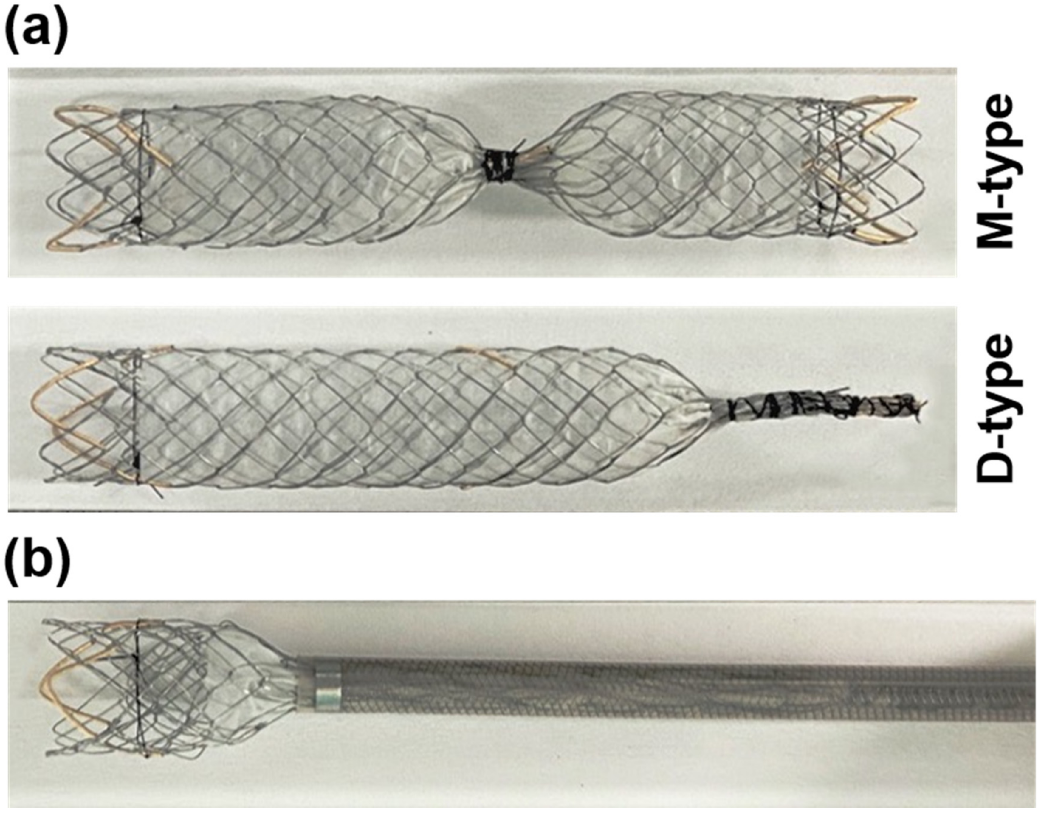
2.1. Preparation of Occlusion SEMSs

The ureteral occlusion SEMSs were prepared as previously described [14,15]. Briefly, the SEMSs were internally and partially coated with a 50-μm-thick silicone membrane. The middle or distal portion of the SEMS was constricted using a nylon thread, according to the stent type (Figure 1a). When fully expanded, the occlusion SEMSs were 8 mm in diameter and 60 mm in length. The delivery system consisted of a 10-Fr sheath and a pusher catheter (Figure 1b).

2.2. Design of the 3D-Printed Phantom

The 3D-printed phantom (Any-med Solution, Seoul, Korea) was manufactured using an injection mold and the liquid silicone rubber technique. After a straight tubular structure with a single side branch was manufactured using Rhino (Rhino 7, Washington, DC, USA), liquid silicone (MED16-6606, Pennsylvania, PA, USA) was injected into the prepared mold for printing. The lengths of the phantom and side branch were 200 mm and 50 mm, respectively. The inner diameter of the phantom was 7 mm, and the wall thickness was 0.2 mm.

2.3. In Vitro Study Using 3D-Printed Phantom

The M- (n = 6) or D-type (n = 6) occlusion SEMSs were placed into the straight part of the phantom under fluoroscopic guidance (Portable Digital Fluoroscopy System; MeteoR, NanoFocusRay Co., Iksan, Korea). A mixed solution of saline and a contrast medium (Telebrix 300; Guerbet, Paris, France) (1:1) was connected to the proximal end of the straight part of the phantom, and a manometer (Yamasu, Honzo, Japan) was connected to the side branch to monitor the pressure. The solution was infused at a rate of 1.5 mL/min into the phantom, with an occlusion SEMS inside, and continuously observed under fluoroscopic guidance to evaluate the bursting pressure causing leakage of contrast through the occlusion SEMS. The experiment was terminated if there was any contrast leakage or stent migration.

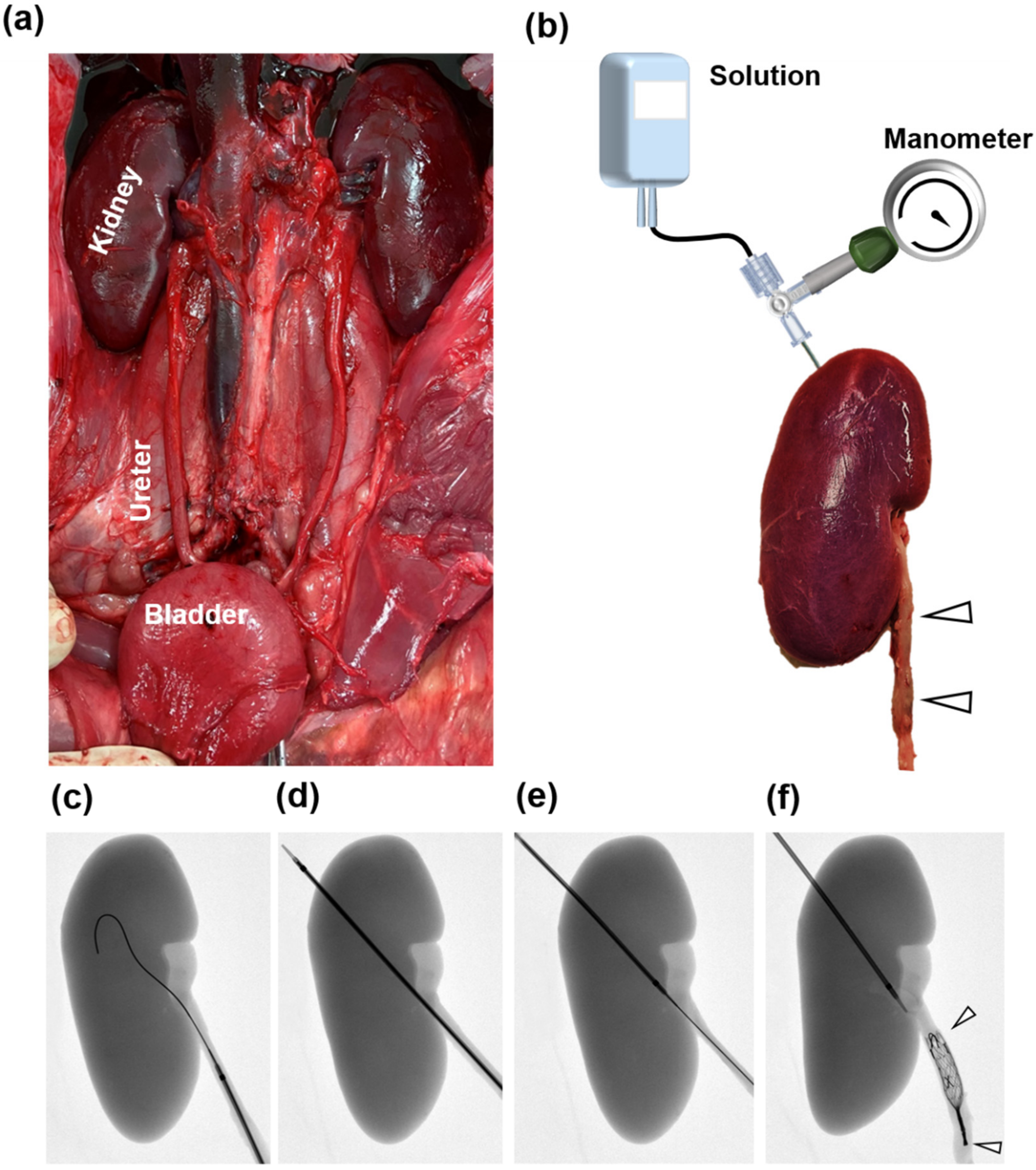
2.4. Ex Vivo Study Design

This study was approved by the Institutional Animal Care and Use Committee of the Asan Institute for Life Sciences (2020-12-189) and conformed to US National Institutes of Health guidelines for humane handling of laboratory animals. In total, 12 porcine urinary tracts, including the kidney, ureter, and bladder, from six pigs weighing 43.1–44.0 kg (mean, 43.6 kg) (Yorkshire; Orient Bio, Seongnam, Korea) were used. The kidney, ureter, and bladder were surgically explored under general anesthesia to evaluate the occlusive properties of the occlusion SEMSs (Figure 2a). All pigs were immediately euthanized by administering 75–150 mg/kg potassium chloride.

2.5. Occlusive SEMS Placement and Experimental Set-Up

The M- (n = 6) and D-type (n = 6) occlusion SEMSs were randomly placed into the extracted porcine ureter, six on the right side and the remaining six on the left side. A 6-Fr Neff catheter (Cook, Bloomington, IN, USA), with a guidewire, was retrogradely inserted through the distal ureteral orifice, and the guidewire, with the catheter, was advanced into the renal pelvis (Figure 2c). The renal parenchyma was punctured from the renal pelvis to the renal surface using the Neff catheter (Cook) to create nephrostomy (Figure 2d). With the wire in place, the Neff catheter was removed. The Neff catheter was inserted at the renal surface with the tip of the Neff catheter positioned in the renal pelvis (Figure 2e). The 10-Fr delivery system, loaded with occlusion SEMS, was retrogradely inserted with an 80-cm-long guide wire (Terumo, Tokyo, Japan) through the distal ureteral orifice. The occlusion SEMS was carefully deployed at the proximal ureter. The pressure bag and the manometer were connected to the Neff catheter using a 3-way cock (Figure 2b). The mixed solution was infused into the renal pelvis at a rate of 1.5 mL/min. Intrapelvic pressure was monitored and recorded at 1-min intervals until the blowout. The mixed solution was continuously observed under fluoroscopic guidance to evaluate the presence of contrast blowout or leakage. The experiment was terminated once blowout or leakage was seen.

2.6. Study Definitions

The occlusive properties were assessed by comparing the pressure levels at blowout and leakage between the two types of occlusion SEMSs. “Blowout” was defined as the pressure at the point of contrast leakage into the renal parenchyma. “Leakage” was defined as the pressure at the point of contrast leakage through the occlusion SEMS.

2.7. Statistical Analysis

Data are expressed as means ± standard deviations. Significance was determined using Student’s t-test. A two-sided p < 0.05 was considered to indicate statistical significance. Statistical analyses were performed using SPSS (version 27.0; SPSS, IBM, New York, NY, USA).

3. Results

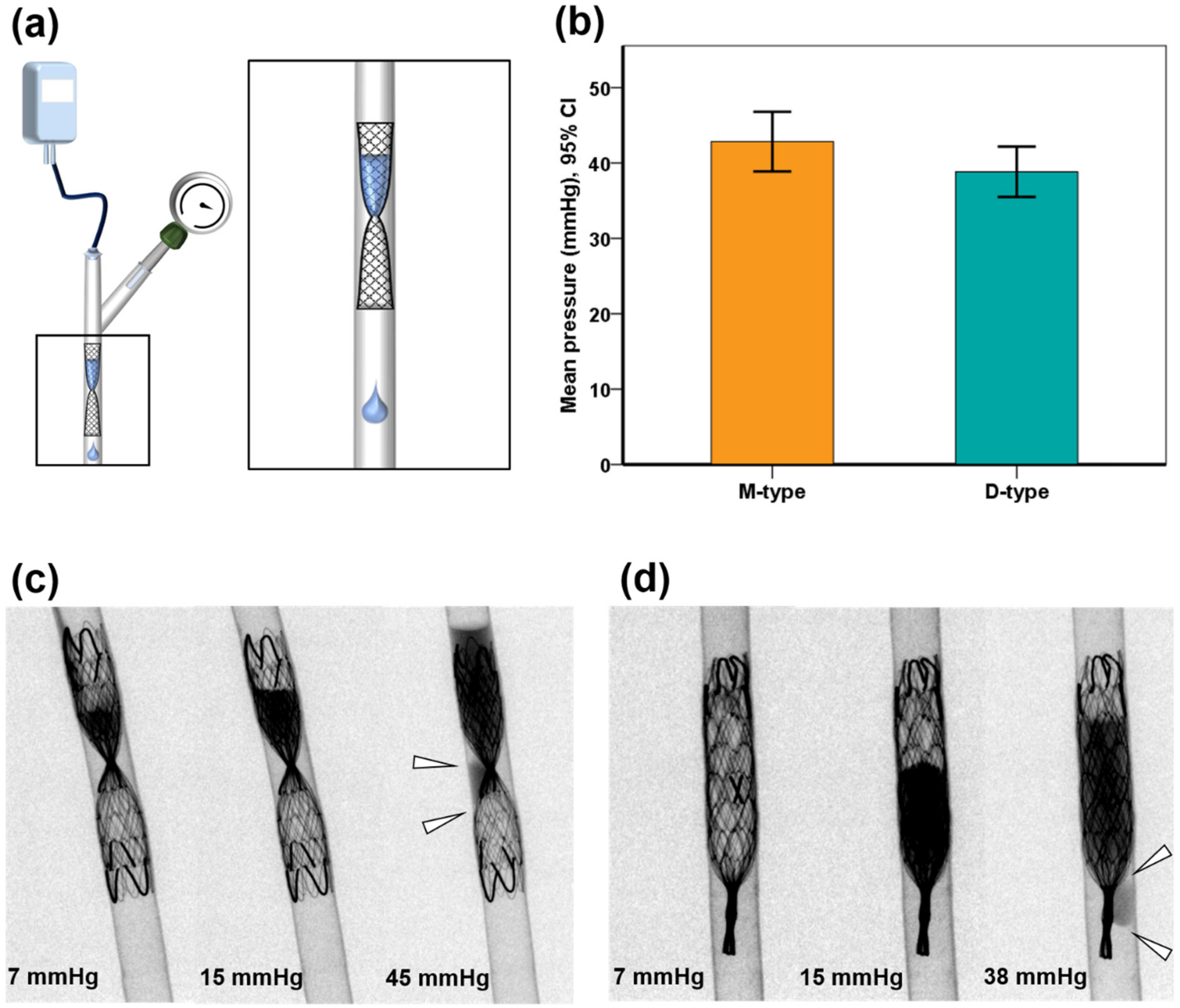
3.1. Occlusive Properties in 3D-Printed Phantom

All occlusion SEMSs were successfully placed into the 3D-printed phantom. The bursting pressure causing leakage of contrast medium through the occlusion SEMS was 38–47 mmHg (mean ± standard deviation [SD], 42.8 ± 3.8 mmHg) in the M-type, and 33–42 mmHg (mean ± SD, 38.8 ± 3.8 mmHg) in the D-type. The bursting pressure in the M-type was relatively higher than that in the D-type, without statistical difference (p = 0.075) (Figure 3). Migration did not occur during the in vitro experiment.

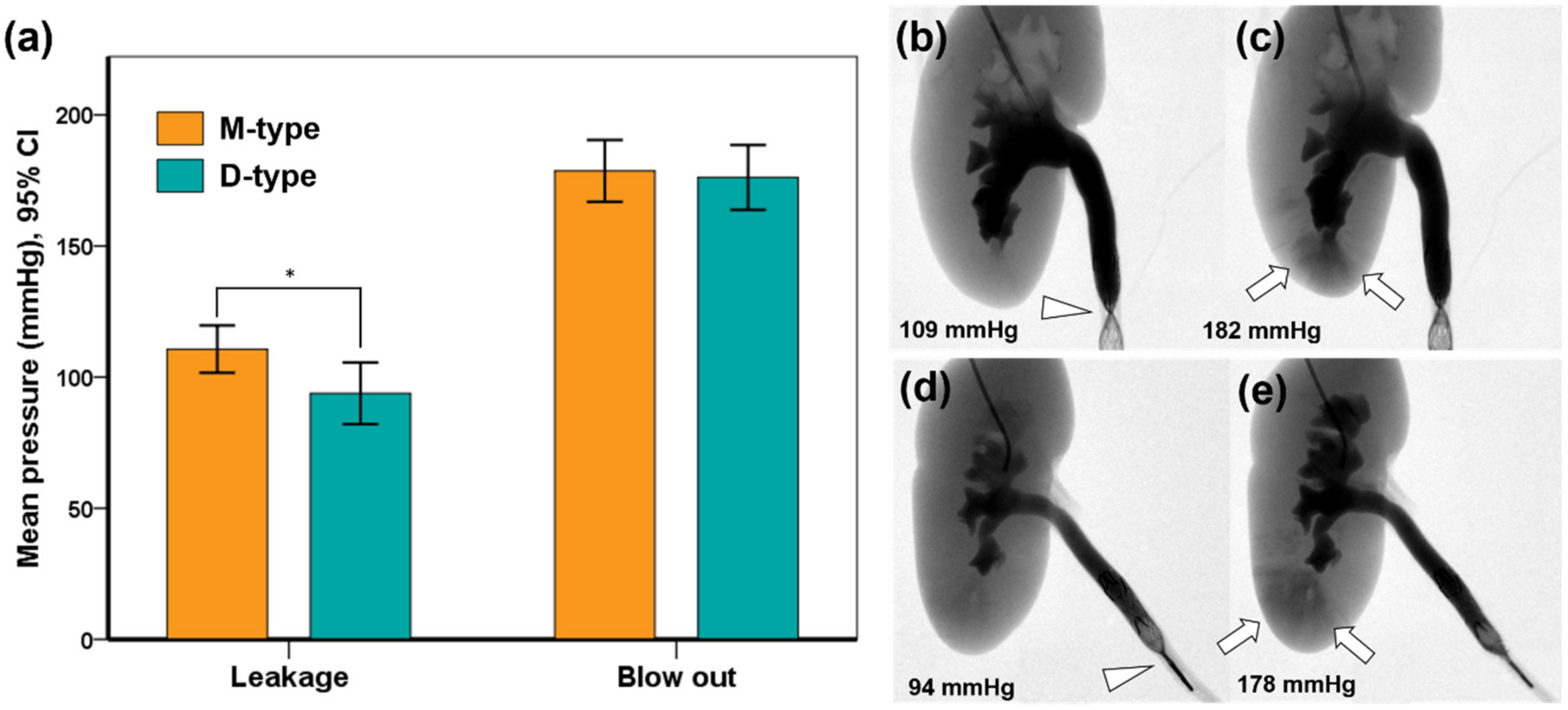
3.2. Occlusive Properties in the Extracted Porcine Urinary Tract

Transrenal nephrostomy and occlusion SEMS placement were technically successful in all extracted urinary tracts, without any procedure-related complications. As the ureter expanded due to the increased pressure, contrast leakage occurred through all the occlusion SEMSs. The mean (±SD) bursting pressures causing leakage through the M-type occlusion SEMS (110.7 ± 8.6 mmHg [range, 99–124 mmHg]) was significantly higher than that of the D-type occlusion SEMS (93.8 ± 11.2 mmHg [range, 82–110 mmHg], p = 0.015). However, the mean (± SD) pressures of blowout did not differ between M-types (178.7 ± 11.2 mmHg [range, 164–194 mmHg]) and D-types (176.2 ± 11.8 mmHg [range, 159–188 mmHg], p = 0.715) (Figure 4). Migration of the occlusion SEMS did not occur during the ex vivo experiments.

4. Discussion

In this study, M- and D-type occlusion SEMSs showed a similar efficacy in occlusive properties in the 3D phantom study; however, superiority of M-type was observed in the ex vivo porcine urinary tract model. Considering that the intrapelvic pressure measured for hydronephrosis in clinical practice, or in the vivo porcine kidney, is about 50–60 mmHg (converted from 68–81 cm H2O) in the literature [21,22], the two types of occlusion SEMS used in this study are considered to be very effective in terms of their occlusive properties, as the burst pressures causing leakage through the occlusion SEMS ranged from 99–124 mmHg, for M-type, and 82–110 mmHg for D-type. Therefore, it is suggested that the design and manufacture of the occlusion SEMS used can be applied to the occlusion SEMS required for actual clinical use.
Over recent decades, a variety of minimally invasive techniques, using different materials, have been introduced for management of ureteral leakage [6,7,10,11,17]. Despite advances in methodology and embolic materials for ureteral occlusion, the primary success rates range from 40% to 100%, and material-related complications can occur in some patients [6,9,10,11,12,13]. Re-intervention requiring additional embolization has been reported in 66.7% of the embolized ureters [7]. Migration of the embolic materials was noted, and recurrent symptoms were subsequently recorded [4]. As shown in previous clinical studies, and the current investigation, occlusion SEMS can easily work with other embolic materials to enhance occlusive properties, with lower complication and recurrence rates [14,15].
The notable point of our ex vivo study design is that, instead of piercing the kidney with a catheter and fixing it, retrogradely inserting a catheter to pierce the kidney can make a hole at once, without damaging the kidney. As a result, the experiment can be carried out easily. In general, in an experiment in which a catheter is inserted through the kidney, when an instrument, such as a guide wire or catheter, is moved or removed, damage to the kidney may occur, which may affect the experimental results or cause the experiment to be stopped. Therefore, through the ex vivo study, meaningful experimental results of the ex vivo study itself can be obtained, and trial-and-error problems in subsequent in vivo studies can be reduced.
After the two types of occlusion SEMSs were evaluated in 13 ureters in 12 patients, with various inoperable urinary leakage or fistula, the reintervention rate to manage leakage recurrence with the use of n-butyl cyanoacrylate and microcoil embolization was 7.7% [14]. Although leakage recurrence through placed occlusion SEMS can be managed with additional embolic materials, such as microcoils and NBCA, more robust occlusion SEMS could reduce the reintervention rate. Although there was no significant difference in the bursting pressure between the M-type and D-type in the current phantom model, M-type is preferable, as it is more superior in the bursting pressure in the current ex vivo study [14,15]. A possible reason for the M-type being superior is because its middle portion was intensively constricted using a nylon thread, while in the case of the D-type, the distal portion was constricted at several points. This difference may influence the degree of occlusion of the two occlusion SEMSs and allow the M-type a higher bursting pressure than the D-type. The occlusive properties of the occlusion SEMSs may be affected by multiple factors, including the degree of mucosal invasion, diameter of the ureter, and shape of the SEMS. For example, stents with flares at the end may improve occlusive properties by reducing migration, but are more likely to increase mechanical injuries to the ureteral wall. In this study, stents were designed to have straight ends to minimize mucosal injuries, but the occlusive properties may have been compromised to some degree.
This study has some limitations. First, the number of sample stents was not large enough for a robust analysis. Second, the occlusion SEMSs were not tested in animal models of ureteral leakage. Third, histological evaluation, to assess potential mucosal damage, was not possible in the phantom. Fourth, the bursting pressure causing leakage of the occlusion SEMS was lower in the 3D-printed phantom than in the ex vivo experiments. Although the 3D-printed phantom was made in the same shape as the human ureter, using silicone rubber, their sizes were slightly off; given that the inner diameter of the phantom was 7 mm, and the diameter of the occlusion SEMSs was 8 mm, it seems likely that loosening was not possible. Therefore, even if the phantom is helpful for stent design, it can hardly replace ex vivo or in vivo studies for property assessment. Despite the need for in vivo investigations involving histological analysis to validate the above-mentioned results, this current study supports the premise that the two types of occlusion SEMSs can successfully occlude the urinary tract.

5. Conclusions

M-and D-types of occlusion SEMSs showed similar efficacy in occlusive properties in a 3D phantom study; however, the M-type was slightly superior to the D-type in the ex vivo porcine urinary tract model. Further in vivo experimental studies are required to confirm these experimental results.

Author Contributions

Conceptualization, J.H.S. and J.-H.P.; data curation, J.W.K., H.H.C., D.-S.W. and J.-H.P.; formal analysis, J.W.K., H.H.C. and J.-H.P.; funding acquisition, H.H.C.; investigation, J.W.K., H.H.C. and J.-H.P.; methodology, J.W.K., H.H.C., D.-S.W. and J.-H.P.; project administration, J.W.K., H.H.C. and J.-H.P.; resources, D.-S.W., C.H.Z., D.S.R., J.M.K., Y.P., S.H.K. and J.H.S.; software, H.H.C.; supervision, J.-H.P. and J.H.S.; validation, D.-S.W., J.M.K. and C.H.Z.; visualization, Y.P., D.S.R. and S.H.K.; writing—original draft preparation, J.W.K. and H.H.C.; writing—review and editing, J.W.K., H.H.C., D.-S.W., C.H.Z., S.H.K., Y.P., J.M.K., D.S.R., J.H.S. and J.-H.P. All authors have read and agreed to the published version of the manuscript.

Funding

This study was supported by a grant (2021IE0016) from the Asan Institute for Life Sciences, Asan Medical Center, Seoul, Korea.

Institutional Review Board Statement

This study was approved by the Institutional Animal Care and Use Committee of the Asan Institute for Life Sciences (2020-12-189) and conformed to the US National Institutes of Health guidelines for humane handling of laboratory animals.

Informed Consent Statement

Not applicable.

Data Availability Statement

The data presented in this study are available on request from the corresponding author. The data are not publicly available due to ethical issues.

Conflicts of Interest

The authors declare no conflict of interest. The funders had no role in the design of the study, the collection, analyses, or interpretation of data, the writing of the manuscript, or the decision to publish the results.

References

  1. Kuetting, D.; Meyer, C.; Schild, H.H.; Pieper, C.C. In Vitro Evaluation of the Occlusive Properties of the ArtVentive Endoluminal Occlusion System Occlusion Device for Transrenal Ureteral Occlusion. J. Endourol. 2017, 31, 1084–1089. [Google Scholar] [CrossRef]
  2. Kim, S.K.; Lee, Y.R.; Kyung, M.S.; Choi, J.S. Transrenal ureteral occlusion with the use of microcoils in five patients with ureterovaginal fistulas. Abdom. Imaging 2008, 33, 615–620. [Google Scholar] [CrossRef] [PubMed]
  3. Günther, R.; Marberger, M.; Klose, K. Transrenal ureteral embolization. Radiology 1979, 132, 317–319. [Google Scholar] [CrossRef]
  4. Avritscher, R.; Madoff, D.C.; Ramirez, P.T.; Wallace, M.J.; Ahrar, K.; Morello, F.A., Jr.; Gupta, S.; Murthy, R.; Wright, K.C.; Hicks, M.E. Fistulas of the lower urinary tract: Percutaneous approaches for the management of a difficult clinical entity. Radiographics 2004, 24 (Suppl. S1), S217–S236. [Google Scholar] [CrossRef] [PubMed]
  5. Augustin, A.M.; Koneval, L.; Kalogirou, C.; Kocot, A.; Kickuth, R. Percutaneous transrenal ureteral plug embolization: Is there a need for tissue adhesives? Diagn. Interv. Radiol. 2021, 27, 762–767. [Google Scholar] [CrossRef] [PubMed]
  6. Saad, W.E.; Kalagher, S.; Turba, U.C.; Sabri, S.S.; Park, A.W.; Stone, J.; Angle, J.F.; Matsumoto, A.H. Ureteric embolization for lower urinary tract fistulae: Use of two amplatzer vascular plugs and N-butyl cyanoacrylate employing the “sandwich” technique. Cardiovasc. Interv. Radiol. 2013, 36, 1068–1072. [Google Scholar] [CrossRef]
  7. Jalaeian, H.; Hicks, R.J.; Hartnell, G.G.; d’Othée, B.J. Transrenal Ureteral Occlusion for Palliation of Refractory Urine Leaks Using Vascular Plugs and Liquid Ethylene Vinyl Alcohol. J. Vasc. Interv. Radiol. 2019, 30, 1994–2001. [Google Scholar] [CrossRef] [PubMed]
  8. Schild, H.H.; Günther, R.; Thelen, M. Transrenal ureteral occlusion: Results and problems. J. Vasc. Interv. Radiol. 1994, 5, 321–325. [Google Scholar] [CrossRef]
  9. Papanicolaou, N.; Pfister, R.C.; Yoder, I.C. Percutaneous occlusion of ureteral leaks and fistulae using nondetachable balloons. Urol. Radiol. 1985, 7, 28–31. [Google Scholar] [CrossRef]
  10. Asvadi, N.H.; Arellano, R.S. Transrenal Antegrade Ureteral Occlusion: Clinical Assessment of Indications, Technique and Outcomes. J. Urol. 2015, 194, 1428–1432. [Google Scholar] [CrossRef]
  11. Schild, H.H.; Meyer, C.; Möhlenbroch, M.; Mueller, S.C.; Simon, B.; Kuhl, C.K. Transrenal ureter occlusion with an Amplatzer vascular plug. J. Vasc. Interv. Radiol. 2009, 20, 1390–1392. [Google Scholar] [CrossRef]
  12. Pieper, C.C.; Meyer, C.; Hauser, S.; Wilhelm, K.E.; Schild, H.H. Transrenal ureteral occlusion using the Amplatzer vascular plug II: A new interventional treatment option for lower urinary tract fistulas. Cardiovasc. Interv. Radiol. 2014, 37, 451–457. [Google Scholar] [CrossRef]
  13. Horenblas, S.; Kröger, R.; van Boven, E.; Meinhardt, W.; Newling, D.W. Use of balloon catheters for ureteral occlusion in urinary leakage. Eur. Urol. 2000, 38, 613–617. [Google Scholar] [CrossRef]
  14. Chen, C.; Kim, J.W.; Shin, J.H.; Hong, B.; Li, H.L.; Kang, C.H.; Cho, Y.J.; Chu, H.H. Therapeutic Ureteral Occlusion with Use of Occlusion Stents for Urinary Leakage or Fistula: A Bicentric Study. Cardiovasc. Interv. Radiol. 2020, 43, 1492–1497. [Google Scholar] [CrossRef]
  15. Park, H.J.; Shin, J.H.; Kim, J.W.; Hong, B.S. Postoperative ureteral leak treated using a silicone-covered nitinol stent. Int. Neurourol. J. 2015, 19, 47–50. [Google Scholar] [CrossRef] [Green Version]
  16. Shindel, A.W.; Zhu, H.; Hovsepian, D.M.; Brandes, S.B. Ureteric embolization with stainless-steel coils for managing refractory lower urinary tract fistula: A 12-year experience. BJU Int. 2007, 99, 364–368. [Google Scholar] [CrossRef]
  17. Lingeman, J.E.; Wong, M.Y.; Newmark, J.R. Endoscopic management of total ureteral occlusion and ureterovaginal fistula. J. Endourol. 1995, 9, 391–396. [Google Scholar] [CrossRef]
  18. Shabrang, C.; Kelbach, S.M.; Hsu, D.P.; Zippe, C.D.; Lie, K.T. Therapeutic ureteral occlusion with n-butyl cyanoacrylate glue and an AMPLATZER plug scaffold. J. Vasc. Interv. Radiol. 2012, 23, 428–430. [Google Scholar] [CrossRef]
  19. Flueckiger, F.; Lammer, J.; Klein, G.E.; Hausegger, K.; Lederer, A.; Szolar, D.; Tamussino, K. Malignant ureteral obstruction: Preliminary results of treatment with metallic self-expandable stents. Radiology 1993, 186, 169–173. [Google Scholar] [CrossRef]
  20. Chung, H.H.; Kim, M.D.; Won, J.Y.; Won, J.H.; Cho, S.B.; Seo, T.S.; Park, S.W.; Kang, B.C. Multicenter experience of the newly designed covered metallic ureteral stent for malignant ureteral occlusion: Comparison with double J stent insertion. Cardiovasc. Interv. Radiol. 2014, 37, 463–470. [Google Scholar] [CrossRef]
  21. Bratt, C.G.; Nilsson, S. Intrapelvic pressure and caliceal dilatation in the evaluation of intermittent hydronephrosis. J. Urol. 1987, 137, 845–848. [Google Scholar] [CrossRef]
  22. Pope, J.C.T.; Showalter, P.R.; Milam, D.F.; Brock, J.W., 3rd. Intrapelvic pressure monitoring in the partially obstructed porcine kidney. Urology 1994, 44, 565–571. [Google Scholar] [CrossRef]
Figure 1. Occlusion stent and delivery system. (a) Photographs showing the two types of occlusion self-expandable metallic stents (SEMSs). From top to bottom: M-type and D-type occlusion SEMSs. (b) The delivery system showing a partially deployed occlusion SEMS.
Applsci 12 01516 g001
Figure 2. An image representing an ex vivo experiment. (a) Photograph showing the extracted porcine urinary tract, including the kidney, ureter, and bladder. (b) Schematic image showing the ex vivo experiment set-up with placed occlusion stent (arrowheads) in the ureter. (cf) Fluoroscopic images obtained during the procedure showing technical steps of nephrostomy using the Neff percutaneous access set and placement of an occlusion stent (arrowheads).
Applsci 12 01516 g002
Figure 3. The process of in vitro set-up and the results of experiments. (a) Schematic illustration of the in vitro set-up using the 3D-printed phantom. (b) Graph showing the mean pressure at the point of the contrast leakage in the M-type and D-type occlusion self-expandable metallic stents (SEMSs). (c) Fluoroscopic images obtained during the in vitro experiment using the M-type occlusion SEMS, showing leakage (arrowheads) at 45 mmHg. (d) Fluoroscopic images showing the presence of leakage (arrowheads) at 38 mmHg in the D-type occlusion SEMS in the 3D printed phantom. Note: CI, confidence interval.
Applsci 12 01516 g003
Figure 4. Results of ex vivo experiments. (a) Graphs showing the mean pressures at the point of leakage and blowout in the M-type and D-type occlusion self-expandable metallic stents (SEMSs). Fluoroscopic images obtained during the ex vivo experiment using the M-type occlusion SEMS showing (b) leakage (arrowhead) at 109 mmHg and (c) blowout (arrows) at 182 mmHg. Fluoroscopic images showing (d) leakage (arrowhead) at 94 mmHg and (e) blowout (arrows) at 178 mmHg in the D-type occlusion SEMS. Note: CI, confidence interval. * p < 0.05.
Applsci 12 01516 g004
Publisher’s Note: MDPI stays neutral with regard to jurisdictional claims in published maps and institutional affiliations.

