Article

Monitoring Molecular Properties of a Fluorescence Light-Up Aptamer Using Fluorescence Cross-Correlation Spectroscopy

Biomedical Research Institute, National Institute of Advanced Industrial Science and Technology (AIST), Tsukuba 305-8566, Japan
*
Author to whom correspondence should be addressed.
Appl. Sci. 2022, 12(4), 2002; https://doi.org/10.3390/app12042002
Submission received: 7 January 2022 / Revised: 8 February 2022 / Accepted: 8 February 2022 / Published: 14 February 2022

Abstract

:
Fluorescence light-up aptamers (FLAPs) are tools for RNA imaging, wherein the RNA of interest is appended with a FLAP sequence that can bind to a corresponding small-molecule fluorogen and enhance its fluorescence. The fluorescence properties of FLAPs have mostly been analyzed in bulk and described as the average of a large number of RNA–fluorogen complexes. In this study, we evaluated the feasibility of fluorescence correlation spectroscopy (FCS)- and fluorescence cross-correlation spectroscopy (FCCS)-based quantifications of FLAPs in a solution using Broccoli, a common FLAP, and its corresponding fluorogen, DFHBI-1T. We investigated the folding efficiency, photostability, and photophysical properties of the Broccoli–DFHBI-1T complex using their FCS/FCCS characteristics. With FCS, we observed that the fluorescence was affected by the affinity between Broccoli and DFHBI-1T and the folding (maturation) state of Broccoli RNA. Moreover, the FCCS measurement of ATTO647N-labeled Broccoli and its complex with DFHBI-1T revealed the proportion of the mature Broccoli–DFHBI-1T complex. The current FCS/FCCS-based study of Broccoli–DFHBI-1T provides a model for analyzing FLAPs and their fluorogen pairs at the single-molecule level.

1. Introduction

RNA plays an important role in gene expression and regulation. Studying RNA-mediated biological systems increases the understanding of life processes and helps in disease diagnosis and therapy. Thus, visualization approaches must be developed to reveal cell RNA localization and detect disease-related RNA in solution.
Fluorescence light-up aptamers (FLAPs) are among the most promising candidates for RNA visualization. FLAP is an RNA aptamer that can specifically bind to a corresponding small-molecule fluorogen. Such fluorogens are usually cell-permeable and essentially non-fluorescent. However, they become highly fluorescent upon binding to the partner FLAP. Thus, FLAP technology provides a simple and promising approach for live-cell RNA imaging by fusing a FLAP sequence to the RNA of interest. For example, Broccoli, a 49-base RNA aptamer, is a FLAP that binds to DFHBI-1T and enhances its green fluorescence [1,2,3]. Broccoli–DFHBI-1T was developed as an improved version of Spinach–DFHBI, and is used in many intracellular and extracellular applications because of its compactness and high fluorescence in physiological environments. In many cases, the fluorescence properties of FLAPs, including Broccoli, have been analyzed in bulk and described as an average of a large number of FLAP–fluorogen complexes, except for a few methods, such as super-resolution imaging. Thus, the fluorescence properties of a FLAP complex with a fluorogen have not been analyzed at the molecular level.
Fluorescence correlation spectroscopy (FCS) is a technique that reveals molecular diffusion dynamics and quantifies the number of molecules of fluorescently labeled target molecules at the single-molecule level [4,5,6]. FCS is mainly applied for the investigation of particle size change caused by molecular interaction, enzymatic activity, and aggregation, and for the quantification of fluorescently labeled biomolecules, including RNA, in solutions and living cells [7,8,9,10]. In addition, FCS can measure the brightness of a single molecule or particle. In the case of bulk analysis, the obtained fluorescence intensity is a convolution of molecular concentration and single-molecule brightness. Using FCS, we can obtain information about the molecular brightness and concentration of fluorescent molecules individually.
Fluorescence cross-correlation spectroscopy (FCCS) is a dual-color extension of FCS [11,12]. FCCS can monitor the interaction between two fluorescently labeled molecules directly by observing the coincidence of fluctuations between two fluorescence intensity profiles. The strength of the molecular interaction is described as the dissociation constant (Kd), which is calculated from the concentrations of each target molecule and complex. In FCCS, concentrations of two kinds of target molecule from autocorrelation curves and the concentration of binding complexes from the cross-correlation curve can be used to calculate the Kd value [13,14]. Sasaki et al. applied the FCCS technique to DNA degradation studies in living cells [15]. FCCS can monitor molecular binding directly in solutions and in living cells.
This report evaluates the feasibility of FCS/FCCS measurements of FLAP–fluorogen pairs, using Broccoli–DFHBI-1T as a model in the conventional FCS/FCCS measurement conditions. In addition, we investigated the folding efficiency, photostability, and photophysical properties of Broccoli–DFHBI-1T by taking advantage of FCS/FCCS, which have not been analyzed in detail at the single-molecule level.

2. Materials and Methods

2.1. Preparation of the RNA

The dsDNA template for Broccoli RNA synthesis was prepared using the Klenow reaction with ssDNA (GAGCCCACACTCTACTCGACAGATACGAATATCTGGACCCGACCGTCTCCCTATAGTGAGTCGTATTA and TAATACGACTCACTATAGG), using a Klenow fragment (Takara Bio, Shiga, Japan). Broccoli RNA was transcribed from the dsDNA template via in vitro transcription reaction at 37 °C for 150 min using a ScriptMAX Thermo T7 Transcription Kit (Toyobo, Osaka, Japan) and purified using a Micro Bio-Spin 30 column (Bio-Rad Laboratories, Hercules, CA, USA) to remove unincorporated rNTPs. Then, 5′-end ATTO647N-labeled Broccoli RNA (/5ATTO647NN/rArGrArGrArCrGrGrUrCrGrGrGrUrCrCrArGrArUrArUrUrCrGrUrArUrCrUrGrUrCrGrArGrUrArGrArGrUrGrUrGrGrGrCrUrC) was chemically synthesized (Integrated DNA Technologies, Coralville, IA, USA).

2.2. Sample Preparation

For FCS and FCCS measurements, RNA and DFHBI-1T were mixed in 4-(2-hydroxyethyl)-1-piperazineethanesulfonic acid(HEPES) buffer (40 mM HEPES pH 7.4, 100 mM KCl, 1 mM MgCl2). DFHBI-1T was purchased from Lucerna (Brooklyn, NY, USA). The RNA and DFHBI-1T concentrations are indicated in the figures and figure legends. Samples were prepared for all measurements, unless specified otherwise.

2.3. FCS and FCCS Measurement

FCS and FCCS experiments were performed using a TCS SP8 X SMD confocal microscope system (Leica Microsystems GmbH, Wetzlar, Germany) with a white-light laser using an acousto-optical beam splitter (AOBS) and cooled HyD detectors. The laser power was measured using a microscope power sensor (S170C; Thorlabs, Inc., NJ, USA) and an optical power and energy meter console (PM400; Thorlabs, Inc., Newton, NJ, USA). The actual laser output was confirmed at the sample position. The system was equipped with a water-immersion objective lens (HC PL Apo 86x/1.20 W motCORR). Broccoli–DFHBI-1T, AcGFP, ATTO488, and fluorescein were excited by a 488 nm laser, and fluorescence signals were collected at 500–750 nm for FCS and 500–610 nm for FCCS (green channel). ATTO647N was excited by a 633 nm laser, and fluorescence signals were collected at 650–750 nm (red channel). The wavelengths of laser lines were tuned using AOBS. Measurements were taken at room temperature (20 °C) in a µ-Slide 18 Well Glass Bottom (Ibidi, Gräfelfing, Germany). The glass surfaces of the wells were treated with 20% N101 blocking reagent (NOF Corporation, Tokyo, Japan). Each sample solution (50 μL) was placed on the glass surface of a well and measured for 60–120 s at 100 μm above the glass surface.
Data analysis was performed using the SymPhoTime 64 software (Picoquant, Berlin, Germany). The fluorescence autocorrelation functions of the green and red channels, Gg(τ) and Gr(τ), and fluorescence cross-correlation function, Gc(τ), were calculated using the following equation:
G x τ = I i t · I j t + τ I i t I j t 1
where τ denotes the time delay; Ii is the fluorescence intensity of the green channel (i = g) or red channel (i = r), detected in the laser-illuminated observation volume (confocal volume); Gg(τ) and Gr(τ) denote the autocorrelation functions of green (i = j = x = g) or red (i = j = x = r); and Gc(τ) denotes the cross-correlation function (i = g, j = r, x = c).
The acquired auto/cross-correlation curves were fitted to a one-component model, as follows:
G τ = 1 N 1 + τ τ D 1 1 + 1 s 2 τ τ D 1 / 2
where N is the average number of fluorescent particles in the confocal volume defined by the radial radius ω and axial radius z, and s is the structural parameter representing the ratio s = z/ω. The diffusion time (τD) corresponds to the average time for the diffusion of fluorescent particles across the confocal volume, which reflects the size of the particles.
For the quantitative analysis of the cross-correlation amplitude, we introduced the relative cross-correlation amplitude (RCA) [15], as follows:
RCA g = N g r N g = G c 0 G r 0 ,   RCA r = N g r N r = G c 0 G g 0
where Ng, Nr, and Ngr denote the number of green-labeled molecules, red-labeled molecules, and the molecules labeled with both (complex of green and red labeled molecules), respectively. The effective confocal volume (Veff) of each channel was estimated by the reference measurement of ATTO488 and ATTO655, with known diffusion coefficients (D = 400 µm2/s and 426 µm2/s, respectively) [16,17], using the following equation:
V e f f = π 3 2   s   4 D τ D 3 2
The effective volume for cross-correlation was estimated as the root mean square of the effective volume values of the green and red channels.

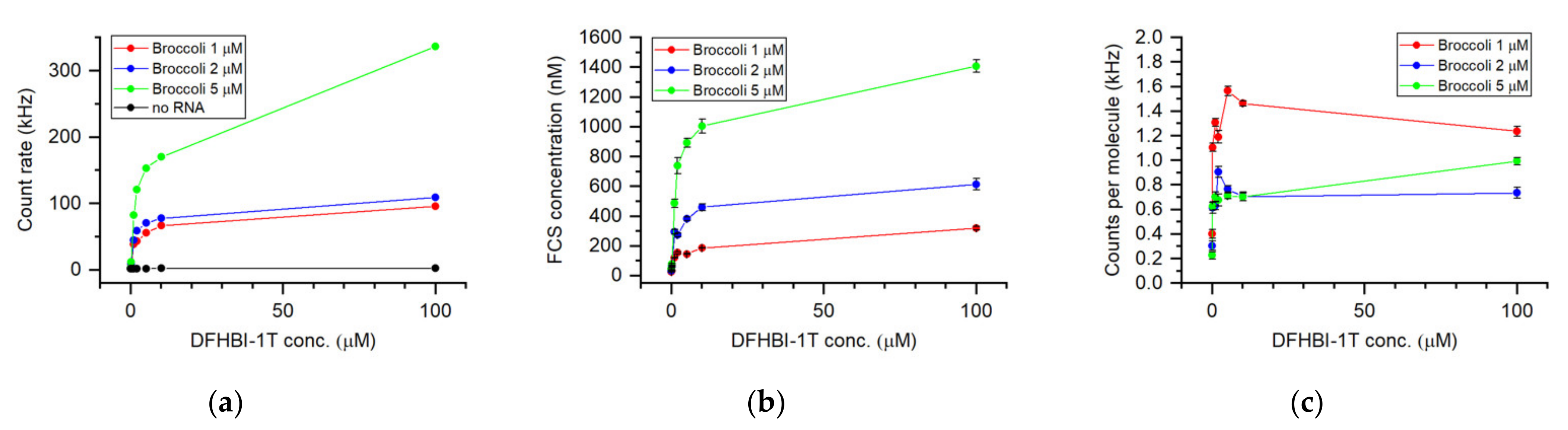
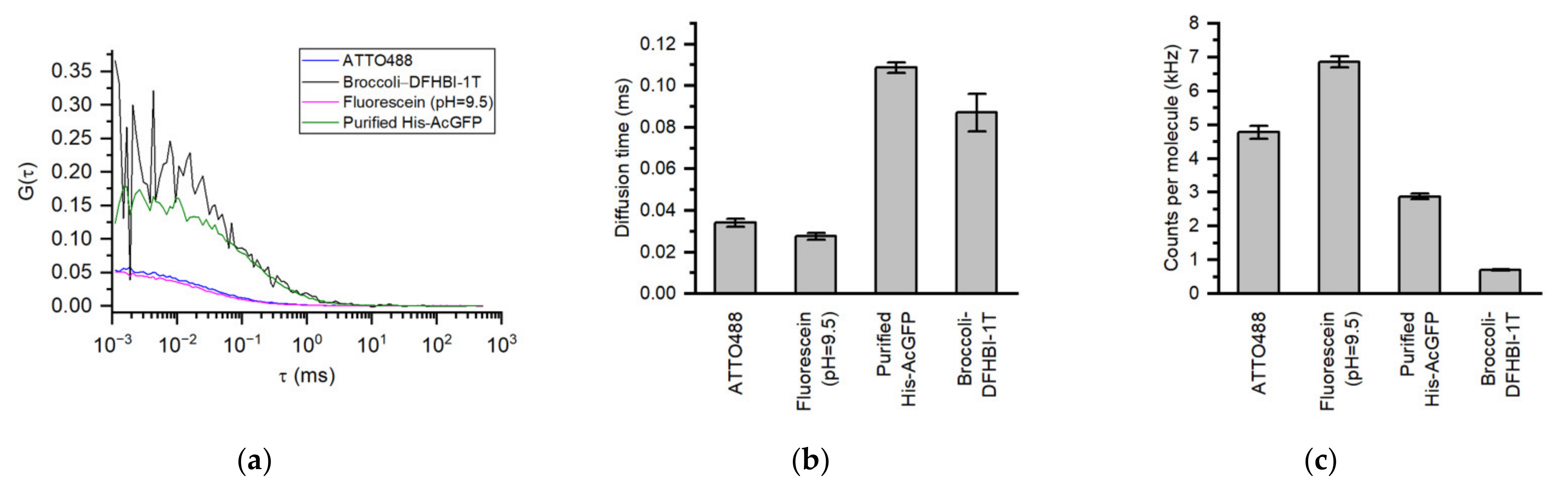
3. Results and Discussion

To determine whether FLAPs can be applied to fluorescence correlation spectroscopy measurements, we performed the FCS measurements of Broccoli–DFHBI-1T as a model with various laser powers. Clear autocorrelation curves were obtained at a relatively high laser power (Figure 1a), and the model equation for one-component diffusion fitted the curves well. At a moderate laser power, the fluorescence signal decreased within 60 s, after which the signal plateaued at two-thirds of the initial value and was not completely quenched (Figure 1b), thus indicating that the decrease in fluorescence was due to a reversible reaction rather than irreversible photobleaching. DFHBI, an analog of DFHBI-1T, undergoes cis-to-trans photoisomerization upon excitation and attenuates its fluorescence [18,19]. Since trans-DFHBI reverts to cis-DFHBI because of thermal relaxation, the fluorescence reaches a plateau at equilibrium during reversible photoisomerization. Therefore, we hypothesized that reversible photoisomerization occurs in Broccoli–DFHBI-1T. To verify this hypothesis, a second measurement was carried out with the same sample, 5 min after first measurement. The signal completely recovered to the initial value of the first measurement, and a similar decay was then observed. The results are consistent with previous reports, and indicate that DFHBI-1T is involved in reversible cistrans photoisomerization. Moreover, Broccoli–DFHBI-1T had sufficient brightness to be applied to FCS to carry out single-molecule-level analysis.
Next, we monitored the number of fluorescent molecules detected by forming a complex of Broccoli and DFHBI-1T. The FCS measurements were carried out using a mixture of Broccoli–DFHBI-1T at different concentrations (Figure 2). Figure 2a shows the average photon count rate of the FCS measurements. The count rate increased depending on the DFHBI-1T concentration and the Broccoli concentration in the samples. The molar concentration of the fluorescent Broccoli–DFHBI-1T complex was then calculated from the molecular number detected in the effective confocal volume (Figure 2b). The effective confocal volume was estimated to be 0.40 fL via the diffusion measurement of the ATTO488 reference solution. FCS can monitor single-molecule brightness as counts per molecule (CPM) (Figure 2c). The CPM showed a plateau value when the concentration of DFHBI-1T was markedly higher than that of Broccoli. This plateau CPM value indicates that the FCS measurements in this concentration range are stable, and that the determined concentration values are reliable. The concentration of the fluorescent Broccoli–DFHBI-1T complex estimated by FCS was 10% to 20% of the prepared Broccoli concentration. Since the Kd value of Broccoli–DFHBI-1T was proposed to be 360 nM [1], the observed difference in concentration cannot be explained only by the degree of Broccoli–DFHBI-1T binding. This difference can be attributed to imperfect folding and the fluctuation of the folding state of Broccoli RNA molecules.
Although the results of FCS measurements (Figure 2) indicated that the actual fraction of the Broccoli–DFHBI-1T complex is less than that estimated from its Kd value and the concentrations of each component, there was no direct evidence. Thus, we attempted FCCS measurements to directly monitor and quantify the molecular interactions between Broccoli and DFHBI-1T. ATTO647N-labeled Broccoli RNA (1 µM) was chemically synthesized and measured with DFHBI-1T (1 µM or 10 µM) using FCCS. The autocorrelation curves in the green and red channels and cross-correlation curves between them were successfully obtained (Figure 3a,b). The concentrations of total RNA (red channel, Cred), fluorescent Broccoli–DFHBI-1T (green channel, Cgreen), and the fraction of the complex that fluoresced green and red (cross-correlation, Ccomplex) were determined. Table 1 shows the Ccomplex, Cgreen, Cred, Kd, and the folding efficiency, derived from the RCA values with the standard deviation (n = 3). In the case of Broccoli–DFHBI-1T, DFHBI-1T fluoresced only when making a complex with Broccoli; thus, Cgreen and Ccomplex show similar values. This result indicates that the labeling efficiency of ATTO647N-Broccoli was approximately 100%. The estimated concentrations of RNA from the FCS/FCCS were lower than the prepared concentrations. This is possibly due to the difference in the quantification method and adsorption to the tubes and glass surface. Using actual FCCS concentrations and the prepared DFHBI-1T concentration (1 µM), the apparent Kd between Broccoli and DFHBI-1T was calculated as 6472 nM. However, this is different from the reported Kd value (360 nM) [1]. We consider that this difference in Kd values is due to the folding efficiency of Broccoli. Since the total RNA was used for the calculation, the presence of misfolded RNAs that cannot induce DFHBI-1T fluorescence can directly affect the obtained Kd value (Figure 3c). Assuming that the Kd value is 360 nM, as reported previously, the estimated Ccomplex in our measurement condition can be expected to be 170 nM. By comparing it with the measured Ccomplex (37.9 nM) in our FCCS measurement, we calculated the folding efficiency of Broccoli RNAs as 22.2%. A similar folding efficiency (24.1%) was obtained when DFHBI-1T was 10 μM. Filonov et al. [1] reported that the folding efficiency of Broccoli without the tRNA scaffold was 40–50%; our measured value was lower, probably due to differences in the pre- and post-Broccoli sequences. The gap between the pre-measured and actual concentrations of Broccoli RNA in samples would be another reason. Filonov et al. calculated the folding efficiency of Broccoli using the pre-measured concentration. However, the actual RNA concentration in a sample should be vary from the pre-measured one because RNA is easily adsorbed on tubes and chips, as we observed in this study (Table 1, Cred). Thus, we directly measured the total concentration of Broccoli RNA in a sample using ATTO647N dye fused to the RNA. As the folding efficiency is calculated based on the total RNA concentration, the difference in the concentration measurement can directly lead to the different values. We believe that our study provides valuable information in the structural study of Broccoli RNA in solution.
This study directly measured total Broccoli RNA labeled with ATTO647N and Broccoli–DFHBI-1T complexes that fluoresced simultaneously. In bulk analysis, the actual concentration of target molecules in the system is difficult to determine. In contrast, the absolute molecular number can be obtained, and the value can be converted into concentrations in FCS/FCCS. This is the advantage of the FCS/FCCS technique for investigating concentration-dependent phenomena, such as molecular interactions. Therefore, it was used to evaluate the interaction of a typical FLAP (Broccoli–DFHBI-1T) and understand its folding efficiency.
Next, we compared the brightness and size of Broccoli–DFHBI-1T to conventional fluorescent molecules. Fluorescein (pH 9.5) and purified 6xhis-AcGFP solutions were prepared and measured under the same conditions as the Broccoli–DFHBI-1T measurement (Figure 4a). The obtained diffusion time of Broccoli–DFHBI-1T was slightly smaller than that for 6xhis-AcGFP (Figure 4b). This result indicated that the size of Broccoli–DFHBI-1T was approximately 3 nm. The obtained CPM values show that the brightness of AcGFP was four times that of Broccoli–DFHBI-1T under our measurement conditions (excitation: 488 nm, emission: 500–750 nm) (Figure 3c). However, the result is not consistent with a previous report, where the reported quantum yield (Qy = 0.41) of Broccoli–DFHBI-1T [20] was half of that of AcGFP (Qy = 0.82), and the molar extinction coefficient (ε = 28,900) of Broccoli–DFHBI-1T [20] was similar to that of AcGFP (ε = 32,500). This may be due to the excitation efficiency at 488 nm. Furthermore, the folding state of Broccoli fluctuates dynamically, and the fluorescence emission rate of a single molecule is unstable, depending on the transient folding state. If the Broccoli RNA molecule and Broccoli–DFHBI-1T complex does not form an appropriate structure, the complex cannot brightly fluoresce, which affects the single-molecule measurement of the FCS.

4. Conclusions

This study demonstrated the advantages of FCS/FCCS techniques in investigating the properties of a FLAP and its fluorogen. In addition, we suggested the importance of using single-molecule measurements to evaluate the folding state of FLAPs.
We suggest that FCS/FCCS can be used to compare the performance of various FLAP–fluorogen pairs, including Mango–TO1-Biotin [21,22], Corn–DFHO [23], SiRA–SiR [24], Pepper–HBC [25], and RhoBAST–TMR-DN [26], and to optimize buffer conditions. Using turn-on FLAP probes, such as BRD probes [27], which respond to the RNA of interest, FCS/FCCS enables the specific detection and quantification of host endogenous RNAs and viral RNAs.
The FCS/FCCS technique should be applicable to the measurement of FLAPs expressed in living cells. The interactions between intracellular RNAs (e.g., mRNA and ncRNA) and proteins can be quantified in living cells using a combination of FLAPs and fluorescent proteins. The combination of FLAPs and the FCS technique should open a new avenue in the field of intracellular signal transduction and regulation.

Author Contributions

Conceptualization, A.S. and Y.F.; Methodology, A.S. and Y.F.; Validation, Y.F. and A.S.; Analysis, A.S.; Investigation, A.S. and Y.F.; Resources, Y.F.; Writing—Original Draft Preparation, A.S. and Y.F.; Writing—Review and Editing, Y.F. and A.S.; Visualization, Y.F. and A.S.; Supervision, A.S.; Project Administration, A.S.; Funding Acquisition, A.S. and Y.F. All authors have read and agreed to the published version of the manuscript.

Funding

This research was partly funded by the Japan Society for the Promotion of Science (JSPS), grant numbers 19K06590 and 19K15720, and the competitive operational subsidy of the Biomedical Research Institute, AIST.

Informed Consent Statement

Not applicable.

Data Availability Statement

Not applicable.

Conflicts of Interest

The authors declare no conflict of interest.

References

  1. Filonov, G.S.; Moon, J.D.; Svensen, N.; Jaffrey, S.R. Broccoli: Rapid selection of an RNA mimic of green fluorescent protein by fluorescence-based selection and directed evolution. J. Am. Chem. Soc. 2014, 136, 16299–16308. [Google Scholar] [CrossRef] [PubMed] [Green Version]
  2. Truong, L.; Ferré-D’Amaré, A.R. From fluorescent proteins to fluorogenic RNAs: Tools for imaging cellular macromolecules. Protein Sci. 2019, 28, 1374–1386. [Google Scholar] [CrossRef] [PubMed]
  3. Neubacher, S.; Hennig, S. RNA Structure and Cellular Applications of Fluorescent Light-Up Aptamers. Angew. Chem. Int. Ed. Engl. 2019, 58, 1266–1279. [Google Scholar] [CrossRef] [PubMed]
  4. Elson, E.L.; Magde, D. Fluorescence correlation spectroscopy. I. Conceptual basis and theory. Biopolymers 1974, 13, 1–27. [Google Scholar] [CrossRef]
  5. Magde, D.; Elson, E.L.; Webb, W.W. Fluorescence correlation spectroscopy. II. An experimental realization. Biopolymers 1974, 13, 29–61. [Google Scholar] [CrossRef]
  6. Rigler, R.; Mets, U.; Widengren, J.; Kask, P. Fluorescence Correlation Spectroscopy with High Count Rate and Low-Background—Analysis of Translational Diffusion. Eur. Biophys. J. Biophys. Lett. 1993, 22, 169–175. [Google Scholar] [CrossRef]
  7. Mateu-Regué, À.; Christiansen, J.; Bagger, F.O.; Hellriegel, C.; Nielsen, F.C. Unveiling mRNP composition by fluorescence correlation and cross-correlation spectroscopy using cell lysates. Nucleic Acids Res. 2021, 49, e119. [Google Scholar] [CrossRef]
  8. Imamura, H.; Sasaki, A.; Honda, S. Fate of a Stressed Therapeutic Antibody Tracked by Fluorescence Correlation Spectroscopy: Folded Monomers Survive Aggregation. J. Phys. Chem. B 2017, 121, 8085–8093. [Google Scholar] [CrossRef]
  9. Sasaki, A.; Yamamoto, J.; Kinjo, M.; Noda, N. Absolute Quantification of RNA Molecules Using Fluorescence Correlation Spectroscopy with Certified Reference Materials. Anal. Chem. 2018, 90, 10865–10871. [Google Scholar] [CrossRef] [Green Version]
  10. Kinjo, M.; Rigler, R. Ultrasensitive Hybridization Analysis Using Fluorescence Correlation Spectroscopy. Nucleic Acids Res. 1995, 23, 1795–1799. [Google Scholar] [CrossRef]
  11. Rigler, R.; Földes-Papp, Z.; Meyer-Almes, F.-J.; Sammet, C.; Völcker, M.; Schnetz, A. Fluorescence cross-correlation: A new concept for polymerase chain reaction. J. Biotechnol. 1998, 63, 97–109. [Google Scholar] [CrossRef]
  12. Bacia, K.; Kim, S.A.; Schwille, P. Fluorescence cross-correlation spectroscopy in living cells. Nat. Methods 2006, 3, 83–89. [Google Scholar] [CrossRef]
  13. Tiwari, M.; Mikuni, S.; Muto, H.; Kinjo, M. Determination of dissociation constant of the NFκB p50/p65 heterodimer using fluorescence cross-correlation spectroscopy in the living cell. Biochem. Biophys. Res. Commun. 2013, 436, 430–435. [Google Scholar] [CrossRef]
  14. Sadaie, W.; Harada, Y.; Matsuda, M.; Aoki, K. Quantitative in vivo fluorescence cross-correlation analyses highlight the importance of competitive effects in the regulation of protein-protein interactions. Mol. Cell Biol. 2014, 34, 3272–3290. [Google Scholar] [CrossRef] [Green Version]
  15. Sasaki, A.; Kinjo, M. Monitoring intracellular degradation of exogenous DNA using diffusion properties. J. Control Release 2010, 143, 104–111. [Google Scholar] [CrossRef]
  16. Dertinger, T.; Pacheco, V.; von der Hocht, I.; Hartmann, R.; Gregor, I.; Enderlein, J. Two-focus fluorescence correlation spectroscopy: A new tool for accurate and absolute diffusion measurements. ChemPhysChem 2007, 8, 433–443. [Google Scholar] [CrossRef]
  17. Culbertson, C.T.; Jacobson, S.C.; Michael Ramsey, J. Diffusion coefficient measurements in microfluidic devices. Talanta 2002, 56, 365–373. [Google Scholar] [CrossRef]
  18. Wang, P.; Querard, J.; Maurin, S.; Nath, S.; Le Saux, T.; Gautier, A.; Jullien, L. Photochemical properties of Spinach and its use in selective imaging. Chem. Sci. 2013, 4, 2865–2873. [Google Scholar] [CrossRef]
  19. Han, K.Y.; Leslie, B.J.; Fei, J.; Zhang, J.; Ha, T. Understanding the photophysics of the spinach-DFHBI RNA aptamer-fluorogen complex to improve live-cell RNA imaging. J. Am. Chem. Soc. 2013, 135, 19033–19038. [Google Scholar] [CrossRef] [Green Version]
  20. Li, X.; Wu, J.; Jaffrey, S.R. Engineering Fluorophore Recycling in a Fluorogenic RNA Aptamer. Angew. Chem. Int. Ed. Engl. 2021, 60, 24153–24161. [Google Scholar] [CrossRef]
  21. Dolgosheina, E.V.; Jeng, S.C.; Panchapakesan, S.S.; Cojocaru, R.; Chen, P.S.; Wilson, P.D.; Hawkins, N.; Wiggins, P.A.; Unrau, P.J. RNA mango aptamer-fluorophore: A bright, high-affinity complex for RNA labeling and tracking. ACS Chem. Biol. 2014, 9, 2412–2420. [Google Scholar] [CrossRef]
  22. Autour, A.; Jeng, S.Y.C.; Cawte, A.D.; Abdolahzadeh, A.; Galli, A.; Panchapakesan, S.S.S.; Rueda, D.; Ryckelynck, M.; Unrau, P.J. Fluorogenic RNA Mango aptamers for imaging small non-coding RNAs in mammalian cells. Nat. Commun. 2018, 9, 656. [Google Scholar] [CrossRef]
  23. Song, W.; Filonov, G.S.; Kim, H.; Hirsch, M.; Li, X.; Moon, J.D.; Jaffrey, S.R. Imaging RNA polymerase III transcription using a photostable RNA-fluorophore complex. Nat. Chem. Biol. 2017, 13, 1187–1194. [Google Scholar] [CrossRef] [PubMed] [Green Version]
  24. Wirth, R.; Gao, P.; Nienhaus, G.U.; Sunbul, M.; Jäschke, A. SiRA: A Silicon Rhodamine-Binding Aptamer for Live-Cell Super-Resolution RNA Imaging. J. Am. Chem. Soc. 2019, 141, 7562–7571. [Google Scholar] [CrossRef] [PubMed]
  25. Chen, X.; Zhang, D.; Su, N.; Bao, B.; Xie, X.; Zuo, F.; Yang, L.; Wang, H.; Jiang, L.; Lin, Q.; et al. Visualizing RNA dynamics in live cells with bright and stable fluorescent RNAs. Nat. Biotechnol. 2019, 37, 1287–1293. [Google Scholar] [CrossRef] [PubMed]
  26. Sunbul, M.; Lackner, J.; Martin, A.; Englert, D.; Hacene, B.; Grün, F.; Nienhaus, K.; Nienhaus, G.U.; Jäschke, A. Super-resolution RNA imaging using a rhodamine-binding aptamer with fast exchange kinetics. Nat. Biotechnol. 2021, 39, 686–690. [Google Scholar] [CrossRef] [PubMed]
  27. Furuhata, Y.; Kobayashi, M.; Maruyama, R.; Sato, Y.; Makino, K.; Michiue, T.; Yui, H.; Nishizawa, S.; Yoshimoto, K. Programmable RNA detection with a fluorescent RNA aptamer using optimized three-way junction formation. RNA 2019, 25, 590–599. [Google Scholar] [CrossRef]
Figure 1. Fluorescence correlation spectroscopy (FCS) measurements of Broccoli–DFHBI-1T (1 µM each). (a) Autocorrelation curves of Broccoli–DFHBI-1T with various laser powers. The autocorrelation curves were obtained after the fluorescence signal plateaued. (b) Fluorescence intensity profiles during two sequential FCS measurements (60 s, thrice each) of the same sample. Excitation laser power was 15 µW.
Applsci 12 02002 g001
Figure 2. Fitting analysis of Broccoli–DFHBI-1T for different concentrations of Broccoli and DFHBI-1T. (a) Count rate. (b) FCS concentration calculated from the number particles in the measurement volume. (c) Counts per molecule (CPM). Red line, 1 µM Broccoli; blue line, 2 µM Broccoli; green line, 5 µM Broccoli; black line, no RNA control measurement (only in (a)). Error bars show standard deviation in fitting analysis (derived via Bootstrap method).
Applsci 12 02002 g002
Figure 3. Direct measurement of the interaction between ATTO647N-labeled Broccoli RNA and DFHBI-1T using fluorescence cross-correlation spectroscopy (FCCS). Autocorrelation curves of green channel (Broccoli–DFHBI-1T fluorescence, excitation: 488 nm, 80 µW) and red channel (ATTO647N fluorescence, excitation: 633 nm, 2 µW), and cross-correlation curve between two channels with the 10 µM (a) and 1 µM (b) concentration of DFHBI-1T. (c) Diagram of possible states of ATTO647N-labeled Broccoli RNA and DFHBI-1T in the measurement system.
Applsci 12 02002 g003
Figure 4. FCS measurements of Broccoli–DFHBI-1T, His-tagged recombinant AcGFP, ATTO488, and fluorescein with the same measurement conditions. (a) Autocorrelation curves of Broccoli–DFHBI-1T (black), His-tagged recombinant AcGFP (green), ATTO488 (blue), and fluorescein (magenta). (b) Comparison of diffusion times of the samples. (c) Comparison of molecular brightness (CPM) of the samples. The diffusion time and CPM values are shown as mean ± standard deviation (n = 3).
Applsci 12 02002 g004
Table 1. Relative cross-correlation amplitude (RCA), measured concentrations, apparent Kd in the FCCS measurement, and estimated folding efficiency of Broccoli molecules. * Values when Broccoli’s folding efficiency is not considered.
RCA Gc(0)/Gr(0)RCA Gc(0)/Gg(0)Cgreen
(nM)
Cred
(nM)
Ccomplex
(nM)
Apparent Kd
from FCCS
(nM)
Estimated Ccomplex
Assuming
Kd = 360 nM (nM)
Folding Efficiency
(Ccomplex/Estimated Ccomplex)
Broccoli-ATTO647N (1 µM)
/DFHBI-1T (10 µM)
0.987
± 0.041
0.198
± 0.011
75.3
± 0.0
260.5
± 5.1
60.6
± 2.5
42,961 *251.30.241
Broccoli-ATTO647N (1 µM)
/DFHBI-1T (1 µM)
0.923
± 0.053
0.137
± 0.009
50.2
± 2.7
245.0
± 8.3
37.9
± 3.6
6472 *170.80.222
Publisher’s Note: MDPI stays neutral with regard to jurisdictional claims in published maps and institutional affiliations.

