Article

Influence of 2% Chlorhexidine on the Bond Strength of Three Adhesive Systems on Primary Molars: An In Vitro Study

by
Laura Velayos-Galán
1,*,
Pedro Molinero-Mourelle
2,3,
Pablo Sevilla
2,
Manrique Fonseca
3,
María Rosa Mourelle-Martínez
1 and
Vicente Vera-González
2
1
Department of Dental Clinical Specialties, School of Dentistry, Complutense University of Madrid, 28040 Madrid, Spain
2
Department of Conservative Dentistry and Buccofacial Prostheses, School of Dentistry, Complutense University of Madrid, 28040 Madrid, Spain
3
Department of Reconstructive Dentistry and Gerodontology, School of Dental Medicine, University of Bern, 3010 Bern, Switzerland
*
Author to whom correspondence should be addressed.
Appl. Sci. 2022, 12(6), 2964; https://doi.org/10.3390/app12062964
Submission received: 17 February 2022 / Revised: 9 March 2022 / Accepted: 9 March 2022 / Published: 14 March 2022

Abstract

:
The hydrolysis of the collagen matrix by metalloproteinases (MMPs) is one of the paradigms that currently arouses most interest due to its close relationship with a decrease in bond strength (BS) and consequent restoration failure. Chlorhexidine 2% has demonstrated its ability to inhibit MMPs’ activity in the permanent dentition, improving the duration of resin–dentine, but there are few studies on deciduous dentition and its possible repercussions. Aim: To determine the influence of 2% chlorhexidine digluconate (CHX) as a dentine pretreatment on the BS of three adhesive systems on primary molars. Methods: 128 primary extracted molars were assigned to eight groups at random. BS in vitro was recorded by micropush-out test, and analyzed by two-way ANOVA. Results: BS values oscillated from 15.01 MPa to 20.41 MPa. There was no statistically relevant variation between the BS total mean of those adhesive groups that had received CHX pretreatment versus those that did not. Adper Prompt L-Pop was the self-etching adhesive with the best BS. Adper Scotchbond 1XT was the total-etch adhesive with the best BS values. Conclusions: Application of 2% chlorhexidine for 60 s as dentine pretreatment did not affect the immediate BS of several adhesive systems used in primary dentition.

1. Introduction

Direct restorations as fillings in the primary dentition are the main restorative procedure in children’s daily practice [1,2]. Successful care of the child is not only essential to complete treatments, but also to establish a favorable basis for the patient’s future dental care acceptance throughout life; however, it is a constant challenge to the skill and experience of the practitioner, and the current trend is to simplify operative procedures, thus shortening treatment times is the aim [2].
New dental adhesives, which decrease the clinical steps, are becoming the strategy of choice in pediatric restorative dentistry [3,4]. Despite great advances in adhesive technology in the last few decades, one of the unresolved dilemmas is the deterioration of the resin/dentin union with time, leading to a very short clinical longevity of composite resins [3,4], a problem caused by the activity of the metalloproteinases (MMPs) of the fundamental substance our organism.
Adhesion to dentine substrate is significantly more complicated than bonding to enamel [5,6]. Dentine contains a higher moisture content and a lower inorganic content, properties that make durable bonding difficult [5,6]. Additional relevant factors are the chemical and microstructural characteristics of the substrates of the deciduous and permanent dentition [5,6]. The hybrid layers created on primary teeth are thicker than those obtained on permanent teeth with identical etching time [6]. This suggests that the dentine in deciduous teeth is more acid-etch reactive because of its reduced mineral content [6]. The incomplete infiltration of the resin monomers into previously demineralized dentine would expose the underlying collagen fibrils, which are subject to hydrolytic breakdown, leading to hybrid layer softening and possible reduction of bond strength, which could be more pronounced in temporary dentine [5,6].
Dentine comprises collagenolytic enzymes, matrix MMPs and cysteine cathepsins [5]. MMPs are a set of zinc- and calcium-dependent enzymes which control natural and pathological processes of collagen-based structures [7]. Various studies have shown that endogenous metalloproteinases, such as collagenases (MMP-8) and gelatinases (MMP-2 and -9), have a role in the development of decay and in altering the hybrid layer that forms in the bonding process [8,9,10]. Due to the dentine mineralization process in the dentinogenesis stage, they are stored latently in the extracellular matrix, but may be reactivated by demineralization of the dentine [5]. As the inorganic structure around the collagen fibrils in dentine is removed by decay and/or acid etching, the collagen fibrils are uncovered and this manifests the MMPs’ activity, initiating the breakdown process [7]. Similar to an enzymatic pathway, the abnormal execution of MMPs might be stopped by the utilization of specific inhibitors, preserving the collagen fibers’ structural integrity, and potentially slowing down the breakdown of the hybrid layer [7].
Nowadays, the approaches to enhance the longevity of resin–dentin bonds, and to prevent restoration fails, are based on the capacity to suppress the activity of endogenous enzymes in dentine [11,12]. Several synthetic inhibitors of MMPs such as EDTA, benzalkonium chloride and chlorhexidine have been proposed. Among others, 2% chlorhexidine (CHX) is a broad-spectrum antiseptic and antimicrobial that bonds to amino acids in dentin and continuously inhibits bacteria over many hours [13]. In addition to its known antiseptic and antimicrobial action, CHX also acts on dentin as a potent inhibitor of MMPs [13]. Thus, a 2% concentration of chlorhexidine digluconate, used as a dentine pretreatment, can inhibit 99% of the collagenolytic activity of metalloproteinases [14,15,16]. CHX efficiently inhibits MMP-2, -9 and -8, and cysteine cathepsins, and could be used as a supplementary approach to rehydrate dentin to maintain the moisture needed to keep the collagen network reactive, allowing for greater hybrid layer integrity over time and improvement in the durability of polymeric restorations [5,11,12]. However, chlorhexidine pretreatment can become a challenge as it may interfere with the dentin bonding process. While numerous studies have documented the impact of CHX on bond strength in permanent teeth, data on its effect in primary dentition are lacking [2,17,18,19]. In view of the flexible application of CHX in conservative dentistry, it is appropriate to research the effect of the application of 2% CHX on the bond strength in deciduous teeth [5].
Based on the null hypothesis that prior use of CHX 2% during the adhesive process may not interfere with the bond strength obtained by the different adhesive systems in the primary dentition, it is necessary to evaluate the use of this inhibitor of the collagenolytic activity of metalloproteinases, to establish the most suitable adhesive protocol for paediatric dentistry clinical practice. The purpose of the current research was to compare the micropush-out BS of different adhesive systems in the primary dentition, with and without the prior application of 2% CHX.

2. Materials and Methods

2.1. Study Design

One hundred and twenty-eight removed human maxillary and mandibular first and second primary molars, not decayed and stored for less than six months according to UNE-EN ISO 3696:1996 [20], were recorded after informed consent was received from the patients‘ parents.
The trial was carried out in compliance with national regulatory guidelines, following the ethical requirements for the development of medical research on human subjects as mentioned in the Declaration of Helsinki [21] and with the approval of the Clinic San Carlos Hospital Ethics Committee of Madrid, Spain (registration code of the study: CI 20/147-E).

2.2. Specimens Preparation

Following the European Standard for Adhesion in Dentistry UNE-EN ISO 29022:2013 [22] for the better handling of molars, specimens and the subsequent removal of occlusal enamel were achieved by a two-step sequential flattening process under running water, using silicon carbide abrasive papers P120 and P400, and a polishing machine Struers® Dap-7 (Struers® GmbH, Willich, Germany). On the exposed dentine surfaces, the same calibrated operator (L.V.-G.) performed Black’s class I cavity preparations (1.6 mm diameter and 3.5 mm depth); dimensions were verified with dental digital caliper (Mestra®, Sondika, Spain) and millimeter probe (Hu-Friedy®, Frankfort, Germany), respectively, with long-stemmed tungsten carbide round bur H1.316.014 (Komet® Dental, Lemgo, Germany) at high speed with continuous water cooling. The prepared specimens were maintained in laboratory water quality 3 at room temperature 20 ± 0.5 °C, and with a relative humidity 31 ± 5%, and were used in the bonding process within 4 h [22].

2.3. Specimen Treatments

The molars were randomly, based on a table of random numbers (Microsoft® Excel® 2010, version 2010 14.0, Redmond, MS, USA), and equally assigned to eight treatment groups (n = 16), after which the corresponding adhesive processes were performed.
The specimens were embedded in a dental self-curing acrylic (Tab 2000™, Kerr Dental®, Detroit, MI, USA) block (25 × 10 mm) prior to treatment. Specifications of the groups and adhesive processes can be found in Table 1, while the materials employed are described in Table 2. The different adhesives were treated as instructed by the manufacturer.
On the required groups, acid etching was performed by applying 35% orthophosphoric acid for 7 s, and then washing with plenty of water and subsequent drying (without desiccation) with air spray. Volumes of 1.5 µL of a 2% water-based CHX were brushed on the corresponding groups for 60 s and these were dried gently with a sterile cotton ball. The fillings were performed by the same operator by using bulk fill nano-hybrid composite with a single increment. Subsequent light curing was performed for 10 s with a 1470 mW/cm2 light intensity (Elipar™ Deep Cure LED Curing Light, 3M ESPE, St Paul, MN, USA). In accordance with ISO 29022:2013, molars were preserved in water at a temperature of 37 ± 2 °C for 24 ± 2 h before sectioning.

2.4. Bond Strength Evaluation

From each specimen, a resin–dentine disc with a cross section about 1 mm thick with respect to its lengthwise axis was created with a low-speed saw (Exact® Cutting Unit 400C, Exact Tools Oy, Helsinki, Finland) with water irrigation. A mark indicating the coronal-oriented area was made on each disc.
Each disc was fixed to the bottom of the micropush-out device by placing the restoration in the hole of the plate. A cylindrical punch (0.8 mm diameter) made of steel from a universal testing machine (Hounsfield test equipment® HTE, Croydon, UK), exerted force in the crown-apical direction until failure load (expressed in newtons (N)) was reached, with a speed of 0.5 mm/min [23]. Failure load values obtained were registered using the advanced testing software Metrotest (TechLab Systems, Itasca, IL, USA): software for control and management of the data in a single piece of test equipment. The statistical trend package for Metrotest allows representation of the results of tests in the form of an XY graph.
The BS was calculated in MPa: failure load (N)/bonding interface area (mm2). The bonding interface area was computed considering the following mathematical formula: A = 2πrh [24].

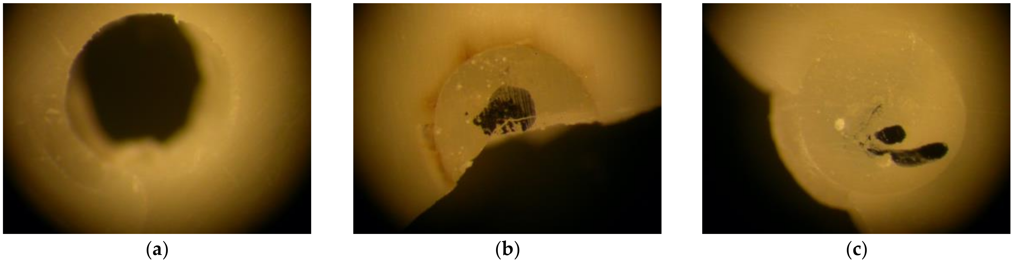
2.5. Failure Type Evaluation

The adhesive failure types were assessed at 9.2× magnification, using a Stereomicroscope Leica® MZ12 (Leica® microsystems GmbH, Wetzlar, DE), by an independent and calibrated examiner (P.S.) and catalogued as cohesive (C), adhesive (A) or mixed (M) failures, as shown in Figure 1.
The training session included a theoretical explanation, followed by a discussion of a series of images of the different types of adhesive failure.

2.6. Statistical Analysis

Mean values and standard deviations (SD) were calculated per group. The Kolmogorov–Smirnov test was chosen to check the normal pattern of the data. Subsequently, and after confirmation of the normality of the variables, the data analysis was carried out by means of a parametric test. The study of the variable resistance to adhesion was carried out using two-way ANOVA and subsequent pairwise comparison performed using Tukey´s post hoc test, while the chi-square test was chosen for the study of the mode of failure.
The statistical analysis was conducted with IBM® SPSS® Statistics 25 (SPSS Inc., Chicago, IL, USA) software. The level of significance was set at α = 0.005.

3. Results

The distribution was normal and homogeneous for the adhesion strength data. The total mean value is 17.99 MPa (95% CI: 16.82–19.17 MPa) with a standard deviation of ±6.72 MPa.
The normality of the distribution of the variables was accepted, and the two-way ANOVA test showed the absence of a statistically significant difference, with p > 0.05 (p-value = 0.716), between the total means of the pretreatment groups with and without CHX; these were very similar to each other, as shown in Table 3 and in Figure 2. The effect size R2 = 0.001 also showed no indication of a possible relationship that would justify the use of CHX as a differential factor affecting BS.
There were statistically significant differences p < 0.05 (p-value = 0.028), with a moderate effect size R2 = 0.073, between the different types of adhesives. As a result, the mean BS value is higher in the Adper Prompt L-pop groups, 20.27 ± 5.89 MPa, as shown in Table 4, although the Tukey’s post hoc paired t-test determines that this difference is not significant, p > 0.05 (p-value = 0.921).
As for the failure type, the results did not demonstrate statistical significance, with p > 0.05. However, a moderate effect size (R2 = 0.068) indicated differences in failure types by group. More adhesive failures were observed in the PL (43.8%) and PL-C (37.5%) groups. Cohesive failures were more abundant in the S-C (56.3%) and Sa (62.5%) groups. Mixed failures were more evenly distributed in all groups, as shown in Table 5.

4. Discussion

This study demonstrated that there was no statistically significant difference between the overall mean bond strength recorded in the CHX-applied groups versus the non-CHX-applied groups. Adper Prompt L-Pop self-etch adhesive and Adper Scotchbond 1XT total-etch adhesive were the best performers both when applied without CHX, giving mean BS values of 20.41 ± 7.05 MPa and 19.85 ± 7.47 MPa, respectively, and when applied with CHX, giving mean BS values of 20.13 ± 4.69 MPa and 18.59 ± 5.13 MPa, respectively. Therefore, the null hypotheses was accepted.
There are very few studies carried out using the novel adhesion test proposed in this study; only a single study carried out in 2021 by Del Rio and collaborators [23] coincides in the analysis of adhesion strength with this same resistance test, though it was disparate in terms of the non-use of CHX during the adhesive process in deciduous dentition and the higher percentage of orthophosphoric acid used in acid etching. If we compare the data provided by these authors [23] for universal adhesives with self-etch strategies, with a value of 13.66 ± 2.81 MPa, versus the values of our study for this same category of adhesive of 17.28 ± 9.48 MPa when applied without CHX pretreatment and 16.15 ± 8.53 MPa on average both when CHX was applied and when CHX was not applied, we do not observe large discrepancies; similarity also in the data with respect to universal adhesives with total-etch strategies with the value of 14.98 ± 3.96 MPa similar to 15.28 ± 4.45 MPa when applied without CHX pretreatment and 16.34 ± 4.90 MPa on average both when CHX was applied and when not applied, provided by the present study. Regarding self-etching adhesives, we also show the similarity of our results with respect to the study carried out in 2021, which provided a BS value of 14.48 ± 2.89 MPa, consistent with 20.41 ± 7.05 MPa when this type of adhesive was applied without CHX and 20.27 ± 5.89 MPa on average both when CHX was applied and when it was not applied, in our study.
If the exploration of the data is extended to other types of studies, despite the use of different adhesion tests, but coinciding in the inclusion of CHX in the adhesive process, data that show similarity with the same study groups in our research, are those provided by Vieira and collaborators in 2003 [19], which ranged between 19.88 ± 1.04 MPa and 17.99 ± 1.15 MPa in their analysis of universal total-etch adhesion strategies with the incorporation of CHX. Similar findings have been reported in other studies, such as that of Abdelmegid et al. in 2018 [25], with values ranging from 14.13 ± 2.09 MPa to 16.45 ± 2.41 MPa in universal self-etching adhesives.
The current study showed that CHX use during the bonding process in primary dentition did not compromise the immediate BS. The results of this analysis confirm the results of other studies that evaluated dentin rehydration with 2% CHX following the bonding process [26,27,28,29]. However, other studies have reported that after an ageing period equivalent to 6 months the application of CHX 2% as a pretreatment increased the bond strength with an application time of 30 or 60 s [7,30]. Earlier in vitro and in vivo research on permanent and deciduous teeth demonstrated that the administration of chlorhexidine at levels from 0.12% to 2% did not produce detrimental effects on the immediate BS of the dentine substrate and the polymeric material [19,26]. Regarding the application and drying of CHX, no differences have been found between the various methods, the most common being microbrush application and drying with absorbent paper.
On the other hand, the current study showed significant differences between the bond strengths of the different adhesives used, regardless of whether CHX was used or not as a pretreatment. Thus, the Scotchbond Universal self-etch (16.15 ± 8.53 MPa) and Scotchbond Universal total-etch (16.34 ± 4.90 MPa) groups showed lower BS values relative to the Adper Scotchbond 1XT (19.22 ± 6.34 MPa) and Adper Prompt L-Pop (20.27 ± 5.89 MPa) groups. Moreover, the present study showed that the BS differed according to the type of adhesive used. Previous studies showed that etch and rise adhesive systems achieved higher BS values than self-etch adhesives [31,32,33]. Self-etching systems benefit most from chlorhexidine application. Demineralized (etched) dentine is almost 80% more effective than mineralized (non-etched) dentine in terms of this application of chlorhexidine. Through acid etching, the peritubular dentine is lost, the dentinal tubules are enlarged, and at the same time their water content increases, and this will allow the incorporation of the chlorhexidine within the dentine matrix [9,31,34,35].
Currently, one of the biggest goals of adhesive dentistry is to improve the ageing resistance of the adhesive interface, making restorations more clinically durable [36].
The durability of the adhesive interface is strongly linked to the hybrid layer quality, that is, the entire exposed collagen and the quality of the resin coat created [7,31]. The conservation of the correct state of the hybrid layer, through the incorporation of CHX to maintain its balance, is a key point in slowing down the loss of BS [31].
CHX has been researched as an important inhibitor of MMPs and shows no negative impact on the BS between resin and dentine [7]. Chlorhexidine 2% is the concentration that has been researched the most and had the strongest inhibitory impact on MMPs [12,31], which is related to the mechanism of CHX cation chelation [7] and calcium sequestration [26]. In addition, its high substantivity thanks to the delivery of molecules with a positive charge to the target area, and the absence of side effects such as an unpleasant taste and brown spots after short-term application, as mentioned in the analysis by Chang et al., 2010 [37], make it an important adjunct in bonding procedures.
In previous studies, Hamdan-Nassar et al., 2019, Loguercio et al., 2009 and Stanislawczuk et al., 2009 mentioned that the high substantivity of CHX allowed the demineralized dentinal collagen fibrils to bind in a very short period of 15 to 60 s, which was sufficient to provide long-term favorable bonding characteristics [31,38,39], with a 60 s application being effective for deciduous teeth [29,30]. However, it should be noted that CHX only prevents collagen degradation; the polymer (resin layer formed) is still liable to absorb water and consequently swell, whereby leaching of the polymer occurs, creating gaps re-exposing the collagen fiber network susceptible to further degradation by MMPs [7], leading to the recurrence of caries and lesions.
The bond strength measurement method is regarded as a robust indicator of the durability of direct adhesive restorations. In addition, different test methods can be considered for BS analysis [36]. However, other variables that may influence these results need to be considered. In clinical practice, some variables can have a significant influence on bond strength, such as saliva [40] or blood [41] contamination. These conditions should be tested in future studies. The micropush test may give more precise information on the impact of different adhesives on BS in comparison to the standard shear bond test, as it involves the elimination of the curing composite [36].
The advantages of using CHX 2% prior to conditioning have been previously proven; however, its use during the adhesive process increases the number of adhesive steps, which is in contrast to the proposed simplified clinical procedures for pediatric dentistry. All studies agree on the benefit of including the chlorhexidine pretreatment step, even though this step slightly increases the clinical time of the restoration [7].
The main mission of the authors in this study was to establish an overview of the behavior of all types of adhesive systems available to date in the primary dentition. The data obtained could help to focus future studies on those adhesive groups that showed the best results in this dentition, such as the self-etch and total-etch strategies, as they were the ones that showed the highest BS values. Futures lines of research could be directed towards the in vitro study of the immediate bond strength and at 6 months after the application of CHX, of self-etch and total-etch adhesives, both from these and other commercial companies. Likewise, as mentioned above, all those in vivo studies that direct their research efforts to the study of the influence not only of MMPs as an intrinsic factor in the possible failure of restorations in the primary dentition, but also the inclusion of other extrinsic factors such as saliva and blood and the implementation of measurement methods for them, would also be of great importance.
A limitation of the present study is that only the immediate bond strength was tested, which did not allow conclusions to be drawn on the impact of CHX on the longer-term bond strength. At the same time, it must be considered that this is an in vitro setup, and its reproducibility in the oral environment is difficult.

5. Conclusions

Considering the obtained results in the present in vitro study:
  • The application of 2% chlorhexidine for 60 s as dentine pretreatment, with the aim of inhibiting the activity of MMPs, did not impact the immediate bond strength of the different adhesive systems used in primary dentition.
  • Self-etch and total-etch adhesive systems were shown to have the best BS values in primary dentition, in contrast to universal adhesive systems, which have the lowest BS values.
  • Bonding strategies in the field of paediatric restorative dentistry should be directed towards simplified, effective and efficient adhesive protocols. This study sheds light on this field, corroborating the proposal that the addition of an extra step in the bonding process, without slowing down the process, improves the prospects of these restorations over time, without decreasing their quality. Nevertheless, further clinical studies are needed to confirm these findings.

Author Contributions

L.V.-G., P.M.-M., P.S., M.F., M.R.M.-M. and V.V.-G. contributed to the study, writing, review and editing of the manuscript. Conceptualization and methodology, L.V.-G., P.M.-M., P.S., V.V.-G. and M.R.M.-M.; software, L.V.-G. and P.S.; validation, P.M.-M.; formal analysis, L.V.-G., V.V.-G. and M.R.M.-M.; investigation, L.V.-G.; resources, P.M.-M.; data curation, L.V.-G. and P.S.; writing—original draft preparation, L.V.-G. and P.S.; writing—review and editing, L.V.-G., P.M.-M., P.S. and M.F.; visualization, L.V.-G., P.M.-M., P.S. and M.F.; supervision, V.V.-G. and M.R.M.-M.; project administration, L.V.-G., V.V.-G. and M.R.M.-M.; funding acquisition, V.V.-G. and M.R.M.-M. All authors have read and agreed to the published version of the manuscript.

Funding

This research received no external funding.

Institutional Review Board Statement

The trial was carried out in compliance with national regulatory guidelines, following the ethical requirements for the development of medical research on human subjects as mentioned in the Declaration of Helsinki [20] and with the approval of the Clinic San Carlos Hospital Ethics Committee of Madrid, Spain (registration code of the study: CI 20/147-E).

Informed Consent Statement

Signed informed consent was obtained from all parents or legal guardians of the child patients.

Data Availability Statement

Data to support the conclusions of this study are made available through the corresponding author, L.V.-G.

Acknowledgments

The help and collaboration of the Postgraduate Paediatric Dentistry Program (Complutense University of Madrid, Spain) have played a very important part in the collection of the samples.

Conflicts of Interest

The authors have no conflicts of interest to declare.

References

  1. Lenzi, T.L.; Raggio, D.P.; Soares, F.Z.M.; de Oliveira Rocha, R. Bonding Performance of a Multimode Adhesive to Artificially induced Caries-affected Primary Dentin. J. Adhes. Dent. 2015, 17, 125–131. [Google Scholar] [PubMed]
  2. Coelho, A.; Amaro, I.; Apolónio, A.; Paula, A.; Saraiva, J.; Ferreira, M.; Marto, C.; Carrilho, E. Effect of Cavity Disinfectants on Adhesion to Primary Teeth—A Systematic Review. Int. J. Mol. Sci. 2021, 22, 4398. [Google Scholar] [CrossRef] [PubMed]
  3. Van Meerbeek, B.; Peumans, M.; Poitevin, A.; Mine, A.; Van Ende, A.; Neves, A.; De Munck, J. Relationship between bond-strength tests and clinical outcomes. Dent. Mater. 2010, 26, e100–e121. [Google Scholar] [CrossRef] [PubMed]
  4. Pashley, D.H.; Tay, F.R.; Breschi, L.; Tjäderhane, L.; Carvalho, R.M.; Carrilho, M.; Tezvergil-Mutluay, A. State of the art etch-and-rinse adhesives. Dent. Mater. 2011, 27, 1–16. [Google Scholar] [CrossRef] [PubMed] [Green Version]
  5. Manfro, A.R.G.; Reis, A.; Loguercio, A.D.; Imparato, J.C.P.; Raggio, D.P. Effect of different concentrations of chlorhexidine on bond strength of primary dentin. Pediatr. Dent. 2012, 34, 11–15. [Google Scholar]
  6. Lenzi, T.L.; Soares, F.; Rocha, R. Degradation of resin-dentin bonds of etch-and-rinse adhesive system to primary and permanent teeth. Braz. Oral Res. 2012, 26, 511–515. [Google Scholar] [CrossRef] [Green Version]
  7. Leitune, V.; Portella, F.; Bohn, P.V.; Collares, F.M.; Samuel, S.M.W. Influence of chlorhexidine application on longitudinal adhesive bond strength in deciduous teeth. Braz. Oral Res. 2011, 25, 388–392. [Google Scholar] [CrossRef] [Green Version]
  8. Breschi, L.; Mazzoni, A.; Ruggeri, A.; Cadenaro, M.; Di Lenarda, R.; De Stefano Dorigo, E. Dental adhesion review: Aging and stability of the bonded interface. Dent. Mater. 2008, 24, 90–101. [Google Scholar] [CrossRef]
  9. Kim, J.; Uchiyama, T.; Carrilho, M.; Agee, K.A.; Mazzoni, A.; Breschi, L.; Carvalho, R.M.; Tjäderhane, L.; Looney, S.; Wimmer, C.; et al. Chlorhexidine binding to mineralized versus demineralized dentin powder. Dent. Mater. 2010, 26, 771–778. [Google Scholar] [CrossRef] [Green Version]
  10. Breschi, L.; Cammelli, F.; Visintini, E.; Mazzoni, A.; Vita, F.; Carrilho, M.; Cadenaro, M.; Foulger, S.; Mazzoti, G.; Tay, F.R.; et al. Influence of chlorhexidine concentration on the durability of etch-and-rinse dentin bonds: A 12-month in vitro study. J. Adhes. Dent. 2009, 11, 191–198. [Google Scholar]
  11. Tjäderhane, L.; Nascimento, F.D.; Breschi, L.; Mazzoni, A.; Tersariol, I.; Geraldeli, S.; Tezvergil-Mutluay, A.; Carrilho, M.; Carvalho, R.M.; Tay, F.R.; et al. Strategies to prevent hydrolytic degradation of the hybrid layer—A review. Dent. Mater. 2013, 29, 999–1011. [Google Scholar] [CrossRef] [Green Version]
  12. Montagner, A.; Sarkis-Onofre, R.; Pereira-Cenci, T.; Cenci, M. MMP Inhibitors on Dentin Stability. J. Dent. Res. 2014, 93, 733–743. [Google Scholar] [CrossRef] [Green Version]
  13. Carrilho, M.R.; Carvalho, R.M.; Sousa, E.N.; Nicolau, J.; Breschi, L.; Mazzoni, A.; Tjäderhane, L.; Tay, F.R.; Agee, K.; Pashley, D.H. Substantivity of chlorhexidine to human dentin. Dent. Mater. 2010, 26, 779–785. [Google Scholar] [CrossRef] [Green Version]
  14. Carrilho, M.; Geraldeli, S.; Tay, F.; de Goes, M.; Carvalho, R.; Tjäderhane, L.; Reis, A.; Hebling, J.; Mazzoni, A.; Breschi, L.; et al. In vivo Preservation of the Hybrid Layer by Chlorhexidine. J. Dent. Res. 2007, 86, 529–533. [Google Scholar] [CrossRef]
  15. Visse, R.; Nagase, H. Matrix metalloproteinases and tissue inhibitors of metalloproteinases: Structure, function, and biochemistry. Circ. Res. 2003, 92, 827–839. [Google Scholar] [CrossRef] [Green Version]
  16. Bourd-Boittin, K.; Fridman, R.; Fanchon, S.; Septier, D.; Goldberg, M.; Menashi, S. Matrix metalloproteinase inhibition impairs the processing, formation and mineralization of dental tissues during mouse molar development. Exp. Cell Res. 2005, 304, 493–505. [Google Scholar] [CrossRef]
  17. ElKassas, D.W.; Fawzi, E.M.; El Zohairy, A. The effect of cavity disinfectants on the micro-shear bond strength of dentin adhesives. Eur. J. Dent. 2014, 8, 184–190. [Google Scholar] [CrossRef]
  18. Suma, N.K.; Shashibhushan, K.K.; Reddy, V.S. Effect of Dentin Disinfection with 2% Chlorhexidine Gluconate and 0.3% Iodine on Dentin Bond Strength: An in vitro Study. Int. J. Clin. Pediatr. Dent. 2017, 10, 223–228. [Google Scholar] [CrossRef]
  19. De Sousa Vieira, R.; Da Silva, I.A. Bond strength to primary tooth dentin following disinfection with a chlorhexidine solution: An in vitro study. Pediatr. Dent. 2003, 25, 49–52. [Google Scholar]
  20. AENOR. Water for Laboratory Analysis. Specification and Test Methods. UNE-EN ISO 3696:1996. 1996. Available online: https://tienda.aenor.com/norma-une-en-iso-3696-1996-n0013433 (accessed on 2 February 2022).
  21. World Medical Association. World Medical Association Declaration of Helsinki: Ethical Principles for Medical Research Involving Human Subjects. JAMA 2013, 310, 2191. [Google Scholar] [CrossRef] [Green Version]
  22. AENOR. Dentistry. Adhesion. Notched-Edge Shear Bond Strength Test. UNE-EN ISO 29022:2013. 2013. Available online: https://www.une.org/encuentra-tu-norma/busca-tu-norma/norma?c=N0052311 (accessed on 2 February 2022).
  23. Del Río Carrillo, R.M.; Molinero-Mourelle, P.; Vera, V.; Romero Villaba, P.; Casañas, E.; Fonseca, M.; Vera-Gonzalez, V.; Mourelle-Martinez, M.R. Micro Push-Out Bond Strength of Resin Composite to Dentin in Primary Dentition Using Three Universal Adhesives with Different pH: An In Vitro Study. Appl. Sci. 2021, 11, 6349. [Google Scholar] [CrossRef]
  24. Ghajari, M.F.; Ghasemi, A.; Badiee, M.; Abdolazimi, Z.; Baghban, A.A. Microshear Bond Strength of Scotchbond Universal Adhesive to Primary and Permanent Dentin: A Six-Month In Vitro Study. Front. Dent. 2019, 16, 173–180. [Google Scholar] [CrossRef]
  25. Abdelmegid, F.Y. Bond Strength to primary teeth dentin following disinfection with 2% chlorhexidine. Int. J. Oral Dent. Health 2018, 4, 49. [Google Scholar]
  26. Manfro, A.R.G.; Reis, A.; Loguercio, A.D.; Imparato, J.C.P.; Raggio, D.P. Effect of chlorhexidine concentration on the bond strength to dentin in primary teeth. Rev. Odonto Ciênc. 2010, 25, 88–91. [Google Scholar] [CrossRef] [Green Version]
  27. Lenzi, T.L.; Tedesco, T.K.; Soares, F.; Loguercio, A.D.; Rocha, R.D.O. Chlorhexidine does not increase immediate bond strength of etch-and-rinse adhesive to caries-affected dentin of primary and permanent teeth. Braz. Dent. J. 2012, 23, 438–442. [Google Scholar] [CrossRef] [Green Version]
  28. Ricci, H.A.; Sanabe, M.E.; Costa, C.A.D.S.; Hebling, J. Effect of chlorhexidine on bond strength of two-step etch-and-rinse adhesive systems to dentin of primary and permanent teeth. Am. J. Dent. 2010, 23, 128–132. [Google Scholar]
  29. Ricci, H.A.; Sanabe, M.E.; Costa, C.A.D.S.; Pashley, D.H.; Hebling, J. Chlorhexidine increases the longevity of in vivo resin-dentin bonds. Eur. J. Oral Sci. 2010, 118, 411–416. [Google Scholar] [CrossRef]
  30. Lenzi, T.L.; Tedesco, T.; Soares, F.; Loguercio, A.D.; Rocha, R. Chlorhexidine application for bond strength preservation in artificially-created caries-affected primary dentin. Int. J. Adhes. Adhes. 2014, 54, 51–56. [Google Scholar] [CrossRef]
  31. Hamdan-Nassar, T.; Bellot-Arcís, C.; Paredes-Gallardo, V.; García-Sanz, V.; Pascual-Moscardó, A.; Almerich-Silla, J.M.; Montiel-Company, J.M. Effect of 2% Chlorhexidine Following Acid Etching on Microtensile Bond Strength of Resin Restorations: A Meta-Analysis. Medicina 2019, 55, 769. [Google Scholar] [CrossRef] [Green Version]
  32. Nishitani, Y.; Yoshiyama, M.; Wadgaonkar, B.; Breschi, L.; Mannello, F.; Mazzoni, A.; Carvalho, R.M.; Tjaderhane, L.; Tay, F.R.; Pashley, D.H. Activation of gelatinolytic/collagenolytic activity in dentin by self-etching adhesives. Eur. J. Oral Sci. 2006, 114, 160–166. [Google Scholar] [CrossRef]
  33. Zheng, P.; Zaruba, M.; Attin, T.; Wiegand, A. Effect of different matrix metalloproteinase inhibitors on microtensile bond strength of an etch-and-rinse and a self-etching adhesive to dentin. Oper. Dent. 2015, 40, 80–86. [Google Scholar] [CrossRef] [Green Version]
  34. Carvalho, R.M.; Yoshiyama, M.; Brewer, P.D.; Pashley, D.H. Dimensional changes of demineralized human dentine during preparation for scanning electron microscopy. Arch. Oral Biol. 1996, 41, 379–386. [Google Scholar] [CrossRef]
  35. Frassetto, A.; Breschi, L.; Turco, G.; Marchesi, G.; Di Lenarda, R.; Tay, F.; Pashley, D.H.; Cadenaro, M. Mechanisms of degradation of the hybrid layer in adhesive dentistry and therapeutic agents to improve bond durability—A literature review. Dent. Mater. 2016, 32, e41–e53. [Google Scholar] [CrossRef]
  36. El Mourad, A.M. Assessment of Bonding Effectiveness of Adhesive Materials to Tooth Structure using Bond Strength Test Methods: A Review of Literature. Open Dent. J. 2018, 12, 664–678. [Google Scholar] [CrossRef] [Green Version]
  37. Chang, Y.-E.; Shin, D.-H. Effect of Chlorhexidine Application Methods on Microtensile Bond Strength to Dentin in Class I Cavities. Oper. Dent. 2010, 35, 618–623. [Google Scholar] [CrossRef]
  38. Loguercio, A.D.; Stanislawczuk, R.; Polli, L.G.; Costa, J.A.; Michel, M.D.; Reis, A. Influence of chlorhexidine digluconate concentration and application time on resin-dentin bond strength durability. Eur. J. Oral Sci. 2009, 117, 587–596. [Google Scholar] [CrossRef]
  39. Stanislawczuk, R.; Amaral, R.C.; Zander-Grande, C.; Gagler, D.; Reis, A.; Loguercio, A.D. Chlorhexidine-containing Acid Conditioner Preserves the Longevity of Resin-dentin Bonds. Oper. Dent. 2009, 34, 481–490. [Google Scholar] [CrossRef]
  40. Zeppieri, I.L.; Chung, C.H.; Mante, F.K. Effect of saliva on shear bond strength of an orthodontic adhesive used with moisture-insesitive and self-etching primers. Am. J. Orthod. Dentofac. Orthop. 2003, 124, 414–419. [Google Scholar] [CrossRef]
  41. Cacciafesta, V.; Sfondrini, M.F.; Scribante, A.; De Angelis, M.; Klersy, C. Effects of blood contamination on the shear bond strengths of conventional and hydrophilic primers. Am. J. Orthod. Dentofac. Orthop. 2004, 126, 207–212. [Google Scholar] [CrossRef]
Figure 1. Failure types: (a) Adhesive (A); (b) Cohesive (C); (c) Mixed (M).
Applsci 12 02964 g001
Figure 2. Comparative vision of BS (measured in MPa) by adhesive type and pretreatment. The error bars indicate the total mean standard deviation (SD) of the groups without CHX, and the groups with CHX, respectively.
Applsci 12 02964 g002
Table 1. Groups and according adhesive processes.
GroupAdhesive Process
Scotchbond Universal self-etch (S)Adhesive, light cure 10 s
CHX and Scotchbond Universal total-etch (S-C)CHX 60 s, dry, adhesive, light cure 10 s
Scotchbond Universal total-etch (Sa)Etching 7 s, wash, adhesive, light cure 10 s
CHX and Scotchbond Universal total-etch (Sa-C)Etching 7 s, wash, CHX 60 s, dry, adhesive, light cure 10 s
Adper Prompt L-Pop (PL)Adhesive, light cure 10 s
CHX and Adper Prompt L-Pop (PL-C)CHX 60 s, dry, adhesive, light cure 10 s
Adper Scotchbond 1XT (XTa)Etching 7 s, wash, adhesive, light cure 10 s
CHX and Adper Scotchbond 1XT (XTa-C)Etching 7 s, wash, CHX 60 s, dry, adhesive, light cure 10 s
Table 2. Materials used in the study.
MaterialTypeCompositionManufacturer
Scotchbond™ UniversalUniversal adhesiveMDP phosphate monomer, di-methacrylate resins, HEMA, Vitrebond copolymer, filler, ethanol, water, initiators, silane3M ESPE, St Paul, MN, USA
Adper™ Prompt™ L-Pop™Self-Etch AdhesiveMethacrylate phosphoric esters, Bis-GMA, camphorquinone, stabilizers, water, HEMA, polyalkenoic acid3M ESPE, St Paul, MN, USA
Adper™ Scotchbond™ 1XTTotal-Etch AdhesiveHEMA, polyalkenoic acid copolymer, Bis-GMA, water-camphorquinone, ethanol3M ESPE, St Paul, MN, USA
Scotchbond™ EtchantEtchant GelWater, 35% phosphoric acid, synthetic amorphous silica, polyethylene glycol, NUC-Aluminum oxide3M ESPE, St Paul, MN, USA
Tetric EvoCeram® Bulk FillLight-curing nano-hybrid compositeBis-GMA, urethane dimethacrylate, ytterbium trifluoride, Bis-EMAIvoclar Vivadent, AG Gabathuler, Schaan, Liechtenstein
Canal Pro CHX 2%PretreatmentChlorhexidine digluconate 2%Coltène, Langenau, Germany
Table 3. BS descriptive analysis depending on the type of adhesive and pretreatment.
Bond Strength (MPa)
Type of Adhesive
Mean (SD)Total Mean (SD)
PretreatmentScotchbond Universal self-etchScotchbond Universal total-etchAdper Prompt L-PopAdper Scotchbond 1XT
without CHX17.28 (9.48)15.28 (4.45)20.41 (7.05)19.85 (7.47)18.21 (7.45)
with CHX15.01 (7.61)17.39 (5.24)20.13 (4.69)18.59 (5.13)17.78 (5.95)
SD: standard deviation.
Table 4. BS descriptive analysis depending on the type of adhesive.
Bond Strength (MPa)
Type of Adhesive
Mean (SD)Total Mean (SD)
Scotchbond Universal self-etchScotchbond Universal total-etchAdper Prompt L-PopAdper Scotchbond 1XT
16.15 (8.53)16.34 (4.90)20.27 (5.89)19.22 (6.34)17.99 (6.72)
SD: standard deviation.
Table 5. Type of failure descriptive analysis by adhesive type and pretreatment.
Type of AdhesivePretreatmentGroupType of Failure
% (n° Cases)
AdhesiveCohesiveMixed
Scotchbond Universal self-etchwithout CHXS18.8% (3)43.8% (7)37.5% (6)
with CHXS-C12.5% (2)56.3% (9)31.3% (5)
Scotchbond Universal total-etchwithout CHXSa6.3% (1)62.5% (10)31.3% (5)
with CHXSa-C31.3% (5)31.3% (5)37.5% (6)
Adper Prompt L-popwithout CHXPL31.3% (5)37.5% (6)31.3% (5)
with CHXPL-C25% (4)31.3% (5)43.8% (7)
Adper Scotchbond 1XTwithout CHXXTa43.8% (7)18.8% (3)37.5% (6)
with CHXXTa-C37.5% (6)12.5% (2)50% (8)
Publisher’s Note: MDPI stays neutral with regard to jurisdictional claims in published maps and institutional affiliations.

