Systematic Review

Orthognathic versus Camouflage Treatment of Class III Malocclusion: A Systematic Review and Meta-Analysis

by
Mohammad Khursheed Alam
1,*,
Shifat A Nowrin
2,
Fazal Shahid
2,
Hassan AlHarby
3,
Huda Abutayyem
4,
Haytham Jamil Alswairki
5 and
Sally Kamal El-Din Mohamed
6
1
Orthodontics, Preventive Dentistry Department, College of Dentistry, Jouf University, Sakaka 72345, Saudi Arabia
2
Department of Orthodontics, Shifa College of Dentistry, Shifa Tameer-e-Millat University, Islamabad 44000, Pakistan
3
Orthodontics Department, College of Dentistry, Riyadh Elm University, Riyadh 13511, Saudi Arabia
4
Department of Orthodontics, College of Dentistry, Ajman University, Ajman 346, United Arab Emirates
5
School of Dental Sciences, Universiti Sains Malaysia, Gelugor 11800, Malaysia
6
Researcher and Consultant, Department of Orthodontic and Pediatric Dentistry, National Research Center, Giza 12622, Egypt
*
Author to whom correspondence should be addressed.
Appl. Sci. 2022, 12(7), 3314; https://doi.org/10.3390/app12073314
Submission received: 20 February 2022 / Revised: 17 March 2022 / Accepted: 23 March 2022 / Published: 24 March 2022

Abstract

:
This systematic review (SR) and meta-analysis (MA) aimed to compare the treatment effects of orthognathic surgery and orthodontic camouflage treatment in adult subjects with Class III malocclusion (C-III-M). Two independent reviewers conducted the literature search comprehensively from 1990 to 8 November 2021. PubMed, Science Direct, Scopus, EBSCOhost and Google Scholar databases were included for literature search. Moreover, a manual search of references from relevant studies was performed. Based on the literature search, a total of six articles were selected for SR and three articles fulfilled the criteria of a MA. Standardized mean difference (SMD) was used to evaluate and compare the treatment effects of orthodontic camouflage treatment and orthognathic surgery in adult C-III-M patients. Test for overall effect from the studies was used to estimate the treatment effect of C-III-M. However, Significant heterogeneity and publication bias was apparent among the selected studies. More research on the treatment effects of orthodontic camouflage treatment and orthognathic surgery in C-III-M is necessary to conclude possible interactions with better specificity.

1. Introduction

Class III malocclusion (C-III-M) is usually featured with either protrusive mandible or retrusive maxilla, sometimes with a pattern of both features. The treatment of this phenomenon in adults is always challenging, as it mostly requires surgical intervention. Many surgical procedures that have been proposed since Hullihen first stated the surgical correction in the mandible for C-III-M 1849 [1]. Later on, sagittal split ramus osteotomy was considered the most preferable approach which was introduced by Trauner and Obwegeser in 1957 as with this surgical technique, mandible could adjust in three dimensions as per the treatment plan [2]. However, C-III-M not only ensue due to prognathic mandible but also due to retrognathic maxilla. Therefore, to correct the maxillary position Obwegeser also introduced the LeFort I osteotomy in 1969 [3]. Some patients require surgery in both aches based on the extent of malocclusion. Only surgical procedures could not correct the facial aesthetics without the orthodontic treatment in many types of malocclusions. Hence, orthodontic procedures exhibit a vital role in the treatment of C-III-M along with surgical intervention [4].
Though orthognathic surgery provides the most stable occlusion, borderline C-III-M could be treated only with orthodontic camouflage treatment. In orthodontic camouflage treatment, skeletal problems disguise with the dentoalveolar compensation, whilst the function, aesthetics and occlusion are allowing for an enhancement [4,5]. Camouflage treatment was initially familiarized between the 1930s and 1940s while the extraction technique became trendy to correct the skeletal malocclusion. The main idea of camouflage treatment in C-III-M cases comprises retroclination of the lower incisor teeth and proclination of upper incisors to correct the dental occlusion. Though the skeletal and facial appearance are mostly compromised [6], some important parameters need to be considered before treating the camouflage treatment in the C-III-M, such as facial aesthetics, anterior-posterior position, the thickness of mandibular symphysis and anteroposterior discrepancy. Firstly, the extension of the facial aesthetics and its importance to the patient’s perspective should be assessed before starting any camouflage treatment [7]. Next, whether the inclination of the incisors of both arches allows the orthodontic camouflage treatment for correcting the C-III-M and the mandibular symphysis should tolerate the extensive retraction of incisors. Finally, the most important parameter is an anterior-posterior discrepancy. Although all the parameters may fall in the acceptable range, camouflage treatment may not perform due to the severe discrepancy [7].
Patient compliance is another major factor, sometimes patients oppose the surgical treatment plan due to many factors so, camouflage treatment remains the only choice for treating the C-III-M cases. The orthodontic camouflage treatment aims to disguise the skeletal discrepancies with dental compensation. With the help of first or second premolar extractions, orthodontic camouflage treatment allows for an amendment in aesthetics, occlusion and functions [5,8]. Many authors reported case reports regarding different treatment modalities for correcting C-III-M [9,10,11,12,13]. Few researchers have endeavored to specify cephalometric standards in terms of the most specific treatment plan C-III-M patients [14,15,16]. Previously, many systematic reviews and meta-analyses were performed on C-III-M. However, most of them were focused on prevalence rate [17], genetic factor [18], treatment stability [19], treatment effectiveness of different myofunctional appliances [20,21,22]. A SR of the orthognathic surgery and camouflage treatment was conducted on Class II malocclusion; nevertheless, none of the systematic reviews was found that compared the treatment effects of these two techniques on C-III-M. Moreover, from the literature search, no well-defined guidelines have been found for adults to indicate the best treatment plan. Therefore, this article presents a SR and MA comparing orthognathic and camouflage treatment in C-III-M.

2. Materials and Methods

2.1. Search Strategy

The PRISMA guideline was followed to conduct this SR (Figure 1) and the SR protocol was registered with PROSPERO (CRD42021289960). Five databases (PubMed, EBSCO, Science Direct, Google Scholar and Scopus) were searched using a PICO-based search strategy, from 1990 up to November 2021 [23] (Table 1). There were no limitations executed on sample size, population and duration of orthodontic treatment. All the references of selected articles were checked to avoid any related study unnoticed throughout the database search. The search was restricted to English-language articles.
The following criteria were followed to include the published studies from the database search: (1) C-III-M, (2) adult patients, (3) comparison of orthognathic surgery and orthodontic camouflage treatment in C-III-M, (4) Observational and interventional studies to assess the treatment intervention. Besides, (1) any types of review articles as comparison data could not be attained for SR, (2), studies with duplication of data (3) studies with craniofacial deformities (such as cleft lip and palate) due to complex treatment planning and (4) studies without sufficient data, (5) case reports and case series were excluded from this study. All these aforementioned inclusion and exclusion criteria were followed by two reviewers to select the published studies for this SR. When two reviewers agreed to all selected studies were included for this SR. In case of disagreement, it was reached a consensus by the third investigator.
One of the reviewers extracted all the data from the selected studies and rechecked the data by the second reviewer. A few information was extracted from the selected studies; such as authors and year of the study, sample size, population, design of the study, treatment modalities (surgery and camouflage), skeletal and dental measurements, the outcome of the study.

2.2. Quality Analysis

Modification of the Downs and Black assessment and checklist was used to evaluate the risk of bias [24]. Two reviewers independently assessed the methodologic quality. Scores of the quality assessment were calculated from zero to 32 where zero was considered lowest and 32 contemplated as the highest quality study. The scoring system is further divided into 0–16, 17–24, 25–32 was considered low, moderate and high, respectively [25].

2.3. Quantitative Analysis

Data from the selected studies were classified into two categories: skeletal and dental measurements. Revman Software version 5.4 was used to conduct the MA. Standardized mean difference (SMD) from both treatment modalities with the 95% confidence interval (CI) were obtained from the selected studies to estimate the measurements between two treatment groups. Each study was checked for the suspected outliers. Clinical heterogeneity was evaluated by scrutinizing the type of interventions, participants and outcome of the selected studies. I2 was used to check the statistical heterogeneity. The I2 < 50% indicated a lack of heterogeneity across the studies and I2 > 50% were considered a significant heterogeneity among the studies. Fixed effects model was carried out when the statistical heterogeneity was low and random effects model conceded with high statistical heterogeneity. A funnel plot was observed to detect the publication bias. An asymmetric funnel plot indicated the possible selection bias.

2.4. Statistical Analysis

IBM SPSS Statistics (version 27.0) for macOS (IBM Co., Armonk, NY, USA) and Revman Software (version 5.4) were used to conduct the statistical analyses. Interobserver agreement was estimated by Cohen’s kappa analysis. Kappa score 0.21–0.40, 0.41–0.60, 0.61–0.80, 0.81–1 representing poor, fair, moderate, substantial and near-perfect agreement, respectively [26].

3. Results

3.1. Selection of Studies

The PRISMA statement has been used to guide the SR. A total of 2998 published studies were classified from different search engines: 121 from PubMed, 1188 from Scopus, 1620 from Science Direct, 20 from EBSCOhost and 49 from Google Scholar after confirming by two individual reviewers. After removing duplicates, 198 studies were selected for screening. A total of 177 articles were excluded due to study design (Either case reports or series of cases reports). The remaining 21 studies with full text were evaluated for inclusion in this SR and 15 were excluded due to inclusion criteria. the gray literature has been searched; however, it did not change the number of selected studies. In the end, six articles were included in the SR (Table 2) and three articles (bottom 3 from Table 2) were selected for MA. All included articles for this study compared the cephalometric measurement between Camouflage and surgery groups. However, two [27,28] among six articles assessed only the pre-treatment radiograph; whereas others evaluated both pre-treatment and post-treatment radiographs and compared between both treatment groups.

3.2. Quality Assessment

The risk of bias in the included studies was appraised using the modified Downs and Black bias assessment checklist. Only two articles were scored 19 which represents the medium quality study, and the other four studies score 13, 14 and 15 which is evidence of low-quality articles (Table 3). Both reviewers scored the selected articles likewise for quality assessment and Cohen’s kappa score showed the absolute level of inter-rater agreement. Table 3 showed that most of the studies described the quality of reporting clearly. However, half of the studies did not mention the exact p-value [15,29,30]. The score of external validity was low for all studies. The internal validity Bias section was measured appropriately by all the selected studies except the blind participants which are due to the retrospective study design. Only two studies followed the blind assessors [27,30]. The selection bias section scored low in all the studies due to the study design and only two studies conducted the power analysis [27,31].

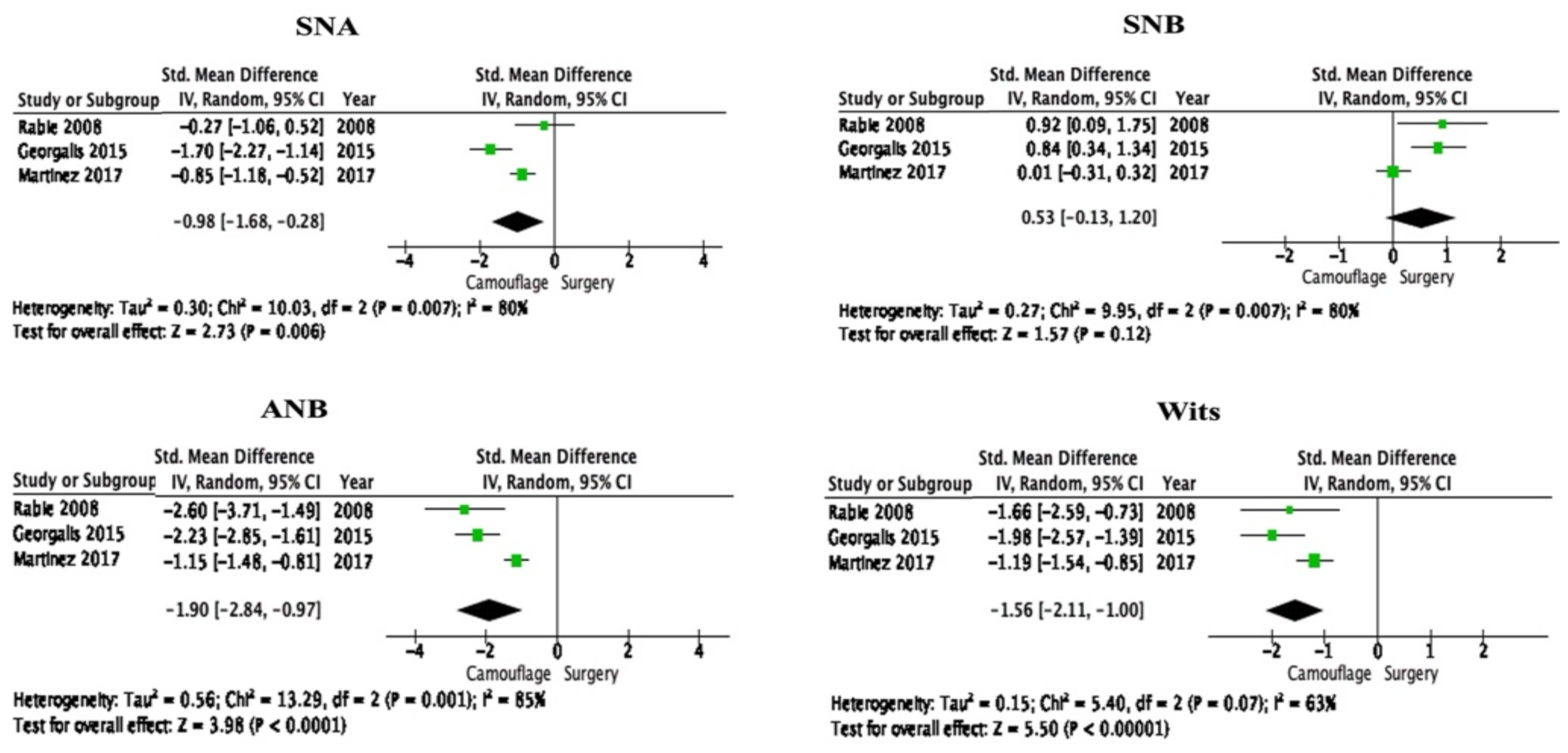
3.3. Meta-Analysis from Selected Studies

MA was performed on skeletal (SNA, SNB, ANB and Withs appraisal) and dental measurements (L1-MDP and IIA). The details of these parameters are shown in Table 4 and Figure 2. It showed that the overall effect between camouflage and surgical treatment in all skeletal and dental measurements was statistically significant (p < 0.05) except SNB. However, all the variables showed higher heterogeneity (I2 ≥ 50%). Therefore, the overall effect could not reflect the true difference. Both fixed and random-effects models were planned, but the only random-effects model was used to conduct the MA and subgroup analyses since the level of heterogeneity of the included studies remained significant. One study on each step was omitted for sensitivity analysis to observe any impact on the pooled effect. For SNA, Wits appraisal and L1-MDP, if the study Georgalis et al. (2015) omitted it reduced the heterogeneity but did not alter the overall effect. Similarly, omitting Martinez et al. (2017) from SNB, and Rabie et al. (2008) form IIA reduced the heterogeneity without altering the overall outcome (Figure 3 and Figure 4).

3.4. Publication Bias

Graphical assessment of the funnel plot was assessed for publication bias. Statistical test for publication bias was not appropriate due to the insignificant numbers (less than ten) of the study included for MA. Funnel plots showed asymmetry, which may be due to the small number of selected studies for the MA.

4. Discussion

This SR and MA compared the orthodontic surgery and orthodontic camouflage treatment in C-III-M. This study included a total of six studies for the qualitative analysis after conducting the literature search through different databases. This insignificant number of selected studies indicates that further research needs to be conducted concerning C-III-M to overcome the constraint of qualitative and quantitative secondary systematic studies.
This current SR included only the non-randomized retrospective observational studies. A randomized clinical trial would not be ethical to compare the surgery and camouflage treatment as the patients have the right to know and give consent about the surgical procedure. Therefore, randomly selecting a patient for surgery or non-surgery is not an option in these types of studies. This non-randomization process is one of the reasons that most of the studies scored low in the quality assessment. Only two studies scored 19 which is considered moderate quality. Power analysis is another explanation due to the lower scores as power analysis consists of a ‘5’ score in the modified Downs and Black assessment [25]. Retrospective studies are mostly available of data-dependent, yet a sample size calculation would strengthen the study design than the studies which followed convenience sample techniques. Due to the lack of compliance with any methodological guideline, studies may introduce bias caused by methodological inaccuracies [32].
Studies that compared the different cephalometric measurements between the camouflage and surgical treatment of C-III-M were included in this SR. However, two studies among the six selected studies were compared only to the pre-treatment cephalometric radiographs [27,28]. Therefore, these two studies were excluded from the MA. Another study compared the pre-treatment and post-treatment radiographs to compare the long-term stability and satisfaction; therefore, to eliminate the heterogeneity this study was also excluded from the MA as it slightly deviated from the aim of this SR [29]. Only the studies which assessed the pre-treatment and post-treatment cephalometric measurements for assessing the treatment effect were included for MA [15,30,31]. Although the aim of all included studies was similar, the variation was observed in the measurement of cephalometric variables. The most common skeletal variables (SNA, SNB, ANB and Wits appraisal) were measured by all the included studies. Martinez et al. (2017) examined additional skeletal variable facial angle (FA) [31], and Rabie et al. (2008) added maxillary mandibular plane [15].
Rabie et al. (2008) found no significant difference among all the skeletal variables included in the MA between the post-treatment orthodontic group and post-treatment surgical group except the ANB [15]. In addition, Georgalis and Woods (2015) discovered a significant difference concerning all variables except SNB between the two groups [30]. On the other hand, Martinez et al. (2017) did not find a significant difference in Wits appraisal between these two groups [31]. All the mean skeletal variables decreased in both treatment procedures except the SNA observed increased by both Martinez et al. (2017) and Georgalis and Woods (2015) in surgical treatment [30,31].
The most inconsistency was noted in dental variables. Only L1-MDP and IIA were common dental measurements among these three studies. Georgalis and Woods (2015) examined overjet (OJ), upper incisor to Sella Nasion plane (U1-SN), upper incisor to palatal plane (U1-Pal), upper incisor to Nasion-Point A (U1-SA) and lower incisor to Nasion B point (L1-NB) along with L1-MDP and IIA [30]. On the other hand, Rabie et al. (2008) measured additional U1-SN for upper incisor which is coherent with Georgalis and Woods (2015); however, Martinez et al. (2017) measured U1-Pal for upper incisor dental measurement [15,30,31]. Hence, only IIA and L1-MDP were included in the forest plot. All three studies included in the MA showed significant differences in L1-MDP and IIA in both treatment groups [15,30,31]. Therefore, it is obvious that dental changes are consistent in all the included studies where L1-MDP increases in surgical treatment and decreases in camouflage treatment. Moreover, IIA decreases in surgical treatment and increases in camouflage treatment. Georgalis and Woods (2015) found a decrease in IIA in camouflage treatment, though significant differences were observed between both groups [30].
Although surgical treatment is more stable and provides a better outcome in C-III-M [2,3], the current MA showed conflicting outcomes. Therefore, statistical heterogeneity test I2 was observed which indicated significant heterogeneity in the selected study and ultimately suggest bias in the analyzed study [33]. Though clinical heterogeneity is constantly persisting in the MA which may be due to the sample size, study design, intervention and outcome of the selected studies. This quantitative analysis also denoted clinical heterogeneity based on the ethnic background as selected three studies in this MA were performed in three different continents Asia, Europe and Oceania. Sample size would be another issue as there is a variety of sample sizes in the included studies. In general, studies included in this SR did not commence with similar treatment groups due to the non-randomized nature of the studies. Moreover, there was limited information on whether the sample represented the entire population or not. In addition, only one study performed the sample size calculation [31]. Therefore, further studies are compulsory where these issues need to be addressed. Other than the forest plots, visual observation of funnel plots is also showing publication bias.
All the studies recruited patients who were showing skeletal C-III-M before starting the orthodontic treatment with ANB value <0°. However, C-III-M can be classified as mild, moderate and severe. Rabie et al. (2008) and Georgalis and Woods (2015) mentioned recruiting borderline C-III-M patients in their study [15,30], whereas other studies only mentioned the ANB value [27,28,29,31]. ANB value <0° could include mild, moderate and severe types of C-III-M. Therefore, the treatment plan would vary based on the nature of the malocclusion. The patients in both groups should present similar characteristics before starting any comparison studies [34]; however, this MA only involved non-randomized observational studies where pre-treatment characteristics should not impact the overall effect.
Treatment of C-III-M in adult patients has always been a challenge for orthodontists [35]. Therefore, assessing and comparing the cephalometric variables in camouflage and orthodontic surgery to estimate the treatment effect is important for appropriate treatment planning depending on the patients’ profile. It is imperative to emphasize all the cephalometric variables with large sample size. Cephalometric dental values are as important as skeletal values as in camouflage both upper and lower dentition change in the greater value to compensate the skeletal deformities [36].
As per our best knowledge, this is the first SR and MA which compared the treatment effects of orthognathic surgery and camouflage treatment of C-III-M. Different databases were searched extensively to include a maximum number of studies for this systematic review. Only six previous studies assessed the treatment effects of orthognathic surgery and camouflage treatment of C-III-M. However, only three studies were included for the MA. Though the most important skeletal variables could evaluate from this MA, the dental and soft tissue variables were not fully explained. This SR fails to assess any changes in the upper dentition and soft tissue changes due to the lack of similar measurements observed in the selected studies. Therefore, it is understandable that more studies related to this topic are necessary. The most important cephalometric variables for skeletal, dental and soft tissue measurements should be extensively included in future studies for better comparison of the orthognathic surgery and camouflage treatment of C-III-M.

5. Conclusions

The quality of evidence in comparing the treatment effect of orthognathic surgery and orthodontic camouflage treatment of adult patients is low. Moreover, the overall effect of quantitative analysis could not reflect the true difference due to the significant heterogeneity. Therefore, further clinical studies with similar pre-treatment patient profiles and with larger sample sizes are recommended.

Author Contributions

M.K.A. designed the study and contributed to writing the manuscript, S.A.N. and F.S. analyzed the data, report the data and M.K.A., S.A.N., F.S., H.A. (Huda Abutayyem), H.A. (Hassan AlHarby), H.J.A. and S.K.E.-D.M. contributed to write and review the manuscript. All authors have read and agreed to the published version of the manuscript.

Funding

Deanship of Scientific Research at Jouf University research grant no. DSR-2021-01-0386.

Institutional Review Board Statement

Not applicable.

Informed Consent Statement

Not applicable.

Data Availability Statement

Not applicable.

Acknowledgments

The authors extend their appreciation to the Deanship of Scientific Research at Jouf University for funding this work through research grant no. DSR-2021-01-0386.

Conflicts of Interest

The authors declare no conflict of interest.

References

  1. Poulton, D.; Ware, W. The American academy of oral roentgenology joins our journal. Oral Surg. Oral Med. Oral Pathol. 1959, 12, 389–390. [Google Scholar]
  2. Trauner, R.; Obwegeser, H. The surgical correction of mandibular prognathism and retrognathia with consideration of genioplasty: Part I. Surgical procedures to correct mandibular prognathism and reshaping of the chin. Oral Surg. Oral Med. Oral Pathol. Oral Radiol. 1957, 10, 677–689. [Google Scholar]
  3. Obwegeser, H.L. Surgical correction of small or retrodisplaced maxillae the “dish-face” deformity. Plast. Reconstr. Surg. 1969, 43, 351–365. [Google Scholar]
  4. Proffit, W.; Fields, H.; Sarver, D. Biomechanics, Mechanics, and Contemporary Orthodontic Appliances, Contemporary Orthodontics; Mosby, Elsevier: St. Louis, MO, USA, 2007; pp. 689–693. [Google Scholar]
  5. Troy, B.A.; Shanker, S.; Fields, H.W.; Vig, K.; Johnston, W. Comparison of incisor inclination in patients with Class III malocclusion treated with orthognathic surgery or orthodontic camouflage. Am. J. Orthod. Dentofac. Orthop. 2009, 135, 146. [Google Scholar]
  6. Burns, N.R.; Musich, D.R.; Martin, C.; Razmus, T.; Gunel, E.; Ngan, P. Class III camouflage treatment: What are the limits? Am. J. Orthod. Dentofac. Orthop. 2010, 137, 9–13. [Google Scholar]
  7. Araujo, M.T.D.S.; Squeff, L.R. Orthodontic camouflage as a treatment alternative for skeletal Class III. J. Orthod. 2021, 26, e21bbo4. [Google Scholar]
  8. Proffit, W.R.; Fields, H.; Sarver, D. Diagnosis and Treatment Planning, Contemporary Orthodontics; Mosby, Elsevier: St. Louis, MO, USA, 2007; Volume 160, pp. 207–217. [Google Scholar]
  9. Kondo, E.; Arai, S. Nonsurgical and non extraction treatment of a skeletal class III adult patient with severe prognathic mandible. World J. Orthod. 2005, 6, 233–247. [Google Scholar]
  10. Kuroda, Y.; Kuroda, S.; Alexander, R.G.; Tanaka, E. Adult Class III treatment using a J-hook headgear to the mandibular arch. Angle Orthod. 2010, 80, 336–343. [Google Scholar]
  11. Lübberink, G.; Nienkemper, M.; Wilmes, B.; Ludwig, B.; Drescher, D. Nonsurgical treatment of a mature adult Class III patient. J. Clin. Orthod. 2014, 48, 697–702. [Google Scholar]
  12. Miguel, J.A.M.; Zanardi, G. Class III camouflage using skeletal anchorage and Pendex appliance. Prog. Orthod. 2011, 12, 73–83. [Google Scholar]
  13. Nakamura, Y.; Miyamoto, Y.; Kanzaki, H.; Wada, S. Orthodontic Treatment of an Adult Class III Malocclusion with Severe Transverse Dental Compensation by Remaining of Buccal Crossbite. Int. J. Orthod. Milwaukee 2015, 26, 29–35. [Google Scholar] [PubMed]
  14. Stellzig-Eisenhauer, A.; Lux, C.J.; Schuster, G. Treatment decision in adult patients with Class III malocclusion: Orthodontic therapy or orthognathic surgery? Am. J. Orthod. Dentofac. Orthop. 2002, 122, 27–37. [Google Scholar]
  15. Rabie, A.-B.M.; Wong, R.W.; Min, G. Treatment in borderline Class III malocclusion: Orthodontic camouflage (extraction) versus orthognathic surgery. Open Dent. J. 2008, 2, 38–48. [Google Scholar] [PubMed] [Green Version]
  16. Kerr, W.; Miller, S.; Dawber, J. Class III malocclusion: Surgery or orthodontics? Br. J. Orthod. 1992, 19, 21–24. [Google Scholar] [PubMed]
  17. Hardy, D.K.; Cubas, Y.P.; Orellana, M.F. Prevalence of angle class III malocclusion: A systematic review and meta-analysis. Open J. Epidemiol. 2012, 2, 75–82. [Google Scholar]
  18. Dehesa-Santos, A.; Iber-Diaz, P.; Iglesias-Linares, A. Genetic factors contributing to skeletal class III malocclusion: A systematic review and meta-analysis. Clin. Oral Investig. 2021, 25, 1587–1612. [Google Scholar]
  19. Lin, Y.; Guo, R.; Hou, L.; Fu, Z.; Li, W. Stability of maxillary protraction therapy in children with Class III malocclusion: A systematic review and meta-analysis. Clin. Oral Investig. 2018, 22, 2639–2652. [Google Scholar]
  20. Yang, X.; Li, C.; Bai, D.; Su, N.; Chen, T.; Xu, Y.; Han, X. Treatment effectiveness of Fränkel function regulator on the Class III malocclusion: A systematic review and meta-analysis. Am. J. Orthod. Dent. Orthop. 2014, 146, 143–154. [Google Scholar]
  21. Chatzoudi, M.I.; Ioannidou-Marathiotou, I.; Papadopoulos, M.A. Clinical effectiveness of chin cup treatment for the management of Class III malocclusion in pre-pubertal patients: A systematic review and meta-analysis. Prog. Orthod. 2014, 15, 62. [Google Scholar]
  22. Zhang, W.; Qu, H.-C.; Yu, M.; Zhang, Y. The effects of maxillary protraction with or without rapid maxillary expansion and age factors in treating class III malocclusion: A meta-analysis. PLoS ONE 2015, 10, e0130096. [Google Scholar]
  23. Moher, D.; Liberati, A.; Tetzlaff, J.; Altman, D.G.; PRISMA Group. Preferred reporting items for systematic reviews and meta-analyses: The PRISMA statement. PLoS Med. 2009, 6, 336–341. [Google Scholar]
  24. Downs, S.; Black, N. The feasibility of creating a checklist for the assessment of the methodological quality both of randomised and non-randomised studies of health care interventions. JECH 1998, 52, 377–384. [Google Scholar]
  25. Raposo, R.; Peleteiro, B.; Paço, M.; Pinho, T. Orthodontic camouflage versus orthodontic-orthognathic surgical treatment in class II malocclusion: A systematic review and meta-analysis. Int. J. Oral Maxillofac. Surg. 2018, 47, 445–455. [Google Scholar] [PubMed] [Green Version]
  26. Landis, J.R.; Koch, G.G. The measurement of observer agreement for categorical data. Biometrics 1977, 33, 159–174. [Google Scholar] [PubMed]
  27. Eslami, S.; Faber, J.; Fateh, A.; Sheikholaemmeh, F.; Grassia, V.; Jamilian, A. Treatment decision in adult patients with class III malocclusion: Surgery versus orthodontics. Prog. Orthod. 2018, 19, 28. [Google Scholar]
  28. Tseng, Y.-C.; Pan, C.-Y.; Chou, S.-T.; Liao, C.-Y.; Lai, S.-T.; Chen, C.-M.; Chang, H.-P.; Yang, Y.-H. Treatment of adult Class III malocclusions with orthodontic therapy or orthognathic surgery: Receiver operating characteristic analysis. Am. J. Orthod. Dentofac. Orthop. 2011, 139, 485–493. [Google Scholar]
  29. Xiong, X.; Yu, Y.; Chen, F. Orthodontic camouflage versus orthognathic surgery: A comparative analysis of long-term stability and satisfaction in moderate skeletal Class III. OJST 2013, 3, 89–93. [Google Scholar]
  30. Georgalis, K.; Woods, M.G. A study of Class III treatment: Orthodontic camouflage vs. orthognathic surgery. Aust. Orthod. J. 2015, 31, 138–148. [Google Scholar]
  31. Martinez, P.; Bellot-Arcís, C.; Llamas, J.M.; Cibrian, R.; Gandia, J.L.; Paredes-Gallardo, V. Orthodontic camouflage versus orthognathic surgery for class III deformity: Comparative cephalometric analysis. Int. J. Oral Maxillofac. Surg. 2017, 46, 490–505. [Google Scholar]
  32. Wu, F.; Wang, L.; Huang, Y.; Guo, W.; Liu, C.; Li, S. Interleukin-1β+ 3954 polymorphisms and risk of external apical root resorption in orthodontic treatment: A meta-analysis. Genet. Mol. Res. 2013, 12, 4678–4686. [Google Scholar]
  33. Nowrin, S.A.; Jaafar, S.; Ab Rahman, N.; Basri, R.; Alam, M.K.; Shahid, F. Association between genetic polymorphisms and external apical root resorption: A systematic review and meta-analysis. Korean J. Orthod. 2018, 48, 395–404. [Google Scholar] [PubMed]
  34. Higgins, J.P.; Thomas, J.; Chandler, J.; Cumpston, M.; Li, T.; Page, M.J.; Welch, V. Cochrane Handbook for Systematic Reviews of Interventions; John Wiley & Sons: Hoboken, NJ, USA, 2019. [Google Scholar]
  35. Jamilian, A.; Cannavale, R.; Piancino, M.G.; Eslami, S.; Perillo, L. Methodological quality and outcome of systematic reviews reporting on orthopaedic treatment for class III malocclusion: Overview of systematic reviews. J. Orthod. 2016, 43, 102–120. [Google Scholar] [PubMed]
  36. Kabbur, K.J.; Hemanth, M.; Patil, G.; Sathyadeep, V.; Shamnur, N.; Harieesha, K.; Praveen, G. An esthetic treatment outcome of orthognathic surgery and dentofacial orthopedics in class II treatment: A cephalometric study. J. Contemp. Dent. Pract. 2012, 13, 602–606. [Google Scholar] [PubMed]
Figure 1. PRISMA guideline for the selection of studies.
Applsci 12 03314 g001
Figure 2. Cephalometric parameters used in this study.
Applsci 12 03314 g002
Figure 3. Forest plots for sagittal measurements.
Applsci 12 03314 g003
Figure 4. Forest plot of dental measurements.
Applsci 12 03314 g004
Table 1. PICOS format.
VariablesDescription
ParticipantsClass III malocclusion
InterventionsOrthognathic surgery
ComparisonsCamouflage treatment
OutcomesSkeletal and dental measurements from lateral cephalogram
Study designsObservational and interventional studies
Table 2. List of included studies.
Study (Year)CountrySample SizeStudy DesignInterventionOutcome
CamouflageSurgical
Eslami et al. (2018) [27]Iran65Retrospective3629Wits appraisal and Holdway H angle could be utilized to determine the treatment modality in borderline C-III-M.
Tseng et al. (2011) [28]Taiwan80Retrospective4040Minimum six cephalometric measurements are required to attain effective diagnosis between nonsurgical and surgical treatment in C-III-M.
Xiong et al. (2013) [29]China46Retrospective2521Camouflage treatment could be a choice of treatment for patients who denied orthognathic surgery
Georgalis et al. (2015) [30]Australia67Retrospective3136Treatment of C-III-M is expected to complete with slightly proclined upper incisors in both surgery and camouflage treatment.
Martinez et al. (2017) [31]Spain156Retrospective7779Three skeletal and two dental measurements showed a significant difference between surgical and camouflage treatment groups.
Rabie et al. (2008) [32]Hong Kong25Retrospective1312For patients with borderline C-III-M, the Holdway angle could be a reliable guide to determine the modality of treatment.
Table 3. Modified Downs and Black bias assessment.
AssessmentEslami et al. (2018) [27]Georgalis et al. (2015) [30]Martinez et al. (2017) [31]Rabie et al. (2008) [15]Tseng et al. 2011 [28]Xiong et al. (2013) [29]
Reporting (Q1–Q10)
Q1: Aim clearly described111111
Q2: Outcomes clearly described111111
Q3: Patients’ characteristics clearly described111111
Q4: Interventions clearly described111111
Q5: Principal confounders clearly described001111
Q6: Main findings clearly described111111
Q7: Random variability for the main outcome provided111111
Q8: Adverse events reported111111
Q9: Lost to follow up reported000110
Q10: Actual p value reported101010
Score8798108
External Validity (Q11–Q13) and Internal Validity-Bias (Q14–Q20)
Q11: Sample asked to participate representative of the population000100
Q12: Sample agreed to participate representative of the population000000
Q13: Staff participating representative to the patient’s environment000000
Q14: Attempt to blind participants000000
Q15: Attempt to blind assessors110000
Q16: Data dredging results stated clearly111111
Q17: Analysis adjusted for length of follow up000001
Q18: Appropriate statistics111111
Q19: Reliable compliance111111
Q20: Accurate outcome111111
Score554545
Internal Validity–Confounding (selection bias) (Q21–Q26) and Power (Q27)
Q21: Same population111111
Q22: Participants recruited at the same time000000
Q23: Randomised?000000
Q24: Adequate allocation concealment?000000
Q25: Adequate adjustment for confounders?000100
Q26: Loss of follow-up reported?000000
Q27: Power calculation505000
Score616211
Total score191319151514
1, 5; Yes, 0; no/unable to determine.
Table 4. List of parameters used in the meta-analysis.
VariablesDetails
SNAThe relation between the maxilla and the cranial base. Angle: Sella-Nasion-Point A
SNBThe relation between the mandible and the cranial base. Angle: Sella-nasion-Point B
ANBRelation between the maxilla and the mandible. Angle: Point A-Nasion-Point B
Wits appraisalThe sagittal disparity in the occlusal plane between Maxilla and Mandible. Distance between projections from points A and B, drawn perpendicular to the Occlusal plane (O)
L1-MDPThe angle between the mandibular plane and the axis of the lower incisor. The angle between the lower incisor axes and mandibular plane
IIAThe posterior angle between the upper and lower incisor axes
Publisher’s Note: MDPI stays neutral with regard to jurisdictional claims in published maps and institutional affiliations.

