Article

Exploring the Strategy of Fusing Sucrose Synthase to Glycosyltransferase UGT76G1 in Enzymatic Biotransformation

College of Biotechnology and Pharmaceutical Engineering, Nanjing Tech University, Nanjing 211816, China
*
Authors to whom correspondence should be addressed.
These authors contributed equally to this work.
Appl. Sci. 2022, 12(8), 3911; https://doi.org/10.3390/app12083911
Submission received: 29 January 2022 / Revised: 4 April 2022 / Accepted: 8 April 2022 / Published: 13 April 2022
(This article belongs to the Topic Frontier in Biocatalysis and Biotransformations)
(This article belongs to the Section Applied Biosciences and Bioengineering)

Abstract

:
Uridine diphosphate glycosyltransferases (UGTs) as fine catalysts of glycosylation are increasingly used in the synthesis of natural products. Sucrose synthase (SuSy) is recognized as a powerful tool for in situ regenerating sugar donors for the UGT-catalyzed reaction. It is crucial to select the appropriate SuSy for cooperation with UGT in a suitable way. In the present study, eukaryotic SuSy from Arabidopsis thaliana (AtSUS1) helped stevia glycosyltransferase UGT76G1 achieve the complete conversion of stevioside (30 g/L) into rebaudioside A (RebA). Position of the individual transcription units containing the genes encoding AtSUS1 and UGT76G1 in the expression plasmid has an effect, but less than that of the fusion order of these genes on RebA yield. Fusion of the C-terminal of AtSUS1 and the N-terminal of UGT76G1 with rigid linkers are conducive to maintaining enzyme activities. When the same fusion strategy was applied to a L637M-T640V double mutant of prokaryotic SuSy from Acidithiobacillus caldus (AcSuSym), 18.8 ± 0.6 g/L RebA (a yield of 78.2%) was accumulated in the reaction mixture catalyzed by the fusion protein Acm-R3-76G1 (the C-terminal of AcSuSym and the N-terminal of UGT76G1 were linked with (EAAAK)3). This work would hopefully reveal the potential of UGT-SuSy fusion in improving the cascade enzymatic glycosylation.

Graphical Abstract

1. Introduction

Glycosylation of small molecular compounds refers to the addition of glycosyl residues to the C-, O-, N-, and S-positions of some small molecules, such as antibiotics, vitamins, and spices, to improve their physical chemistry and biology characteristics [1,2]. The formation of glycoside bonds during glycosylation is popularly obtained by chemical or enzymatic methods. Compared with complicated chemical manipulation, enzyme-mediated glycosylation is capable of recognizing certain chemical groups to synthesize specific products with simple steps and not introducing extra-toxic chemical reagents due to the splendid region- and stereoselectivity of enzymes, especially in the glycosylation of compounds with complex conformations. In nature, glycosylation of small molecular compounds is usually catalyzed by glycosyltransferases (GTs), among which uridine diphosphate glycosyltransferases (UGTs) are pivotal members [3,4]. Uridine diphosphate (UDP)-activated sugar molecules are widely used as donors in glycosylation of natural products, such as steviosides [5], triterpenoids [6], flavonoids [7], and ginsenosides [8]. UGTs also contribute to the metabolic engineering applications for producing these natural products [9]. However, such glycosylation reactions are restricted by catalytic abilities of GTs and the availability of nucleoside diphosphate (NDP)-sugar donor [10,11].
Sucrose synthase (SuSy, EC 2.4.1.13) belonging to the GT family, which reversibly catalyzes NDP and sucrose to produce NDP-glucose and fructose, is one of the most promising candidates addressing the supply of NDP-glucose [12]. Most eukaryotic SuSysmainly participate in the physiological metabolism of plants, especially for the synthesis or decomposition of sucrose [13,14]. A few SuSys have also been found in prokaryotes-like bacteria in recent years, which have higher thermal stability and prefer adenosine diphosphate (ADP), while plant-derived SuSys prefer UDP [13]. SuSys could work with UGTs in vitro to constitute the coupling system for regenerating UDP-glucose (UDPG) that is provided to the glycosylation reaction catalyzed by UGTs [14]. The one-pot two-enzyme reactions by coupling the activities of UGT and SuSy (UGT-SuSy) were applied to numerous chemical compounds such as C-glycosylation of phloretin [11], O-glycosylation of stevioside, and quercetin [15,16]. Generally, the catalytic efficiency of free enzymes in the reaction that would be affected by several factors can be enhanced to some extent by organizing them into a multi-enzyme complex using co-immobilization technology [17], scaffolding technology [18], or fusion technology [19]. Among them, gene fusion technology, widely used in protein soluble expression, purification, imaging, and biopharmaceuticals, is also an effective and relatively simple strategy for setting up a multi-enzyme catalytic system in vitro [20].
UGT76G1, a UGT was initially discovered in Stevia rebaudiana for the conversion of stevioside into rebaudioside A (RebA), a sweetener with low calories and intense sweetness [21]. As has been known so far in the biosynthesis of steviol glycosides (SGs), UGT76G1 uses UDPG as the glycosyl donor to form the β-1,3-glycosidic bond at the steviol C13- and C19-bound glucose involving eight different reactions [5]. It is the only enzyme directly responsible for the production of rebaudioside M, which is generally recognized as the best SG sweetener [22]. In our previous research, we have set up a UGT-SuSy reaction involving UGT76G1 and a SuSy from Arabidopsis thaliana (AtSUS1) [23]. UDPG and UDP in crude extracts from the recombinant cells were used without additional supply [15,24]. In the present study, we try to compare the performance of four different originated SuSys in cooperation with UGT76G1, as well as the coupling details, such as the gene construct strategy of two enzymes. As a result, a fusion of SuSy and UGT was investigated for increased effective production of RebA from stevioside. This is the application of fusion technology to improve catalytic efficiency in SGs synthesis.

2. Materials and Methods

2.1. Plasmids and Strains

Nucleotide sequences respectively encoding UGT76G1 (Q6VAB4), AtSUS1 (P49040), StSUS1 (P10691), AtSUS3 (Q9M111), and AcSuSy (A0A059ZV61) were codon-optimized for expression in Escherichia coli and synthesized by Genscript Biotechnology Co., Ltd. (Nanjing, China). pRSFDuet-1 (Novagen) was used as the backbone for constructing recombinant plasmids. The double mutant of AcSuSy (L637M-T640V) was made using a Mut Express® II Fast Mutagenesis Kit V2 (Vazyme Biotech Co., Ltd., Nanjing, China), and the resultant clone was named as AcSuSym. More details regarding the plasmid and strain constructs used in this study are shown in Table 1. The linkers adopted for fusion constructs are shown in Table 2.

2.2. Expression of Recombinant UGT and SuSy in E. coli

A total of 100 mL of auto-induction medium (15 g/L tryptone, 25 g/L yeast extract, 10 g/L NaCl, 2 g/L glucose, and 0.5 g/L lactose) containing 50 mg/L kanamycin was inoculated with 1mL overnight culture of the recombinant strains grown in LB (Luria–Bertani) medium (tryptone 10 g/L, yeast extract 5 g/L, and NaCl 10 g/L) with 50 mg/L kanamycin, and incubated at 37 °C with shaking at 200 rpm for 2 h, then transferred to 25 °C for another 20–30 h.
The cell pellets were harvested after washing twice and then disrupted with a Sonifier (Ningbo Scientz Biotechnology Co. Ltd., Ningbo, China) in the potassium phosphate buffer (100 mM, pH 7.2). The supernatant was obtained by centrifugation at 4 °C and used as the crude enzyme extract.

2.3. Determination of Glycosyltransferase and SuSy Activities

Enzyme activities of glycosyltransferase and SuSy were assayed as previously described [15,23]. One unit (U) of glucosyltransferase activity was defined as the amount of enzyme that produced 1 μmol of RebA from stevioside per min, and one unit (U) of SuSy activity was defined as the amount of enzyme releasing 1 μmol of reducing sugar per min under the given assay conditions.

2.4. Enzymatic Synthesis of RebA

To convert stevioside into RebA, the reaction mixtures (20 mL) containing 20 g/L (or 30 g/L) stevioside, 60 g/L (or 90 g/L) sucrose, an appropriate amount of total protein from the crude enzyme extract, and potassium phosphate buffer (100 mM, pH 7.2) were generally incubated at 30 °C for 24 h with shaking at 200 rpm. For convenience of description, the reactions were named after the recombinant strains, from which the crude extracts were prepared.
For the reactions with Acm-R3-76G1, the effects of various factors including temperature, pH, ratios of stevioside to sucrose, and crude enzyme concentrations were investigated in 100 mM potassium phosphate buffer. The reactions were performed at 20 °C, 30 °C, 40 °C, 50 °C, and 60 °C, or at a pH of 6, 7.2, 8, and 9; or with different mass ratios of stevioside to sucrose (1:3, 1:5, 1:20, 1:30, 1:40, and 1:50; 20 g/L stevioside), or with the crude enzyme of 5, 8, 10, 12, and 15 mg/mL. Only the mentioned factor was changed each time, while others remained constant. To test the effect of UDP supply on the reactions of Acm-76G1 and Acm-R3-76G1, UDP was added as described.
The collected reaction mixtures were appropriately diluted and inactivated at 95 °C for 5 min. Then, the supernatants were obtained after centrifugation and filtered before analysis by high-performance liquid chromatography (HPLC).

2.5. HPLC Analysis

The samples were analyzed by HPLC (UltiMate 3000, Dionex China Limited, Beijing, China) equipped with a Cosmosil packed colum-C18 (COSMOSIL, 250 mm × 4.6 mm) as previously described [15]. The RebA yield was calculated as follows: RebA yield (%) = C (RebA)/C (St). C (St) represents the initial molar concentration of stevioside, and C (RebA) represents the increased RebA molar concentration after reaction. In the case of the relative RebA yield, the highest yield of the sample in the tested group was regarded as 100%.

2.6. Prediction of Protein Solubility

The solubilities of UGT, SuSy, and their fusion proteins were calculated using the Wilkinson–Harrison solubility model [25], which involved the calculation of a canonical variable (CV) for proteins. The probability of solubility or insolubility was calculated by the following formula: 0.4934 + 0.276 | C V C V | 0.0392 ( C V C V ) 2 , where CV′ = 1.71; and C V = λ 1 ( N + G + P + S n ) + λ 2 | ( R + K ) ( D + E ) n 0.03 | , n, total number of amino acids in protein; λ1, 15.43; λ2, −29.56; N, G, P, S, R, K, D, E, number of Asn, Gly, Pro, Ser, Arg, Lys, Asp, or Glu residues. If CVCV’ is greater than 0, it predicts that the protein is insoluble. If CVCV′ is less than 0, it predicts that the protein is soluble.

3. Results and Discussion

3.1. Selection of SuSys for Constructing One-Pot Two-Enzyme Systems

UGT-SuSy system has been widely applied in glycosylation reactions, especially for natural products [16,26,27]. Most research focuses on the enzyme engineering of UGTs and SuSys [24,28]. Few has been done with the collaborative relationship between these two enzymes. As is known, various SuSys had different abilities to synthesize UDPG, which could have a significant impact on the glycosylation reaction [12]. Firstly, four SuSys, that is, AtSUS1 and AtSUS3 from A. thaliana [29,30,31], StSUS1 from Solanum tuberosum [32,33], and AcSuSy from Acidithiobacillus caldus [13,34], were co-expressed with UGT76G1, respectively, and the crude extracts prepared from the corresponding strains were used to set up four individual one-pot two-enzyme reactions, which are called At1-76G1, St1-76G1, Ac-76G1, and At3-76G1. All enzymes were successfully expressed, although inclusion still dominated (Figure S1). As is shown in Table 3, At1-76G1 and At3-76G1, including SuSys from the same source (A. thaliana), had a high yield of RebA. After 8 h, stevioside was almost converted to RebA by At1-76G1. The RebA yield for the reaction catalyzed by St1-76G1 ranked third (66.8%). Unexpectedly, in the reaction mixture of Ac-76G1 containing prokaryotic AcSuSy that has the highest specific activity (59.2 mU/mg, above 5-fold of StSUS1) among the four SuSys, the yield of RebA was even less than 1%. The UGT activity in Ac-76G1 (114.7 mU/mg) was evidently higher than that in St1-76G1 (88.8 mU/mg), suggesting that it was AcSuSy that led to the worse performance of Ac-76G1.

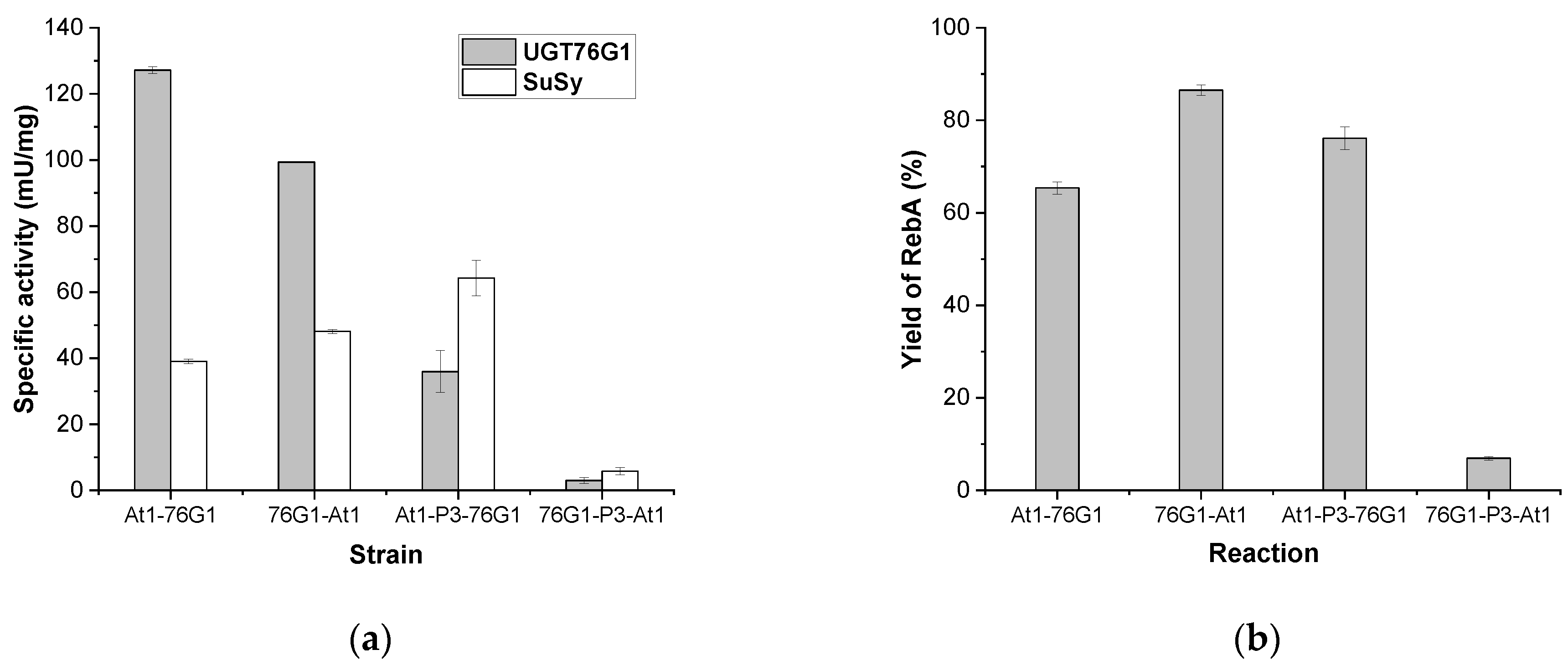
3.2. Comparison of the Construct Strategy for Expressing AtSUS1 and UGT76G1

In comparison to different construct strategies to generate the recombinant strain for enzyme preparation, At1-76G1 was selected, since AtSUS1 has the higher specific activity (47.6 mU/mg) than that of the other two eukaryotic SuSys. To construct At1-76G1, genes encoding AtSUS1 and UGT76G1 were inserted in NcoI/EcoRI and NdeI/XhoI of pRSFDuet-1, respectively. Another strain, named 76G1-At1, was obtained by transformed the plasmid pRSF-76G1-AtSUS1 in which the genes encoding AtSUS1 and UGT76G1 were inserted in NdeI/XhoI and NcoI/EcoRI of pRSFDuet-1, respectively. That means that these two genes belonging to an individual transcription unit exchanged their cloning positions. Although the protein soluble expression patterns look similar (Figure S2), the specific activities of two enzymes obtained from At1-76G1 and 76G1-At1 were not the same, especially for UGT76G1 (Figure 1a). The yield of RebA was 86.5% for 76G1-At1, which was 1.3 times of At1-76G1 (65.4%), but At1-76G1 has higher UGT activity than 76G1-At1 (Figure 1). These results indicated that the arrangement of the gene sequence of AtSUS1 and UGT76G1 in the expression plasmid may exert an influence on RebA production, probably because the activity ratio of UGT and SuSy is a critical factor under the condition that both enzyme activities were not low.
Recent studies show gene fusion strategies promote the folding, stability, bioactivity, and soluble expression of the fused proteins [35,36,37]. In theory, the fusion of two enzymes will shorten the space distance of enzymes, which is more conducive to the conversion of substrates. It was reported that a linker encoding for amino acids Pro-Pro-Pro (PPP) had been successfully applied in a fusion construction of the flavonoid O-glucosyltransferase OsUGT3 and AtSUS1 [38]. Therefore, we introduced the PPP linker between AtSUS1 and UGT76G1 in At1-76G1 and 76G1-At1, which were named At1-P3-76G1 and 76G1-P3-At1, respectively. It was shown that the fusion order of these two enzymes had a great influence on the soluble expression of fusion proteins, and thus, on the activities of UGT and SuSy, and the synthesis of RebA catalyzed by the fusion enzymes (Figure S3a and Figure 1). When the C-terminal of AtSUS1 was fused with the N-terminal of UGT76G1(At1-P3-76G1), the yield of RebA was 76.2%, which was 1.2 times that of At1-76G1 at 5 h under the same conditions. It was still not better than that of 76G1-At1 (86.5%). However, when the N-terminal of AtSUS1 was fused with the C-terminal of UGT76G to form the fusion protein 76G1-P3-At1, the RebA yield of 76G1-P3-At1 was only 6.9%.
Merely exchanging the two encoding genes on the expression plasmids caused a similar RebA yield between At1-76G1 and 76G1-At1 (Figure 1b). However, the fusion proteins with the PPP linker showed distinct differences compared to the free enzyme systems. The specific activity of UGT76G1 significantly decreased for At1-P3-76G1 (less than half of At1-76G1 and 76G1-At1), but the production of RebA was comparable with At1-76G1 and 76G1-At1. The specific activities of both UGT and SuSy declined sharply for 76G1-P3-At1, leading to almost no accumulation of RebA in the glycosyltransferase-catalyzed reaction. Therefore, the fusion order of UGT and SuSy has an important effect on their enzyme activity and productivity. The fusion of the C-terminal of UGT76G1 and the N-terminal of AtSUS1 (76G1-P3-At1) would probably pull the active sites of these two enzymes too close and affect their spatial conformations, resulting in low enzyme activities. However, the fusion of the C-terminal of AtSUS1 and the N-terminal of UGT76G1 (At1-P3-76G1) has less effect on their active conformations.

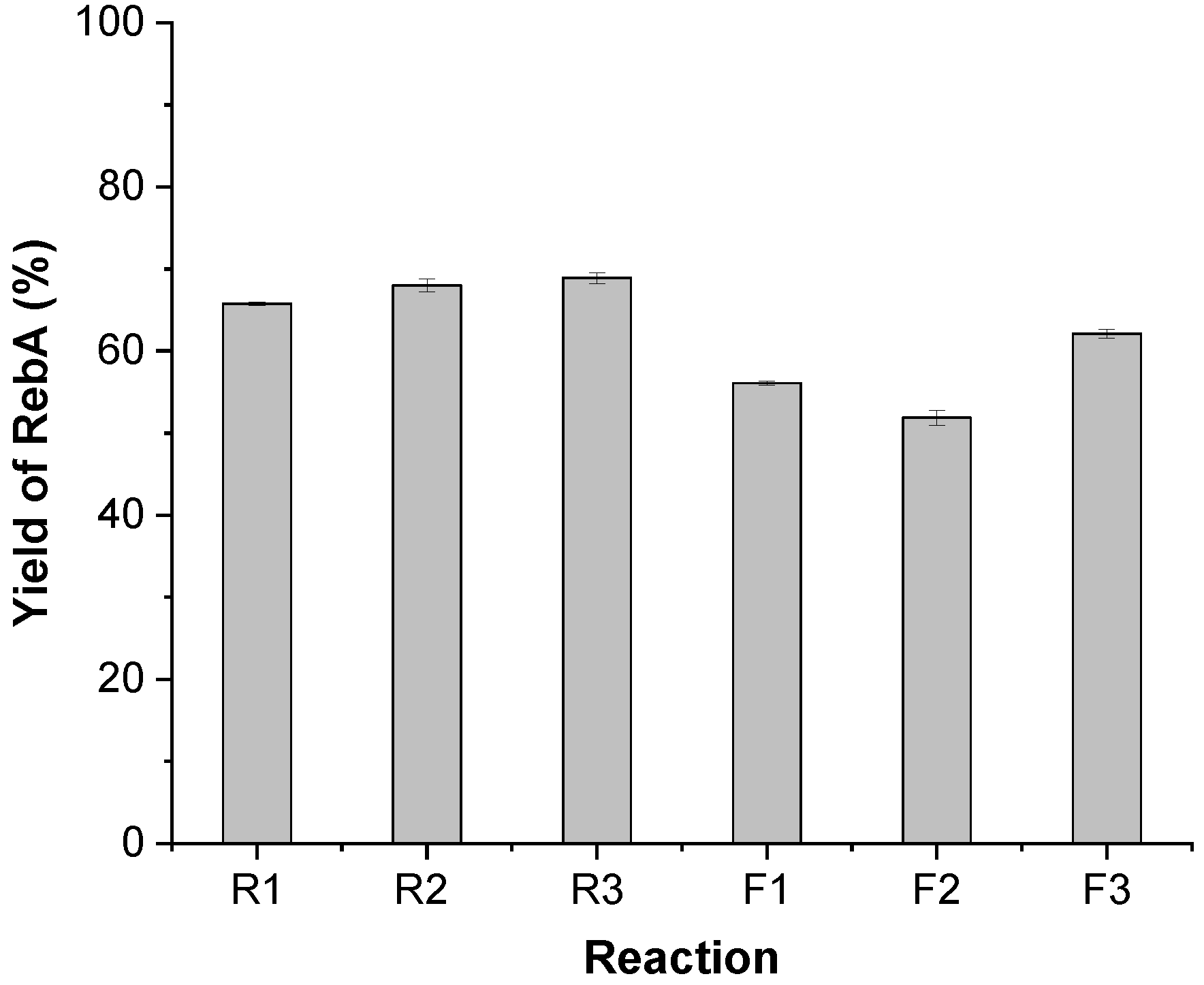
3.3. Fusion of AtSUS1 and UGT76G1 by Rigid and Flexible Linkers

Efforts have been made to clarify the properties of linkers in various fusion proteins [39,40]. Empirical linkers of a fusion construct are mainly divided into flexible, rigid, and in vivo cleavable linkers [41]. Rigid linkers and flexible linkers are the most popular applied linkers for fusion constructs [42,43,44]. To investigate the potential of fusion protein linkers in the UGT-SuSy reactions, three rigid linkers (EAAAK)n (n = 1, 2, 3) and three flexible linkers (GGGGS)n (n = 1, 2, 3) were fused between the C-terminal of AtSUS1 and N-terminal of UGT76G1 based on the above results. The new fusion constructs were named R1, R2, R3, F1, F2, and F3, respectively.
As is shown in Figure S3b and Figure 2, each fusion protein was expressed and had the ability to catalyze stevioside into RebA. Generally, the rigid linkers worked better than the flexible linkers, and the longer rigid linker worked better than the shorter one. RebA yield of R3 was about 2.4 times of R1 after reaction for 2 h (data not shown). At 16 h, the RebA yield of R3 reached 68.9%. We also compared the R3 with At1-P3-76G1 (Table 4) and found they almost had the same RebA productivity in 3 h. The linker PPP belongs to the rigid linkers as well [45]. As a result, the rigid protein linkers tending to maintain the respective conformations of two fusion partners are more applicable in the fusion construct linking the C-terminal of AtSUS1 and the N-terminal of UGT76G1 for RebA synthesis.

3.4. Fusion of the Prokaryotic SuSy with UGT76G1

As shown in Table 3, only a small amount of RebA was detected in the reaction catalyzed by Ac-76G1, but AcSuSy showed the best specific activity among four SuSys under the investigated conditions. In the assays of SuSy activity, 500 mM sucrose and 10 mM UDP were added [15], which would not available in the one-pot two-enzyme reactions that used 175.3 or 262.9 mM (60 or 90 g/L) sucrose, and the UDP and UDPG from the cell lysate. Afterward, a double mutant of AcSuSy (L637M-T640V), which has the reduced Km value to UDP [28] and named AcSuSym in this study, was coupled with UGT76G1 instead of AcSuSy. It was shown in HPLC analysis, that only a small amount of RebA was synthesized in the reaction catalyzed by Ac-76G1 (Figure S4a), while stevioside had been mostly converted into RebA in the reaction catalyzed by Acm-76G1 in 12 h (Figure S4b). The RebA concentration of Acm-76G1 was about seven times that of Ac-76G1, indicating that the mutant (AcSuSym) can effectively improve RebA productivity in UGT-SuSy reactions.
Then, another fusion construct named Acm-R3-76G1 was created by fusing the C-terminal of AcSuSym and the N-terminal of UGT76G1 with the rigid linker (EAAAK)3. To enhance the catalytic ability to produce RebA, the influence factors in terms of reaction temperature, pH, substrate ratio, and enzyme concentrations on the reaction catalyzed by Acm-R3-76G1 were investigated.
Temperature is critical for enzymatic reactions; the speed of the glycosylation reaction is positively related to temperature in a certain range. Increasing reaction temperature will accelerate the glycosylation of stevioside, but it will also shorten the half-life of the enzyme, making the enzyme unstable and prone to inactivation. Considering the temperature preferences of bacterial AcSuSym and plant origin UGT76G1, the temperature for Acm-R3-76G1 was set in the range of 20 °C to 60 °C. As was shown in Figure 3a, the yield of RebA continued to rise at 20–50 °C under the same conditions, and reached the peak at 50 °C, which was 9 times that at 20 °C. However, the yield dropped rapidly at 60 °C, possibly because the high temperature led to the inactivation of the fusion protein Acm-R3-76G1.
SuSys involve in a reversible reaction, whose direction was affected by pH [12]. The reactions catalyzed by Acm-R3-76G1 were conducted at a pH range of 6–9. As was shown in Figure 3b, the highest yield of RebA was obtained at pH 7.2, which was 1.85-fold that at pH 9. Under the experimental conditions in this study, pH preference of Acm-R3-76G1 was pH 7.2 > pH 6 > pH 8 > pH 9 for RebA formation.
As for the substrate mass ratios of stevioside to sucrose, it looked like the higher amount of sucrose used, the more RebA was generated below the ratio of 1:20 (stevioside: sucrose) (Figure 3c). In the reactions catalyzed by Acm-R3-76G1, the yield was relatively low when the initial substrate ratio was 1:3 or 1:5; it reached about 54.4% at the ratio set to 1:30. While the substrate ratio was 1:20, and sucrose was up to 400 g/L, the yield reached the highest at the reaction of 16 h, which was 4.2 times that at the ratio of 1:3.
The product yield is positively relevant to the enzyme concentration in the glycosylation reactions (Figure 3d). The increase in crude enzyme concentration accelerated the accumulation of RebA. At 50 °C and 7.2, using the mass ratio (stevioside: sucrose) 1:20, the RebA yield (17.0 g/L) in the reaction mixture with 15 mg/mL of the crude enzyme was 1.8 times higher than that with 5 mg/mL crude extract, and stevioside was almost completely converted after reaction of 8 h.
Compared with UGT-SuSy systems composed of eukaryotic SuSys, the reactions using prokaryotic SuSys, such as Acm-76G1 and Acm-R3-76G1, prefer higher temperature and higher sucrose concentration. The Km value of AcSuSy for UDP is 7.8 mM. After mutation, a significantly reduced Km value for UDP (up to 60 times) was achieved for AcSuSym. However, it is still above 0.1 mM, higher than that of most plant SuSys [28]. In the UGT-SuSy reactions set up in this study, the required UDP came from the cell lysate. At an elevated temperature (50 °C), with the extension of reaction time, the degradation of UDP cannot be ignored. As indicated in Figure 4, 2 mM of UDP was enough to support the reactions catalyzed by Acm-76G1 and Acm-R3-76G1, while a lower efficiency was found in the control reactions that only used UDP in the cell lysate. However, Acm-R3-76G1 that was catalyzed with the fusion protein always outperformed Acm-76G1 with the free enzymes. RebA accumulation in Acm-R3-76G1 without UDP supply was even more than Acm-76G1 with 2 mM UDP. After reaction for 2 h, 13.5 g/L RebA was detected in Acm-R3-76G1 containing 2 mM UDP, which indicated the initial reaction speed was relatively fast. After that, RebA increased slowly, reaching 18.8 ± 0.6 g/L (a yield of 78.2%) at 12 h.
In contrast, the prokaryotic AcSuSym had better performance than plant SuSys in the fusions. From the conversion results, the rigid linker (EAAAK)3 that pulls close the C-terminal of AcSuSym and the N-terminal of UGT76G1 did not seem to affect enzyme activities. In addition, the increased soluble expression of Acm-R3-76G1 could be observed by SDS-PAGE (Figure S5). It is known that enzymes are the key factors that affect reaction efficiency. Soluble expression is the premise of obtaining active enzymes. As predicted in Table 5, UGT76G1 is probably insoluble when it is overexpressed in E. coli. However, AtSUS1 and AcSuSym are predicted to have more probability of solubility, which increases the solubility of the fusion constructs when they are fused with UGT76G1, respectively. Among 76G1-P3-At1, At1-P3-76G1, R3 (At1-R3-76G1), and Acm-R3-76G1, Acm-R3-76G1 has the maximum probability of solubility (56%). Therefore, the technology of enzyme fusion will also improve the soluble expression of fusion partners.

4. Conclusions

The UGT-SuSy cascade system is expected to be successfully applied to the large-scale industrial production of SGs. Among AtSUS1 and AtSUS3 from A. thaliana, StSUS1 from S. tuberosum, and AcSuSy from A. caldus, AtSUS1 was the best candidate to set up the reaction coupling with Stevia glycosyltransferase UGT76G1. Arrangement of the gene sequence of AtSUS1 and UGT76G1 on the expression plasmid may affect the expression of enzymes, thus affecting the yield of RebA from stevioside. Fusions of the C-terminal of SuSy and N-terminal of UGT76G1 (At1-P3-76G1) with the rigid linker (EAAAK)3 seem to have a less negative effect on the active conformation of enzymes. However, the fusion containing AtSUS1 and UGT76G1 was not superior to the reactions with their free enzymes, probably due to the low Km (UDP) value of plant SuSys. The space effect created by fusion worked in the case that prokaryotic AcSuSym was applied, which maintained a high Km (UDP) value (above 0.1 mM). When 2 mM UDP was added to the reaction catalyzed by Acm-R3-76G1, the highest RebA yield of 78.2% (18.8 ± 0.6 g/L) was obtained from 20 g/L stevioside after reaction for 12 h.

Supplementary Materials

The following supporting information can be downloaded at: https://www.mdpi.com/article/10.3390/app12083911/s1, (Figures S1–S3 and S5) SDS-PAGE analysis of enzyme expression in the different recombinant strains; (Figure S4) HPLC analysis of the 12-h reaction mixtures catalyzed by Ac-76G1 and Acm-76G1 at 30 °C (PDF).

Author Contributions

Conceptualization, Y.L.; Methodology, Y.L. and H.J.; Investigation, Y.T., P.S. and R.C.; Data curation, Y.T. and P.S.; Writing—original draft preparation, Y.T., P.S. and Y.L.; Writing—review and editing, Y.T. and Y.L.; Supervision, Y.L. and H.J.; Project administration, Y.L.; Funding acquisition, Y.L. All authors have read and agreed to the published version of the manuscript.

Funding

This research was funded by the National Key R&D Program of China (2021YFC2101500), NSFC (21878155), the Jiangsu Synergetic Innovation Center for Advanced Bio-manufacture, and PAPD.

Institutional Review Board Statement

Not applicable.

Informed Consent Statement

Not applicable.

Data Availability Statement

The data presented in this study are available in the submitted article.

Conflicts of Interest

The authors declare no conflict of interest.

Abbreviations

ADP, adenosine diphosphate; GT, glycosyltransferase; HPLC, high-performance liquid chromatography; NDP, nucleoside diphosphate; RebA, rebaudioside A; SG, steviol glycoside; St, stevioside; SuSy, sucrose synthase; UDP, uridine diphosphate; UDPG, UDP-glucose; UGT, uridine diphosphate glycosyltransferase.

References

  1. Bowles, D.; Isayenkova, J.; Lim, E.K.; Poppenberger, B. Glycosyltransferases: Managers of small molecules. Curr. Opin. Plant Biol. 2005, 8, 254–263. [Google Scholar] [CrossRef] [PubMed]
  2. Nidetzky, B.; Gutmann, A.; Zhong, C. Leloir glycosyltransferases as biocatalysts for chemical production. ACS Catal. 2018, 8, 6283–6300. [Google Scholar] [CrossRef]
  3. De Bruyn, F.; Maertens, J.; Beauprez, J.; Soetaert, W.; De Mey, M. Biotechnological advances in UDP-sugar based glycosylation of small molecules. Biotechnol. Adv. 2015, 33, 288–302. [Google Scholar] [CrossRef] [PubMed]
  4. Danby, P.M.; Withers, S.G. Advances in enzymatic glycoside synthesis. ACS Chem. Biol. 2016, 11, 1784–1794. [Google Scholar] [CrossRef]
  5. Olsson, K.; Carlsen, S.; Semmler, A.; Simon, E.; Mikkelsen, M.D.; Moller, B.L. Microbial production of next-generation stevia sweeteners. Microb. Cell Factories 2016, 15, 207. [Google Scholar] [CrossRef] [Green Version]
  6. Rahimi, S.; Kim, J.; Mijakovic, I.; Jung, K.H.; Choi, G.; Kim, S.C.; Kim, Y.J. Triterpenoid-biosynthetic UDP-glycosyltransferases from plants. Biotechnol. Adv. 2019, 37, 107394. [Google Scholar] [CrossRef]
  7. Xiao, J.B.; Muzashvili, T.S.; Georgiev, M.I. Advances in the biotechnological glycosylation of valuable flavonoids. Biotechnol. Adv. 2014, 32, 1145–1156. [Google Scholar] [CrossRef]
  8. Kim, Y.J.; Zhang, D.B.; Yang, D.C. Biosynthesis and biotechnological production of ginsenosides. Biotechnol. Adv. 2015, 33, 717–735. [Google Scholar] [CrossRef]
  9. Wang, P.P.; Wang, J.L.; Zhao, G.P.; Yan, X.; Zhou, Z.H. Systematic optimization of the yeast cell factory for sustainable and high efficiency production of bioactive ginsenoside compound K. Syn. Syst. Biotechnol. 2021, 6, 69–76. [Google Scholar] [CrossRef]
  10. Lim, E.K.; Ashford, D.A.; Hou, B.K.; Jackson, R.G.; Bowles, D.J. Arabidopsis glycosyltransferases as biocatalysts in fermentation for regioselective synthesis of diverse quercetin glucosides. Biotechnol. Bioeng. 2004, 87, 623–631. [Google Scholar] [CrossRef]
  11. Bungaruang, L.; Gutmann, A.; Nidetzky, B. Leloir glycosyltransferases and natural product glycosylation: Biocatalytic synthesis of the C-glucoside nothofagin, a major antioxidant of redbush herbal tea. Adv. Synth. Catal. 2013, 355, 2757–2763. [Google Scholar] [CrossRef] [PubMed] [Green Version]
  12. Schmolzer, K.; Gutmann, A.; Diricks, M.; Desmet, T.; Nidetzky, B. Sucrose synthase: A unique glycosyltransferase for biocatalytic glycosylation process development. Biotechnol. Adv. 2016, 34, 88–111. [Google Scholar] [CrossRef] [PubMed]
  13. Diricks, M.; De Bruyn, F.; Van Daele, P.; Walmagh, M.; Desmet, T. Identification of sucrose synthase in nonphotosynthetic bacteria and characterization of the recombinant enzymes. Appl. Microbiol. Biotechnol. 2015, 99, 8465–8474. [Google Scholar] [CrossRef] [PubMed]
  14. Rupprath, C.; Schumacher, T.; Elling, L. Nucleotide deoxysugars: Essential tools for the glycosylation engineering of novel bioactive compounds. Curr. Med. Chem. 2005, 12, 1637–1675. [Google Scholar] [CrossRef] [PubMed]
  15. Chen, L.L.; Sun, P.; Zhou, F.F.; Li, Y.; Chen, K.Q.; Jia, H.H.; Yan, M.; Gong, D.C.; Ouyang, P.K. Synthesis of rebaudioside D, using glycosyltransferase UGTSL2 and in situ UDP-glucose regeneration. Food Chem. 2018, 259, 286–291. [Google Scholar] [CrossRef] [PubMed]
  16. Sun, P.; Cai, R.; Chen, L.; Li, Y.; Jia, H.; Yan, M.; Chen, K. Natural product glycosylation: Biocatalytic synthesis of quercetin-3,4′-O-diglucoside. Appl. Biochem. Biotechnol. 2019, 190, 464–474. [Google Scholar] [CrossRef]
  17. Jia, F.; Narasimhan, B.; Mallapragada, S. Materials-based strategies for multi-enzyme immobilization and co-localization: A review. Biotechnol. Bioeng. 2014, 111, 209–222. [Google Scholar] [CrossRef]
  18. Li, Y.; Cirino, P.C. Recent advances in engineering proteins for biocatalysis. Biotechnol. Bioeng. 2014, 111, 1273–1287. [Google Scholar] [CrossRef]
  19. Andre, C.; Kim, S.W.; Yu, X.H.; Shanklin, J. Fusing catalase to an alkane-producing enzyme maintains enzymatic activity by converting the inhibitory byproduct H2O2 to the cosubstrate O2. Proc. Natl. Acad. Sci. USA 2013, 110, 3191–3196. [Google Scholar] [CrossRef] [Green Version]
  20. Zhang, Y.H.P. Substrate channeling and enzyme complexes for biotechnological applications. Biotechnol. Adv. 2011, 29, 715–725. [Google Scholar] [CrossRef]
  21. Richman, A.; Swanson, A.; Humphrey, T.; Chapman, R.; McGarvey, B.; Pocs, R.; Brandle, J. Functional genomics uncovers three glucosyltransferases involved in the synthesis of the major sweet glucosides of Stevia rebaudiana. Plant J. 2005, 41, 56–67. [Google Scholar] [CrossRef] [PubMed]
  22. Prakash, I.; Markosyan, A.; Bunders, C. Development of next generation Stevia sweetener: Rebaudioside M. Foods 2014, 3, 162–175. [Google Scholar] [CrossRef] [PubMed]
  23. Wang, Y.; Chen, L.L.; Li, Y.; Li, Y.Y.; Yan, M.; Chen, K.Q.; Hao, N.; Xu, L. Efficient enzymatic production of rebaudioside A from stevioside. Biosci. Biotechnol. Biochem. 2016, 80, 67–73. [Google Scholar] [CrossRef] [PubMed]
  24. Chen, L.L.; Cai, R.X.; Weng, J.Y.; Li, Y.; Jia, H.H.; Chen, K.Q.; Yan, M.; Ouyang, P.K. Production of rebaudioside D from stevioside using a UGTSL2 Asn358Phe mutant in a multi-enzyme system. Microb. Biotechnol. 2020, 13, 974–983. [Google Scholar] [CrossRef] [PubMed]
  25. Davis, G.D.; Elisee, C.; Newham, D.M.; Harrison, R.G. New fusion protein systems designed to give soluble expression in Escherichia coli. Biotechnol. Bioeng. 1999, 65, 382–388. [Google Scholar] [CrossRef]
  26. Liu, H.; Tegl, G.; Nidetzky, B. Glycosyltransferase co-immobilization for natural product glycosylation: Cascade biosynthesis of the C-glucoside nothofagin with efficient reuse of enzymes. Adv. Synth. Catal. 2021, 363, 2157–2169. [Google Scholar] [CrossRef]
  27. Chu, J.; Yue, J.; Qin, S.; Li, Y.; Wu, B.; He, B. Biocatalysis for rare ginsenoside Rh2 production in high level with co-immobilized UDP-glycosyltransferase Bs-YjiC mutant and sucrose synthase AtSuSy. Catalysts 2021, 11, 132. [Google Scholar] [CrossRef]
  28. Diricks, M.; Gutmann, A.; Debacker, S.; Dewitte, G.; Nidetzky, B.; Desmet, T. Sequence determinants of nucleotide binding in sucrose synthase: Improving the affinity of a bacterial sucrose synthase for UDP by introducing plant residues. Protein Eng. Des. Sel. 2017, 30, 143–150. [Google Scholar] [CrossRef] [Green Version]
  29. Bieniawska, Z.; Barratt, D.H.P.; Garlick, A.P.; Thole, V.; Kruger, N.J.; Martin, C.; Zrenner, R.; Smith, A.M. Analysis of the sucrose synthase gene family in Arabidopsis. Plant J. 2007, 49, 810–828. [Google Scholar] [CrossRef]
  30. Masada, S.; Kawase, Y.; Nagatoshi, M.; Oguchi, Y.; Terasaka, K.; Mizukami, H. An efficient chemoenzymatic production of small molecule glucosides with in situ UDP-glucose recycling. FEBS Lett. 2007, 581, 2562–2566. [Google Scholar] [CrossRef] [Green Version]
  31. Baroja-Fernandez, E.; Munoz, F.J.; Li, J.; Bahaji, A.; Almagro, G.; Montero, M.; Etxeberria, E.; Hidalgo, M.; Sesma, M.T.; Pozueta-Romero, J. Sucrose synthase activity in the sus1/sus2/sus3/sus4 Arabidopsis mutant is sufficient to support normal cellulose and starch production. Proc. Natl. Acad. Sci. USA 2012, 109, 321–326. [Google Scholar] [CrossRef] [PubMed] [Green Version]
  32. Romer, U.; Schrader, H.; Gunther, N.; Nettelstroth, N.; Frommer, W.B.; Elling, L. Expression, purification and characterization of recombinant sucrose synthase 1 from Solanum tuberosum L. for carbohydrate engineering. J. Biotechnol. 2004, 107, 135–149. [Google Scholar] [CrossRef] [PubMed]
  33. Sauerzapfe, B.; Engels, L.; Elling, L. Broadening the biocatalytic properties of recombinant sucrose synthase 1 from potato (Solanum tuberosum L.) by expression in Escherichia coli and Saccharomyces cerevisiae. Enzym. Microb. Technol. 2008, 43, 289–296. [Google Scholar] [CrossRef]
  34. Schmolzer, K.; Lemmerer, M.; Gutmann, A.; Nidetzky, B. Integrated process design for biocatalytic synthesis by a Leloir glycosyltransferase: UDP-glucose production with sucrose synthase. Biotechnol. Bioeng. 2017, 114, 924–928. [Google Scholar] [CrossRef] [PubMed]
  35. Sabourin, M.; Tuzon, C.T.; Fisher, T.S.; Zakian, V.A. A flexible protein linker improves the function of epitope-tagged proteins in Saccharomyces cerevisiae. Yeast 2007, 24, 39–45. [Google Scholar] [CrossRef] [PubMed] [Green Version]
  36. Amet, N.; Wang, W.; Shen, W.C. Human growth hormone-transferrin fusion protein for oral delivery in hypophysectomized rats. J. Control. Release 2010, 141, 177–182. [Google Scholar] [CrossRef] [Green Version]
  37. Amet, N.; Lee, H.F.; Shen, W.C. Insertion of the designed helical linker led to increased expression of tf-based fusion proteins. Pharm. Res. 2009, 26, 523–528. [Google Scholar] [CrossRef] [Green Version]
  38. Son, M.H.; Kim, B.G.; Kim, D.H.; Jin, M.; Kim, K.; Ahn, J.H. Production of flavonoid O-glucoside using sucrose synthase and flavonoid O-glucosyltransferase fusion protein. J. Microbiol. Biotechnol. 2009, 19, 709–712. [Google Scholar] [CrossRef] [Green Version]
  39. Argos, P. An investigation of oligopeptides linking domains in protein tertiary structures and possible candidates for general gene fusion. J. Mol. Biol. 1990, 211, 943–958. [Google Scholar] [CrossRef]
  40. George, R.A.; Heringa, J. An analysis of protein domain linkers: Their classification and role in protein folding. Protein Eng. Des. Sel. 2002, 15, 871–879. [Google Scholar] [CrossRef] [Green Version]
  41. Chen, X.Y.; Zaro, J.L.; Shen, W.C. Fusion protein linkers: Property, design and functionality. Adv. Drug Deliv. Rev. 2013, 65, 1357–1369. [Google Scholar] [CrossRef] [PubMed] [Green Version]
  42. Chang, S.Y.; Chu, J.L.; Guo, Y.L.; Li, H.; Wu, B.; He, B.F. An efficient production of high-pure xylooligosaccharides from corncob with affinity adsorption-enzymatic reaction integrated approach. Bioresour. Technol. 2017, 241, 1043–1049. [Google Scholar] [CrossRef] [PubMed]
  43. Kusters, K.; Pohl, M.; Krauss, U.; Olcucu, G.; Albert, S.; Jaeger, K.E.; Wiechert, W.; Oldiges, M. Construction and comprehensive characterization of an EcLDCc-CatIB set-varying linkers and aggregation inducing tags. Microb. Cell Factories 2021, 20, 49. [Google Scholar] [CrossRef] [PubMed]
  44. Savickaite, A.; Druteika, G.; Sadauskas, M.; Malunavicius, V.; Lastauskiene, E.; Gudiukaite, R. Study of individual domains’ functionality in fused lipolytic biocatalysts based on Geobacillus lipases and esterases. Int. J. Biol. Macromol. 2021, 168, 261–271. [Google Scholar] [CrossRef]
  45. Grawe, A.; Ranglack, J.; Weyrich, A.; Stein, V. IFLinkC: An iterative functional linker cloning strategy for the combinatorial assembly and recombination of linker peptides with functional domains. Nucleic Acids Res. 2020, 48, e24. [Google Scholar] [CrossRef]
Figure 1. RebA production catalyzed by the free enzymes of UGT76G1 and AtSUS1, and their fusion proteins. (a) The special activities of UGT and SuSy. (b) RebA yield of four reactions catalyzed by At1-76G1, 76G1-At1, At1-P3-76G1, and 76G1-P3-At1. The reaction mixture (20 mL), consisting of approximately 5 mg/mL of the crude extract, 20 g/L stevioside, and 60 g/L sucrose, was incubated at pH 7.2 and 30 °C for 5 h.
Applsci 12 03911 g001
Figure 2. The influence of different types of linkers on the yield of RebA. The reaction mixture (20 mL), consisting of approximately 3 mg/mL of the crude extract, 20 g/L stevioside, and 60 g/L sucrose, was incubated at pH 7.2 and 30 °C for 16 h.
Applsci 12 03911 g002
Figure 3. Effects of temperature (a), pH (b), substrate ratios (c), and crude enzyme concentrations (d) on RebA synthesis catalyzed by Acm-R3-76G1.
Applsci 12 03911 g003
Figure 4. The influence of UDP (2 mM) supplement on RebA production catalyzed by Acm-76G1 and Acm-R3-76G1. The reaction mixture (10 mL), consisting of approximately 15 mg/mL of the crude extract, 20 g/L stevioside, 400 g/L sucrose, and a certain concentration of UDP, was incubated at pH 7.2 and 50 °C.
Applsci 12 03911 g004
Table 1. Strains and plasmids used in this study.
StrainsPlasmidsPlasmid Description
At1-76G1pRSF-AtSUS1-76G1Genes encoding AtSUS1 and UGT76G1 inserted in NcoI/EcoRI and NdeI/XhoI of pRSFDuet-1, respectively
St1-76G1pRSF-StSUS1-76G1The coding region of AtSUS1 was replaced by that of StSUS1 in pRSF-AtSUS1-76G1
Ac-76G1pRSF-AcSuSy-76G1The coding region of AtSUS1 was replaced by that of AcSuSy in pRSF-AtSUS1-76G1
At3-76G1pRSF-AtSUS3-76G1The coding region of AtSUS1 was replaced by that of AtSUS3 in pRSF-AtSUS1-76G1
76G1-At1pRSF-76G1-AtSUS1Genes encoding UGT76G1 and AtSUS1 inserted in NcoI/EcoRI and NdeI/XhoI of pRSFDuet-1, respectively
At1-P3-76G1pRSF-AtSUS1-P3-76G1The coding regions of AtSUS1 and UGT76G1 are fused with a linker of PPP in pRSF-AtSUS1-76G1
76G1-P3-At1pRSF-76G1-P3-AtSUS1The coding regions of UGT76G1 and AtSUS1 are fused with a linker of PPP in pRSF-76G1-AtSUS1
R1pRSF-AtSUS1-R1-76G1The coding regions of AtSUS1 and UGT76G1 are fused with an R1 linker in pRSF-AtSUS1-76G1
R2pRSF-AtSUS1-R2-76G1The coding regions of AtSUS1 and UGT76G1 are fused with an R2 linker in pRSF-AtSUS1-76G1
R3pRSF-AtSUS1-R3-76G1The coding regions of AtSUS1 and UGT76G1 are fused with an R3 linker in pRSF-AtSUS1-76G1
F1pRSF-AtSUS1-F1-76G1The coding regions of AtSUS1 and UGT76G1 are fused with an F1 linker in pRSF-AtSUS1-76G1
F2pRSF-AtSUS1-F2-76G1The coding regions of AtSUS1 and UGT76G1 are fused with an F2 linker in pRSF-AtSUS1-76G1
F3pRSF-AtSUS1-F3-76G1The coding regions of AtSUS1 and UGT76G1 are fused with an F3 linker in pRSF-AtSUS1-76G1
Acm-76G1pRSF-AcSuSym-76G1Mutations of L637M and T640V in the encoding region of AcSuSy (AcSuSym) were made in pRSF-AcSuSy-76G1
Acm-R3-76G1pRSF-AcSuSym-R3-76G1The coding regions of AcSuSym and UGT76G1 are fused with an R3 linker in pRSF-AcSuSym-76G1
Note: All the above plasmids were transformed into E. coli BL21 (DE3)-competent cells, resulting in the corresponding strains. Details about linkers are shown in Table 2.
Table 2. Details of the linkers in fusion proteins.
LinkerAmino AcidsEncoding Sequence (5′-3′)
P3PPPCACCACCACCACCACCAC
R1EAAAKGAGGCGGCGGCGAAG
R2(EAAAK)2GAGGCTGCTGCGAAGGAAGCGGCGGCGAAA
R3(EAAAK)3GAGGCTGCTGCGAAGGAAGCGGCGGCGAAAGAGGCGGCGGCGAAG
F1GGGGSGGTGGCGGTGGCAGC
F2(GGGGS)2GGTGGCGGTGGCAGCGGTGGCGGTGGCAGC
F3(GGGGS)3GGTGGCGGTGGCAGCGGTGGCGGTGGCAGCGGTGGCGGTGGCAGC
Table 3. Effect of the coupled SuSy and UGT76G1 on RebA synthesis.
Strains Used for the Crude Extract PreparationSpecific ActivityRebA
UGT76G1
(mU/mg)
SuSy
(mU/mg)
Concentration
(g/L)
Yield
(%)
At1-76G1125.1 ± 10.547.6 ± 0.235.2 ± 1.699.3
At3-76G1104.4 ± 4.813.7 ± 0.331.6 ± 2.790.5
St1-76G188.8 ± 9.311.6 ± 0.624.6 ± 0.466.8
Ac-76G1114.7 ± 5.759.2 ± 0.40.63 ± 0.040.8
Note: The reaction mixture (20 mL), consisting of approximately 5 mg/mL of the crude extract, 30 g/L stevioside, and 90 g/L sucrose, was incubated at pH 7.2 and 30 °C for 8 h.
Table 4. The influence of P3 and R3 linkers on RebA production.
ReactionRebA (g/L)
1 h3 h
At1-P3-76G13.5 ± 0.29.2 ± 0.5
At1-R3-76G13.3 ± 0.18.8 ± 0.5
Note: The reaction mixture (20 mL), consisting of approximately 5 mg/mL of the crude extract, 20 g/L stevioside, and 60 g/L sucrose, was incubated at pH 7.2 and 30 °C for 3 h.
Table 5. Prediction of the solubility of proteins.
ProteinMW (kDa)Size (aa)Probability of Solubility
or Insolubility
UGT76G152.045857% insoluble
AtSUS193.080860% soluble
AcSuSym91.379463% soluble
76G1-P3-At1147.4128950% soluble
At1-P3-76G1145.3126952% soluble
R3 (At1-R3-76G1)146.4128154% soluble
Acm-R3-76G1144.6126656% soluble
Note: aa, amino acids, MW, molecular weight, Da, dalton.
Publisher’s Note: MDPI stays neutral with regard to jurisdictional claims in published maps and institutional affiliations.

