Article

Ajwa-Dates (Phoenix dactylifera)-Mediated Synthesis of Silver Nanoparticles and Their Anti-Bacterial, Anti-Biofilm, and Cytotoxic Potential

1
Department of Medical Laboratories, College of Applied Medical Sciences, Qassim University, Buraydah 51452, Saudi Arabia
2
Department of Basic Health Sciences, College of Applied Medical Sciences, Qassim University, Buraydah 51452, Saudi Arabia
3
Departments of Biotechnology and Bioinformatics, Yogi Vemana University, Kadapa 516005, Andhra Pradesh, India
4
Department of Epidemic Disease Research, Institutes of Research and Medical Consultations (IRMC), Imam Abdulrahman Bin Faisal University, Dammam 31441, Saudi Arabia
5
Department of Medical Laboratory Technology, Faculty of Applied Medical Sciences, University of Tabuk, Tabuk 71491, Saudi Arabia
6
Department of Emergency Medical Services, Faculty of Health Sciences, AlQunfudah, Umm Al-Qura University, Makkah 21912, Saudi Arabia
7
Department of Biology, College of Science, University of Hafr Al Batin, Hafr Al Batin 31991, Saudi Arabia
8
Department of Applied Medical Sciences, Applied College, Qassim University, Unayzah 51911, Saudi Arabia
*
Authors to whom correspondence should be addressed.
Appl. Sci. 2022, 12(9), 4537; https://doi.org/10.3390/app12094537
Submission received: 22 March 2022 / Revised: 13 April 2022 / Accepted: 26 April 2022 / Published: 29 April 2022

Abstract

:
Green nanotechnology is the evolution of cost-effective and environmentally friendly processes for the production of metal-based nanoparticles due to medicinal importance and economic value. The aim of the present study was to biosynthesize and characterize silver nanoparticles (AgNPs) using the seed extract of Ajwa dates (Aw). The anti-bacteriostatic activity of biosynthesized Aw–AgNPs against Gram-positive and Gram-negative bacterial strains was evaluated. The anti-biofilm activity was examined by the tissue culture plate method. Lastly, the anti-cancer potential of Aw–AgNPs was investigated against the human breast cancer cell line HCC712. UV–visible absorption spectra exhibited the plasmon resonance peak at 430 nm, with the solution undergoing rapid color changes that verified the existence of biosynthesized silver nanoparticles in the solution. TEM and SEM images illustrated that the Aw–AgNPs were spherical and between 15 and 80 nm in diameter. The reduction and stabilization of Aw–AgNPs was due to the functional groups present in the biomolecules of the Ajwa seeds, as identified by FTIR. The Aw–AgNPs exhibited significant anti-bacterial activity against all the tested bacterial strains. Moreover, the Aw–AgNPs efficiently hampered the biofilm formation of the bacterial strains and exhibited cytotoxicity at various concentrations. Overall, these findings suggest that biosynthesized Aw–AgNPs may be used as a potential therapeutic formulation against bacterial infections and breast cancer.

1. Introduction

Previous studies have shown the critical role of metal-based nanoparticles in the health sciences, including their anti-bacterial, anti-fungal, and anti-viral activity, as well as their potential use in the therapy of several diseases [1]. Silver nanoparticles (AgNPs), which have unique optical and electronic properties and function in numerous ways (e.g., catalysis, degradation of environmental pollutants, biosensors, cancer therapy, and anti-bacterial activity), have received substantial attention in the fields of nanoscience and nanotechnology [2]. Applications of AgNPs in health management, including dentistry, drug delivery, and tumor imaging, as well as in every day uses, such as bio-labeling, coating for solar energy absorption, catalysis, electronics, and the food industry, have been reported [3,4]. Additionally, silver nanoparticles are demonstrated to play a role in anti-biofilm activity and inhibit the growth of pathogenic bacteria [5]. Biofilm is a natural survival strategy that is used by various pathogens to protect them from antibiotics, biocides, and other physical or chemical challenges [6]. Biofilm is also considered an issue in the persistence of various infections [6,7,8]. Furthermore, studies have confirmed the potential role of nanoparticles in anti-cancer activity due to their ability to modulate biological activities [9]. Of further importance, natural compounds are suggested to possess fewer toxic manifestations and greater efficacy in the treatment of various types of cancers [10,11].
Many synthetic approaches have been established to synthesize metal nanoparticles, such as the photochemical, sovothermal, sonochemical, and spin-coating methods [12,13,14,15]. However, interest in biosynthesis, rather than physical and chemical synthesis, has grown due to the ever-increasing demand to produce clean and nontoxic chemicals, as well as biocompatible and environmentally agreeable solvents [2]. Moreover, biological synthesis is advantageous not only because of its eco-friendlier manner with respect to some of the physicochemical strategies but it has also been shown to produce a much larger quantity of NPs [16]. For this reason, biological methods that use aqueous extracts derived from plant materials have emerged as viable alternatives to the chemical and physical synthesis approaches that are still widely used as of today [17]. Thus far, plants, plant extracts, plant tissue, fruits, microorganisms, and marine algae have been used for green synthesis of metal nanoparticles [18]. During the biosynthesis of nanoparticles, plant extracts may work both as reducing agents as well as stabilizing agents [19], and this method is rapid and compatible with the large-scale synthesis of metal nanoparticles in comparison to other methods [20].
Date palm (Phoenix dactylifera) is one of humankind’s oldest cultivated plants, and it plays a noteworthy role in the everyday life of humans; it also has important health implications. Al-Alawi et al. reported the health-promoting aspects of date fruit extracts, their applications to the pharmaceutical industry, and their use for the synthesis of natural-compound-based industrial products [21]. Dates also contain various ingredients, including polyphenolic compounds that exhibit anti-inflammatory, hepatoprotective, and anti-cancer properties [22]. Moreover, they have been reported to possess anti-diabetic, hypolipidemic, and antioxidant properties [23]. This is of relevance given that the eco-friendly synthesis of AgNPs using palm date seeds has been recently proposed and was shown to be an effective strategy for improving the activity of anti-inflammatory drugs [24]. This was also touched upon by Khatami and Pourseyedi [10], and Ansari et al. [3] demonstrated the anti-fungal, anti-bacterial, and catalytic activities of AgNPs synthesized by date palm. However, the wider range of biomedical applications for these eco-friendly nanoparticles, including their anti-cancer and anti-biofilm potential, have yet to be examined and should be investigated if they are to be utilized to their full potential.
The aims of this study are to (i) biosynthesize AgNPs using Ajwa date seed extract; (ii) characterize biosynthesized AgNPs by different analytical techniques; (iii) assess the anti-bacterial activity of biosynthesized AgNPs against Gram-negative and Gram-positive bacteria; (iv) evaluate the anti-biofilm activity against biofilm-producing bacteria; and (v) examine their cytotoxic effects.

2. Methods

2.1. Materials

Ajwa dates (Phoenix dactylifera) were obtained from a local market in Qassim, Saudi Arabia. Analytical grade silver nitrate (AgNO3) was procured from Sigma-Aldrich Company (St. Louis, MO, USA). Breast cancer cell line (HCC712) was acquired from the National Centre for Cell Science (NCCS), Pune, India. Other reagents and culture media were purchased from Hi-Media (Mumbai, India). Bacterial cultures of Klebsiella pneumoniae (MTCC 618), Escherichia coli (MTCC 40), Staphylococcus aureus (MTCC 3160), Pseudomonas aeruginosa (MTCC 1688), and Enterococcus faecalis (MTCC 439) were obtained from the Institute of Microbial Technology, IMTECH, Chandigarh, India.

2.2. Biosynthesis of Silver Nanoparticles (AgNPs)

Aqueous Ajwa date seed extract was prepared by taking 2.5 g of the seed powder in 100 mL of distilled water in a conical flask. The solution was heated to 70–80 °C for 15–20 min in a water bath. The solution was filtered at room temperature and stored at 4 °C until further analysis [2]. The seed extract (0.5 mL) was added to 50 mL of silver nitrate (0.1 M) and the reaction mixture was maintained overnight under dark conditions at room temperature. A gradual color change from pale yellow to tan-brownish indicated the formation of AgNPs. The AgNPs from the Ajwa dates (Aw–AgNPs) were collected and purified by centrifugation at 8000 rpm and then rinsed three times with deionized water. The Aw–AgNPs were dried under vacuum to obtain a powder. The resultant product was utilized for further characterization analysis.

2.3. Characterization of Aw–AgNPs

UV–Vis spectrophotometers characterization: The preparation of Aw–AgNPs was observed and the reduction of Ag+ ions was monitored by using a UV–Vis spectrophotometer (Thermo-Scientific Evolution 201) operating at wavelengths of 300–700 nm. To obtain the UV–visible spectra of the Aw–AgNPs sample, 2 mL of the diluted supernatant was loaded in a quartz cuvette with a 1 cm path length and inserted in a UV–Vis spectrophotometer.
FTIR characterization: Functional compounds associated with seed extract are involved in the reduction of silver ions into silver nanoparticles, which was confirmed by FTIR spectrometer (Perkin Elmer, UK). The spectra were recorded in the wavenumber range of 4000 to 500 cm−1 by crushing the small amount of sample with the KBr powder.
SEM-EDAX characterization: The morphology and size of the Aw–AgNPs was examined by Scanning electron microscopy using a FESEM, Supra 55—Carl Zeiss, Germany. The conductive resin tape was used to cover the SEM copper plate, and particles were dispersed and gold-coated on the tape. The elemental analysis of Aw–AgNPs was carried out by Energy-dispersive X-ray spectroscopy (EDAX). The EDAX attachment on the SEM provided chemical analysis of the minute particles and confirmed the presence of specific elements.
TEM characterization: The morphology and size of the Aw–AgNPs particles were further confirmed by Transmission Electron Microscopy (JEOL JEM-10100). Aw–AgNPs samples were drop-coated on carbon-coated copper grids mounted on blotting paper and left to dry in the air for five minutes.

2.4. Particle Size and Zeta Potential Analysis

The particle size and polydispersity index were observed by using principle of Dynamic Light Scattering technique through the Malvern Zetasizer Nano-ZS90.

2.5. Evaluation of Anti-Bacterial Activity

The anti-bacterial activity of the Aw–AgNPs was evaluated using the agar well-diffusion method against the following Gram-negative and Gram-positive bacterial strains: Klebsiella pneumoniae, Escherichia coli, Staphylococcus aureus, Pseudomonas aeruginosa, and Enterococcus faecalis, respectively. The plates were incubated at 37 °C for 24 h and the inhibition zone was measured in mm [25]. Penicillin was used as positive control. In brief, the above-mentioned bacterial strains were grown at 37 °C for 24 h in the required medium and diluted to acquire a colony forming unit 105 (CFUS/mL). Further, 100 µg/mL concentration of Aw–AgNPs was added to the overnight grown bacterial culture and again incubated at 37 °C for 24 h.

2.6. Detection of Anti-Biofilm Activity

The anti-biofilm activity of Aw–AgNPs was investigated by comparing the biofilm-producing bacterial strains in the presence and absence of Aw–AgNPs. Biofilm formation was carried out by the tissue culture plate (TCP) method. The TCP assay was completed as described by Christensen et al. [8], a method that is considered the standard test for the detection of biofilm formation of different pathogens. Optical density (OD) of the stained adherent biofilm was measured by a micro-ELISA plate reader operating at a wavelength of 570 nm. The experiment was performed in triplicate. An average of the OD values of sterile medium, used as a negative control, was determined and subtracted from all test values, which included bacterial strains with and without the Aw–AgNPs. The OD of the sample was converted to percentage of biofilm inhibition and calculated as follows: percentage of biofilm inhibition = 100 − (OD of the test/OD of the control) × 100 [26].

2.7. Anti-Cancer Activity of Aw–AgNPs

2.7.1. Preparation of Cell Culture and Maintenance

The breast cancer cell lines (HCC-712), both with and without Aw–AgNPs, were maintained in Dulbecco’s minimal essential medium (DMEM) with specific concentrations of antibiotic penicillin–streptomycin. After treatment, cells were incubated for 24 h at 37 °C in 5% CO2.

2.7.2. Cell Viability Assay

The cytotoxic effect of Aw–AgNPs against breast cancer cell lines was determined by an MTT (3-[4,5-dimethylthiazol-2-yl]-2,5-diphenyltetrazolium bromide) assay. The Aw–AgNPs were evaluated against HCC-712 cell lines (1 × 10⁶ cells/mL) and the culture was seeded in sterile 96-flat-bottom-well plates. Before the examination, the cells were attached to the 96-well plates for 72 h in 200 μL of DMEM with 10% fetal bovine serum (FBS). The stock solutions of nanoparticles (5 mg/mL) and seed extract (5 mg/mL) were prepared in sterile distilled water and diluted to the required concentrations (25, 50, 100, 150, 200, and 500 μg/mL) using the cell culture medium and incubated for 24 h in a 5% CO2 atmosphere. After 24 h, the cells were washed with phosphate-buffered saline (PBS) followed by addition of 100 μL of MTT, and the cells were again incubated for 3–4 h at 37 °C in 5% CO2. Thereafter, the formazan crystals were dissolved in 200 μL of dimethyl sulfoxide (DMSO) and the optical density of each well was observed. The quantity of formazan product, as measured in a calorimetric assay at 570 nm, and the growth inhibition rates were calculated as follows: percentage of growth inhibition = A570 of treated cells/A570 of control cells × 100.

2.7.3. Measurement of Cyto-Morphological Changes in HCC-712

The HCC-712 cells were treated with various concentrations (25, 50, 100, 200, and 500 µg/mL) of Aw–AgNPs and incubated at 37 °C in 5% CO2. After an overnight incubation period, the cells were washed with the standard protocol using PBS and the cytotoxic effect and morphological changes were observed by optical microscopy.

2.8. Statistical Analysis

The anti-bacterial, anti-biofilm, and anti-cancerous experiments were performed in at least triplicate, and the means, standard deviations, and error bars were calculated using Microsoft Excel. The error bars indicate the 95% confidence interval. GraphPad Prism software, version 6.0 (La Jolla, CA, USA) was used to perform the statistical analysis by using one-way and two-way ANOVA; * denotes significant difference of positive control with test samples at p < 0.05 and ** p < 0.01.

3. Results and Discussion

3.1. UV–Vis Spectrophotometry

The biosynthesis of AgNPs from Ajwa dates seed extract was confirmed by UV–Vis spectroscopy, which is a commonly used method to characterize biosynthesized metal nanoparticles. The maximum absorption was observed at 432 nm (Figure 1), which validated the presence of biosynthesized AgNPs in solution. Similar results emerged from a study by Ansari and Alzohairy [3] recorded the peak at 429 nm for AgNPs synthesized from date seed extract [2]. Farhadi et al. 2017 showed that, with the decrease in the amount of date extract, the size of AgNPs became smaller and temperature was indirectly proportional to the size of AgNPs [2]. A conversion from a pale-yellow color to a tan-brownish color was noticed due to surface plasmon resonance phenomenon, which acts as an indicator that the synthesis of AgNPs in the reaction mixture was successful.

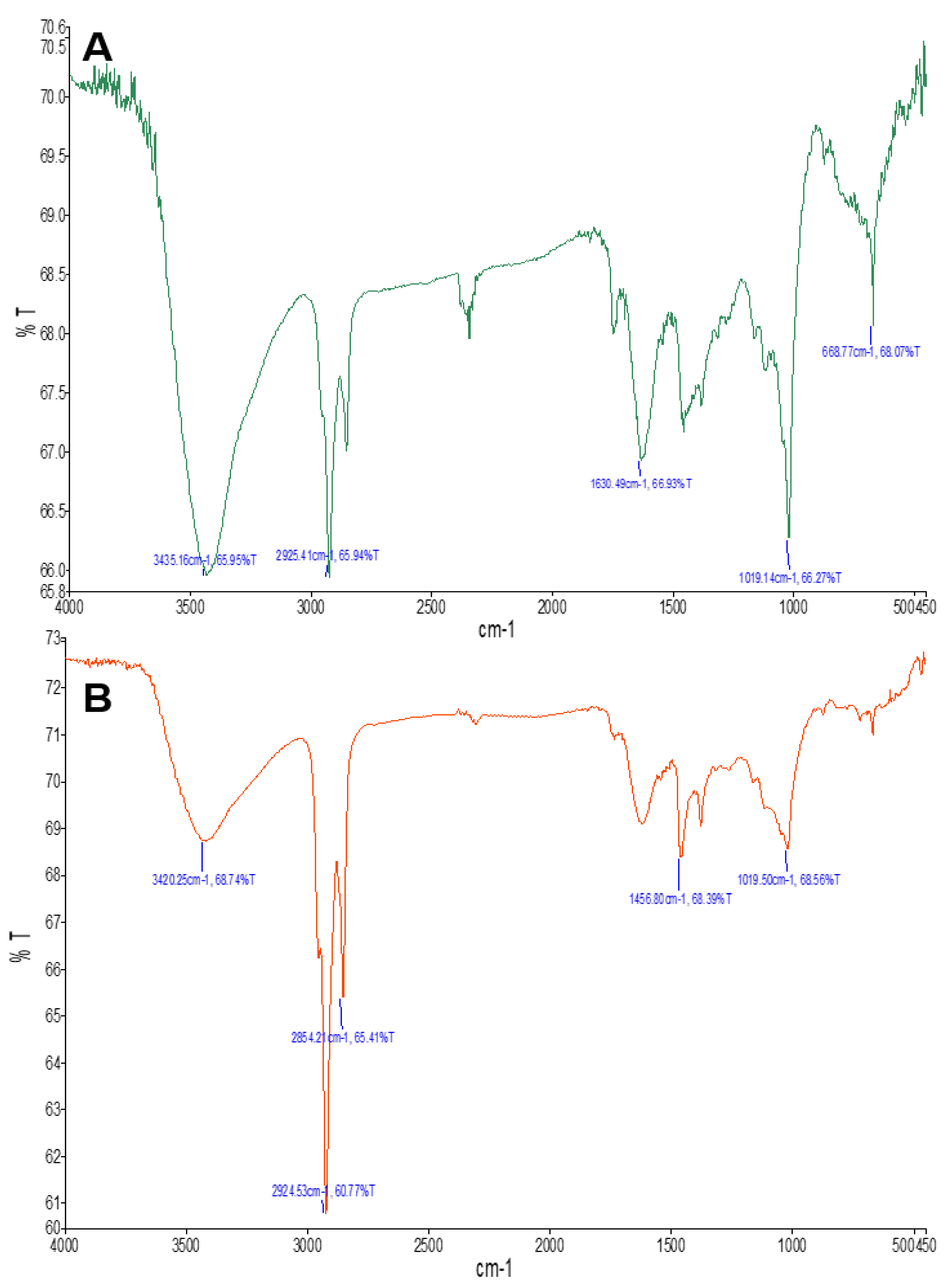
3.2. FTIR Spectroscopy
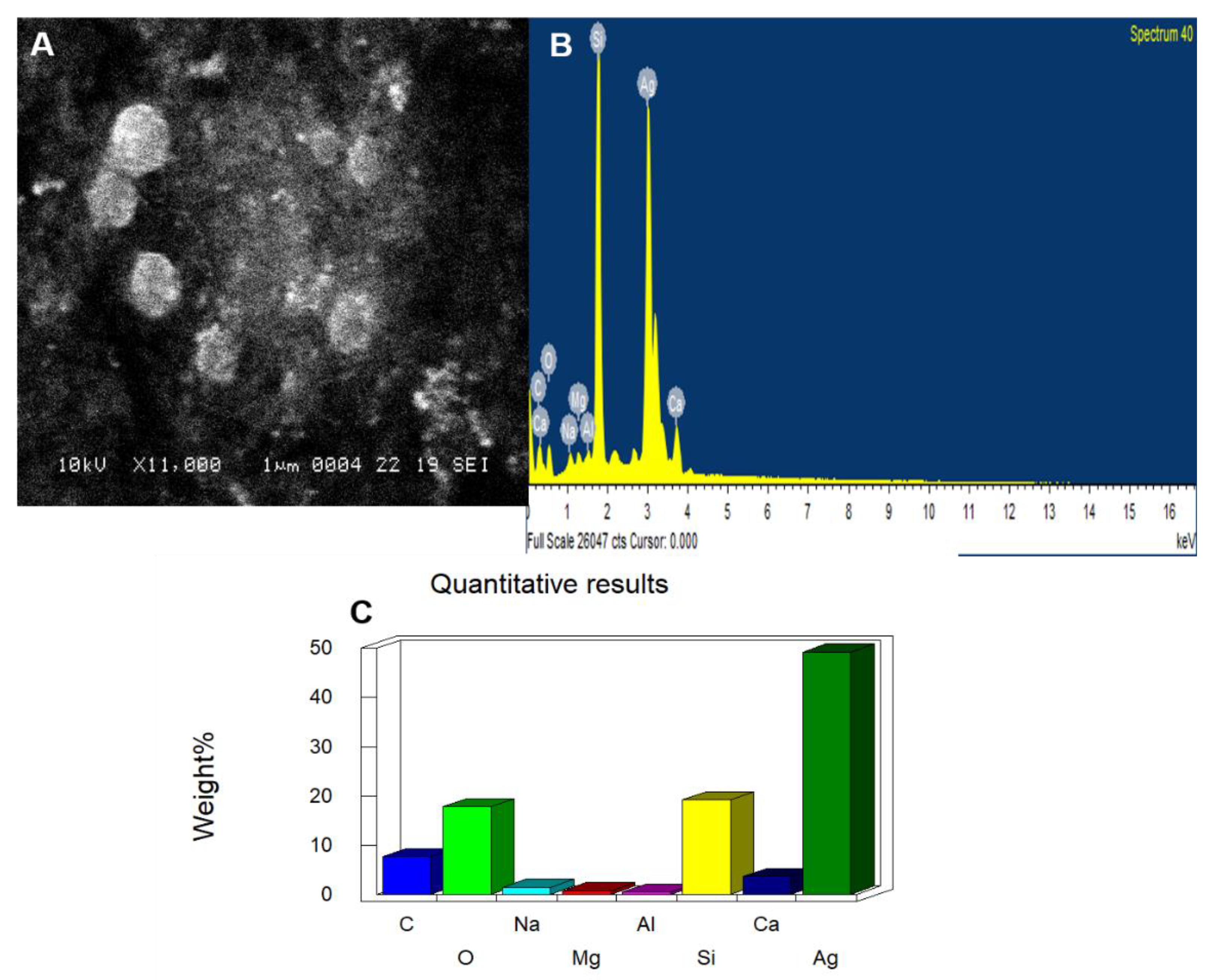
FTIR spectroscopy was performed to identify the biomolecules that were responsible for the capping and efficient stabilization of AgNPs. The key peaks, wavenumbers, and interpretation of the probable functional groups are displayed in Figure 2 and Table 1 of the FTIR spectra of seed extract and Aw–AgNPs. The intensity of the banding is decreased due to silver ion reduction in biosynthesized nanoparticles (Figure 2B). The band at 1019.06 cm−1 indicates the N-H bonding vibration of amides. The bands between 1019 and 668 cm−1 in the seed extract illustrates the C=C stretching mode in aromatic compounds and indicates the occurrence of certain aromatic compounds such as flavonoids. Ajwa seed powder consists of various bioactive compounds like flavonoids, terpenoids, alkaloids, glycosides etc. Researchers also demonstrated the presence of hydroxyl, carboxyl, and carbonyl functional groups in carbohydrates, flavonoids, tannins, and phenolic acids of Ajwa date fruit and seed extracts which may be accountable for the reduction of the Ag+ ions and stabilization of AgNPs [2,10]. The role of flavonoids and phenolic compounds in the reaction mechanism during formation of AgNPs could be derived from their electron or hydrogen donor properties, with the keto form present on the backbone of flavonoid compounds being responsible for the reduction of Ag+ to Ag.

3.3. SEM and EDAX

The SEM image highlighted that the formed nanoparticles were spherical in shape and were fine and uniform in size (Figure 3A). The biosynthesized AgNPs from various plant extracts have also been described previously [27,28,29]. Our results are consistent with those obtained by Narayanaswamy et al., [25] who showed that the AgNPs prepared from leaf extracts of Clitoria ternatea and Solanum nigrum were mostly spherical in shape and 10 to 50 nm in diameter. The elemental composition of Aw–AgNPs was analyzed by EDAX. In the acquired EDAX spectrum, all the elements showed a unique set of peaks with different atomic structures (Figure 3C). The EDAX elemental composition presented in Figure 3B,C highlights strong signal of silver element with weight % of 50. AgNPs have been synthesized from same source in the study by Farhadi et al. and this is reflected in our EDAX results [2].

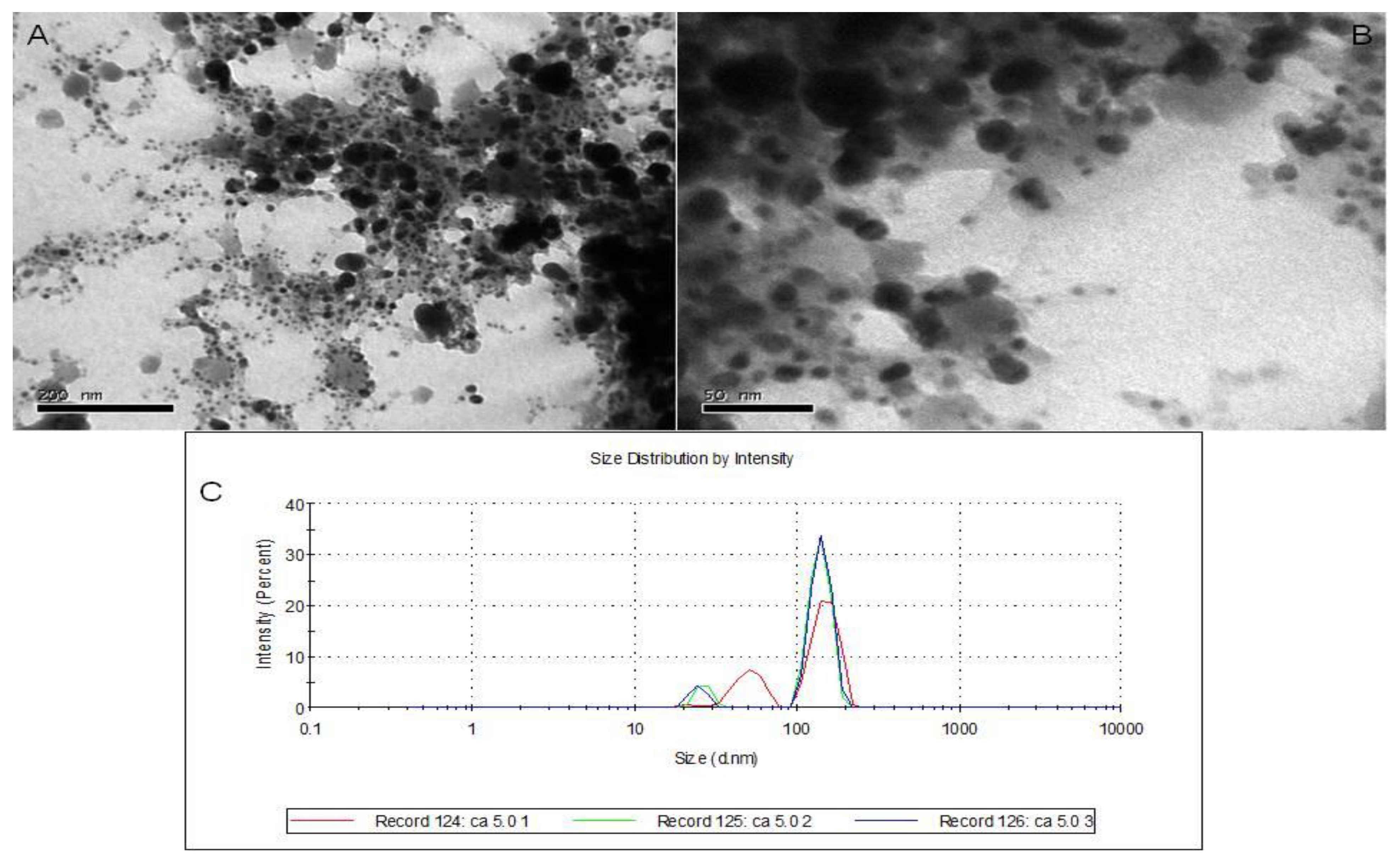
3.4. TEM Analysis and DLS

The shape of the synthesized AgNPs was spherical and the diameter of biosynthesized AgNPs, as characterized by TEM, were in the range of 15 to 80 nm with a narrow size distribution pattern (Figure 4A,B). This confirms the findings obtained by our SEM analysis and are similar to those observed by Farhadi et al. who determined the size of biosynthesized date fruit AgNPs to be in a range between 25 and 60 nm [2]. The particle size distribution of AgNPs of Ajwa in relation to intensity (percent) was monitored through Dynamic light scattering analysis (Figure 4C). From this figure we concluded that the inconsistency in the size of AgNPs is because of few agglomerations in sample. Supporting studies were done to determine mean size of nanoparticles in aqueous solutions utilizing Dynamic light scattering. TEM is one of the most powerful methods for obtaining direct structural and size information of nanoparticles.

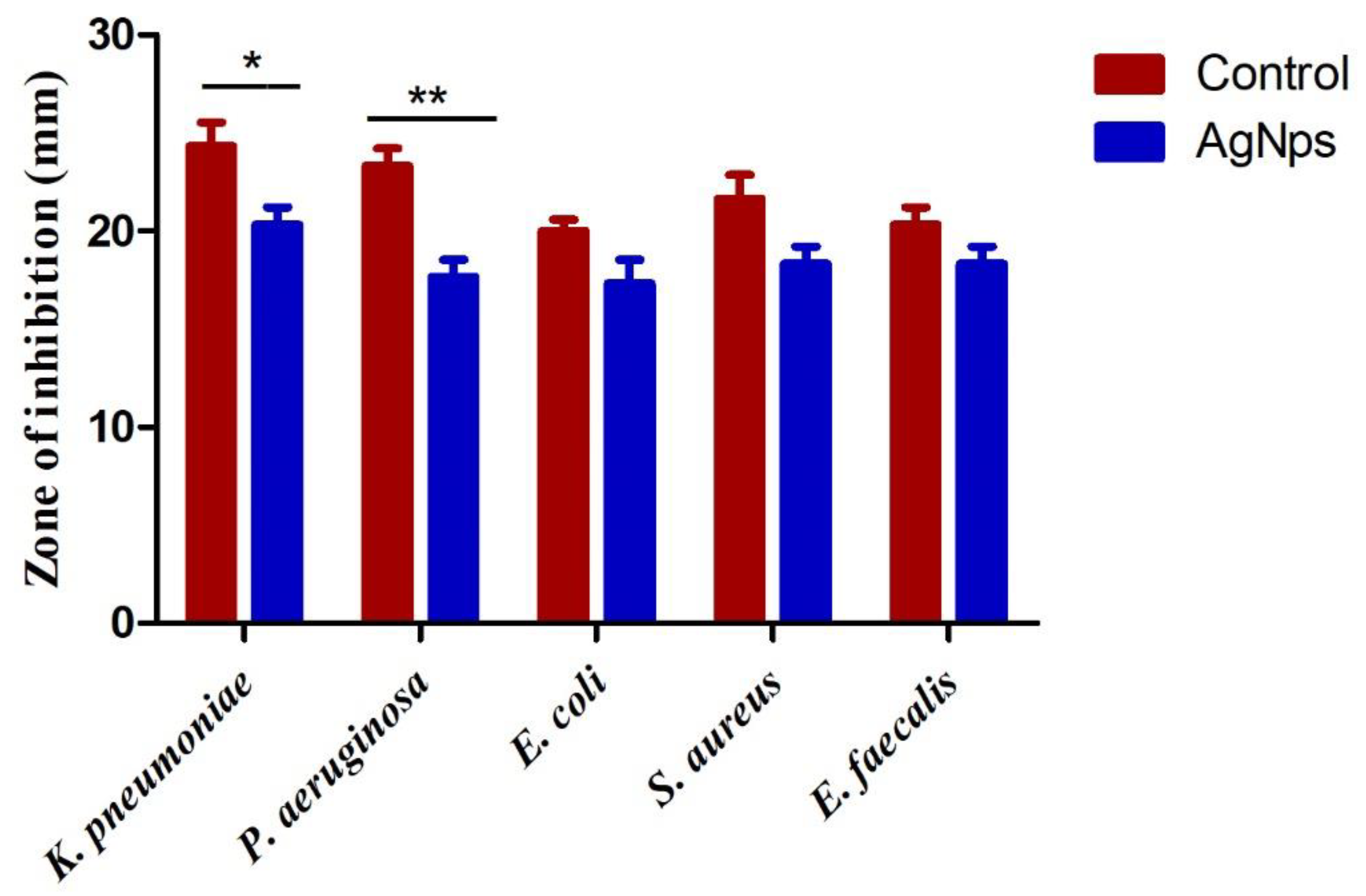
3.5. Assessment of Anti-Bacterial Activity

The activity of Aw–AgNPs against bacterial strains was observed at 100 µg/mL, as indicated in Figure 5. These results show that Aw–AgNPs display an evident significant effect against the tested bacterial strains. These finding are supported by an earlier study that showed that the highest antimicrobial activity was exhibited against Staphylococcus epidermidis, whereas the lowest antimicrobial activity was noted against Bacillus cereus and Escherichia coli [2]. In this study Aw–AgNPs displayed evident anti-bacterial activity, with significant (p < 0.05, p < 0.01) impacts on Gram-negative than Gram-positive bacteria. Similarly, a study by Narayanaswamy et al. showed that biosynthesized AgNPs from the leaf of two different plants presented very strong bactericidal effects against Pseudomonas aeruginosa, Escherichia coli, Staphylococcus aureus, and Streptococcus viridians [30]. Aw–AgNPs exhibited significantly higher inhibition of Staphylococcus aureus and Escherichia coli than the Sukkari dates seed extract AgNPs reported by Salem [31]. Similarly, anti-bacterial activity of AgNPs assessed by broth dilution method shows that the MIC of AgNPs synthesized by date seed were found 1.56 µg/mL against A. baumannii (ATCC 19606) and K. pneumonia (PCI 602) [10]. In the present work, synthesized Aw–AgNPs showed a strong anti-bacterial effect and was similar to that observed with the use of penicillin, as indicated in Figure 5, highlighting its possible utility as an anti-bacterial agent. Our study exhibited significantly higher 20, 18, 17, 19, 19 mm zone of inhibition against Klebsiella pneumonia, Pseudomonas aeruginosa, Escherichia coli, Staphylococcus aureus and Enterococcus faecalis when compared with the Al-Tamimi et al. [32].

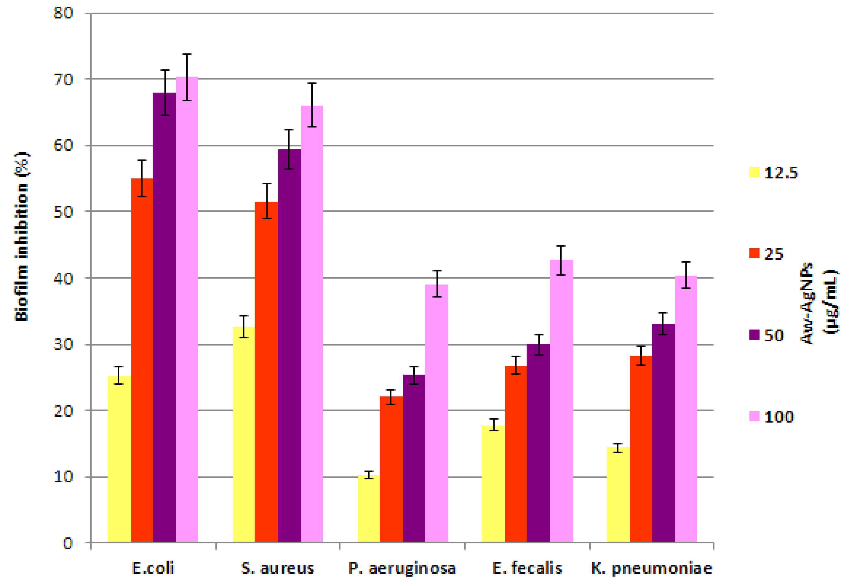
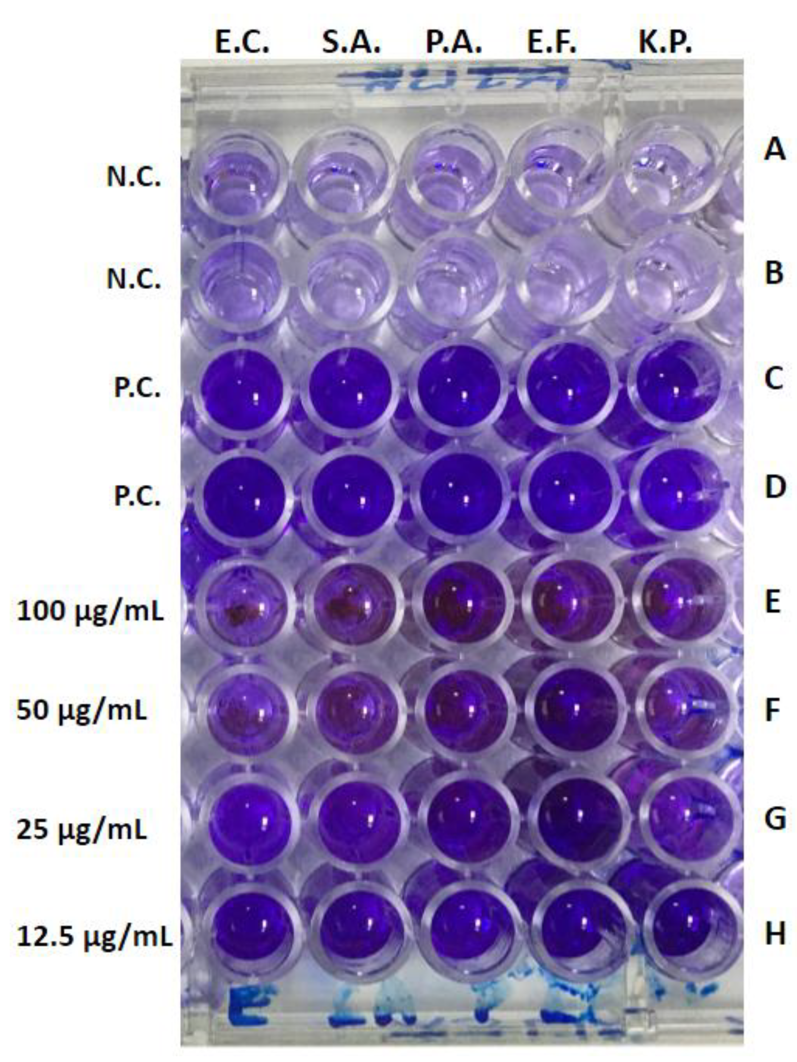
3.6. Anti-Biofilm Activity

Although the anti-biofilm activity of AgNPs synthesized by various plant extracts has been well documented in the literature, reports on the anti-biofilm potential of nanoparticles synthesized by date seed extract are scanty. The anti-biofilm potential of Aw–AgNPs against various bacterial strains is shown in Figure 6 and Figure 7. It has been observed that Aw–AgNPs inhibits the biofilm formation of E. coli and S. aureus by 54.97% and 51.62%, respectively, at a 25 μg/mL concentration. Conversely, the same concentration of Aw–AgNPs showed lower activity against the biofilm of Pseudomonas aeruginosa, Enterococcus faecalis, and Klebsiella pneumonia. The findings from this study suggest that Aw–AgNPs could reduce the biofilm formed by Gram-negative as well as Gram-positive bacteria (Figure 7). It was also observed that a 50 µg/mL concentration of Aw–AgNPs significantly inhibited the biofilm formation in E. coli and S. aureus, while a 100 µg/mL concentration of Aw–AgNPs is able to inhibit the biofilm formation in the rest of the bacterial strains. Previous studies also suggested the same pattern of anti-biofilm activity in AgNPs [7]. Al-Tamimi et al. [32] reported the inhibition of the biofilm activity of the Ajwa dates of 70.5% and 54.19% against S. aureus and Salmonella strains. In our study, we reported significant 70, 66, 39, 43, 41% inhibition against E. coli, S. aureus, P. aeruginosa, Enterococcus faecalis, and Klebsiella pneumonia. Furthermore, Aw–AgNPs at a dose of 50 μg/mL prevented 30 to 60% of the biofilm formation and diminished the growth of the bacteria itself. Kalishwaralal et al. also reported similar results against biofilm-producing Gram-positive and Gram-negative bacteria and observed that 100 μM of AgNPs resulted in a 40 to 70% reduction in biofilm [33].

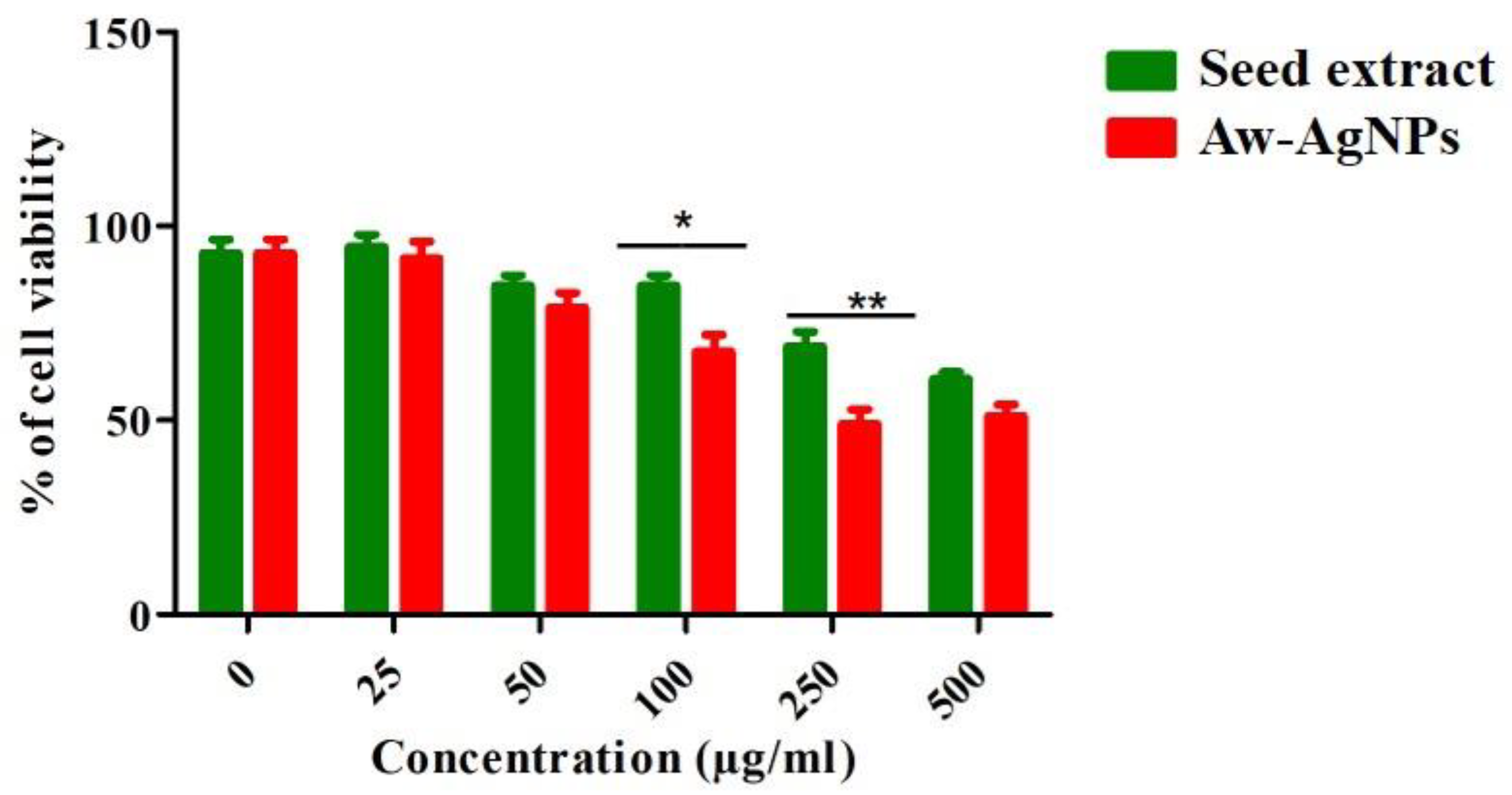
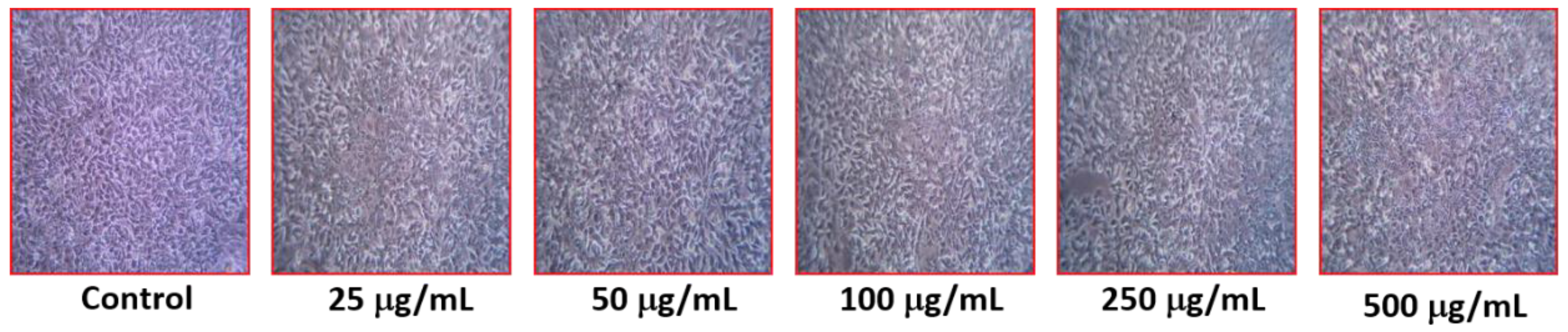
3.7. Effect of Aw–AgNPs against Breast Cancer Cell Line HCC712

The cytotoxicity of Aw–AgNPs was evaluated against breast cancer cell line HCC712 in the range of 25 to 500 µg/mL. Aw–AgNPs exhibited a dose-dependent response against HCC712 cells. Our findings showed that the percentage of cell viability decreased with increasing Aw–AgNPs concentration and significant cytotoxicity was observed at 100 and 250 µg/mL (Figure 8 and Figure 9). Khateef et al. [27] reported the cytotoxic effect of AgNPs prepared from Buchanania axillaris extracts against human breast cancer cells (MCF-7). They found that, as the concentration of AgNPs increased, cytotoxicity increased, which is similar to our results. We earlier demonstrated the cytotoxic effect of green synthesized AgNPs from Terfezia claveryi against MCF-7, and this is also supportive of the cytotoxicity results against the HCC712 cell line observed in this study [28]. Significant cytotoxicity against human breast cancer cell line HCC712 by using Nigella sativa AgNPs was reported by Almatroudi et al. [5]. A recent study has also reported the cytotoxic activity on a human squamous cell carcinoma cell line (HSC-2) by using Ajwa date pit extract [34], in which they reported that the 0.63 mg/mL concentration significantly decreased the percentage of cell viability, which correlates with the findings of our present study.

4. Conclusions

The current study reveals that the Ajwa date extract may be a viable source for the biosynthesis of AgNPs. The activity of these biosynthesized AgNPs against Gram-negative and Gram-positive bacteria shows that they are able to provide the anti-bacterial activity as well as to fortify the biomedicine value of the Ajwa extract. Furthermore, this study demonstrated that Aw–AgNPs showed a noteworthy cytotoxic effect against HCC712 breast cancer cells. Thus, this bio-green process for the synthesis of AgNPs is simple, rapid, and may be effective in several biomedical applications, including the control of various bacterial infections, as well as the treatment of cancer.

Author Contributions

Conceptualization, K.S.A., H.K. and A.A.; methodology, H.K., M.A., R.K. and M.A.A.; software, H.K., M.A. and M.A.A.; validation, M.A., M.A.A. and E.A.A.; formal analysis, K.S.A., H.K., M.A., M.A.K., M.A.A., M.H.A. and N.M.A.; investigation, K.S.A., A.H.R., F.A., R.K., N.M.A. and A.A.; resources, K.S.A., H.K., R.K. and A.A.; data curation, M.A., M.A.K., A.H.R., R.K. and B.Y.A.; writing—H.K., M.A., M.A.K., A.H.R. and B.Y.A.; writing—review and editing, H.K., M.A., M.A.A., M.A.K., A.H.R., N.M.A. and B.Y.A.; visualization, F.A., E.A.A., M.H.A. and N.M.A.; supervision, K.S.A., H.K., M.A.K., A.H.R., M.A.A. and A.A.; project administration, H.K. and A.A.; funding acquisition, H.K. and A.A. All authors have read and agreed to the published version of the manuscript.

Funding

This research received no external funding.

Institutional Review Board Statement

Not Applicable.

Informed Consent Statement

Not Applicable.

Data Availability Statement

Reported in this study.

Acknowledgments

The researchers would like to thank the Deanship of Scientific Research, Qassim University for funding the publication of this project.

Conflicts of Interest

The authors declare no conflict of interest.

References

  1. Salleh, A.; Naomi, R.; Utami, N.D.; Mohammad, A.W.; Mahmoudi, E.; Mustafa, N.; Fauzi, M.B. The Potential of Silver Nanoparticles for Antiviral and Antibacterial Applications: A Mechanism of Action. Nanomaterials 2020, 10, 1566. [Google Scholar] [CrossRef] [PubMed]
  2. Yaqoob, A.A.; Umar, K.; Ibrahim, M.N.M. Silver nanoparticles: Various methods of synthesis, size affecting factors and their potential applications—A review. Appl. Nanosci. 2020, 10, 1369–1378. [Google Scholar] [CrossRef]
  3. Ansari, M.A.; Alzohairy, M.A. One-pot facile green synthesis of silver nanoparticles using seed extract of Phoenix dactylifera and their bactericidal potential against MRSA. Evid.-Based Complement. Altern. Med. 2018, 2, 1860280. [Google Scholar] [CrossRef] [PubMed] [Green Version]
  4. Lewis, K. Riddle of biofilm resistance. Antimicrob. Agents Chemother. 2001, 45, 999–1007. [Google Scholar] [CrossRef] [PubMed] [Green Version]
  5. Almatroudi, A.; Khadri, H.; Azam, M.; Rahmani, A.H.; Al Khaleefah, F.K.; Khateef, R.; Ansari, M.A.; Allemailem, K.S. Antibacterial, Antibiofilm and Anticancer Activity of Biologically Synthesized Silver Nanoparticles Using Seed Extract of Nigella sativa. Processes 2020, 8, 388. [Google Scholar] [CrossRef] [Green Version]
  6. Kora, A.J.; Rastogi, L. Enhancement of antibacterial activity of capped silver nanoparticles in combination with antibiotics, on model gram-negative and gram-positive bacteria. Bioinorg. Chem. Appl. 2013, 8, 871097. [Google Scholar] [CrossRef] [PubMed]
  7. Palanisamy, N.K.; Ferina, N.; Amirulhusni, A.N.; Zain, Z.M.; Hussaini, J.; Ping, L.J.; Durairaj, R. Antibiofilm properties of chemically synthesized silver nanoparticles found against Pseudomonas aeruginosa. J. Nanobiotechnol. 2014, 12, 1–7. [Google Scholar] [CrossRef] [PubMed] [Green Version]
  8. Christensen, G.; Simpson, W.; Younger, J.J.; Baddour, L.; Barrett, F.; Melton, D.M.; Beachey, E. Adherence of coagulase negative Staphylococci to plastic tissue cultures: A quantitative model for the adherence of staphylococci to medical devices. J. Clin. Microbiol. 1985, 22, 996–1006. [Google Scholar] [CrossRef] [Green Version]
  9. Ashokkumar, S.; Ravi, S.; Velmurugan, S. Green synthesis of silver nanoparticles from Gloriosa superba L. leaf extract and their catalytic activity. Spectrochim. Acta A Mol. Biomol. Spectrosc. 2013, 115, 388–392. [Google Scholar] [CrossRef]
  10. Khatami, M.; Shahram Pourseyedi, S. Phoenix dactylifera (date palm) pit aqueous extract mediated novel route for synthesis high stable silver nanoparticles with high antifungal and antibacterial activity. IET Nanobiotechnol. 2015, 9, 184–190. [Google Scholar] [CrossRef]
  11. Chenthamara, D.; Subramaniam, S.; Ramakrishnan, S.G.; Krishnaswamy, S.; Essa, M.M.; Lin, F.H.; Qoronfleh, M.W. Therapeutic efficacy of nanoparticles and routes of administration. Biomater. Res. 2019, 23, 20. [Google Scholar] [CrossRef] [PubMed]
  12. Michitaka, O.; Naoki, T. Photoreduction of Rhodium (III) Ions in Water with Ultraviolet Light Aiming to Prepare the Dispersions of Ultrafine Particles. Chem. Lett. 1990, 19, 489–492. [Google Scholar] [CrossRef]
  13. Khademalrasool, M.; Farbod, M. A simple and high yield solvothermal synthesis of uniform silver nanowires with controllable diameters. J. Nanostruct. 2015, 5, 415–422. [Google Scholar]
  14. Mizukoshi, Y.; Okisu, K.; Maeda, Y.; Yamamoto, T.A.; Oshima, R.; Nagata, Y. Sonochemical preparation of bimetallic nanoparticles of gold/palladium in aqueous solution. J. Am. Phys. Chem. B 1997, 101, 7033–7037. [Google Scholar] [CrossRef]
  15. Darvishi, S.; Borghei, S.M.; Hashemizadeh, S.A. Structural, optical and electrical properties of silver nanoparticles deposited by spin coating method. J. Nanostruct. 2012, 2, 501–504. [Google Scholar]
  16. Khatami, M.; Alijani, H.; Nejad, M.; Varma, R. Core@ shell nanoparticles: Greener synthesis using natural plant products. Appl. Sci. 2018, 8, 411. [Google Scholar] [CrossRef] [Green Version]
  17. Phongtongpasuk, S.; Poadang, S.; Yongvanich, N. Environmental-friendly method for synthesis of silver nanoparticles from dragon fruit peel extract and their antibacterial activities. Energy Procedia 2016, 89, 239–247. [Google Scholar] [CrossRef] [Green Version]
  18. Banerjee, U.C.; Mittal, A.K.; Chisti, Y. Research review paper: Synthesis of metallic nanoparticles using plant extract. Biotechnol. Adv. 2013, 31, 346–356. [Google Scholar]
  19. Kumar, V.; Yadav, S.K. Plant-mediated synthesis of silver and gold nanoparticles and their applications. Chem. Technol. Biotechnol. 2009, 84, 151–157. [Google Scholar] [CrossRef]
  20. Malik, P.; Shankar, R.; Malik, V.; Sharma, N.; Mukherjee, T.K. Green chemistry based benign routes for nanoparticle synthesis. Nanoparticle 2014, 2014, 302429. [Google Scholar] [CrossRef] [Green Version]
  21. Al-Alawi, R.A.; Al-Mashiqri, J.H.; Al-Nadabi, J.S.M.; Al-Shihi, B.I.; Baqi, Y. Date Palm Tree (Phoenix dactylifera L.): Natural Products and Therapeutic Options. Front. Plant Sci. 2017, 8, 845. [Google Scholar] [CrossRef] [PubMed] [Green Version]
  22. Khan, F.; Khan, T.J.; Kalamegam, G.; Pushparaj, P.N.; Chaudhary, A.; Abuzenadah, A.; Kumosani, T.; Barbour, E.; Al-Qahtani, M. Anti-cancer effects of Ajwa dates (Phoenix dactylifera L.) in diethylnitrosamine induced hepatocellular carcinoma in Wistar rats. BMC Complement. Altern. Med. 2017, 17, 418. [Google Scholar] [CrossRef] [PubMed]
  23. Hasan, M.; Mohieldein, A. In Vivo Evaluation of Anti Diabetic, Hypolipidemic, Antioxidative Activities of Saudi Date Seed Extract on Streptozotocin Induced Diabetic Rats. J. Clin. Diagn. Res. 2016, 10, FF06–FF12. [Google Scholar] [CrossRef] [PubMed]
  24. Sirry, S.M.; Samah Ali, S.; Abdelaziz, A.; Mohamed, A. Biosynthesis of silver nanoparticles using the extracts of palm date seeds (Phoenix dactylifera L.) and its application in increasing the anti-inflammatory effect of piroxicam drug. Adv. Nat. Sci. Nanosci. Nanotechnol. 2020, 11, 035017. [Google Scholar] [CrossRef]
  25. CLSI. Performance Standards for Antimicrobial Susceptibility Testing, Fifteenth Informational Supplement, CLSI Document; 2006 M100-S16, vol 26-3; M7-A7, vol 26-2; M2-A9, vol 26-1; CLSI: Wayne, PA, USA, 2006. [Google Scholar]
  26. Jadhav, S.; Shah, R.; Bhave, M.; Palombo, E.A. Inhibitory activity of yarrow essential oil on listeria planktonic cells and biofilms. Food Control 2013, 29, 125–130. [Google Scholar] [CrossRef]
  27. Das, G.; Patra, J.K.; Debnath, T.; Ansari, A.; Shin, H.-S. Investigation of antioxidant, antibacterial, antidiabetic, and cytotoxicity potential of silver nanoparticles synthesized using the outer peel extract of Ananas comosus (L.). PLoS ONE 2019, 14, e0220950. [Google Scholar] [CrossRef] [Green Version]
  28. Erdogan, O.; Abbak, M.; Demirbolat, G.M.; Birtekocak, F.; Aksel, M.; Pasa, S.; Ozge, C. Green synthesis of silver nanoparticles via Cynara scolymus leaf extracts: The characterization, anticancer potential with photodynamic therapy in MCF7 cells. PLoS ONE 2019, 14, e0216496. [Google Scholar] [CrossRef] [Green Version]
  29. Hamouda, R.A.; Hussein, M.H.; Abo-Elmagd, R.A.; Bawazir, S.S. Synthesis and biological characterization of silver nanoparticles derived from the cyanobacterium Oscillatoria limnetica. Sci. Rep. 2019, 9, 13071. [Google Scholar] [CrossRef]
  30. Narayanaswamy, K.; Athimoolam, R.; Ayyavoo, J. Green Synthesis of Silver Nanoparticles Using Leaf Extracts of Clitoria ternatea and Solanum nigrum and Study of Its Antibacterial Effect against Common Nosocomial Pathogens. J. Nanosci. 2015, 2015, 928204. [Google Scholar] [CrossRef] [Green Version]
  31. Salem, S.H. Biosynthesis of Silver Nanoparticles from Date (Phoenix dactylifera) Seeds Extract and Evaluation of Antibacterial Activity against Pathogenic Bacteria. Orient. J. Chem. 2020, 36, 1189–1193. [Google Scholar]
  32. Al-Tamimi, A.; Alfarhan, A.; Rajagopal, R. Antimicrobial and anti-biofilm activities of polyphenols extracted from different Saudi Arabian date cultivars against human pathogens. J. Infect. Public Health 2021, 14, 1783–1787. [Google Scholar] [CrossRef] [PubMed]
  33. Kalishwaralal, K.; BarathManiKanth, S.; Pandian, S.R.; Deepak, V.; Gurunathan, S. Silver nanoparticles impede the biofilm formation by Pseudomonas aeruginosa and Staphylococcus epidermidis. Colloids Surf. B Biointerfaces 2010, 79, 340–344. [Google Scholar] [CrossRef] [PubMed]
  34. Shahbaz, K.; Asif, J.A.; Liszen, T.; Nurul, A.A.; Alam, M.K. Cytotoxic and Antioxidant Effects of Phoenix dactylifera L. (Ajwa Date Extract) on Oral Squamous Cell Carcinoma Cell Line. BioMed Res. Int. 2022, 2022, 5792830. [Google Scholar] [CrossRef] [PubMed]
Figure 1. UV spectrum of the AgNPs synthesized from Ajwa date seed extract. Values expressed as mean ± SE of the mean (n = 3). (Inset: change in color from pale yellow to tan-brownish color).
Applsci 12 04537 g001
Figure 2. FTIR spectrum of Ajwa dates. (A) Seed extract; (B) AgNPs in the range of 4000 to 400 cm−1.
Applsci 12 04537 g002
Figure 3. Characterization of AgNPs. (A) SEM image of AgNPs; (B) EDAX spectrum of AgNPs; (C) quantitative results obtained by EDAX from Ajwa date extract.
Applsci 12 04537 g003
Figure 4. Characterization of AgNPs. (A) TEM image of AgNPs. The scale bar corresponds to 200 nm. (B) Closer view of the AgNPs. The scale bar corresponds to 50 nm. (C) Particle size distribution obtained by dynamic light scattering (DLS).
Applsci 12 04537 g004
Figure 5. Assessment of anti-bacterial activity of Aw–AgNPs (100 µg/mL) and positive control, penicillin. Values expressed as mean ± SE of the mean (n = 3). * Denotes significant difference of positive control (penicillin) with test samples at p < 0.05 and ** p < 0.01.
Applsci 12 04537 g005
Figure 6. Anti-biofilm activity, represented by biofilm inhibition (%), of silver nanoparticles (ug/mL) synthesized from Ajwa dates extract against different standard bacterial strains. Significant difference (p < 0.05) was observed by one-way ANOVA test.
Applsci 12 04537 g006
Figure 7. Image showing the screening of biofilm inhibition in the presence of Aw–AgNPs. Rows (A,B), negative control (NC): culture media only, no bacterial colony, and no Aw–AgNPs; Row (C,D), positive control (PC): culture media plus 5 different bacterial colonies placed accordingly in different wells and no Aw–AgNPs; Row (EH): culture media plus 5 different bacterial colonies placed accordingly in different wells, as well as various concentrations (100 to 12.5 μg/mL) of AgNPs synthesized from Ajwa date extract. EC = indicates Escherichia coli; SA indicates Staphylococcus aureus; PA indicates Pseudomonas aeruginosa; EF indicates Enterococcus faecalis; KP indicates Klebsiella pneumoniae.
Applsci 12 04537 g007
Figure 8. MTT assay results confirming the in vitro cytotoxicity of Aw–AgNPs against HCC 712 human breast cancer cell lines. * Denotes significant difference of control with test samples at p < 0.05 and ** p < 0.01.
Applsci 12 04537 g008
Figure 9. In vitro anti-cancer activity of Aw–AgNPs against HCC 712 human breast cancer cell lines at different concentrations (25–500 µg/mL).
Applsci 12 04537 g009
Table 1. FTIR analysis and probable functional groups.
Aw–AgNPs Wave Number (cm−1)Probable Functional GroupCompound Class
3420N-H StretchAmines
2854C-H StretchAlkanes
2985C-H StretchAlkanes
1030C-O stretchEster
1019C-4-OH stretchTypical for glucose residue of disaccharides
668C=C stretchAlkene
Publisher’s Note: MDPI stays neutral with regard to jurisdictional claims in published maps and institutional affiliations.

