Article

Trypsin Depolarizes Pacemaker Potentials in Murine Small Intestinal Interstitial Cells of Cajal

Division of Longevity and Biofunctional Medicine, School of Korean Medicine, Pusan National University, Yangsan 50612, Korea
*
Author to whom correspondence should be addressed.
Appl. Sci. 2022, 12(9), 4755; https://doi.org/10.3390/app12094755
Submission received: 2 March 2022 / Revised: 3 May 2022 / Accepted: 6 May 2022 / Published: 9 May 2022

Abstract

:
Interstitial cells of Cajal (ICCs) generate pacemaker potentials in the gastrointestinal (GI) tract. In this study, the effects of trypsin on pacemaker potentials in murine small intestinal ICCs were examined. We used whole-cell patch-clamp analysis. The results of whole-cell patch-clamp analysis revealed that trypsin dose-dependently depolarized pacemaker potentials and decreased their amplitude. Treatments with the antagonists of neurokinin1 (NK1) and NK2 receptors (SR-140333 and SR-48968, respectively) slightly inhibited the trypsin-induced responses. However, treatment with the combination of SR-140333 and SR-48968 completely inhibited trypsin-induced responses. Trypsin slightly depolarized pacemaker potentials and increased their amplitude after the intracellular application of GDP-β-S. Additionally, incubation in external Ca2+-free solution inhibited trypsin-induced responses. In the presence of U-73122, staurosporine, Go6976, or xestospongin C, trypsin did not depolarize the pacemaker’s potentials. However, trypsin depolarized the pacemaker potentials in the presence of rottlerin. Finally, HC067047, a TRPV4 inhibitor, did not affect the trypsin-induced responses. These results suggest that trypsin depolarized pacemaker potentials through NK1 and NK2 receptors in the murine small intestinal ICCs, with this effect being dependent on the G protein, phospholipase C, protein kinase C, inositol triphosphate pathways, and extracellular Ca2+ but being independent of the TRPV4 pathway. Hence, trypsin-mediated GI motility regulation must be considered for prokinetic drug developments.

1. Introduction

Protease-activated receptors (PARs), which belong to the family of seven transmembrane-spanning G protein-coupled receptors (GPCRs), are activated by the serine protease-mediated proteolytic cleavage of the N termini [1]. Among the four known types of PARs (PAR1–4) [2], PAR2 is activated by trypsin, while PAR1, PAR3, and PAR4 are activated by thrombin [3].
Several cells of the gastrointestinal (GI) tract, such as the smooth muscle cells, endothelial cells, enteric neurons, and immune cells, express PARs [4,5]. PARs exhibit various physiological functions in the GI tract. In particular, PARs regulate the glandular exocrine secretion in the GI system [6,7]. PAR2 exerts both pro-inflammatory and anti-inflammatory effects in pancreatitis and related pain [8,9] and gastrointestinal mucosal inflammation/damage [10,11]. PAR1, PAR2, and PAR4, which are expressed in the smooth muscle cells and their adjacent cells, regulate the motility of the smooth muscles [12,13]. The roles of PARs in GI motility regulation are complex and vary depending on the species and organs. PAR2 and PAR1 agonists elicit a strong contraction in isolated mouse gastric longitudinal smooth muscle strips, whereas they elicit a transient relaxation in carbachol-contracted muscles [13,14]. Additionally, PAR2 and PAR1 agonists elicit a transient relaxation followed by a contraction in the isolated mouse small intestine [13]. Various fundamental and clinical studies have reported the role of PAR2 in colonic pain [15,16].
In contrast to receptors activated through ligand–receptor interactions, PAR2 is activated by a proteolytic mechanism in which the PAR2 agonist (i.e., trypsin) binds and cleaves the amino terminus of the receptor [3]. The activation of PAR2 by trypsin increases inositol triphosphate (IP3) production through Gαq/11 coupling [6,11]. Trypsin, a serine protease, is a widely studied protease for which its role in intestinal digestion has been previously reported [1].
Interstitial cells of Cajal (ICCs) are considered electrical pacemakers for the generation of slow waves in the GI tract. The loss of ICCs can cause severe gut diseases, including dysmotility, in both humans and animals [17,18,19]. Therefore, ICCs are reported to be pharmacological targets for regulating GI motility. Sung et al. [20] suggested that PARs regulate the excitability of colonic muscles and activate different conductance pathways in each cell type of the smooth muscle cell-ICC-platelet-derived growth factor receptor α-positive cell syncytium. Additionally, PAR1 and PAR2 modulate the pacemaker activity of colonic ICCs through the phospholipase C (PLC)-dependent [Ca2+]i release pathway [21]. However, the mechanism underlying the trypsin-PAR2 signaling-mediated regulation of small intestinal ICCs has not been elucidated.
In this study, the effects of trypsin and PAR2 activation on the pacemaker potentials in murine small intestinal ICCs were investigated using whole-cell patch-clamp techniques.

2. Materials and Methods

2.1. Ethical Statement

All animal care and experimental procedures were performed according to the guidelines of the Institutional Animal Care and Use Committee at Pusan National University (Busan, Korea; Approval no. PNU-2020-2831).

2.2. Preparation of ICCs and ICC Clusters

Male and female mice (aged 3–6 days with a bodyweight of 2.0–2.3 g) were purchased from the Institute of Cancer Research (ICR, Samtako Bio Korea Co., Ltd., Osan, Korea). The animals were maintained under controlled conditions (temperature, 21 ± 3 °C; relative humidity (RH), 50 ± 6%; circadian cycle, 12 h light–dark cycle). The small intestines were excised and dissected along the mesenteric border. The luminal contents were removed using a Krebs-Ringer bicarbonate solution. The mucosae were obtained after sharp dissection. Small tissue strips of the intestinal muscle were equilibrated with Ca2+-free Hank’s solution (containing 5.36 mM KCl, 125 mM NaCl, 0.34 mM NaOH, 0.44 mM Na2HCO3, 10 mM glucose, 2.9 mM sucrose, and 11 mM HEPES; pH 7.4) for 30 min. The cells were dispersed in an enzyme solution containing collagenase (Worthington Biochemical, Lakewood, NJ, USA) and cultured in smooth muscle growth medium (Clonetics, San Diego, CA, USA) supplemented with 2% antibiotic/antimycotic solution (Gibco, Grand Island, NY, USA) and 5 ng/mL−1 murine stem cell factor (Sigma-Aldrich, St. Louis, MO, USA) at 37 °C in a 95% O2-5% CO2 incubator. To isolate ICCs, the ICCs cultured for <12 h were incubated with anti-c-kit antibodies (1:50; eBioscience, San Diego, CA, USA) for 20 min [22].

2.3. Patch-Clamp Experiments

The whole-cell patch-clamp method was used to record the effects of trypsin on the pacemaker potentials of ICCs. The composition of the physiological salt solution used to bathe the cultured ICC clusters was 5 mM KCl, 135 mM NaCl, 2 mM CaCl2, 10 mM glucose, 1.2 mM MgCl2, and 10 mM HEPES (adjusted to pH 7.4 with NaOH) [23]. Meanwhile, the composition of the pipette solution used to examine pacemaker potentials was 140 mM KCl, 5 mM MgCl2, 2.7 mM K2ATP, 0.1 mM NaGTP, 2.5 mM creatine phosphate disodium, 5 mM HEPES, and 0.1 mM EGTA (adjusted to pH 7.2 with KOH). The patch-clamp experiments were performed using Axopatch I-D and Axopatch 200B amplifiers (Axon Instruments, Burlingame, CA, USA). Command pulses were applied using pClamp software (version 6.1; Axon Instruments). The data were filtered at 5 kHz with Axopatch 200B amplifiers. The results were analyzed using pClamp and Origin software version 6.0 (Microcal, Northampton, MA, USA). All experiments were performed at 30–32 °C.

2.4. Drugs

AC55541 (selective PAR2 agonist) [24], SR-140333 (NK1 receptor antagonist) [25], staurosporine (a broad-spectrum protein kinase C (PKC) inhibitor) [26], Go6976 (a calcium-dependent PKC α/β inhibitor) [27], rottlerin (a calcium-independent PKC δ inhibitor) [28], HC067047 (selective TRPV4 antagonist) [29], and thapsigargin (inhibitor of Ca2+-ATPase in the endoplasmic reticulum) [30] were purchased from Tocris Bioscience (Bristol, UK). Trypsin, U-73122 (PLC inhibitor) [31], xestospongin C (a selective and membrane-permeable inhibitor of IP3 receptor) [32], guanosine 5′-diphosphate (GDP)-β-S (for the inactivation of G protein-binding proteins) [33,34], and other drugs were purchased from Sigma-Aldrich. The stock solutions of all drugs were prepared in distilled water or dimethyl sulfoxide (DMSO) and stored at −20 °C. The final concentration of DMSO in the bath solution was less than 0.1%. At this concentration, DMSO did not affect the results. Additionally, the addition of these chemicals did not alter the pH of the bath solution.

2.5. Statistical Analysis

All results are expressed as mean ± standard error of mean (n refers to the number of cells used in the experiments). The means of different groups were analyzed using one-way analysis of variance, followed by Bonferroni’s post hoc test. All statistical analyses were performed using Prism 6.0 (GraphPad Software Inc., San Diego, CA, USA) and Origin version 8.0 (OriginLab Corporation, Northampton, MA, USA). Differences were considered significant at p < 0.05.

3. Results

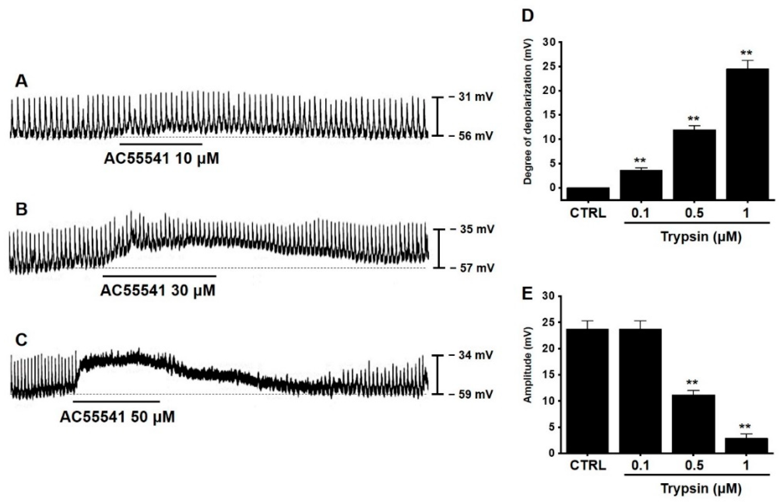
3.1. Effects of Trypsin on Pacemaker Potentials in Murine Small Intestinal ICCs

The pacemaker potentials of cultured ICC clusters were examined using the whole-cell patch-clamp technique. The ICC clusters formed network-like structures after culturing for <12 h and generated spontaneous rhythmic contractions [35]. In the current clamp mode (I = 0), the murine small intestinal ICCs generated pacemaker potentials with mean resting membrane potential and mean amplitude of −57.2 ± 2.3 and 23.8 ± 1.6 mV, respectively (Figure 1). Trypsin (0.1–1 μM) dose-dependently depolarized the pacemaker potentials and decreased their amplitudes (Figure 1A–C). The mean degrees of depolarization in the presence of 0.1, 0.5, and 1 μM trypsin were 5.4 ± 0.8 (p < 0.01), 12.7 ± 1.1 (p < 0.01), and 26.0 ± 1.6 mV (p < 0.01), respectively (Figure 1D, n = 15), while the mean amplitudes were 21.0 ± 1.5 (p < 0.01), 12.3 ± 1.0 (p < 0.01) and 2.4 ± 0.5 mV (p < 0.01) (Figure 1E, n = 15), respectively. These findings suggest that trypsin dose-dependently depolarizes the pacemaker potentials in murine small intestinal ICCs.

3.2. Effects of AC55541 on Pacemaker Potentials in Murine Small Intestinal ICCs

Next, the effect of AC55541 on pacemaker potentials in murine small intestinal ICCs was examined. The ICCs were incubated with the PAR2 agonist AC55541 [24]. AC55541 (10–50 μM) dose-dependently depolarized the pacemaker potentials and decreased their amplitude (Figure 2A–C). The mean degrees of depolarization in the presence of 10, 30, and 50 μM AC55541 were 3.6 ± 0.5 (p < 0.01), 11.9 ± 0.9 (p < 0.01), and 24.5 ± 1.8 mV (p < 0.01), respectively (Figure 2D, n = 12), while the mean amplitudes were 23.8 ± 1.5, 11.2 ± 0.8 (p < 0.01), and 2.9 ± 0.8 mV (p < 0.01) (Figure 2E, n = 12), respectively. This indicated that trypsin activates PAR2 on murine small intestinal ICCs.

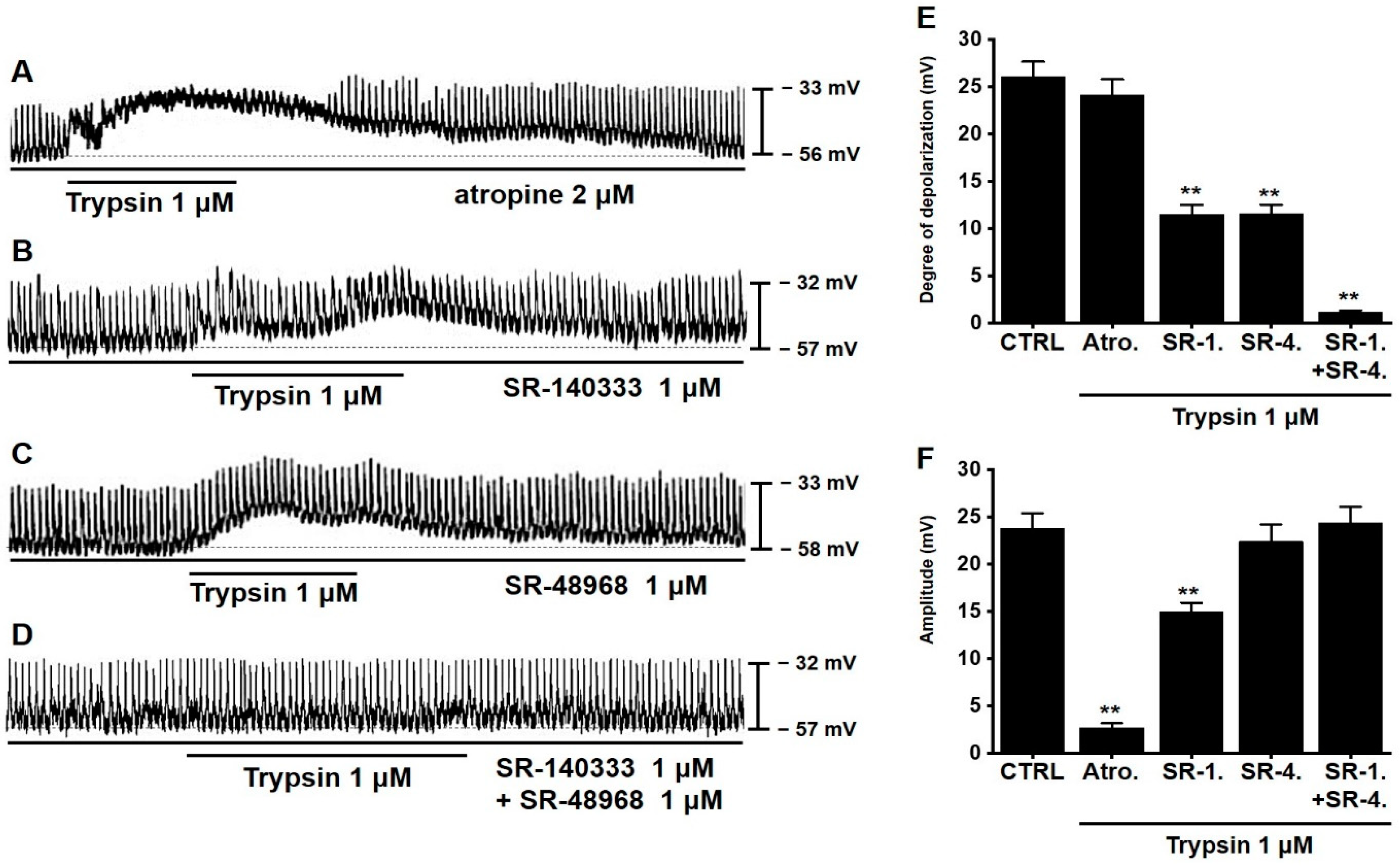
3.3. Involvement of Neurokinin Receptors in Trypsin-Induced Pacemaker Potential Depolarization in Murine Small Intestinal ICCs

To examine the role of cholinergic pathways in trypsin-induced pacemaker potential depolarization in murine small intestinal ICCs, the trypsin-induced responses were compared in the presence and absence of the muscarinic receptor antagonist atropine. Atropine did not affect the pacemaker potentials in murine small intestinal ICCs. Trypsin depolarized the pacemaker potentials in murine small intestinal ICCs in the presence of atropine (Figure 3A). This suggested that the trypsin-induced pacemaker potential depolarization was not regulated by the muscarinic cholinergic system. Next, the roles of neurokinin receptors in trypsin-induced pacemaker potential depolarization in murine small intestinal ICCs were examined. ICCs were treated with the specific neurokinin receptor antagonists SR-140333 (NK1 receptor inhibitor; 1 μM) and SR-48968 (NK2 receptor inhibitor; 1 μM) alone or in combination [36]. In ICCs treated with SR-140333 or SR-48968, trypsin slightly depolarized the pacemaker potentials and decreased their amplitude (Figure 3B,C). However, treatments with the combination of SR-140333 and SR-48968 completely inhibited the trypsin-induced responses (Figure 3D), which suggested that NK1 and NK2 receptors exert an additive effect. The mean degrees of depolarization in the presence of atropine, SR-140333, SR-48968, and SR-140444 + SR-48968 were 24.1 ± 1.7, 11.5 ± 1.0 (p < 0.01), 11.6 ± 0.9 (p < 0.01), and 1.1 ± 0.2 mV (p < 0.01), respectively (Figure 3E, n = 21), while the mean amplitudes were 2.7 ± 0.5 (p < 0.01), 15.0 ± 1.0 (p < 0.01), 22.4 ± 1.8, and 24.3 ± 1.7 mV (Figure 3F, n = 21), respectively. These results suggest that both NK1 and NK2 receptors mediate trypsin-induced pacemaker potential depolarization in murine small intestinal ICCs.

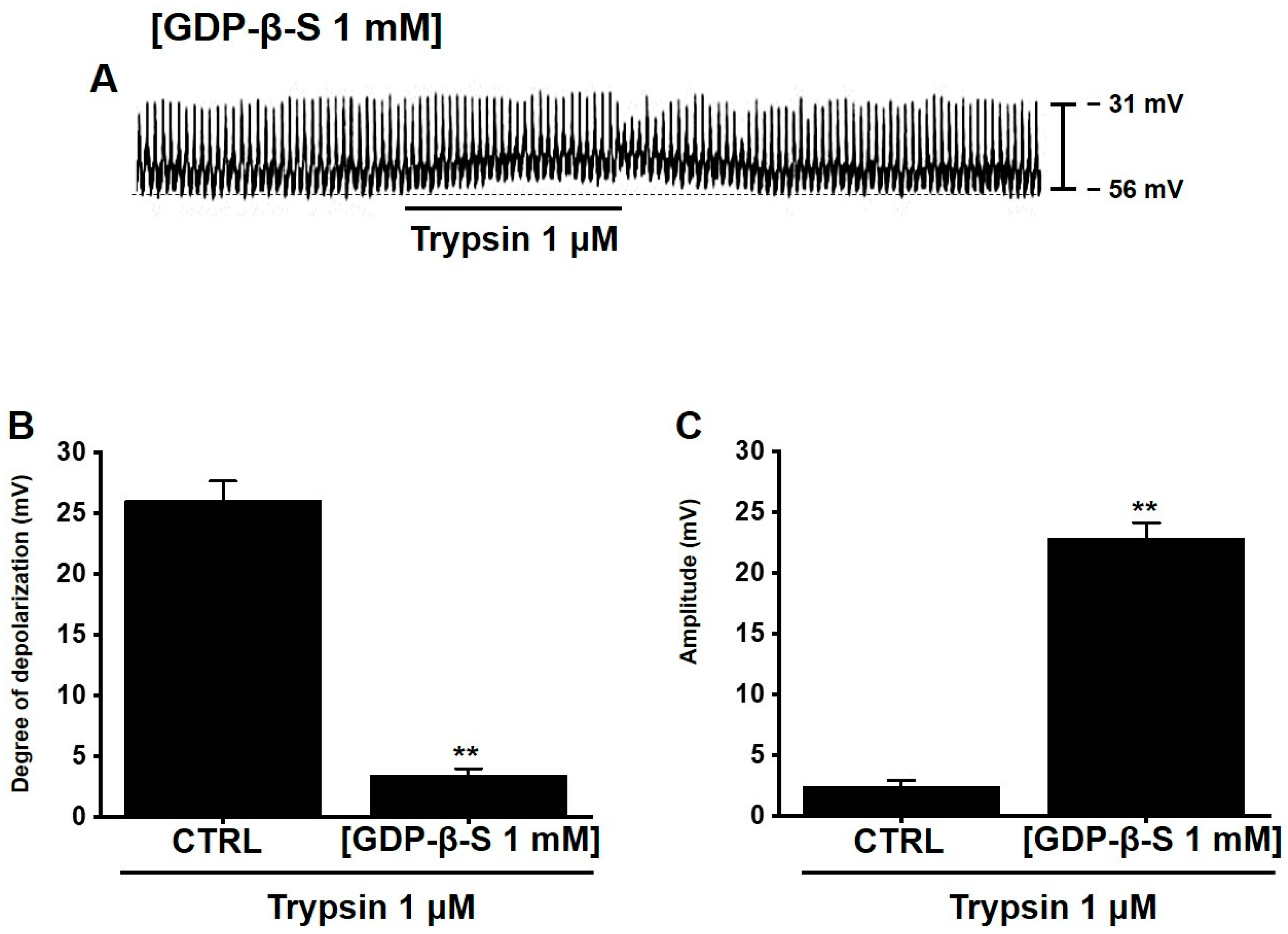
3.4. Involvement of G Proteins in Trypsin-Induced Pacemaker Potential Depolarization in Murine Small Intestinal ICCs

To examine the roles of G proteins in trypsin-induced pacemaker potential depolarization, ICCs were treated with GDP-β-S, which permanently inactivates G protein-binding proteins [33,34]. Trypsin (1 μM) slightly depolarized pacemaker potentials and increased their amplitude after the intracellular application of GDP-β-S (1 mM) (Figure 4A). The mean degree of depolarization and mean amplitude in the presence of GDP-β-S were 3.4 ± 0.5 (p < 0.01) (Figure 4B, n = 6) and 22.8 ± 1.3 mV (p < 0.01) (Figure 4C, n = 6), respectively. These results suggest that G proteins are required for trypsin-induced pacemaker potential depolarization in murine small intestinal ICCs.

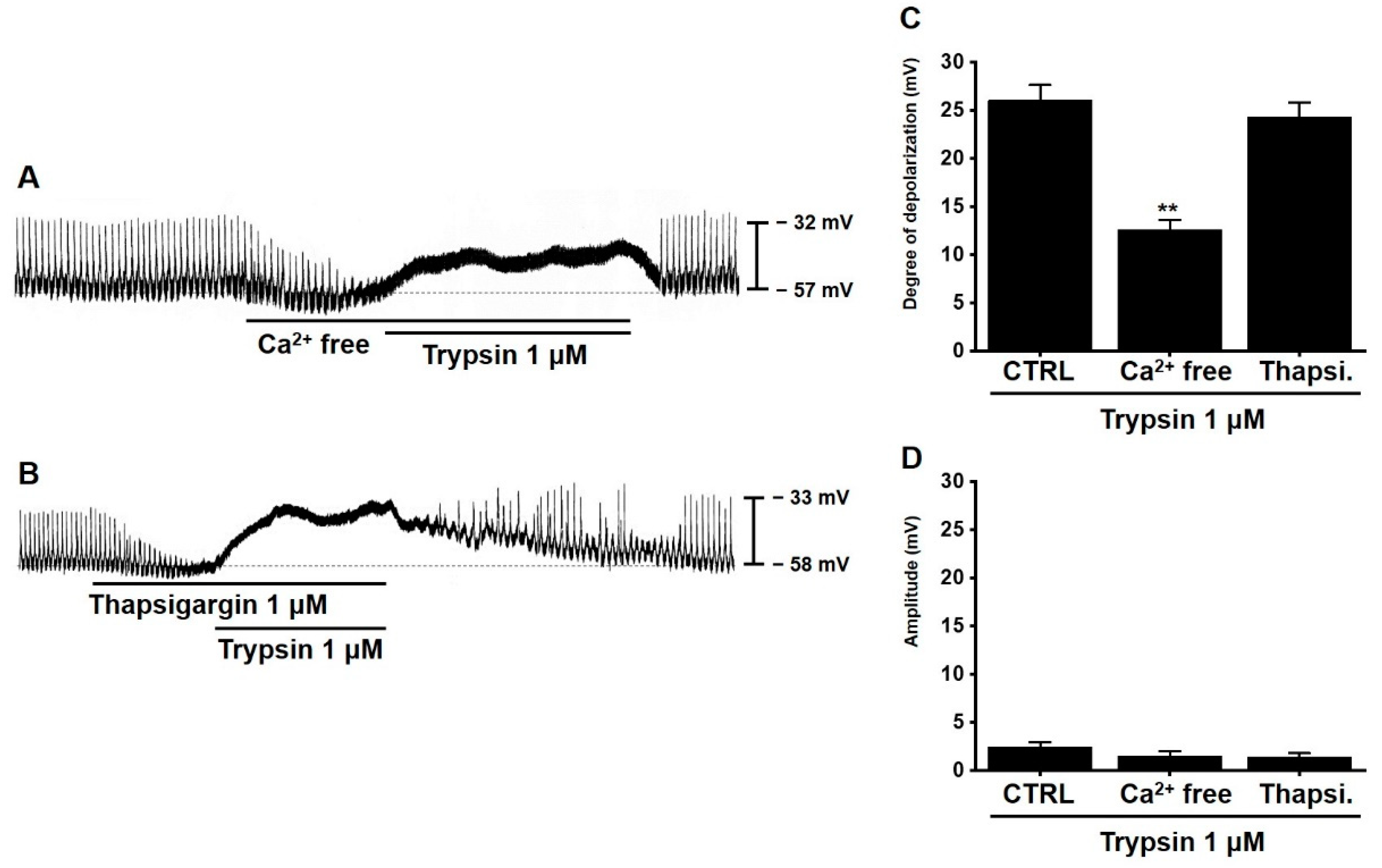
3.5. Involvement of Extracellular and Intracellular Calcium Ions in Trypsin-Induced Pacemaker Potential Depolarization in Murine Small Intestinal ICCs

Intracellular and extracellular Ca2+ are involved in modulating intestinal motility [37]. To investigate the role of Ca2+ in trypsin-induced pacemaker potential depolarization, trypsin-induced depolarization was measured under Ca2+-free conditions or in the presence of thapsigargin, an inhibitor of Ca2+-ATPase in the endoplasmic reticulum. Pre-treatment with external Ca2+-free solution inhibited pacemaker potentials and trypsin-induced pacemaker potential depolarization (Figure 5A). However, pre-treatment with thapsigargin inhibited the pacemaker potential but not the trypsin-induced pacemaker potential depolarization (Figure 5B). The mean degrees of depolarization under external Ca2+-free conditions and in the presence of thapsigargin were 12.5 ± 1.0 (p < 0.01) and 24.3 ± 1.5 mV (Figure 5C, n = 8), respectively, while the mean amplitudes were 1.5 ± 0.5 and 1.4 ± 0.4 mV (Figure 5D, n = 7), respectively. These results suggest that trypsin-induced pacemaker potential depolarization requires extracellular Ca2+ in murine small intestinal ICCs.

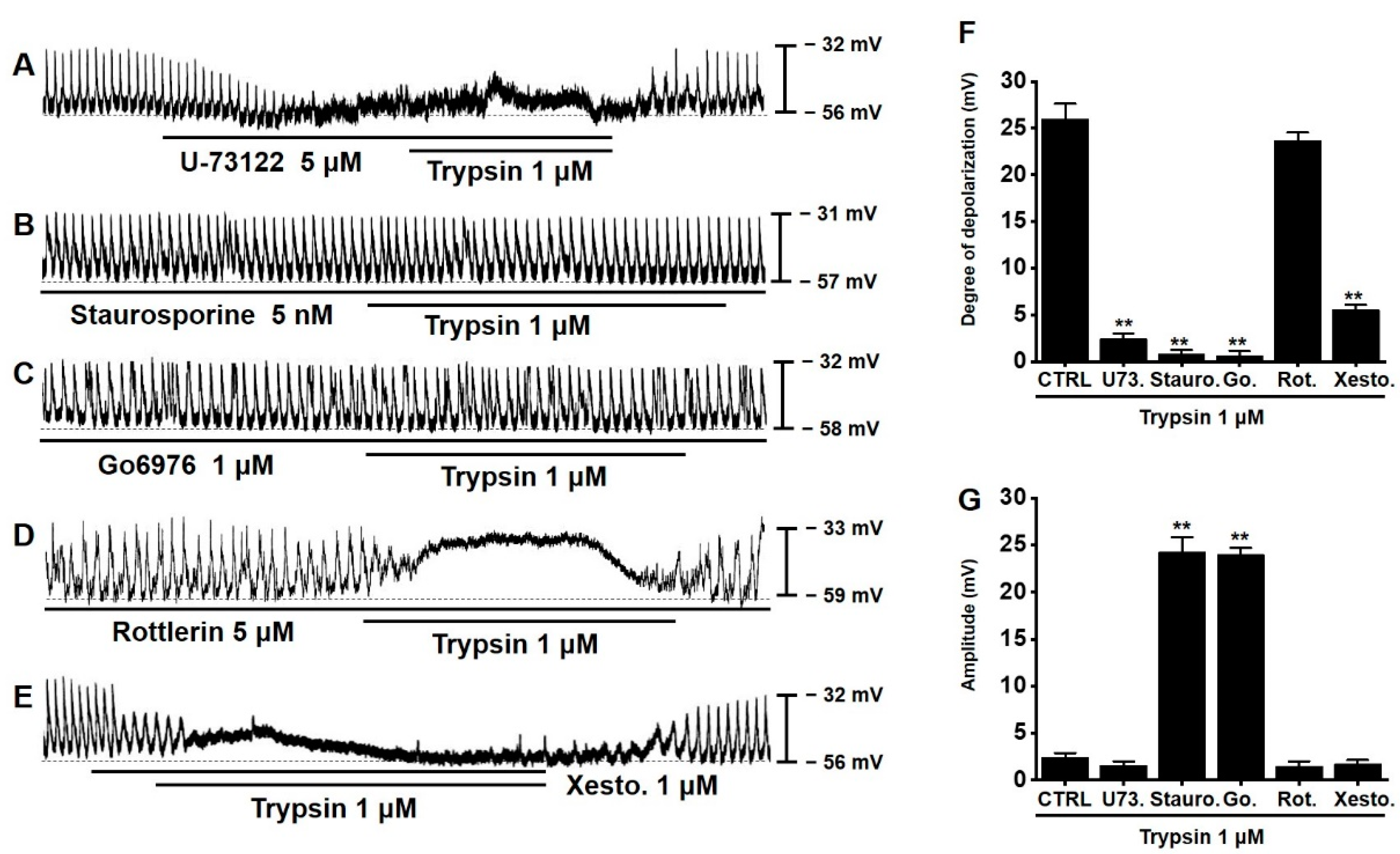
3.6. Involvement of PLC, PKC, and Inositol Triphosphate (IP3) Pathways in Trypsin-Induced Pacemaker Potential Depolarization in Murine Small Intestinal ICCs

Next, the role of the PLC, PKC, and IP3 pathways in trypsin-induced pacemaker potential depolarization was examined using U-73122, staurosporine, Go6976, rottlerin, and xestospongin C. In the presence of U-73122 (5 μM) (Figure 6A), staurosporine (5 nM) (Figure 6B), Go6976 (1 μM) (Figure 6C), and xestospongin C (1 μM) (Figure 6E), trypsin did not depolarize the pacemaker potentials. However, trypsin depolarized the pacemaker potentials in the presence of rottlerin (5 μM) (Figure 6D). The mean degrees of depolarization in the presence of U-73122, staurosporine, Go6976, rottlerin, and xestospongin C were 2.5 ± 0.6 (p < 0.01, n = 7), 0.8 ± 0.4 (p < 0.01, n = 8), 0.6 ± 0.5 (p < 0.01, n = 9), 23.7 ± 0.8 (n = 8), and 5.5 ± 0.6 mV (p < 0.01, n = 6) (Figure 6F), respectively, while the mean amplitudes were 1.6 ± 0.4 (n = 7), 24.3 ± 1.6 (p < 0.01, n = 8), 24.0 ± 0.7 (p < 0.01, n = 9), 1.5 ± 0.5 (n = 8), and 1.7 ± 0.4 mV (n = 6) (Figure 6G), respectively. These results suggest that the PLC, PKC, and IP3 pathways are involved in trypsin-induced pacemaker potential depolarization in murine small intestinal ICCs.

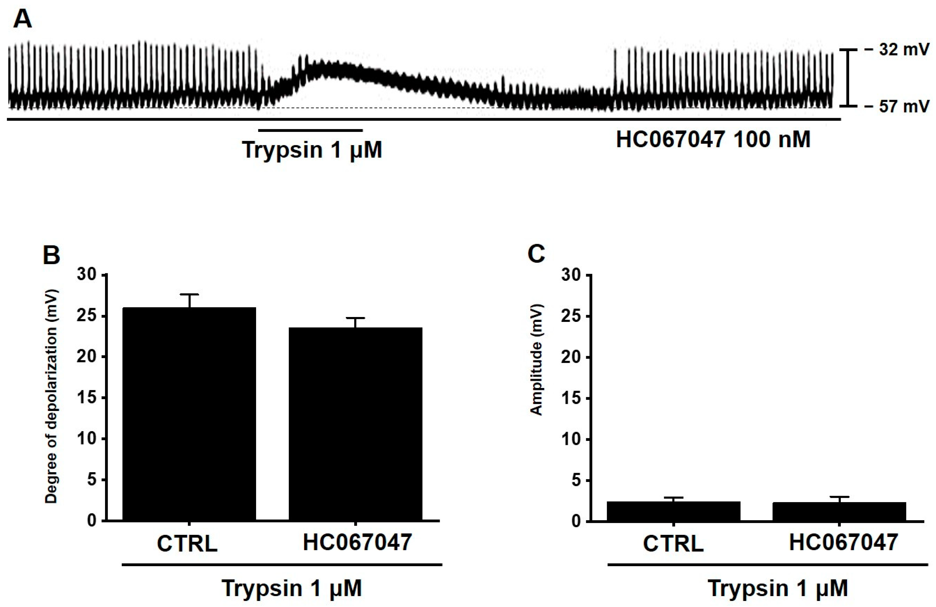
3.7. TRPV4 Is Not Involved in Trypsin-Induced Pacemaker Potential Depolarization in Murine Small Intestinal ICCs

The transient receptor potential (TRP) vanilloids 4 (TRPV4) ion channel is one of the major downstream targets of PAR2 [38,39]. The coupling of PAR2 and TRPV4 is required for sustained inflammatory signaling [40]. Therefore, the role of TRPV4 channels in trypsin-induced pacemaker potential depolarization was investigated using HC067047 (a TRPV4 inhibitor). In the presence of HC067047 (100 nM), trypsin depolarized the pacemaker potentials (Figure 7A). The mean degree of depolarization and mean amplitude in the presence of HC067047 were 23.5 ± 1.2 (Figure 7B, n = 8) and 2.3 ± 0.8 mV (Figure 7C, n = 8), respectively. These results suggest that TRPV4 is not involved in trypsin-induced pacemaker potential depolarization in murine small intestinal ICCs.

4. Discussion

In this study, the mechanism underlying the trypsin-mediated depolarization of pacemaker potentials in murine small intestinal ICCs was examined. Trypsin, a PAR2 agonist, dose-dependently depolarized the pacemaker potentials and decreased their amplitudes in the murine small intestinal ICCs (Figure 1). In addition to trypsin, this study examined the effects of AC55541, another PAR2 agonist, on pacemaker potentials. Consistent with the properties of trypsin, AC55541 dose-dependently depolarized the pacemaker potentials and decreased their amplitude in murine small intestinal ICCs (Figure 2). Trypsin-induced responses were mediated by the additive effects of NK1 and NK2 receptors (Figure 3). Additionally, trypsin-induced responses were dependent on extracellular Ca2+ and the downstream G protein, PLC, PKC, and IP3 signaling pathways. Treatment with GDP-β-S or pre-incubation in Ca2+-free solution inhibited trypsin-induced responses (Figure 4 and Figure 5). Moreover, trypsin did not depolarize the pacemaker potentials in the presence of U-73122, staurosporine, Go6976, or xestospongin C (Figure 6). Additionally, HC067047 did not inhibit trypsin-induced responses (Figure 7). Therefore, the trypsin/PAR2 signaling pathway modulated GI motility by regulating the activity of ICCs in the murine small intestine.
Currently, more than 500 types of proteases have been identified in all mammals. Proteases are reported to be involved in complex physiological processes, including ovulation, intestinal digestion, inflammation, immunity, and wound healing [41]. Trypsin is the major digestive enzyme secreted in the anterior portion of the intestine. Trypsinogen (proform of trypsin) is synthesized by pancreatic acinar cells, secreted into the duodenum, and activated by enterokinases. PARs are a novel subclass of the seven transmembrane-spanning GPCRs [1]. Among the four known types of PARs [2], PAR1, PAR3, and PAR4 are activated by thrombin, while PAR2 is activated by trypsin [3]. The expression of PARs is detected in various tissues, including those of the pancreas, liver, kidney, prostate, ovary, and GI tract and several cells, such as the smooth muscle cells, neurons, fibroblasts, epithelial cells, and neutrophils [42].
PAR2 is involved in various pathophysiological processes of several organs, including the skin [43], cardiovascular system [44,45], pulmonary system [46], and GI tract [2]. In particular, PAR2 is expressed throughout the GI tract, especially in the intestinal cells, nerve cells, and myocytes. This indicates the importance of PAR2 in regulating GI functions and the pathogenesis of GI diseases [47]. PAR2 agonists regulate GI motility by modulating the transport of ions in intestinal epithelial cells [48]. However, the regulatory effects of PAR2 on GI motility vary depending on the location. Although PAR2 promotes contraction in the gastric body [12], contraction and relaxation occur repeatedly in the gastric fundus [14]. PAR2 inhibits spontaneous contractions in the stomach and colon [49]. The results of mouse studies revealed that PAR1 and PAR2 promote GI transit through the L-type Ca2+ channel pathway [50]. Additionally, PAR2 agonists promoted the contraction of murine intestinal smooth muscle, which was dependent on the sensory neural pathways and the NK1 and NK2 receptors [51].
In this study, trypsin-induced cellular responses were dependent on the additive effects of NK1 and NK2 receptors (Figure 3). Generally, GI motility is highly regulated by cholinergic mechanisms. Hence, this study examined the role of the cholinergic system in trypsin-induced pacemaker potential depolarization. Treatment with the muscarinic receptor antagonist atropine did not affect the pacemaker’s potentials. In the presence of atropine, trypsin depolarized the pacemaker’s potentials (Figure 3A), which suggested that the muscarinic cholinergic system was not involved in trypsin-mediated pacemaker potential depolarization. Zao and Shea-Donohue [51] demonstrated that treatment with NK1 and NK2 receptor antagonists decreased trypsin-induced contractions, indicating the involvement of neurokinin receptors in trypsin-induced responses. Therefore, the role of neurokinin receptors in trypsin-induced pacemaker potential depolarization in murine small intestinal ICCs was examined in this study. ICCs were treated with SR-140333 (specific neurokinin receptor NK1 antagonist) and SR-48968 (NK2 antagonist) alone or in combination. In the presence of SR-140333 or SR-48968, trypsin slightly depolarized the pacemaker potentials and decreased their amplitude (Figure 3B,C). However, treatment with the combination of SR-140333 and SR-48968 completely inhibited trypsin-induced responses (Figure 3D). These findings suggest that trypsin-induced responses are dependent on the additive effects of NK1 and NK2 receptors. Tachykinin NK receptors are distributed in the enteric nerves, smooth muscles, and ICCs. Thus, NK receptors may modulate GI motility by regulating the activity of GI cells [52]. Jun et al. [53] suggested that substance P may modulate intestinal motility by stimulating NK 1 receptors of the ICCs. The findings of this study indicated that both NK1 and NK2 receptors mediate trypsin-induced responses. It is known that the NK receptor is involved in trypsin-induced small intestinal smooth muscle contraction [51]. Therefore, considering the results in this paper, it may be thought that when trypsin depolarizes the pacemaker potential of ICC, this stimulation excites smooth muscle and eventually causes small intestine contraction. It can also be seen that NK receptors are involved in all these processes. Furthermore, trypsin did not depolarize the pacemaker potentials in the presence of U-73122, staurosporine, Go6976, or xestospongin C. However, trypsin depolarized the pacemaker potentials in the presence of rottlerin (Figure 6). Trypsin (a PAR-2 selective serine protease) stimulates [3H]inositol phosphate formation and Ca2+ mobilization through PLC isoforms coupled to the heterotrimeric G proteins Gαq/11 [2]. Additionally, GPCRs coupled to Gαq/11 activate p42/44 MAP kinase through PKC [54]. Swystun et al. [55] suggested that trypsin-induced increases in the Na+ and Cl short-circuit currents and transepithelial resistance were inhibited in human bronchial and nasal epithelial cells upon treatment with the PLC inhibitor U-73122. Therefore, the effects of trypsin may be regulated by PLC. Consistently, this study demonstrated that treatment with U-73122 completely inhibited the trypsin-induced responses. In the presence of staurosporine (a broad-spectrum PKC inhibitor) and Go6976 (a calcium-dependent PKC α/β inhibitor), trypsin did not depolarize the pacemaker’s potentials (Figure 6B,C). However, trypsin depolarized the pacemaker potentials in the presence of rottlerin (a calcium-independent PKC δ inhibitor) (Figure 6D). These findings indicate that PKC-dependent mechanisms were involved in trypsin-induced responses and that Ca2+ regulation was critical for these mechanisms. In various tissues, PAR2 coupled with PLC hydrolyzes PIP2 into IP3 and diacylglycerol (DAG). IP3 increases the levels of [Ca2+]i, whereas DAG activates PKC [6,11]. In this study, trypsin did not depolarize the pacemaker potentials in the presence of xestospongin C, an IP3 receptor blocker (Figure 6E). This indicated that IP3 receptors mediate trypsin-induced responses. Additionally, the members of the TRP ion channels, including TRPV1, TRPV4, and TRPA1, are the major downstream targets of PAR2 [36,37]. The activation of these non-selective cation channels stimulates the influx of extracellular Ca2+ ions and the release of neuropeptides in peripheral tissues and the spinal cord, which leads to the induction of neurogenic inflammation and pain [56]. Recently, TRPV4 was demonstrated to contribute to the response of HEK cells to PAR2 agonists by modulating the expression of TRP channels. The activation of TRPV4 prolongs PAR2 signaling [38]. In this study, trypsin depolarized the pacemaker potentials in the presence of HC067047, a TRPV4 inhibitor (Figure 7A). This indicated that TRPV4 is not involved in trypsin-induced pacemaker potential depolarization in murine small intestinal ICCs. Inflammation-related PAR2 activates TRPV4. However, the activities of PAR2 and TRPV4 are not correlated in ICCs. The reasons for these differential properties are unknown. We hypothesized that the differential properties may be attributed to differences in cells and tissues and the pathophysiological environments. However, further studies are needed to verify this hypothesis.
ICCs are the pacemaker cells involved in GI motility [17,18,19]. Therefore, ICCs serve as a good model for studying GI motility. In this study, trypsin depolarized the pacemaker potentials in murine small intestinal ICCs through tachykinin receptors. This indicated that trypsin acts on ICCs and increases GI motility by stimulating the smooth muscle cells or neurons. Sung et al. [20] suggested that trypsin promotes transient hyperpolarization and relaxation of colonic muscles The relaxation responses were followed by excitatory responses. The inhibitory phase, which was inhibited by apamin, was mediated by the activation of small conductance calcium-activated potassium channels in PDGFRα+ cells. Therefore, the authors suggested that PAR agonists can regulate GI motility through the induction of integrated responses generated by the smooth muscle cell-ICC-PDGFRα+ cell syncytium. Shin et al. [21] suggested that PAR modulated the pacemaker activity of colonic ICCs through the PLC-dependent [Ca2+]i release pathway and the activation of tyrosine kinase, JNK, and hyperpolarization-activated cyclic nucleotide. This study demonstrated that trypsin promotes small intestine motility by depolarizing pacemaker potentials in the small intestinal ICCs.

5. Conclusions

This study demonstrated that trypsin/PAR2 signaling regulates ICR murine small intestinal motility by modulating the pacemaker activity of ICCs. Trypsin and AC55541 (PAR2 agonists) dose-dependently depolarized the pacemaker potentials and decreased their amplitude through tachykinin NK1 and NK2 receptors. The trypsin/PAR2 signaling-regulated pacemaker potential depolarization is dependent on extracellular Ca2+ and the G protein, PLC, PKC, and IP3 signaling pathways but not on the TRPV4 signaling pathways (Figure 8). Thus, trypsin/PAR2 signaling regulates intestinal motility by modulating the activity of small intestinal ICCs. In general, trypsin is a digestive enzyme that is critical for good health. In addition, the results of this paper indicate that trypsin may play a role in regulating gastrointestinal motility. Therefore, it is considered as one of the good proteases that can treat abnormalities of the digestive system by controlling the concentration of trypsin.

Author Contributions

Conceptualization, B.J.K.; methodology, N.R.C. and J.N.K.; software, J.N.K.; validation, N.R.C.; formal analysis, N.R.C.; investigation, N.R.C. and J.N.K.; resources, N.R.C. and J.N.K.; data curation, N.R.C. and J.N.K.; writing—original draft preparation, N.R.C.; writing—review and editing, B.J.K.; supervision, B.J.K.; project administration, B.J.K. All authors have read and agreed to the published version of the manuscript.

Funding

This study was supported by Basic Science Research Program through the National Research Foundation of Korea (NRF) funded by the Ministry of Education (2021R1I1A3042479).

Institutional Review Board Statement

All animal care and experiments were conducted in accordance with the guidelines issued by the Institutional Animal Care and Use Committee (IACUC) at Pusan National University (Busan, Republic of Korea; approval no. PNU-2020-2831).

Informed Consent Statement

No applicable.

Data Availability Statement

The datasets used and/or analyzed during the current study are available from the corresponding author upon reasonable request.

Conflicts of Interest

The authors declare that they have no competing interest.

References

  1. Kim, K.K.; Turner, R.; Khazan, N.; Kodza, A.; Jones, A.; Singh, R.K.; Moore, R.G. Role of trypsin and protease-activated receptor-2 in ovarian cancer. PLoS ONE 2020, 15, e0232253. [Google Scholar] [CrossRef] [PubMed]
  2. Macfarlane, S.R.; Seatter, M.J.; Kanke, T.; Hunter, G.D.; Plevin, R. Proteinase-activated receptors. Pharmacol. Rev. 2001, 53, 245–282. [Google Scholar] [PubMed]
  3. Darmoul, D.; Gratio, V.; Devaud, H.; Laburthe, M. Protease-activated receptor 2 in colon cancer: Trypsin induced MAPK phosphorylation and cell proliferation are mediated by epidermal growth factor receptor transactivation. J. Biol. Chem. 2004, 279, 20927–20934. [Google Scholar] [CrossRef] [PubMed] [Green Version]
  4. Vergnolle, N. Review article: Proteinase-activated receptors—novel signals for gastrointestinal pathophysiology. Aliment. Pharmacol. Ther. 2000, 14, 257–266. [Google Scholar] [CrossRef]
  5. Amadesi, S.; Bunnett, N. Protease-activated receptors: Protease signaling in the gastrointestinal tract. Curr. Opin. Pharmacol. 2004, 4, 551–556. [Google Scholar] [CrossRef]
  6. Bohm, S.K.; Kong, W.; Bromme, D.; Smeekens, S.P.; Anderson, D.C.; Connolly, A.; Kahn, M.; Nelken, N.A.; Coughlin, S.R.; Payan, D.G.; et al. Molecular cloning, expression and potential functions of the human proteinase-activated receptor-2. Biochem. J. 1996, 314, 1009–1016. [Google Scholar] [CrossRef] [Green Version]
  7. Singh, V.P.; Bhagat, L.; Navina, S.; Sharif, R.; Dawra, R.; Saluja, A.K. PAR-2 protects against pancreatitis by stimulating exocrine secretion. Gut 2007, 56, 958–964. [Google Scholar] [CrossRef] [Green Version]
  8. Kawabata, A.; Matsunami, M.; Tsutsumi, M.; Ishiki, T.; Fukushima, O.; Sekiguchi, F.; Kawao, N.; Minami, T.; Kanke, T.; Saito, N. Suppression of pancreatitis-related allodynia/hyperalgesia by proteinase-activated receptor-2 in mice. Br. J. Pharmacol. 2006, 148, 54–60. [Google Scholar] [CrossRef] [Green Version]
  9. Matej, R.; Housa, D.; Olejar, T. Acute pancreatitis: Proteinase activated receptor-2 as Dr. Jekyll and Mr. Hyde. Physiol. Res. 2006, 55, 467–474. [Google Scholar] [CrossRef]
  10. Kawabata, A. PAR-2: Structure, function and relevance to human diseases of the gastric mucosa. Expert. Rev. Mol. Med. 2002, 2002, 1–17. [Google Scholar] [CrossRef]
  11. Ossovskaya, V.S.; Bunnett, N.W. Protease-activated receptors: Contribution to physiology and disease. Physiol. Rev. 2004, 84, 579–621. [Google Scholar] [CrossRef] [PubMed] [Green Version]
  12. Kawabata, A.; Kuroda, R.; Nishikawa, H.; Kawai, K. Modulation by protease-activated receptors of the rat duodenal motility in vitro: Possible mechanisms underlying the evoked contraction and relaxation. Br. J. Pharmacol. 1999, 128, 865–872. [Google Scholar] [CrossRef] [PubMed] [Green Version]
  13. Sekiguchi, F.; Hasegawa, N.; Inoshita, K.; Yonezawa, D.; Inoi, N.; Kanke, T.; Saito, N.; Kawabata, A. Mechanisms for modulation of mouse gastrointestinal motility by proteinase-activated receptor (PAR)-1 and -2 in vitro. Life Sci. 2006, 78, 950–957. [Google Scholar] [CrossRef] [PubMed]
  14. Cocks, T.M.; Sozzi, V.; Moffatt, J.D.; Selemidis, S. Protease activated receptors mediate apamin-sensitive relaxation of mouse and guinea pig gastrointestinal smooth muscle. Gastroenterology 1999, 116, 586–592. [Google Scholar] [CrossRef]
  15. Vergnolle, N.; Bunnett, N.W.; Sharkey, K.A.; Brussee, V.; Compton, S.J.; Grady, E.F.; Cirino, G.; Gerard, N.; Basbaum, A.I.; Andrade-Gordon, P.; et al. Proteinase-activated receptor-2 and hyperalgesia: A novel pain pathway. Nat. Med. 2001, 7, 821–826. [Google Scholar] [CrossRef]
  16. Kawao, N.; Ikeda, H.; Kitano, T.; Kuroda, R.; Sekiguchi, F.; Kataoka, K.; Kamanaka, Y.; Kawabata, A. Modulation of capsaicin-evoked visceral pain and referred hyperalgesia by protease-activated receptors 1 and 2. J. Pharmacol. Sci. 2004, 94, 277–285. [Google Scholar] [CrossRef] [Green Version]
  17. Huizinga, J.D.; Thuneberg, L.; Kluppel, M.; Malysz, J.; Mikkelsen, H.B.; Bernstein, A. W/kit gene required for interstitial cells of Cajal and for intestinal pacemaker activity. Nature 1995, 373, 347–349. [Google Scholar] [CrossRef]
  18. Kim, B.J.; Lim, H.H.; Yang, D.K.; Jun, J.Y.; Chang, I.Y.; Park, C.S.; So, I.; Stanfield, P.R.; Kim, K.W. Melastatin-type transient receptor potential channel 7 is required for intestinal pacemaking activity. Gastroenterology 2005, 129, 1504–1517. [Google Scholar] [CrossRef]
  19. Yin, J.; Chen, J.D.Z. Roles of interstitial cells of Cajal in regulating gastrointestinal motility: In Vitro versus in vivo studies. J. Cell. Mol. Med. 2008, 12, 1118–1129. [Google Scholar] [CrossRef] [Green Version]
  20. Sung, T.S.; Kim, H.U.; Kim, J.H.; Lu, H.; Sanders, K.M.; Koh, S.D. Protease-activated receptors modulate excitability of murine colonic smooth muscles by differential effects on interstitial cells. J. Physiol. 2015, 593, 1169–1181. [Google Scholar] [CrossRef] [Green Version]
  21. Shin, D.H.; Kim, M.W.; Choi, S.; Zuo, D.C.; Park, C.G.; Kim, Y.D.; Lee, J.; Cho, J.Y.; So, I.; Jun, J.Y. Regulation of the Pacemaker Activity of Colonic Interstitial Cells of Cajal by Protease-Activated Receptors: Involvement of Hyperpolarization-Activated Cyclic Nucleotide Channels. Pharmacology 2016, 98, 171–182. [Google Scholar] [CrossRef] [PubMed]
  22. Hong, N.R.; Park, H.S.; Ahn, T.S.; Kim, H.J.; Ha, K.T.; Kim, B.J. Ginsenoside Re inhibits pacemaker potentials via adenosine triphosphate-sensitive potassium channels and the cyclic guanosine monophosphate/nitric oxide-dependent pathway in cultured interstitial cells of Cajal from mouse small intestine. J. Ginseng Res. 2015, 39, 314–321. [Google Scholar] [CrossRef] [PubMed] [Green Version]
  23. Moon, S.B.; Choi, N.R.; Kim, J.N.; Kwon, M.J.; Kim, B.S.; Ha, K.T.; Lim, E.Y.; Kim, Y.T.; Kim, B.J. Effects of black garlic on the pacemaker potentials of interstitial cells of Cajal in murine small intestine in vitro and on gastrointestinal motility in vivo. Anim. Cells. Syst. 2022, 26, 37–44. [Google Scholar] [CrossRef] [PubMed]
  24. Seitzberg, J.G.; Knapp, A.E.; Lund, B.W.; Bertozzi, S.M.; Currier, E.A.; Ma, J.; Sherbukhin, V.; Burstein, E.S.; Olsson, R. Discovery of potent and selective small-molecule PAR-2 agonists. J. Med. Chem. 2008, 51, 5490–5493. [Google Scholar] [CrossRef]
  25. Nieuwmeyer, F.; Ye, J.; Huizinga, J.D. Ava[l-Pro9,N-MeLeu10] substance P(7-11) (GR 73632) and Sar9, Met(O2)11 increase distention-induced peristalsis through activation of neurokinin-1 receptors on smooth muscle and interstitial cells of cajal. J. Pharmacol. Exp. Ther. 2006, 317, 439–445. [Google Scholar] [CrossRef]
  26. Lewis, G.; Christiansen, L.; McKenzie, J.; Luo, M.; Pasackow, E.; Smurnyy, Y.; Harrington, S.; Gregory, P.; Veres, G.; Negre, O.; et al. Staurosporine Increases Lentiviral Vector Transduction Efficiency of Human Hematopoietic Stem and Progenitor Cells. Mol. Ther. Methods Clin. Dev. 2018, 9, 313–322. [Google Scholar] [CrossRef] [Green Version]
  27. Grandage, V.L.; Everington, T.; Linch, D.C.; Khwaja, A. Gö6976 is a potent inhibitor of the JAK 2 and FLT3 tyrosine kinases with significant activity in primary acute myeloid leukaemia cells. Br. J. Haematol. 2006, 135, 303–316. [Google Scholar] [CrossRef]
  28. Gschwendt, M.; Müller, H.J.; Kielbassa, K.; Zang, R.; Kittstein, W.; Rincke, G.; Marks, F. Rottlerin, a novel protein kinase inhibitor. Biochem. Biophys. Res. Commun. 1994, 199, 93–98. [Google Scholar] [CrossRef]
  29. Everaerts, W.; Zhen, X.; Ghosh, D.; Vriens, J.; Gevaert, T.; Gilbert, J.P.; Hayward, N.J.; McNamara, C.R.; Xue, F.; Moran, M.M.; et al. Inhibition of the cation channel TRPV4 improves bladder function in mice and rats with cyclophosphamide-induced cystitis. Proc. Natl. Acad. Sci. USA 2010, 107, 19084–19089. [Google Scholar] [CrossRef] [Green Version]
  30. Ding, W.X.; Ni, H.M.; Gao, W.; Hou, Y.F.; Melan, M.A.; Chen, X.; Stolz, D.B.; Shao, Z.M.; Yin, X.M. Differential effects of endoplasmic reticulum stress-induced autophagy on cell survival. J. Biol. Chem. 2007, 282, 4702–4710. [Google Scholar] [CrossRef] [Green Version]
  31. Leitner, M.G.; Michel, N.; Behrendt, M.; Dierich, M.; Dembla, S.; Wilke, B.U.; Konrad, M.; Lindner, M.; Oberwinkler, J.; Oliver, D. Direct modulation of TRPM4 and TRPM3 channels by the phospholipase C inhibitor U73122. Br. J. Pharmacol. 2016, 173, 2555–2569. [Google Scholar] [CrossRef] [PubMed]
  32. Saleem, H.; Tovey, S.C.; Molinski, T.F.; Taylor, C.W. Interactions of antagonists with subtypes of inositol 1,4,5-trisphosphate (IP3) receptor. Br. J. Pharmacol. 2014, 171, 3298–3312. [Google Scholar] [CrossRef] [PubMed]
  33. Komori, S.; Kawai, M.; Takewaki, T.; Ohashi, H. GTP-binding protein involvement in membrane currents evoked by carbachol and histamine in guinea-pig ileal muscle. J. Physiol. 1992, 450, 105–126. [Google Scholar] [CrossRef] [Green Version]
  34. Ogata, R.; Inoue, Y.; Nakano, H.; Ito, Y.; Kitamura, K. Oestradiol-induced relaxation of rabbit basilar artery by inhibition of voltage-dependent Ca channels through GTP-binding protein. Br. J. Pharmacol. 1996, 117, 351–359. [Google Scholar] [CrossRef] [PubMed] [Green Version]
  35. Koh, S.D.; Sanders, K.M.; Ward, S.M. Spontaneous electrical rhythmicity in cultured interstitial cells of Cajal from the murine small intestine. J. Physiol. 1998, 513, 203–213. [Google Scholar] [CrossRef]
  36. Boichot, E.; Germain, N.; Emonds-Alt, X.; Advenier, C.; Lagente, V. Effects of SR 140333 and SR 48968 on antigen and substance P-induced activation of guinea-pig alveolar macrophages. Clin. Exp. Allergy 1998, 28, 1299–1305. [Google Scholar] [CrossRef]
  37. Kirchhoff, P.; Geibel, J.P. Role of calcium and other trace elements in the gastrointestinal physiology. World J. Gastroenterol. 2006, 12, 3229–3236. [Google Scholar] [CrossRef]
  38. Grant, A.D.; Cottrell, G.S.; Amadesi, S.; Trevisani, M.; Nicoletti, P.; Materazzi, S.; Altier, C.; Cenac, N.; Zamponi, G.W.; Bautista-Cruz, F.; et al. Protease-activated receptor 2 sensitizes the transient receptor potential vanilloid 4 ion channel to cause mechanical hyperalgesia in mice. J. Physiol. 2007, 578, 715–733. [Google Scholar] [CrossRef]
  39. Sipe, W.E.; Brierley, S.M.; Martin, C.M.; Phillis, B.D.; Cruz, F.B.; Grady, E.F.; Liedtke, W.; Cohen, D.M.; Vanner, S.; Blackshaw, L.A.; et al. Transient receptor potential vanilloid 4 mediates protease activated receptor 2-induced sensitization of colonic afferent nerves and visceral hyperalgesia. Am. J. Physiol. Gastrointest. Liver Physiol. 2008, 294, G1288–G1298. [Google Scholar] [CrossRef]
  40. Poole, D.P.; Amadesi, S.; Veldhuis, N.A.; Abogadie, F.C.; Lieu, T.M.; Darby, W.; Liedtke, W.; Lew, M.J.; McIntyre, P.; Bunnett, N.W. Protease-activated receptor 2 (PAR2) protein and transient receptor potential vanilloid 4 (TRPV4) protein coupling is required for sustained inflammatory signaling. J. Biol. Chem. 2013, 288, 5790–5802. [Google Scholar] [CrossRef] [Green Version]
  41. Lopez-Otin, C.; Bond, J.S. Proteases: Multifunctional enzymes in life and disease. J. Biol. Chem. 2008, 283, 30433–30437. [Google Scholar] [CrossRef] [PubMed] [Green Version]
  42. Schmidlin, F.; Bunnett, N.W. Protease-activated receptors: How proteases signal to cells. Curr. Opin. Pharmacol. 2001, 1, 575–582. [Google Scholar] [CrossRef]
  43. Steinhoff, M.; Corvera, C.U.; Thoma, M.S.; Kong, W.; McAlpine, B.E.; Caughey, G.H.; Ansel, J.C.; Bunnett, N.W. Proteinase activated receptor-2 in human skin: Tissue distribution and activation of keratinocytes by mast cell tryptase. Exp. Dermatol. 1999, 8, 282–294. [Google Scholar] [CrossRef] [PubMed]
  44. Sabri, A.; Muske, G.; Zhang, H.; Pak, E.; Darrow, A.; Andrade Gordon, P.; Steinberg, S.F. Signaling properties and functions of two distinct cardiomyocyte protease-activated receptors. Circ. Res. 2000, 86, 1054–1061. [Google Scholar] [CrossRef] [Green Version]
  45. Shintani, Y.; Hirano, K.; Nakayama, T.; Nishimura, J.; Nakano, H.; Kanaide, H. Mechanism of trypsin-induced contraction in the rat myometrium: The possible involvement of a novel member of protease-activated receptor. Br. J. Pharmacol. 2001, 133, 1276–1285. [Google Scholar] [CrossRef] [Green Version]
  46. Cocks, T.M.; Moffatt, J.D. Protease-activated receptor-2 (PAR2) in the airways. Pulm. Pharmacol. Ther. 2001, 14, 183–191. [Google Scholar] [CrossRef]
  47. Kong, W.; McConalogue, K.; Khitin, L.M.; Hollenberg, M.D.; Payan, D.G.; Bohm, S.K.; Bunnett, N.W. Luminal trypsin may regulate enterocytes through proteinase-activated receptor 2. Proc. Natl. Acad. Sci. USA 1997, 94, 8884–8889. [Google Scholar] [CrossRef] [Green Version]
  48. Mall, M.; Gonska, T.; Thomas, J.; Hirtz, S.; Schreiber, R.; Kunzelmann, K. Activation of ion secretion via proteinaseactivated receptor-2 in human colon. Am. J. Physiol. Gastrointest. Liver Physiol. 2002, 282, G200–G210. [Google Scholar] [CrossRef] [Green Version]
  49. Corvera, C.U.; Dery, O.; McConalogue, K.; Bohm, S.K.; Khitin, L.M.; Caughey, G.H.; Payan, D.G.; Bunnett, N.W. Mast cell tryptase regulates rat colonic myocytes through proteinase-activated receptor 2. J. Clin. Investig. 1997, 100, 1383–1393. [Google Scholar] [CrossRef] [Green Version]
  50. Kawabata, A.; Kuroda, R.; Nagata, N.; Kawao, N.; Masuko, T.; Nishikawa, H.; Kawai, K. In vivo evidence that proteaseactivated receptors 1 and 2 modulate gastrointestinal transit in the mouse. Br. J. Pharmacol. 2001, 133, 1213–1218. [Google Scholar] [CrossRef] [Green Version]
  51. Zho, A.; Shea-Donohue, T. PAR-2 agonists induce contraction of murine small intestine through neurokinin receptors. Am. J. Physiol. Gastrointest. Liver Physiol. 2003, 285, G696–G703. [Google Scholar] [CrossRef] [PubMed] [Green Version]
  52. Lavin, S.T.; Southwell, B.R.; Murphy, R.; Jenkinson, K.M.; Furness, J.B. Activation of neurokinin 1 receptors on interstitial cells of Cajal of the guinea-pig small intestine by substance P. Histochem. Cell Biol. 1998, 110, 263–271. [Google Scholar] [CrossRef] [PubMed]
  53. Jun, J.Y.; Choi, S.; Yeum, C.H.; Chang, I.Y.; You, H.J.; Park, C.K.; Kim, M.Y.; Kong, I.D.; Kim, M.J.; Lee, K.P.; et al. Substance P induces inward current and regulates pacemaker currents through tachykinin NK1 receptor in cultured interstitial cells of Cajal of murine small intestine. Eur. J. Pharmacol. 2004, 495, 35–42. [Google Scholar] [CrossRef] [PubMed]
  54. Luttrell, L.M. Location, location, location: Activation and targeting of MAP kinases by G protein-coupled receptors. J. Mol. Endocrinol. 2003, 30, 117–126. [Google Scholar] [CrossRef] [PubMed] [Green Version]
  55. Swystun, V.; Chen, L.; Factor, P.; Siroky, B.; Bell, P.D.; Matalon, S. Apical trypsin increases ion transport and resistance by a phospholipase C-dependent rise of Ca2+. Am. J. Physiol. Lung Cell. Mol. Physiol. 2005, 288, L820–L830. [Google Scholar] [CrossRef] [Green Version]
  56. Basbaum, A.I.; Bautista, D.M.; Scherrer, G.; Julius, D. Cellular and molecular mechanisms of pain. Cell 2009, 139, 267–284. [Google Scholar] [CrossRef] [Green Version]
Figure 1. Effects of trypsin on the pacemaker potentials in murine small intestinal interstitial cells of Cajal. (AC) Trypsin (0.1–1 μM) dose-dependently depolarized pacemaker potentials and decreased their amplitudes. Responses to trypsin are summarized in (D,E). Bars represent mean ± standard error of mean. ** p < 0.01 compared with the control. CTRL, control.
Applsci 12 04755 g001
Figure 2. Effects of AC55541 on the pacemaker potentials in murine small intestinal interstitial cells of Cajal. (AC) AC55541 (10–50 μM) dose-dependently depolarized pacemaker potentials and decreased their amplitudes. Responses to AC55541 are summarized in (D,E). Bars represent mean ± standard of mean. ** p < 0.01 compared with the control. CTRL, control.
Applsci 12 04755 g002
Figure 3. Effect of cholinergic muscarinic or tachykinin receptor antagonists on trypsin-induced pacemaker potential depolarization in murine small intestinal interstitial cells of Cajal. (A) Atropine (cholinergic muscarinic receptor antagonist) did not affect trypsin-induced pacemaker potential depolarization. (B) SR-140333 (tachykinin NK1 receptor antagonist) slightly inhibited trypsin-induced pacemaker potential depolarization. (C) SR-48968 (tachykinin NK2 receptor antagonist) slightly inhibited trypsin-induced pacemaker potential depolarization. (D) The combination of SR-140333 and SR-48968 completely inhibited trypsin-induced pacemaker potential depolarization. Responses to trypsin in the presence of atropine, SR-140333, SR-48968, and SR-140333 + SR-48968 are summarized in (E,F). Bars represent mean ± standard of mean. ** p < 0.01 compared with the non-treated controls. CTRL, control. Atro., atropine. SR-1, SR-140333. SR-4, SR-48968.
Applsci 12 04755 g003
Figure 4. Effect of guanosine 5′-diphosphate (GDP)-β-S on trypsin-induced pacemaker potential depolarization in murine small intestinal interstitial cells of Cajal. (A) Trypsin slightly depolarized pacemaker potentials after the intracellular application of GDP-β-S (1 mM). (B,C) Responses to trypsin in the presence of GDP-β-S are summarized. Bars represent mean ± standard of mean. ** p < 0.01 compared with the non-treated controls. CTRL, control.
Applsci 12 04755 g004
Figure 5. Effects of extracellular and intracellular Ca2+ regulation on trypsin-induced pacemaker potential depolarization in murine small intestinal interstitial cells of Cajal. (A) Trypsin slightly depolarized pacemaker potentials under external Ca2+-free conditions. (B) Trypsin depolarized pacemaker potentials in the presence of thapsigargin. (C,D) Responses to trypsin under Ca2+-free conditions and in the presence of thapsigargin are summarized. Bars represent mean ± standard of mean. ** p < 0.01 compared with the non-treated controls. CTRL, control. Thapsi., thapsigargin.
Applsci 12 04755 g005
Figure 6. Effects of U-73122 (an active phospholipase C inhibitor), staurosporine (a broad-spectrum protein kinase C (PKC) inhibitor), Go6976 (a calcium-dependent PKC α/β inhibitor), rottlerin (a calcium-independent PKC δ inhibitor), and xestospongin C (a blocker of inositol triphosphate receptor) on trypsin-induced pacemaker potential depolarization in murine small intestinal interstitial cells of Cajal. (AC) Trypsin did not depolarize pacemaker potentials in the presence of U-73122, staurosporine, and Go6976. (D) In the presence of rottlerin, trypsin depolarized the pacemaker potentials. (E) Trypsin did not depolarize pacemaker potentials in the presence of xestospongin C. Responses to trypsin in the presence of U-73122, staurosporine, Go6976, rottlerin, and xestospongin C are summarized in (F,G). Bars represent mean ± standard of mean. ** p < 0.01, compared with the non-treated controls. CTRL, control; U73., U-73122; Stauro., staurosporine; Go., Go6976; Rot., rottlerin; Xesto., xestospongin C.
Applsci 12 04755 g006
Figure 7. Effect of HC067047, a TRPV4 inhibitor, on trypsin-induced pacemaker potential depolarization in murine small intestinal interstitial cells of Cajal. (A) Trypsin depolarized pacemaker potentials in the presence of HC067047. (B,C) Responses to trypsin in the presence of HC067047 are summarized. Bars represent the mean ± standard error of mean. CTRL, control.
Applsci 12 04755 g007
Figure 8. Schematic representation of the signaling pathway of trypsin-induced ICC depolarization. Trypsin-induced depolarization may be mediated by NK1 and NK2 receptors, G-protein, PLC, PKC, IP3, and external Ca2+-dependent pathways. ICC: Interstitial cells of Cajal.
Applsci 12 04755 g008
Publisher’s Note: MDPI stays neutral with regard to jurisdictional claims in published maps and institutional affiliations.

