Article

Multi-Band Polarization Imaging in a Harsh Sea Fog Environment

1
National and Local Joint Engineering Research Center of Space Optoelectronics Technology, Changchun University of Science and Technology, Changchun 130022, China
2
College of Opto-Electronic Engineering, Changchun University of Science and Technology, Changchun 130022, China
3
Xi’an North Electro-Optic Science & Technology Defense Co., Ltd., Xi’an 710043, China
4
Xi’an Kun Lun Industry (GROUP) Co., Ltd., Xi’an 710005, China
5
Electronic Information Engineering, Changchun University of Science and Technology, Changchun 130022, China
*
Author to whom correspondence should be addressed.
Appl. Sci. 2023, 13(1), 202; https://doi.org/10.3390/app13010202
Submission received: 15 November 2022 / Revised: 18 December 2022 / Accepted: 21 December 2022 / Published: 23 December 2022

Abstract

:
Researchers in many nations are focusing more on the growth and usage of the marine field, and it is apparent that study on the marine field will be the future development trend. The present study adopts the idea of polarization imaging based on liquid crystal phase retarder as a solution to the drawbacks of conventional industrial camera imaging clarity. Various optical thicknesses are employed to characterize the sea fog concentration; an outside optical imaging equipment is constructed for sea fog imaging research; and pictures comprising polarization characteristics may be determined through image processing. Using multi-band as factors, the benefit of polarization imaging in a sea fog environment is assessed objectively using contrast, information entropy, degree of polarization, and other evaluation indices. The results demonstrate that the quality of the polarization image is superior to that of the intensity image and that the outline of the target is more pronounced in the polarization image. Additionally, the polarization imaging effect is better in the 670 nm band, and the polarization contrast is increased by 1.9%. The contrast trend of the polarization picture is impacted by the time period, but it is roughly equivalent to that of the intensity image. This gives a solid platform for target surveys and civic operations under conditions of dense marine fog.

1. Introduction

As a result of the extraction and use of marine resources, study in several aspects of the marine environment has become more specialized. In recent years, the study of polarized light has become a hot topic, and its detection and imaging aspects have attracted interest from all fields. Different target materials produce polarization properties that are determined by their own characteristics [1], and polarization can effectively distinguish scatterers of different materials and surface morphology. Consequently, polarization imaging technology may acquire not only classic intensity imaging data information [2,3] but also multidimensional polarization data.
Polarimetric imaging of the sea fog environment has been the subject of a great deal of local and international study. Mingchun, L., et al. [4] devised a high-spatial-resolution, simultaneous-imaging polarimetry for satellite platform environments while simultaneously obtaining the target Stokes parameters to prevent sea glare, sea fog, atmospheric radiation, etc. Fu, Q., et al. [5] developed a polarization transfer model based on RT3/ PolRadtran (polarized radiative transfer) in a sea fog environment, using the Mie scattering theory and taking into consideration the physical properties of sea fog. The PolRatran (polarized radiative transfer) model for polarization transfer in sea fog environment was tested, and the findings indicate that circularly polarized light has superior polarization retention properties in comparison to linearly polarized light at the same contrast setting. In the visible spectrum, the penetration of incoming light rises with wavelength, and the quantity of salt in sea fog influences the degree of polarization. Liang, J., et al. [6] suggested a polarization defogging approach based on the study of the distribution of the angle of polarization (AOP), which was shown to significantly improve the contrast and visible range of photos captured in a thick fog environment. El Ketara, M., et al. [7] achieved a significant improvement in fog obscuration by employing a novel method based on a clever combination of spectral band and polarization analysis coupled with advanced image processing techniques utilizing an existing visible passive full Stokes polarization imaging camera, “SALSA.” Zhang, X., et al. [8] constructed an experimental setup for real-time polarization imaging using a focal plane long-wave infrared polarization detector on a standard laboratory target and applied it to a shipboard sea surface. Guo, X., et al. [9] suggested a scSE-LinkNet model for daytime sea fog detection that employs residual blocks to encode feature maps and learns sea fog data features by taking into account the spectral and spatial information of nodes.
The majority of extant research on foggy pictures focuses on the origins and consequences of fog production on land. A more comprehensive and systematic treatment of the sea fog environment and imaging elements is lacking. In this research, the polarization state representation of light based on the Stokes vector is applied to the concept of liquid crystal phase delayers to create polarization pictures. In addition, the optical imaging platform is constructed outside for sea fog imaging research, with sea fog serving as the imaging medium. The pictures including polarization parameters are acquired by image processing, and the polarization images are examined primarily from two perspectives: multi-band and optical thickness of the sea fog medium. It offers theoretical and technological assistance for the identification of maritime objects using high-precision imaging.

2. Multi-Band Polarization Imaging Principle

2.1. The Stokes Vector Method

The Stokes vector is characterized by three independent parameters (amplitude E x , amplitude E y , and phase difference δ ) and is the most often measured macroscopic quantity. In order to determine the polarization parameters in the solution of the interaction between the scattering medium and the polarized light, the Stokes vector is used. The Stokes vector consists of four parameters, shown by the matrix below.
s = [ S 0 S 1 S 2 S 3 ] = [ | E x | 2 + | E y | 2 | E x | 2 | E y | 2 2 E x E y cos δ 2 E x E y sin δ ]
where E x and E y are the components of the electric vector in the selected coordinate system in the x and y directions, δ is the difference in the bit phase of the two vibrational components at the time of study, and the “ “ sign denotes the time-averaged value [10]. S 0 represents the sum of the intensities in the X and Y directions, S 1 represents the difference between the intensities in the X and Y directions, S 2 represents the difference between the intensities in the +45 and −45 degree directions, and S 3 indicates whether the light is right (or left) polarized. This collection of four-dimensional vectors may be used to represent the state and degree of polarization of any polarized light. The following is the relationship for totally polarized light:
S 0 2 = S 1 2 + S 2 2 + S 3 2
The above equation demonstrates that these four factors are not entirely independent.
The following relationship is determined for partly polarized light:
S 0 2 > S 1 2 + S 2 2 + S 3 2
The degree of polarization DOP (Degree of Depolarization), as the proportion of fully polarized light in the whole intensity, is expressed by the following equation:
P = S 1 2 + S 2 2 + S 3 2 S 0
In the optical path to be measured, the Stokes vector is produced by introducing a polarizer and phase-delay device (1/4 waveplate) and measuring the modulated light intensity. If the polarization state of the input light is represented by the Stokes vector S i n = [ I , Q , U , V ] T [11] and the polarization state of the corresponding outgoing light after a series of optical devices is represented by the Stokes vector S o u t , then S o u t may be determined by matrix operations as follows:
S o u t = [ S 0 S 1 S 2 S 3 ] = M × S i n = [ m 11 m 12 m 13 m 14 m 21 m 22 m 23 m 24 m 31 m 32 m 33 m 34 m 41 m 42 m 43 m 44 ] × [ S 0 S 1 S 2 S 3 ] i n
where the 4 × 4 matrix M is known as the Mueller matrix and may be expressed as a cascade matrix when several optical polarization components are present.
In the system, the Mueller matrix of the variable liquid crystal phase delay slice is
M L C V R = [ 1 0 0 0 0 cos 2 2 β + sin 2 2 β cos δ cos 2 β sin 2 β ( 1 cos δ ) sin 2 β sin δ 0 cos 2 β sin 2 β ( 1 cos δ ) sin 2 2 β + cos 2 2 β cos δ cos 2 β sin δ 0 sin 2 β cos δ cos 2 β sin δ cos δ ]
where β is the angle between the fast axis and the horizontal direction, and δ is the magnitude of the phase delay.
If the X -directional detector is used, the polarizer’s Mueller matrix is
M 90 = 1 2 [ 1 1 0 0 1 1 0 0 0 0 0 0 0 0 0 0 ]
Then, the new Stokes vector of incident light obtained after the detector and the liquid crystal phase delayer can be expressed as
S o u t = M P 90 × M L C V R × S i n
Determine the β value of the liquid crystal phase delay, alter the voltage value of LCVR (Liquid Crystal Variable Retarder), and get four groups of LCVR phase delay, with each group corresponding to a system Mueller matrix M and a detectable light intensity I . After solving and synthesizing the inverse matrix, it is possible to extract the Stokes parameters of the incident light.
[ S 0 S 1 S 2 S 3 ] = [ A 00 A 01 A 02 A 03 A 10 A 11 A 12 A 13 A 20 A 21 A 22 A 23 A 30 A 31 A 32 A 33 ] × [ I 1 I 2 I 3 I 4 ]
The Stokes vector for this beam is generally found by knowing the four intensity measurements, giving the values of the four Stokes parameters in the following form:
{ S 0 = I x + I y S 1 = I x I y S 2 = 2 I p ( I x + I y ) S 3 = 2 I r ( I x + I y )
where I x , I y , and I p indicate the intensity of light measured when the measured beam travels through a polarization analyzer (line polarization) with 0 , 90 , and 45 , respectively, and I r represents the intensity detected when the beam passes through a 1/4 waveplate with the orientation set to zero. Using the calculation formula, the polarization degree D O P and polarization angle θ of the incident light are determined, and full polarization characteristics for the incident light are detected.
{ D O P = S 1 2 + S 2 2 + S 3 2 S 0 θ = 1 2 arctan ( S 2 S 1 )
It is known that the modulation properties of light may affect the polarization angle and polarization degree of the target’s reflected light, which can be used to describe the target’s polarization properties. And since the polarization state of reflected light penetrating different media is distinct, the polarization angle image and polarization degree image of the target can be obtained by resolving the polarization state of reflected light, so as to achieve the target survey and identification purposes in order to conclude the detection and extraction of effective information on the target.

2.2. Polarization Imaging Principle of Liquid Crystal Phase Retarder Method

LCVR is used for polarization imaging, as seen in Figure 1’s imaging-principle diagram. When a beam of monochromatic linearly polarized light is directed vertically into the LCVR, the incoming light is split into two vibrational components, Slow and Fast, with equal phase and amplitude, since the vibration direction of the light vector is at 45° to the optical axis.
These two components can be expressed as
| E f | = E 0 cos θ = | E s | = E 0 sin θ = 2 2 E 0
where E 0 is the amplitude of monochromatic line polarized light, E f is the amplitude of polarized light vibrating in the fast axis direction, and E s is the amplitude of polarized light vibrating in the slow axis direction, and from Equation (12), it is known that the two vertical components produce a phase difference of 90° when they are emitted from the liquid crystal phase delay.
(1) When a voltage is provided such that the LCVR phase delay is λ / 4 , the phase difference of π / 2 between the two quadrature components propagating along the fast axis and the component propagating along the slow axis following modulation by LCVR may be represented as [12], respectively.
E f = 2 2 E 0 sin ( τ + δ ) E s = 2 2 E 0 sin τ
When these two components are combined, the left-hand circularly polarized light is obtained.
(2) When the applied voltage causes the phase delay of LCVR to be 3 λ / 4 , at this time, the component located in the slow axis propagates a little faster, and a phase difference of 3 π / 2 is generated between the component propagating along the slow axis and the component propagating along the fast axis, which can be expressed as follows, respectively,
E f = 2 2 E 0 sin τ E s = 2 2 E 0 sin ( τ + δ )
When these two components are combined, right circularly polarized light is obtained.
By analogy, when the driving voltage is applied so that the phase delay of LCVR is λ / 2 and λ , the phase difference between the component propagating along the fast axis and the component propagating along the slow axis will be π and 0. When these two orthogonal components are combined, 0° linearly polarized light and 90° linearly polarized light can be obtained.
For the collection of Stokes parameters, this imaging approach employs a voltage-controlled liquid crystal phase delay to modify the polarization device without mechanical rotation. In applications involving polarization measurement, this imaging technique offers a quick reaction time and excellent precision.

2.3. LCVR Phase Delay Calibration

Liquid crystal phase variable retarder LCVR works in the 350 nm–700 nm wavelength range with a through-light aperture of 20 mm and a driving voltage of 0–8 V. The phase delay is dependent on the supply voltage. Calibration is essential in order to precisely determine the connection between the driving voltage and phase delay. In the system, the LCVR phase delay characteristics were calibrated using the light intensity technique, and the light intensity method calibration block diagram is shown in Figure 2.
When the light is incident vertically, the LCVR drive voltage is continuously adjusted and the corresponding light intensity is measured using an optical power meter, and the amount of LCVR phase delay is obtained using Equation (6).
M p = 1 2 ( 1 1 0 0 1 1 0 0 0 0 0 0 0 0 0 0 )
M L C V R = ( 1 0 0 0 0 cos δ 0 sin δ 0 0 1 0 0 sin δ 0 cos δ )
S o u t = M p 1 × M L C V R × M p 2
From Equation (17), it is obtained that
δ = 2 n π ± cos 1 ( 2 I I 0 1 ) , n = 0 , 1 , 2 ,
where I represents the intensity of the outgoing light, I 0 represents the intensity of the light at the phase delay amount δ = 0 . Through the detector, the outgoing light is detected by the power meter, and the relationship between the driving voltage and the phase delay amount may be determined using Equation (18).
The characteristic curves of the measured LCVR drive voltage versus the amount of phase delay are shown in Figure 3.

2.4. Polarization Image Evaluation Index

The polarization picture including polarization information is generated by applying theoretical knowledge to the intensity image, and the assessment of the resulting polarization image is a crucial aspect of the study. This study will employ the standard approach of image assessment index to analyze the quality of polarization photographs, focusing primarily on subjective evaluation and objective evaluation.
(1)
Subjective evaluation
People are often the topic of picture quality assessment, and subjective evaluation is the more prevalent method, which is mostly based on the evaluator’s perception of the image’s integrity and clarity. Image fidelity refers to the difference between the processed image and the original image, whereas clarity is the amount of information the human eye extracts from a picture. However, the human eye lacks sufficient knowledge about the picture, and the subjective sentiments of various evaluators and the appraisal of the image’s virtues and demerits may vary, so it cannot be objectively appraised. In order to describe the influence of polarized pictures, objective assessment indices are used.
(2)
Objective evaluation
Objective assessment entails calculating a certain numerical index using a specified method in order to arrive at a quantitative evaluation of the picture. Objective assessment provides the benefits of high certainty and efficacy and is resistant to subjective human influence. In order to analyze the findings of polarization photographs objectively, the following three metrics are used.
  • Information entropy (EN)
Information entropy may be used to quantify the amount of information included in a picture, and a rise in the information entropy value of an image signifies an increase in the amount of information contained in that image [13]. The resulting image’s information entropy is determined by the following equation.
E N = i = 0 n p i log 2 p i
where n represents the entire number of gray levels and p i represents the proportion of pixels having a gray value of i to the total number of pixels. EN may be used to assess the amount of information contained in a picture, with bigger numbers indicating higher quality and more data.
b.
Average gradient (AG)
The average gradient, which not only measures the blurriness of the picture but also reflects the contrast of the image’s features, characterizes the image’s clarity [14]. When there are more layers in a picture, the average gradient value increases, and the image’s sharpness improves. The definition of the mean gradient is as follows: where M × N is the picture size and P ( i , j ) is the pixel value at ( i , j ) .
A G = 1 ( M 1 ) ( N 1 ) i = 1 M 1 j = 1 N 1 [ [ F ( i + 1 , j ) F ( i , j ) ] 2 + [ F ( i , j + 1 ) F ( i , j ) ] 2 2 ] 1 2
where F is the processed image. The average gradient shows the contrast of the image features, with a bigger AG value indicating a more distinct and high-quality image.
c.
Standard deviation (STD)
The standard deviation of an image reveals the degree of dispersion of the image’s grayscale mean and may also be used to assess the image’s contrast [15]. The greater the STD value, the greater the contrast of the image. The following equation defines the standard deviation.
S T D = i = 1 M j = 1 N ( P ( i , j ) P _ ) 2 M × N
where P _ is the average grayscale of the image and is determined by the following equation:
P _ = 1 M × N i = 1 M j = 1 N P ( i , j )
The link between the value of the evaluation indexes and the evaluation effects may be observed more clearly in Table 1, which displays the objective assessment effects based on image attributes.

3. Multi-Band Polarization Imaging Experiments

The SALSA complete Stokes polarization imager from Bossa Nova, United States, is chosen to capture image data in this work. The SALSA polarization camera uses the technique of liquid crystal phase delay to collect the polarization parameters in time, and it can capture and store the dynamics of every pixel point in every frame. Figure 4 depicts the primary polarization imaging device used in this experiment, whereas Figure 4a,b depict the physical and structural diagrams of the SALSA camera, respectively.
The filters are the supporting equipment of the SALSA camera, and the imaging system is factory-calibrated to assure the excellent quality of the multi-band image effect generated by the optical detecting system. Table 2 lists the pertinent camera characteristics. In this research, three distinct wavelength filters—namely, red (670 nm), green (530 nm), and blue (450 nm)—are used to acquire visible band images.
The experiment was conducted in Aoshan Bay, the First Institute of Oceanography, Ministry of Natural Resources, Jimo District, Qingdao City, Shandong Province, China. The SALSA full Stokes polarization imager from Bossa Nova, USA, was used in the experiment, as shown in Figure 4; a lens with aperture number 1.4 and focal length 75 mm was selected for imaging during the experiment, and during the experiment of multi-band image acquisition, the filter had to be changed manually, and the filter of the appropriate band was rotated in front of the SALSA camera lens to achieve full polarization measurements of the buildings located on the opposite side of the coast at 670 nm, 530 nm, and 450 nm, respectively. In this article, the three bands were examined independently by switching the filters, and the images remained unaltered, demonstrating that sea fog was the primary source of the experimental findings.
In order to test the accuracy of SALSA camera imaging, this work calibrated the polarization imaging for three wavelengths of light in clear weather and sea fog conditions, as shown in Figure 5: a. Clear weather, b. Sea fog environment. In contrast, the shift in visual contrast was found to be minimal during clear weather and substantial during sea fog. It shows that following experimental findings are mostly influenced by sea fog rather than spectral alterations.

4. Experimental Results and Analysis

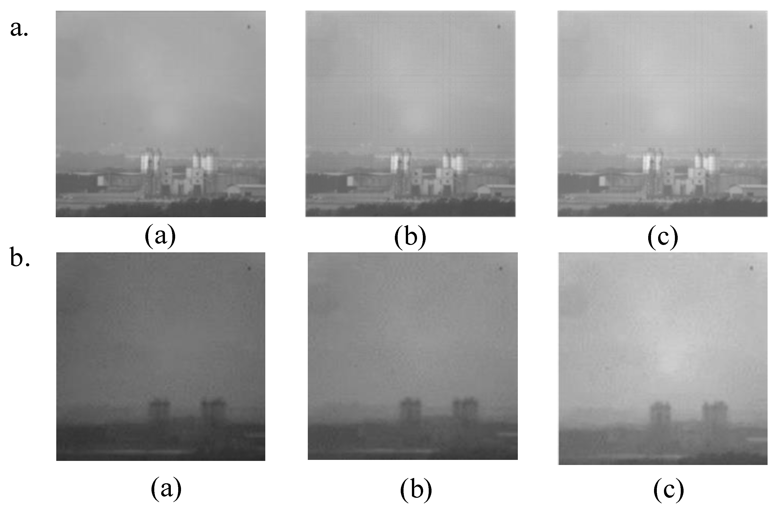
4.1. Analysis of the Effect of Multi-Band on Polarization Imaging

In this experiment, multi-band imaging experiments were performed using the SALSA camera in a sea fog environment with an optical thickness of 0.75, which was obtained by a solar radiometer. Subsequently, the multi-band polarization images were calculated by processing the intensity images in a MATLAB2014a environment. In Figure 6, Figure 7 and Figure 8, Figure (a) shows the multi-band intensity images of the sea spray environment with the bands at 670 nm, 530 nm, and 450 nm, respectively, and Figure (b)~(d) show the polarization images, line polarization images, and polarization angle images of the corresponding bands obtained at 670 nm, 530 nm, and 450 nm, respectively.
From Figure 6, Figure 7 and Figure 8, it can be intuitively seen that the intensity image is blurred, the imaging gray value is high, a lot of information is covered, the contour details are not obvious enough, and the target edge information cannot be clearly seen, which is due to the different absorption and scattering effects of sea fog particles on different wavelengths of light in the complex sea fog environment, resulting in more background noise when imaging. Since the polarization degree image is more effective than the polarization angle image, the edge contour information of the plant can be seen, and the line polarization degree image is less effective than the darker one and has less representation of the details. Therefore, this paper focuses on the polarization degree image. Since the contrast of an image can intuitively reflect the observer’s perception of the image, the image contrast equation is known according to the Weber–Fechner law as follows [16].
C W = I t I b I b
where C W is the target background contrast of the intensity image, I t denotes the target area brightness, and I b is the overall brightness of the background area of the image.
Figure 9 above shows the results of the contrast experiments based on the acquired multi-band intensity images and polarization images of the imaging target in the sea fog environment. Compared to the standard intensity picture, polarization has a powerful fog-transparent capacity to emphasize the target’s outlines, which may significantly increase the target’s contrast. When the contrast of the target polarization image at 450 nm is 7.1%, it has the lowest contrast compared to the other two bands. Compared with the other bands, the contrast of the polarization image obtained by processing is larger than the contrast of the intensity image when the band is 530 nm, and the rise in the value between the contrast is larger compared with the rest of the bands. With the increase of wavelength, the contrast of the polarization image increased by 1.4% at 670 nm compared with that at 450 nm. This is because the scattering intensity is inversely proportional to the wavelength, meaning that the longer the wavelength, the less likely it is to be scattered. When the contrast value of the polarization image was the largest, compared with the polarization image of the rest of the band, the difference between the target and the background in this band was the largest, which was more suitable for the observation of the target.
Figure 10 shows the evaluation indexes of polarization images at 670 nm, 530 nm, and 450 nm. The figure shows that the information entropy, mean gradient, and standard deviation of the polarization images at 670 nm are higher than those of the rest of the bands.

4.2. Analysis of the Effect of Optical Thickness on Polarization Imaging

Due to the inconsistency of the medium concentration throughout the experiment, this part defines the sea fog environment at varying degrees of visibility via various optical thicknesses in order to visually depict the medium concentration. The Beer–Lambert equation may be used to determine the dependency of the transmittance ratio between transmitted and incident light intensity on optical thickness. If the value of the incident light intensity is I o , then the incident light intensity I may be represented as [17] after L distance of transmission in the medium.
I = I o exp ( μ e L ) = I o exp ( τ )
The optical thickness τ = μ e L , where μ e is the extinction coefficient, can be expressed as
μ e = ρ C e = ρ π r o 2 Q e
where Q e is the extinction factor, which can be obtained from the Mie scattering calculation, ρ is the volume concentration of the transmission medium, r o is the isomeric radius of the medium particles, and π r o 2 is the absorption cross section of the medium particles.
Therefore, bringing (24) into (25) yields the relationship between the transmittance T and the medium volume concentration ρ as follows:
T = I / I o = exp ( τ ) = exp ( ρ π r o 2 Q e L )
where the isomeric radius r o of a single medium particle is constant and the extinction factor Q e and the experimental light transmission distance L are constant; therefore, the right-hand side of the equation π r o 2 Q e L is a constant. The above equation shows that the optical thickness τ is proportional to the volume concentration ρ , from which the optical thickness can be calculated.
In this paper, the optical thickness of the sea spray was measured from 5:00 to 16:00 using a solar radiometer, and five sets of data were read for each time period and averaged. The optical thicknesses at different time periods are shown in Figure 11.
In this section, imaging was performed when the optical thickness was 0.75, 0.61, and 0.52, mainly because the visual effect of sea fog with such optical thickness was obvious during the experiment. Figure 12 shows multi-band intensity images with optical thicknesses of 0.75, 0.61 and 0.52, respectively.
As demonstrated in Figure 12, as the optical thickness rises, the polarization picture of each band gets blurrier, yet the polarization image produced may still reveal the edge contour with greater clarity. Since the difference between the visible target and the background is the basis and prerequisite for recognizing the target, and since each target surface has polarization characteristics, the polarization contrast between the target and background can be obtained to compare the polarization characteristics of the target in a more intuitive manner; the polarization contrast is calculated as follows [18]:
C D O P = D O P T D O P B D O P T + D O P B
where C D O P is the polarization contrast between the target and the background, D O P T is the polarization of the target, and D O P B is the polarization of the background. Figure 13 shows the polarization contrast of multi-band images with different optical thicknesses of sea fog, where (a) is the polarization contrast of multi-band intensity images and (b) is the polarization contrast of multi-band polarization images.
The analysis of the experimental histogram reveals that as the value of sea spray optical thickness increases, the polarization contrast decreases and the image blurs, but the polarization contrast in the 670 nm band is greater and more favorable for observation at varying sea spray optical thicknesses. At an optical thickness of 0.75 in this band, the polarization contrast of the polarization image increases from 1.96% to 3.86%, a 1.9% increase compared to the polarization contrast of the intensity image, whereas the bands of 530 nm and 450 nm increase by 1.08% and 0.28%, respectively, indicating that when the optical thickness value is 0.75, the polarization contrast value of the polarization image at 670 nm increases more. The polarization contrast values of the polarization pictures at 670 nm and 530 nm grow almost twice as much as the polarization contrast values of the intensity images as the optical thickness increases. When the optical thickness increased from 0.52 to 0.75, the polarization contrast of the polarization images decreased by approximately 4.55 percent, 4.68 percent, and 5.29 percent at 670 nm, 530 nm, and 450 nm, respectively, due to the increase in the radius of the sea salt aerosol particles, which decreased the polarization of the images.
Figure 14 shows the objective assessment indices of the photographs under varying sea fog optical thicknesses. According to the data in the figure, it is intuitively apparent that the greater the optical thickness, the lower the objective indexes, the poorer the visual effect, and the greater the loss of detail information. Moreover, as the optical thickness increases, the evaluation indexes of the polarized image at 670 nm increase, and it can be assumed that the imaging effect of this band is the best, the brightness of the image is high, and the surface features of the tar are clearly visible. Compared to the other two bands, the image information is more comprehensive the more polarization information of the target is supplied.

5. Conclusions

This paper focuses on the multi-band polarization imaging of a sea fog environment and the analysis of the polarization images obtained from the processing. Through outdoor experiments, the effects of multi-band on target polarization imaging and the effects of sea fog optical thickness on target imaging can be seen. With the increase of sea fog, the polarized light appeared to recede, and the quality of polarization image decreased. When the optical thickness was 0.75, the best effect was achieved in the 670 nm band in the sea fog environment, and the polarization contrast increased by 1.9%, where the polarization contrast values of 670 nm and 530 nm with the increase of optical thickness were about twice as high as the contrast values of intensity images. It provides strong technical and data support for multi-band polarimetric imaging in a sea fog environment. The outdoor experiments in this paper realize the imaging of static targets, followed by image computation and processing analysis; however, the shooting and real-time processing of moving targets in the sea fog environment is the general trend and because of its difficulty should be the focus of future research methods.

Author Contributions

Data curation, Q.F. and N.L.; formal analysis, H.G. and X.L.; investigation, D.G. and Y.Y.; methodology, Q.F.; project administration, J.D. and Q.F.; resources, S.Z.; software, J.Z.; supervision, J.D.; validation, X.L.; visualization, S.Z. and J.Z.; writing—original draft, Q.F.; writing—review and editing, N.L. All authors have read and agreed to the published version of the manuscript.

Funding

National Natural Science Foundation of China (61890963; 61890960; 62127813).

Institutional Review Board Statement

Not applicable.

Informed Consent Statement

Not applicable.

Data Availability Statement

The study did not report any data.

Conflicts of Interest

The authors declare no conflict of interest.

References

  1. Vaughn, I.J.; Hoover, B.G. Noise reduction in a laser polarimeter based on discrete waveplate rotations. Opt. Express 2008, 16, 2091–2108. [Google Scholar] [CrossRef] [PubMed] [Green Version]
  2. Fu, X.; Mehta, Y.; Chen, Y.A.; Lei, L.; Zhu, L.; Barange, N.; Dong, Q.; Yin, S.; Mendes, J.; He, S.; et al. Directional Polarized Light Emission from Thin-Film Light-Emitting Diodes. Adv. Mater. 2021, 33, 2006801. [Google Scholar] [CrossRef] [PubMed]
  3. Kibis, O.V. Floquet theory of spin dynamics under circularly polarized light pulses. Phys. Rev. A 2022, 105, 043106. [Google Scholar] [CrossRef]
  4. Ling, M.; Song, M.; Hong, J.; Sun, X.; Tao, F. Optical design for simultaneous polarization imager based on off-axis three-mirror. Infrared Laser Eng. 2019, 48, 518001. [Google Scholar] [CrossRef]
  5. Fu, Q.; Luo, K.; Song, Y.; Zhang, M.; Zhang, S.; Zhan, J.; Duan, J.; Li, Y. Study of Sea Fog Environment Polarization Transmission Characteristics. Appl. Sci. 2022, 12, 8892. [Google Scholar] [CrossRef]
  6. Liang, J.; Ren, L.; Ju, H.; Zhang, W.; Qu, E. Polarimetric dehazing method for dense haze removal based on distribution analysis of angle of polarization. Opt. Express 2015, 23, 26146–26157. [Google Scholar] [CrossRef]
  7. El Ketara, M.; Breugnot, S. Imaging through haze using multispectral polarization imaging method. In Polarization: Measurement, Analysis, and Remote Sensing XIII; SPIE: Washington, DC, USA, 2018; Volume 10655, pp. 156–173. [Google Scholar]
  8. Zhang, X.; Zhang, Y.; Zhao, H.; Deng, H.Y.; Wang, Y. Research on long wave infrared imaging of sea target. In Eighth Symposium on Novel Photoelectronic Detection Technology and Applications; SPIE: Washington, DC, USA, 2022; Volume 12169, pp. 54–63. [Google Scholar]
  9. Guo, X.; Wan, J.; Liu, S.; Xu, M.; Sheng, H.; Yasir, M. A scSE-LinkNet Deep Learning Model for Daytime Sea Fog Detection. Remote Sens. 2021, 13, 5163. [Google Scholar] [CrossRef]
  10. KU, S.; Mahato, K.K.; Mazumder, N. Polarization-resolved Stokes-Mueller imaging: A review of technology and applications. Lasers Med. Sci. 2019, 34, 1283–1293. [Google Scholar] [CrossRef]
  11. Wang, B.; Dong, L.; Zhao, M.; Wu, H.; Ji, Y.; Xu, W. An infrared maritime target detection algorithm applicable to heavy sea fog. Infrared Phys. Technol. 2015, 71, 56–62. [Google Scholar] [CrossRef]
  12. Wang, Y.; Su, Y.; Sun, X.; Hao, X.; Liu, Y.; Zhao, X.; Li, H.; Zhang, X.; Ju, J.; Tian, J.; et al. Principle and Implementation of Stokes Vector Polarization Imaging Technology. Appl. Sci. 2022, 12, 6613. [Google Scholar] [CrossRef]
  13. Wu, Z.; Guo, L.; Zhu, M. The Image registration method combining Image information entropy and feature points. Infrared Laser Eng. 2013, 42, 2846–2852. [Google Scholar]
  14. Han, P.; Liu, F.; Wei, Y.; Shao, X. Optical correlation assists to enhance underwater polarization imaging performance. Opt. Lasers Eng. 2020, 134, 106256. [Google Scholar] [CrossRef]
  15. Bao, F. Research on Underwater Polarization Imaging and Image Processing Methods; Changchun University of Science & Technology: Changchun, China, 2019. [Google Scholar]
  16. Wang, W.; Chen, Z.; Yuan, X. Simple low-light image enhancement based on Weber-Fechner law in logarithmic space. Signal Process. Image Commun. 2022, 106, 116742. [Google Scholar] [CrossRef]
  17. Yao, L.; Gao, J.; Xiao, K.; Gong, Y. Theory and Testing Technique of Smoke; National Defence Industry Press: Beijing, China, 2004; pp. 37–41.
  18. Zhang, Y.; Gao, K.; Zhou, Y.; Hua, Z.; Han, L. Multi-polarization parameter target detection method based on modulation contrast. In Proceedings of the 2019 International Conference on Optical Instruments and Technology: Optoelectronic Imaging/Spectroscopy and Signal Processing Technology, Beijing, China, 26–28 October 2019; SPIE: Washington, DC, USA, 2020; Volume 11438, pp. 197–206. [Google Scholar]
Figure 1. Principle of LCVR polarization imaging.
Applsci 13 00202 g001
Figure 2. Block diagram showing the concept of phase delay measuring.
Applsci 13 00202 g002
Figure 3. LCVR phase delay characteristic curve.
Applsci 13 00202 g003
Figure 4. Major polarization imaging devices (a) SALSA camera; (b) Structure diagram.
Applsci 13 00202 g004
Figure 5. Polarization imaging at three wavelengths in varied weather a. Sunny weather, b. Sea fog environment (a) 670 nm (b) 530 nm (c) 450 nm.
Applsci 13 00202 g005
Figure 6. 670 nm band image (a) Intensity images (b) Polarization images (c) Line polarization images (d) Polarization angle images.
Applsci 13 00202 g006
Figure 7. 530 nm band image (a) Intensity images (b) Polarization images (c) Line polarization images (d) Polarization angle images.
Applsci 13 00202 g007
Figure 8. 450 nm band image (a) Intensity images (b) Polarization images (c) Line polarization images (d) Polarization angle images.
Applsci 13 00202 g008
Figure 9. Polarization contrast of multi-band targets.
Applsci 13 00202 g009
Figure 10. Evaluation indexes of multi-band target polarization images in sea fog environment.
Applsci 13 00202 g010
Figure 11. Optical thickness of sea spray from solar radiometer measurements.
Applsci 13 00202 g011
Figure 12. Multi-band images with different optical thicknesses (a). Intensity image; (b). Polarization image.
Applsci 13 00202 g012
Figure 13. Contrast of multi-band images with different sea fog visibility (a) Intensity image; (b) Polarization image.
Applsci 13 00202 g013
Figure 14. Objective evaluation index of images with different optical thickness of sea fog (a). 670 nm (b). 530 nm (c). 450 nm.
Applsci 13 00202 g014
Table 1. Evaluation effect.
Evaluation IndicatorsChange of ValueEvaluation Effect
ENIncreaseThe more information the image contains
AGIncreaseThe clearer the image
STDIncreaseImage fullness
Table 2. SALSA imager parameters.
NameParameters
Camera size80 mm × 80 mm × 100 mm
Resolution (pixels)1040 × 1040
Frame rate at maximum resolution12 fps (12 bits), 20 fps (12 bits)
Digital8/12-bit grayscale imaging
Synchronous interface methodUSB Interface
Spectral bandwidth410 nm~685 nm
SoftwareVersion: Salsa 2.3.5
Disclaimer/Publisher’s Note: The statements, opinions and data contained in all publications are solely those of the individual author(s) and contributor(s) and not of MDPI and/or the editor(s). MDPI and/or the editor(s) disclaim responsibility for any injury to people or property resulting from any ideas, methods, instructions or products referred to in the content.

