Article

Inhibition of Listeria monocytogenes by Broth Cultures of Surface Microbiota of Wooden Boards Used in Cheese Ripening

1
Department Pathobiological Sciences, University of Wisconsin-Madison, Madison, WI 53706, USA
2
Microbiology Doctoral Training Program, Department Bacteriology, University of Wisconsin-Madison, Madison, WI 53706, USA
3
Department Food Science, University of Wisconsin-Madison, Madison, WI 53706, USA
4
Department Pathobiological Sciences, Food Research Institute, University of Wisconsin-Madison, Madison, WI 53706, USA
*
Author to whom correspondence should be addressed.
Appl. Sci. 2023, 13(10), 5872; https://doi.org/10.3390/app13105872
Submission received: 30 March 2023 / Revised: 27 April 2023 / Accepted: 4 May 2023 / Published: 10 May 2023
(This article belongs to the Special Issue Food Microbiology: Dairy Products Microbiota)

Abstract

:

Featured Application

Wooden boards are commonly used in the production of artisan cheese. A previous report described the complex bacterial communities present on wooden boards used in cheese ripening. Here, broth cultures of the bacterial communities isolated from these boards were found to inhibit the growth of L. monocytogenes. Pure cultures of Leuconostoc mesenteroides and Staphylococcus equorum isolated from the most inhibitory community also inhibited L. monocytogenes. These findings provide new insights into the potential interactions between the microbiota from cheese ripening and the growth of L. monocytogenes.

Abstract

Listeria monocytogenes is a significant concern in cheese production. It has been assumed by some that wood surfaces pose a greater risk for pathogen contamination during cheese production. The goal of this study was to evaluate the effect of the microbiota obtained from wooden boards used in cheese ripening on the growth of Listeria monocytogenes. Bacterial communities from the surface of wooden boards obtained from cheese-ripening facilities were inoculated into tryptic soy broth (TSB) and incubated at 11 °C for 48 h. These communities (108 CFU/mL stationary phase cells) were co-incubated with 104–105 CFU/mL L. monocytogenes 2203 at 11 °C for up to eight days. At various times, samples were removed, diluted in sterile saline and plated on modified Oxford agar. Bacterial communities from each of the five boards from three different facilities significantly inhibited growth of L. monocytogenes in vitro, compared to growth of L. monocytogenes 2203 alone. Using 16S rRNA analysis, we identified sequences belonging to the genera Carnobacterium, Leuconostoc and Staphylococcus as the most abundant in the communities grown in TSB. Leuconostoc mesenteroides and Staphylococcus equorum isolated from the most inhibitory community significantly inhibited growth of L. monocytogenes in TSB at 11 °C, compared to growth of L. monocytogenes 2203 alone. These findings suggest that some members of the complex microbial communities on wooden boards in cheese aging facilities might inhibit the growth of L. monocytogenes.

1. Introduction

Wood is a natural and sustainable material that has been used in cheese production and ripening for centuries. In the ripening room, wooden boards help moderate hydric balance and serve as a reservoir for microflora important for cheese ripening. Through direct contact between the board and young cheeses, the board microflora contributes a diverse microbial ecosystem that eventually populates the cheeses’ rinds. Over the course of aging, this microbial ecosystem is a key contributor to the distinct color, texture, flavor and aroma of the final cheese product [1]. Previous studies have evaluated the effects of wood in the cheese-processing environment [2,3] and established the ability of wood to provide permeability to air and moisture, which enhance microbial growth.
Listeria monocytogenes contamination is a serious concern in cheese production and has led to costly recalls and rare, but unfortunate, disease outbreaks [4,5]. Wood, because of its porous nature, is presumed to be more difficult to clean than synthetic, smooth materials [1] and, hence, poses a greater risk for harboring pathogenic microflora such as L. monocytogenes. However, prior studies suggest that L. monocytogenes contamination from wood boards is less likely than commonly assumed. For example, Ak et al. [6,7] demonstrated that with reasonable cleaning efforts, wooden cutting boards are unlikely to create undue risk of pathogen cross-contamination. In another study, Zangerl et al. [8] found that heat treatment was effective in eliminating L. monocytogenes from the surface of wooden cheese-ripening boards.
Several previous studies have suggested that the resident indigenous microflora on food-contact surfaces can strongly influence the presence or growth of pathogens such as L. monocytogenes. Not surprisingly, the outcomes of these studies varied depending on the unique conditions of each experiment. Mariani et al. [9] analyzed the fate of multiple strains of L. monocytogenes in the presence of native microflora from wooden cheese-ripening shelves. The authors concluded that the native microflora present on these boards inhibited the growth of L. monocytogenes, an effect that was eliminated when the microflora was inactivated by heat treatment. However, these authors did not further characterize which organisms were responsible for the inhibition of L. monocytogenes. In a separate approach, Ref. [10] reported that some Bacillus isolates inhibited L. monocytogenes biofilm formation, whereas isolates of Pseudomonas and Flavobacterium allowed for the growth of L. monocytogenes on stainless steel surfaces.
In a recent study, our group characterized the indigenous bacterial communities present on wooden boards obtained from cheese-ripening facilities [11]. The purpose of the present study was to determine if broth cultures of these communities can inhibit the growth of L. monocytogenes under laboratory conditions and then further characterize these inhibitory communities using a combination of 16S rRNA amplicon sequencing and co-culturing methods.

2. Materials and Methods

2.1. Microflora Sampling of Wooden Cheese-Ripening Boards

Five wooden boards employed for the purpose of cheese aging were obtained from three different Wisconsin cheese-making facilities. We recognize that a wide spectrum of boards with variables such as plant location/geography, wood type, cheese type, board age, board treatment and so forth would have broadened the design and scope of this study. However, manufacturing plants that utilize such boards are relatively rare. Furthermore, plants that do utilize boards for cheese aging are often reluctant to provide these boards for numerous reasons, including the potential disruption to the plant ripening environment and other unintended costs and consequences. While a more comprehensive study design may serve broader goals within this realm of study, this current study design and findings provide unique discoveries relevant for broader application and inference. Boards A and B (cedar and pine, respectively) were sourced from one facility, and board C (spruce) was obtained from a second facility; both facilities produced surface-ripened cheese. Boards D and E (spruce) were obtained from a third facility and were used for cave- and smear-ripened cheeses, respectively. Surface samples were collected from a randomly selected 64 cm2 area on each board by vigorously scraping the surface with a sterile cell scraper (Biologix Technologies Inc., Monona, WI, USA) and cotton-tipped applicators (Fisher Scientific, Hampton, NH, USA). Stock cultures of the surface sample microbial communities were prepared by inoculating them into 10 mL sterile tryptic soy broth (TSB) and incubating at 11 °C for 24 h (>107 CFU/mL). Aliquots (1 mL) of the resulting cultures were stored at −80 °C in TSB with 10% glycerol (v/v). For each experiment, an aliquot was thawed, and 1 mL was inoculated into 29 mL of TSB.

2.2. Establishing a Growth Curve for Listeria monocytogenes 2203

L. monocytogenes 2203, used as a challenge inoculum in this study, is a clinical isolate from a foodborne disease outbreak associated with Mexican-style cheese [4]. Cells were stored at −20 °C on Micro bank™ Cryobeads (pro-lab Diagnostics, Richmond Hill, ON, Canada). To establish a growth curve for L. monocytogenes 2203, a cryobead was placed into 9 mL of TSB using a sterile loop and incubated overnight at 37 °C with shaking (LabQuake® shaker rotisserie with clips; Model no. 400110; Barnstead/Thermolyne, Dubuque, IA, USA). The bacterial suspension was diluted to the desired concentration (104 CFU/mL) in TSB and confirmed by plating on blood agar (BAP) and modified Oxoid (MOX) agar plates. The bacterial suspension was added to a multi-well plate (0.5 mL per well) (Nunc™, cat no: 144530, Thermo Fisher Scientific, Waltham, MA, USA) and incubated at 11 °C for seven days in a refrigerated unit (Fisher Scientific, Hampton, NH, USA). Samples were removed every 24 h from each well, serially diluted in sterile phosphate buffered saline (PBS) and plated in duplicate on MOX agar. Plates were incubated at 37 °C for 48 h, the colonies were counted and the results expressed as log10 CFU/mL.

2.3. Growth Curves of the Cheese Board Surface Microflora

To establish growth curves for the microbial communities, 1 mL of stock culture (~107 CFU/mL) was thawed and first inoculated into 29 mL of sterile TSB. The resulting microbial suspensions were dispensed (0.5 mL per well) into multi-well plates (Nunc™, cat no: 144530, Thermo Fisher Scientific, Waltham, MA, USA) and incubated at 11 °C for 8 days. At 48 h intervals, determined samples were removed, serially diluted in sterile PBS and plated on Trypticase™ Soy Agar (TSA II) with 5% Sheep Blood (BAP; BD® Biosciences, Franklin Lakes, NJ, USA). The plates were incubated at 37 °C for 24 h (boards A, C and D) or at room temperature (RT) for 48 h (boards B and E). Colonies were counted and the results expressed as log10 CFU/mL.

2.4. Growth of Listeria monocytogenes 2203 in the Presence of the Microbial Communities

To assess the growth of L. monocytogenes 2203 when co-cultured with the microbial communities, the latter were incubated at 11 °C in a multi-well plate for two days to reach stationary phase (108–109 CFU/mL). L. monocytogenes 2203 (104–105 CFU/mL) was then added to each well and incubated at 11 °C for eight days. Samples were removed (0.1 mL) from each well every 48 h, serially diluted in sterile PBS and plated on MOX (to quantify L. monocytogenes 2203) and BAP (to quantify the microbial community). BAP plates (boards A, C and E) and MOX plates (all boards) were incubated at 37 °C for 48 h. BAP plates for boards B and E were incubated at 25 °C for 48 h. Colonies were counted and the results expressed as mean ± SEM log10 CFU/mL.
Representative bacterial colonies isolated on BAP plates from surface samples of board E were sent to the School of Veterinary Medicine diagnostic microbiology service for identification by a MALDI-TOF MS Biotyper (Bruker, Billerica, MA, USA), using the direct transfer (DT) technique according to the manufacturer’s recommendations. A score > 1.7 is consistent with a species-level identification of Gram-positive species. One isolate from community E was identified by MALDI-TOF MS (Wisconsin Veterinary Diagnostic Laboratory) as a probable Leuconostoc mesenteroides (score of 1.860) and a second as Staphylococcus equorum (score of 1.840). These two bacterial species were separately incubated in BHI at 11 °C in a multi-well plate for two days to reach stationary phase (108–109 CFU/mL). L. monocytogenes 2203 (104–105 CFU/mL) was then added to each well and incubated at 11 °C for eight days. Samples were removed from each well every 48 h, serially diluted in sterile PBS and plated on MOX (to quantify L. monocytogenes 2203) and BAP (to quantify the competing bacteria). BAP plates and MOX plates were incubated at 37 °C for 48 h. BAP plates for boards B and E were incubated at 25 °C for 48 h. Colonies were counted and the results expressed as mean ± SEM log10 CFU/mL.

2.5. 16S rRNA Analysis of the Cheese Board Inhibitory Communities

Additionally, 16S rRNA analysis of broth of the cheese board communities was performed as described previously [11]. For each of the microbial communities, aliquots were inoculated into tubes of sterile TSB. L. monocytogenes 2203 (104 CFU/mL) were added to three of these tubes, while the remaining three tubes were left uninoculated to serve as controls. Samples were removed at 0, 4 and 8 days of co-incubation and centrifuged (4500× g) for 15 min at 4 °C (Eppendorf®, Centrifuge 5804R, and rotor: A-4-44, Hauppauge, NY, USA). The resulting cell pellets were washed twice with sterile PBS, suspended in sterile PBS and stored at −80 °C until total DNA extraction was performed [12,13,14]. Briefly, 1 mL of each suspension was transferred to a 2 mL screw-cap tube with 0.5 g of 0.1 mm zirconium beads (Thermo Fisher Scientific, Waltham, MA, USA). A total of 50 μL of 20% sodium dodecyl sulfate (SDS; Promega, Madison, WI, USA) and 700 μL cold equilibrated phenol (Thermo Fisher Scientific, Waltham, MA, USA) were added to each tube and subjected to bead beating for 2 min on a tabletop bead beater (Mini Bead Beater, Biospec Products, Bartlesville, OK, USA). The mixture was then heated in a 60 °C water bath for 10 min before a second 2 min round of bead beating. Tubes were then centrifuged for 10 min at 4 °C on a tabletop centrifuge (11,500× g, Microfuge 20R, Beckman Coulter, Brea, CA, USA). The aqueous layer was washed 2–4 times with 500 μL of cold equilibrated Phenol:Chloroform:isoamyl alcohol (25:24:1) (Thermo Fisher Scientific, Waltham, MA, USA) to remove the lipid layer and then transferred to a new tube. A total of 50 μL of 2M sodium acetate (Thermo Fisher Scientific, Waltham, MA, USA) and 300 μL of isopropanol (Thermo Fisher Scientific, Waltham, MA, USA) were added to the washed aqueous layer, and the DNA was allowed to precipitate overnight at −20 °C. The DNA was then pelleted in the same centrifuge (11,500× g for 20 min at 4 °C), washed twice with 70% ethanol (Decon Labs, Inc., King of Prussia, PA, USA) and then dried overnight in a fume hood. DNA Pellets were re-suspended in 20 μL of elution buffer (Thermo Fisher Scientific, Waltham, MA, USA), quantified using an individual Qubit® 2.0 Fluorometer (Invitrogen, San Diego, CA, USA) and stored at 4 °C. As a negative control, water was used instead of the DNA template for each DNA extraction and PCR reaction and was processed in a similar manner to the surface samples.

2.6. Amplification and Sequencing of Bacterial 16S rRNA

The hypervariable 4 (V4) region of the bacterial 16S rRNA coding region was PCR amplified using barcoded, 1-step sequencing primers for each sample [15]. PCR reactions were performed using varying concentrations (5 ng/μL–10 ng/μL) of total sample DNA, along with 0.5 µL of each primer (forward and reverse), 12.5 μL of 2X Hot Start Ready Mix (KAPA Biosystems, Wilmington, MA, USA) and nuclease-free water (Integrated DNA Technologies (IDT), Coralville, IA, USA) to a total volume of 25 μL. PCR reactions were conducted using the following cycling conditions: initial denaturation of 95 °C for 3 min; 25 cycles of 95 °C for 30 s, 55 °C for 30 s and 72 °C for 30 s; followed by a final extension at 72 °C for 5 min (Thermocycler; C1000 Touch Thermal Cycler, BioRad, Hercules, CA, USA). PCR products were then assessed by gel electrophoresis in a 1.0% low-melt agarose gel (National Diagnostics, Atlanta, GA, USA). Bright bands at ~380 bp were cut out of the gel, placed on a 96-well collection plate and purified using a Zymoclean Gel DNA Recovery Kit (Zymo Research, Irvine, CA, USA). Purified DNA was quantified using a Qubit® 2.0 fluorometer, equimolarly pooled and sequenced on an Illumina MiSeq using a v2 kit (2 × 250 bp) (Illumina, San Diego, CA, USA) with custom sequencing primers as described by Kozich et al. and Li et al. [13,15]. A total of 34 samples were sequenced, including 15 experimental (with L. monocytogenes), 15 controls (without L. monocytogenes) and 4 negative controls (for both DNA extraction and PCR amplification).

2.7. Sequence Processing and Analysis

Sequences were demultiplexed on the Illumina MiSeq system. Further sequence processing was performed using mothur v.1.46.1 [16], following a protocol adapted from Kozich et al. [15]. Briefly, paired end sequences were assembled into continuous segments and poor quality sequences were removed. Sequences were aligned to the SILVA 16S rRNA gene reference database v138, and those not aligning to the v4 region were removed. Pre-clustering was performed (diffs = 2) to reduce error and computational load, and chimeric sequences were detected (UCHIME; [17], http://drive5.com/uchime, accessed on 1 March 2023) and removed. Sequences were taxonomically classified using the SILVA 16S rRNA gene reference database (v138) [18], with a bootstrap value cutoff of 80. Sequences classified as Cyanobacteria, mitochondria, Eukarya or Archaea were removed, and singleton sequences were also removed. The remaining bacterial sequences were grouped into operational taxonomic units (OTUs) at 97% sequence similarity using the opticlust method. Good’s coverage was calculated for each sample, and those with coverage < 95% were removed. Sequences were normalized to 10,753 sequences per sample. Sequences for this study are available in the NCBI’s Sequence Read Archive under BioProject accession PRJNA884361 [asteinberger9 (2020). Asteinberger9/seq\_scripts v1.1. URL https://doi.org/10.5281/zenodo.4270481 (accessed on 1 March 2023)].

2.8. Statistical Analyses

Figures 1, 2 and 6 were created using Python v.3.8.5 [19], whereas Figures 3–6 were generated in R v3.53 [20], using RStudio v1.4.1106 [21] and packages including tidyverse, phyloseq v1.26.1 [22,23]), vegan [24], dplyr ([25]) and ggplot2 [23]. To calculate differences among experimental and control groups, days and boards, the Kruskal–Wallis test was used for alpha diversity and PERMANOVA for beta diversity. Pairwise Wilcoxon tests were performed for pairwise comparisons with FDR-correction applied to resultant p-values. The Kruskal–Wallis test was also used to calculate the difference between the growth of L. monocytogenes with and without the microbial communities (Figures 2 and 6).

3. Results

3.1. Growth Curves for the Wooden Board Microbial Communities and L. monocytogenes 2203

We first determined the growth curves for both the microbial communities recovered from the surface of the wooden ripening boards and for L. monocytogenes 2203 when each was individually inoculated into TSB and incubated at 11 °C for eight days. For all five communities, CFUs increased during the first two days of incubation and then remained stable through the later eight days of incubation (Figure 1A). The growth curve for L. monocytogenes 2203 demonstrated that maximal numbers (>109 CFU/mL) were achieved at four days and remained stable through seven days (Figure 1B).

3.2. Growth of L. monocytogenes 2203 in the Presence of Microbial Communities

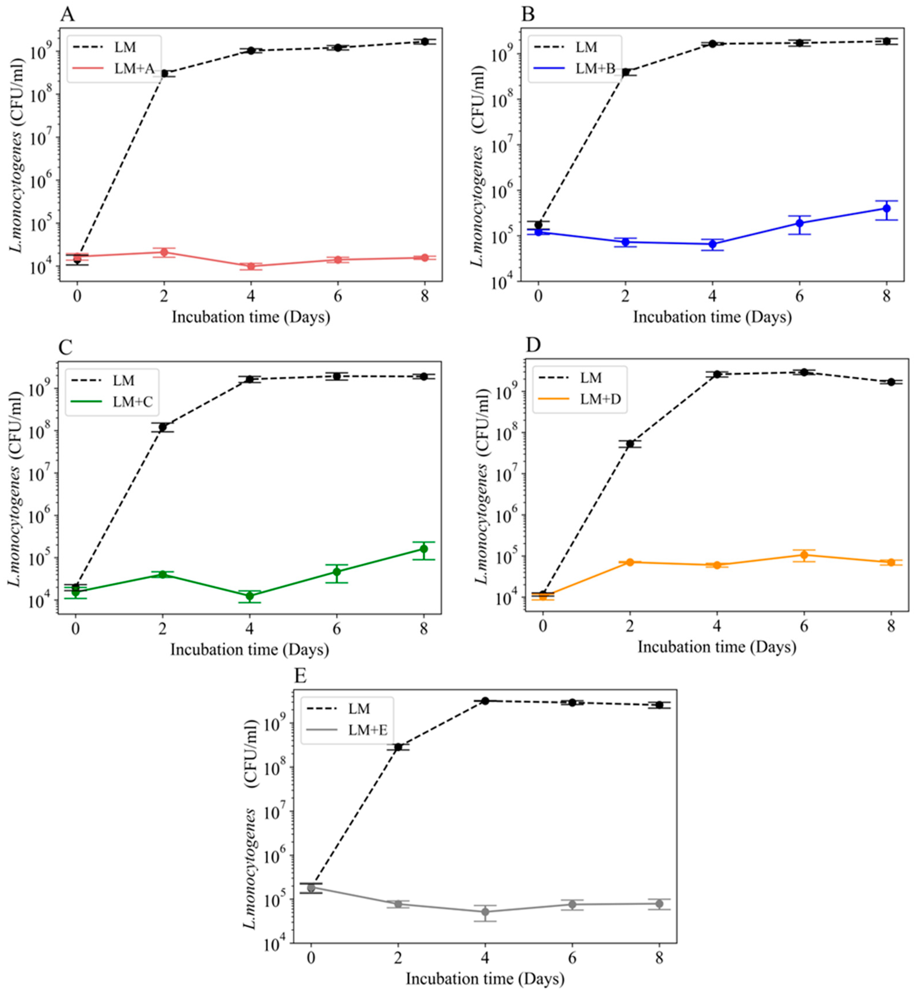
The goal of our study was to evaluate the ability of L. monocytogenes 2203 to grow in the presence of the microbial communities obtained from our five cheese boards. To test this, we inoculated L. monocytogenes 2203 into broth cultures of each cheese board’s surface microbiota, as shown in Figure 2. We found that growth of L. monocytogenes 2203 was significantly inhibited (p < 0.05) by each of the five microbial communities. The greatest inhibition was seen with surface microbial communities from boards A and E and the least with the communities from boards C and D.

3.3. Sequencing Summary

16S rRNA amplicon sequencing yielded 1,222,505 raw sequences, of which 915,465 quality-filtered sequences remained from the thirty samples and four negative controls. Negative controls were removed during normalization, as their sequence counts were less than the normalization cutoff. After normalization, board communities yielded an average of 10,751 ± 2.4 SD quality-filtered sequences per sample, resulting in 101 unique OTUs. Sequencing depths for all samples were satisfactory before and after normalization (Good’s coverage: 99.8 ± 0.0002 SD %; range: 99.88–99.97%) [26], indicating sufficient capture of the species diversity in all samples.

3.4. Diversity and Composition of Cultured Cheese Board Bacterial Communities

To evaluate and compare the bacterial community from each board, alpha (within sample; richness and evenness) and beta (between sample) diversity were first compared among boards. For alpha diversity, no differences were found between board communities for either Chao’s richness or Shannon’s diversity metrics (p > 0.05, Figure 3, Table A1). Beta diversity differed across boards for both the Bray–Curtis dissimilarity and the Jaccard index (p < 0.05, Figure 4, Table A1). Pairwise comparisons found cultured communities from boards A and B, A and D, A and E, B and C, B and D, B and E, C and E and D and E to be distinct for both metrics (p < 0.05) (Table A2), indicating differing community compositions. These results mirror findings from our previous study on the uncultured bacterial communities from the same wooden cheese boards [11].
Next, cultured board bacterial community compositions were compared at the genus level. Relative abundances of the 10 most abundant genera were found to be similar for boards A, B and C, which were distinct from boards D and E, which exhibited similar microbiota to each other (Figure 5). The microbiota of boards D and E were dominated by Leuconostoc (>85–95% relative sequence abundance in each) with lesser contributions from Carnobacterium, Staphylococcus and Enterococcus. The microbiota of boards A, B and C were dominated by Carnobacterium (>87–99% relative sequence abundance) with lesser contributions from Brevibacterium, Enterococcus, Nocardiopsis, Psychrobacter, Prevotella and Staphylococcus.

3.5. Impact of L. monocytogenes 2203 on Cheese Board Bacterial Communities

To evaluate the response of the cultured board communities to L. monocytogenes, bacterial community alpha and beta diversity was compared between experimental (with L. monocytogenes) and control (without L. monocytogenes) samples from the five board communities (A–E). The alpha diversity of the board communities was not found to significantly differ (p > 0.05) between the experimental and control samples for either Shannon’s diversity or Chao’s richness (Figure 3, Table A1). For beta diversity, the Bray–Curtis dissimilarity and Jaccard index metrics were also found to not differ (p > 0.05) between the experimental and control groups (Figure 4, Table A1).
When considering bacterial community composition, we found no substantial change in the abundances of the 10 most abundant OTUs in the experimental samples, compared to the controls using SIMPER (similarity percentages) (Figure 5) (asteinberger9, 2020). We note that OTUs classified in the genus Listeria were not recovered from any sample that was not inoculated with L. monocytogenes 2203, and plating of these samples on MOX agar plates yielded no colonies, further verifying the absence of L. monocytogenes in the uninoculated samples.

3.6. Inhibition of L. monocytogenes 2203 by Specific Cheese Board Microbes

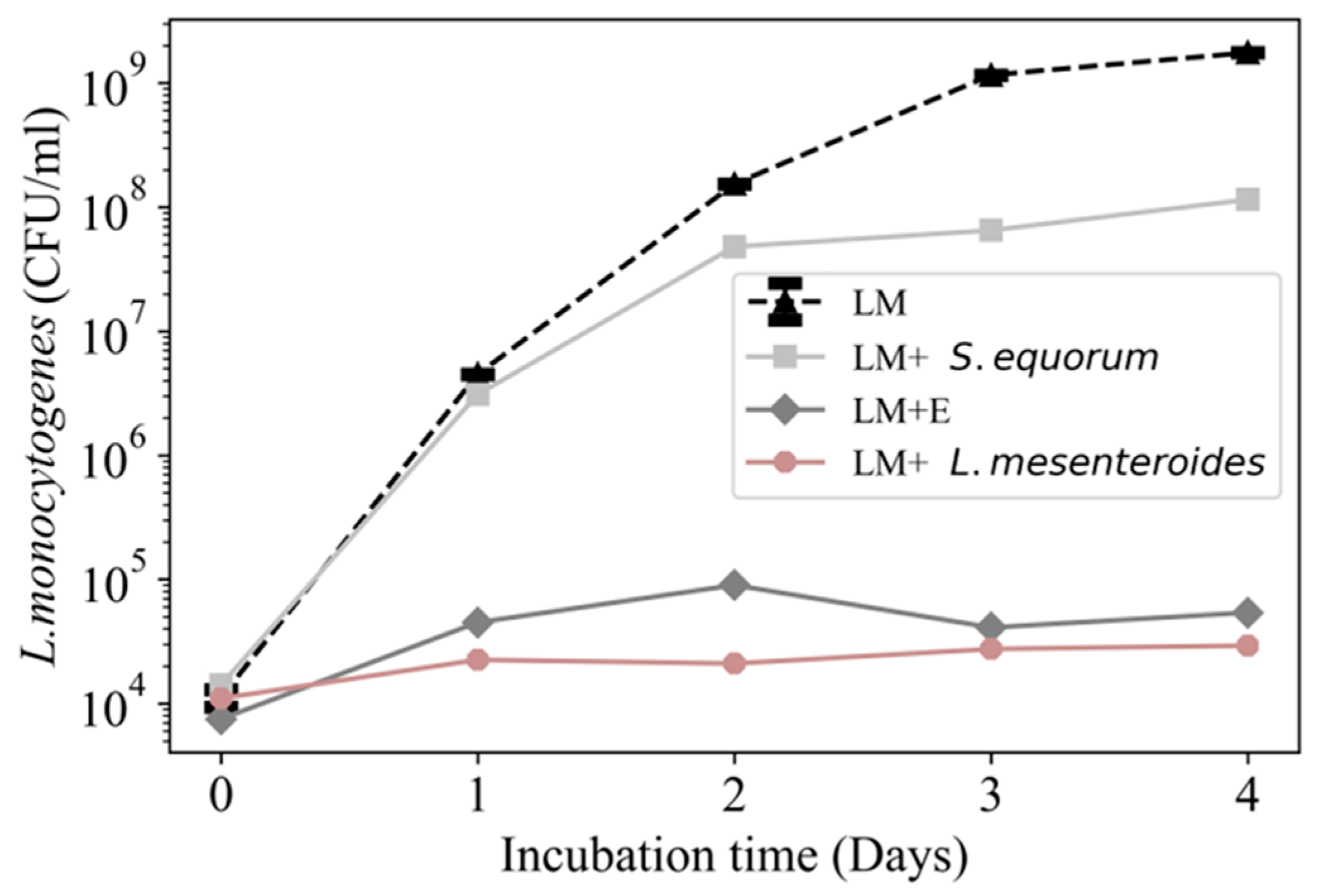
To determine which individual members of these cheese board microbial communities might be inhibitory to L. monocytogenes 2203, we chose to focus on the community from board E (dominated by Leuconostoc and Staphylococcus), which was found to be the most inhibitory of the five board communities (Figure 2 and Figure 5). One isolate from community E was identified by MALDI-TOF MS (School of Veterinary Medicine diagnostic microbiology service) as a probable Leuconostoc mesenteroides (score of 1.860) and a second as Staphylococcus equorum (score of 1.840). Broth cultures of these two bacterial species, along with the total microbial community of board E as a positive control, were assessed for inhibition of L. monocytogenes 2203 growth. As shown in Figure 6, the isolate identified as a probable Leuconostoc mesenteroides completely inhibited the growth of L. monocytogenes 2203, similar to the total microbial community from board E. In contrast, the isolate identified as S. equorum was less inhibitory for the growth of L. monocytogenes 2203 (approximately 1 log10 fewer CFUs than control cultures). We acknowledge that there might be other inhibitory bacterial species present in the community from board E that we did not isolate.

4. Discussion

In this study, we demonstrated that broth cultures of the surface microbiota of wooden boards obtained from three cheese-ripening processes are inhibitory towards L. monocytogenes 2203. In addition, we characterized the resident inhibitory microbiota communities and showed that specific members of these communities may participate in the observed inhibitory effects. These results confirm the “Jameson effect” proposed by Mellefont et al. [27], which posits that a well-established indigenous microbial community can inhibit the growth of L. monocytogenes, and further verify other findings [28], which reported the inhibitory effect of the surface microbiota of smear cheese wooden shelves on L. monocytogenes. We attempted to recreate a condition similar to that used in cheese ripening by performing our experiments in a low-temperature environment. This likely compounds the challenges that L. monocytogenes must overcome when colonizing and establishing on wooden cheese boards. As such, we expected that any persistence of L. monocytogenes in the presence of these inhibitory microbiota would likely be at low numbers, relative to other members of the microbial community. This is supported by our 16S rRNA analysis of the cheese board communities, where samples that were not inoculated with L. monocytogenes 2203 did not contain any OTUs classified as Listeria.
Our analyses of five cheese board microbiotas inoculated with L. monocytogenes 2203 revealed communities dominated by a handful of genera. Two distinct microbiotas were observed, which were dominated by either Carnobacterium or Leuconostoc. Additionally, our diversity analyses indicated that control and experimental sets of all cultured communities were not significantly different from one another (Figure 3, Figure 4 and Figure 5). Several studies have reported that bacteriocins produced by various species of Carnobacterium and Leuconostoc are capable of inhibiting L. monocytogenes. A previous report [29] demonstrated that Carnobacterium can persist at refrigeration temperatures and produce Carnobacterial class IIa bacteriocins that inhibit the growth of various Listeria spp. These authors suggested that Carnobacterium could be used as a protective culture against L. monocytogenes. Surprisingly, we did not isolate colonies of Carnobacterium spp. from our TSB cultures, despite our 16S rRNA analysis indicating that it was a dominant member of the community. This suggests that either it is not a dominant member of our cultures (16S rRNA analyses cannot differentiate live from dead bacterial cells) or our culture conditions were incapable of isolating Carnobacterium.
Leuconostoc is also known to produce bacteriocins with inhibitory effects on L. monocytogenes. For example, ref. [30] showed the antimicrobial effects of a Leuconostoc mesenteroides isolate from goat’s milk and demonstrated that bacteriocins produced by this isolate had a narrow inhibitory spectrum limited to Listeria spp. Our finding that a Leuconostoc mesenteroides isolate from board E was capable of strongly inhibiting L. monocytogenes 2203 (Figure 6) may explain, in part, the inhibitory activity of the microbiota from board E on L. monocytogenes 2203.
Overall, our findings are consistent with studies by other investigators on the use of wood in the production of artisan cheese. An analysis of wooden boards used to produce traditional Sicilian cheeses showed they contained a complex microbiota on their surfaces [31,32], and pathogens such as L. monocytogenes were not detected. Although the use of wood has been called into question for putative hygienic reasons, there have not been foodborne outbreaks associated with its use [33]. Nor does wood appear to be effective at transferring pathogens to cheese. Fresh blocks of wood inoculated with L. monocytogenes poorly transferred the organism to cheese [34], and L. monocytogenes can be eliminated from wood when subjected to heat and chemical sanitization [8,35].
We acknowledge that our study did not directly examine the inhibitory activity of the microbiota on the intact boards. Nor does it provide insights into the inhibitory mechanisms that these communities and isolates employ to limit the growth of L. monocytogenes 2203. Future studies should investigate these mechanisms. We also chose to use a single cheese-associated outbreak isolate of L. monocytogenes for this initial study. Subsequent studies should examine the generalizability of our findings to other relevant isolates of L. monocytogenes.
Despite the limitations of our study, these data provide evidence that the complex microbial communities present on the surface of wooden boards used for cheese ripening might be inhibitory for L. monocytogenes, while also contributing to the desired sensory character of aged cheese. Although these results are intriguing, it is important to note they do not negate the potential risk from undesirable microbes that might be present on a poorly maintained wooden surface.

Author Contributions

The authors made the following contributions to this study: conceptualization, K.W., S.R. and C.C.; methodology, K.W., A.S., S.R. and C.C.; software, K.W. and A.S.; validation, K.W., A.S. and C.C.; formal analysis, K.W. and C.C.; investigation, K.W. and A.S.; resources, C.C., S.R. and G.S.; data curation, K.W. and A.S.; writing—original draft preparation, K.W.; writing—review and editing, all authors; visualization, K.W.; supervision, C.C. and G.S.; project administration, C.C.; funding acquisition, C.C. All authors have read and agreed to the published version of the manuscript.

Funding

This work was supported by a USDA National Institute of Food and Agriculture (Washington, DC, USA) Hatch grant WIS02007 to G. Suen and by the University of Wisconsin–Madison Food Research Institute and the Walter and Martha Renk Endowed Laboratory for Food Safety (University of Wisconsin–Madison) to C. Czuprynski.

Institutional Review Board Statement

Not applicable.

Informed Consent Statement

Not applicable.

Data Availability Statement

Sequences for this study are available in the NCBI’s Sequence Read Archive under BioProject accession PRJNA884361.

Conflicts of Interest

The authors declare no conflict of interest.

Appendix A

Table A1. Statistical analysis of alpha (Shannon and Chao) and beta (Bray–Curtis and Jaccard) diversity. Kruskal–Wallis and permutational analysis of variance (PERMANOVA) tests of different sample sets were used. * < 0.05.
Diversity AnalysisSample Setsp-Value
ShannonKruskalGroup0.06
Category0.13
Days0.5
ChaoKruskalGroup0.6
Category0.4
Days0.2
Bray-CurtisBetadisper Group, days, category>0.05
PERMANOVAGroup<0.05 *
Category0.32
Days<0.05 *
JaccardBetadisperGroup, days, category>0.05
PERMANOVA Group<0.05 *
Category0.29
Days<0.05 *
Table A2. Pairwise comparison of groups (A-E; control and experimental). FDR correction method for both metrics (Bray–Curtis and Jaccard) was used. * p < 0.05.
Group Analysis: Corrected Method Adjusted p-Value (Bray-Curtis)Adjusted p-Value
(Jaccard)
A vs. BPairwise Adonis: fdr0.023 0.023
A vs. C0.2390.238
A vs. D0.003 *0.003 *
A vs. E0.003 *0.003 *
B vs. C0.0220.021
B vs. D0.003 *0.003 *
B vs. E0.003 *0.003 *
C vs. D0.003 *0.003 *
C vs. E0.003 *0.003 *
D vs. E0.003 *0.003 *

References

  1. Aviat, F.; Gerhards, C.; Rodriguez-Jerez, J.; Michel, V.; le Bayon, I.; Ismail, R.; Federighi, M. Microbial safety of wood in contact with food: A review. Compr. Rev. Food Sci. Food Saf. 2016, 15, 491–505. [Google Scholar] [CrossRef]
  2. Dervisoglu, M.; Yazici, F. Ripening changes of Kulek cheese in wooden and plastic containers. J. Food Eng. 2001, 48, 243–249. [Google Scholar] [CrossRef]
  3. di Grigoli, A.; Francesca, N.; Gaglio, R.; Guarrasi, V.; Moschetti, M.; Scatassa, M.L.; Settanni, L.; Bonanno, A. The influence of the wooden equipment employed for cheese manufacture on the characteristics of a traditional stretched cheese during ripening. Food Microbiol. 2015, 46, 81–91. [Google Scholar] [CrossRef] [PubMed]
  4. MacDonald, P.D.M.; Whitwam, R.E.; Boggs, J.D.; MacCormack, J.N.; Anderson, K.L.; Reardon, J.W.; Saah, J.R.; Graves, L.M.; Hunter, S.B.; Sobel, J. Outbreak of Listeriosis among Mexican immigrants as a result of consumption of illicitly produced Mexican-style cheese. Clin. Infect. Dis. 2005, 40, 677–682. [Google Scholar] [CrossRef]
  5. Melo, J.; Andrew, P.W.; Faleiro, M.L. Listeria monocytogenes in cheese and the dairy environment remains a food safety challenge: The role of stress responses. Food Res. Int. 2015, 67, 75–90. [Google Scholar] [CrossRef]
  6. Ak, N.O.; Cliver, D.O.; Kaspar, C.W. Decontamination of plastic and wooden cutting boards for kitchen use. J. Food Prot. 1994, 57, 23–30. [Google Scholar] [CrossRef] [PubMed]
  7. Ak, N.O.; Cliver, D.O.; Kaspar, C.W. Cutting boards of plastic and wood contaminated experimentally with bacteria. J. Food Prot. 1994, 57, 16–22. [Google Scholar] [CrossRef] [PubMed]
  8. Zangerl, P.; Matlschweiger, C.; Dillinger, K.; Eliskases-Lechner, F. Survival of Listeria monocytogenes after cleaning and sanitation of wooden shelves used for cheese ripening. Eur. J. Wood Wood Prod. 2009, 68, 415–419. [Google Scholar] [CrossRef]
  9. Mariani, C.; Oulahal, N.; Chamba, J.-F.; Dubois-Brissonnet, F.; Notz, E.; Briandet, R. Inhibition of Listeria monocytogenes by resident biofilms present on wooden shelves used for cheese ripening. Food Control 2011, 22, 1357–1362. [Google Scholar] [CrossRef]
  10. Bremer, P.J.; Monk, I.A.N.; Osborne, C.M. Survival of Listeria monocytogenes attached to stainless steel surfaces in the presence or absence of Flavobacterium spp. J. Food Prot. 2001, 64, 1369–1376. [Google Scholar] [CrossRef]
  11. Wadhawan, K.; Steinberger, A.J.; Rankin, S.A.; Suen, G.; Czuprynski, C.J. Characterizing the microbiota of wooden boards used for cheese ripening. JDS Commun. 2021, 2, 171–176. [Google Scholar] [CrossRef] [PubMed]
  12. Cox, M.; de Paula, T.; Lin, M.; Hall, M.B.; Suen, G. Transient changes in milk production efficiency and bacterial community composition resulting from near-total exchange of ruminal contents between high- and low-efficiency Holstein cows. J. Dairy Sci. 2017, 100, 7165–7182. [Google Scholar] [CrossRef]
  13. Li, W.; Edwards, A.; Riehle, C.; Cox, M.S.; Raabis, S.; Skarlupka, J.H.; Steinberger, A.J.; Walling, J.; Bickhart, D.; Suen, G. Transcriptomics analysis of host liver and meta-transcriptome analysis of rumen epimural microbial community in young calves treated with artificial dosing of rumen content from adult donor cow. Sci. Rep. 2019, 9, 790. [Google Scholar] [CrossRef] [PubMed]
  14. Stevenson, D.M.; Weimer, P.J. Dominance of Prevotella and low abundance of classical ruminal bacterial species in the bovine rumen revealed by relative quantification real-time PCR. Appl. Microbiol. Biotechnol. 2007, 75, 165–174. [Google Scholar] [CrossRef]
  15. Kozich, J.J.; Westcott, S.L.; Baxter, N.T.; Highlander, S.D.; Schloss, P.D. Development of a dual-index sequencing strategy and curation pipeline for analyzing amplicon sequence data on the MiSeq Illumina sequencing platform. Appl. Environ. Microbiol. 2013, 79, 5112–5120. [Google Scholar] [CrossRef] [PubMed]
  16. Schloss, P.D.; Westcott, S.L.; Ryabin, T.; Hall, J.R.; Hartmann, M.; Hollister, E.B.; Lesniewski, R.A.; Oakley, B.B.; Parks, D.H.; Robinson, C.J.; et al. Introducing mothur: Open-source, platform-independent, community-supported software for describing and comparing microbial communities. Appl. Environ. Microbiol. 2009, 75, 7537–7541. [Google Scholar] [CrossRef]
  17. Edgar, R.C.; Haas, B.J.; Clemente, J.C.; Quince, C.; Knight, R. UCHIME improves sensitivity and speed of chimera detection. Bioinformatics 2011, 27, 2194–2200. [Google Scholar] [CrossRef]
  18. Pruesse, E.; Quast, C.; Knittel, K.; Fuchs, B.M.; Ludwig, W.; Peplies, J.; Glöckner, F.O. SILVA: A comprehensive online resource for quality checked and aligned ribosomal RNA sequence data compatible with ARB. Nucleic Acids Res. 2007, 35, 7188–7196. [Google Scholar] [CrossRef]
  19. van Rossum, G.; Drake, F.L. Python 3 Reference Manual; CreateSpace: Scotts Valley, CA, USA, 2009. [Google Scholar]
  20. R Core Team. R: A Language and Environment for Statistical Computing. 2019. Available online: http://www.R-project.org (accessed on 4 September 2022).
  21. Allaire, J.J. RStudio: Integrated Development Environment for R; RStudio, PBC: Boston, MA, USA, 2019; Available online: http://www.rstudio.com/ (accessed on 4 September 2022).
  22. McMurdie, P.J.; Holmes, S. phyloseq: An R Package for Reproducible Interactive Analysis and Graphics of Microbiome Census Data. PLoS ONE 2013, 8, e61217. [Google Scholar] [CrossRef]
  23. Wickham, H.; Averick, M.; Bryan, J.; Chang, W.; McGowan, L.D.; François, R.; Grolemund, G.; Hayes, A.; Henry, L.; Hester, J.; et al. Welcome to the Tidyverse. J. Open Source Softw. 2019, 4, 1686. [Google Scholar] [CrossRef]
  24. Oksanen, J.; Blanchet, F.G.; Friendly, M.; Kindt, R.; Legendre, P.; McGlinn, D.; Minchin, P.R.; O’Hara, R.B.; Simpson, G.L.; Solymos, P. Vegan: Community Ecology Package, R package Version 2.5–7; R Foundation for Statistical Computing: Vienna, Austria, 2021; Available online: http://CRAN.Rproject.org/package=vegan (accessed on 4 September 2022).
  25. Wickham, H.; Francois, R. dplyr: A Grammar of Data Manipulation, R Package Version 0.4.3.; R Foundation for Statistical Computing: Vienna, Austria, 2015; Available online: http://CRAN.R-project.org/package=dplyr (accessed on 4 September 2022).
  26. Good, I.J.; Toulmin, G.H. The number of new species, and the increase in population coverage, when a sample is increased. Biometrika 1956, 43, 45–63. [Google Scholar] [CrossRef]
  27. Mellefont, L.A.; McMeekin, T.A.; Ross, T. Effect of relative inoculum concentration on Listeria monocytogenes growth in co-culture. Int. J. Food Microbiol. 2008, 121, 157–168. [Google Scholar] [CrossRef] [PubMed]
  28. Guillier, L.; Stahl, V.; Hezard, B.; Notz, E.; Briandet, R. Modelling the competitive growth between Listeria monocytogenes and biofilm microflora of smear cheese wooden shelves. Int. J. Food Microbiol. 2008, 128, 51–57. [Google Scholar] [CrossRef] [PubMed]
  29. Leisner, J.J.; Laursen, B.G.; Prévost, H.; Drider, D.; Dalgaard, P. Carnobacterium: Positive and negative effects in the environment and in foods. FEMS Microbiol. Rev. 2007, 31, 592–613. [Google Scholar] [CrossRef] [PubMed]
  30. Héchard, Y.; Dérijard, B.; Letellier, F.; Cenatiempo, Y. Characterization and purification of mesentericin Y105, an anti-Listeria bacteriocin from Leuconostoc mesenteroides. Microbiology 1992, 138, 2725–2731. [Google Scholar] [CrossRef] [PubMed]
  31. Settanni, L.; Busetta, G.; Puccio, V.; Licitra, G.; Franciosi, E.; Botta, L.; Di Gerlando, R.; Todaro, M.; Gaglio, R. In-depth investigation of the safety of wooden shelves used for traditional cheese Ripening. Appl. Environ. Microbiol. 2021, 87, e01524-21. [Google Scholar] [CrossRef]
  32. Gaglio, R.; Busetta, G.; Gannuscio, R.; Settanni, L.; Licitra, G.; Todaro, M. A multivariate approach to study the bacterial diversity associated to the wooden shelves used for aging traditional Sicilian cheeses. Foods 2022, 11, 774. [Google Scholar] [CrossRef]
  33. Licitra, G.; Caccamo, M.; Valence, F.; Lortal, S. Traditional wooden equipment used for cheesemaking and their effect on quality. In Global Cheesemaking Technology: Cheese Quality and Characteristics; Papademas, P., Bintsis, T., Eds.; Wiley: Hoboken, NJ, USA, 2017; pp. 157–172. [Google Scholar]
  34. Ismail, R.; Aviat, F.; Gay-Perret, P.; Le Bayon, I.; Federighi, M.; Michel, V. An assessment of L. monocytogenes transfer from wooden ripening shelves to cheeses: Comparison with glass and plastic surfaces. Food Control 2017, 73, 273–280. [Google Scholar] [CrossRef]
  35. Imhof, R.; Schwendimann, L.; Riva Scettrini, P. Sanitising wooden boards used for cheese maturation by means of a steam-mediated heating process. J. Consum. Prot. Food Saf. 2017, 12, 255–263. [Google Scholar] [CrossRef]
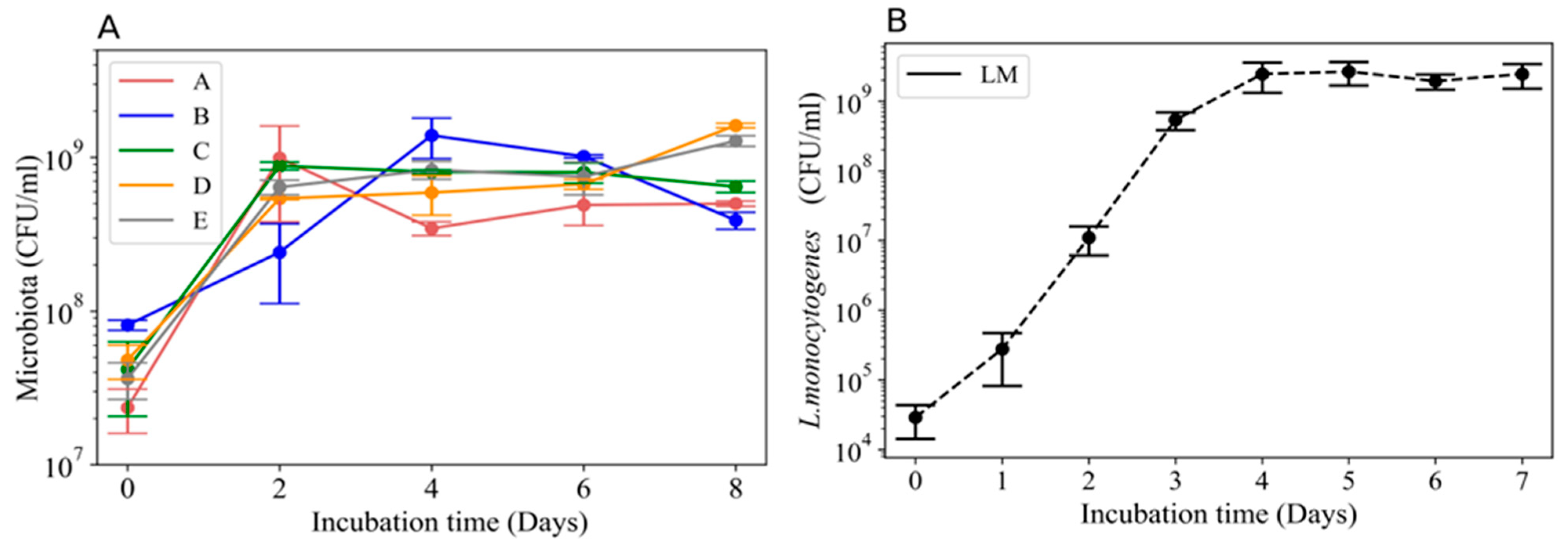
Figure 1. Growth curves for broth cultures of wooden cheese-ripening board microbial communities and L. monocytogenes 2203. (A) Microbial communities from five wooden cheese-ripening boards (A–E) were inoculated into TSB (initial inocula 7.2–7.8 Log10 CFU/mL) and incubated at 11°C. (B) Growth curve for L. monocytogenes 2203 inoculated into TSB (initial inocula 4.1–4.6 Log10 CFU/mL) and incubated at 11 °C. Error bars depict standard error of the mean (n = 4).
Applsci 13 05872 g001
Figure 2. Inhibition of L. monocytogenes 2203 by broth cultures of the microbial communities from five different wooden boards (AE). Board communities were incubated in TSB at 11 °C for 48 h until they reached stationary growth phase (108 CFU/mL), at which point 105 CFU/mL L. monocytogenes 2203 (LM) was added. The cultures were then incubated at 11 °C for 8 days. Samples were removed at 2-day intervals and plated on MOX agar to quantify LM 2203 (mean ± SEM CFU/mL). Dashed lines correspond to LM 2203 alone (positive control) and solid lines to LM 2203 in the presence of each microbial community (n = 6).
Applsci 13 05872 g002
Figure 3. Box plots illustrating alpha diversity measures (Chao richness and Shannon diversity) of broth cultures of the bacterial communities from wooden cheese boards. Median values are indicated in the plots. Using Wilcoxon Rank Sum Tests (with FDR correction), it was determined that surface community alpha diversity did not significantly differ among boards (Group A–E) (p > 0.05), nor did they differ with exposure to L. monocytogenes (Category, p > 0.05), for either Chao’s richness (Chao1) or Shannon’s diversity (Shannon).
Applsci 13 05872 g003
Figure 4. NMDS (nonmetric multidimensional scaling) plots depicting the β diversity of cheese-ripening boards. Beta diversity of each board’s microbiota either with (experimental) or without (control) L. monocytogenes, assessed using (A) Bray–Curtis dissimilarity or (B) Jaccard similarity analysis. Ellipses represent standard error of all samples collected for each respective cheese-ripening board.
Applsci 13 05872 g004
Figure 5. Relative abundance of the 10 most abundant genera in broth cultures from the wooden boards. Samples were assessed using 16S rRNA sequencing. The x-axis labels indicate number of days of sample collection for each board community (AE) incubated with (experimental) or without (control) L. monocytogenes.
Applsci 13 05872 g005
Figure 6. Inhibition of L. monocytogenes 2203 (LM) by L. mesenteroides and S. equorum isolated from an inhibitory microbial community (board E). LM corresponds to LM 2203 alone (negative control); LM+E corresponds to growth of LM 2203 in presence of microbial community of board E (positive control). LM+S. equorum and LM+L. mesenteroides illustrate growth of LM 2203 in the presence of these species that were isolated from the microbiota of board E. Symbols represent the mean ± SEM CFU/mL from a single experiment.
Applsci 13 05872 g006
Disclaimer/Publisher’s Note: The statements, opinions and data contained in all publications are solely those of the individual author(s) and contributor(s) and not of MDPI and/or the editor(s). MDPI and/or the editor(s) disclaim responsibility for any injury to people or property resulting from any ideas, methods, instructions or products referred to in the content.