Segmentation of Acne Vulgaris Images Techniques: A Comparative and Technical Study
Abstract
1. Introduction
2. Methods
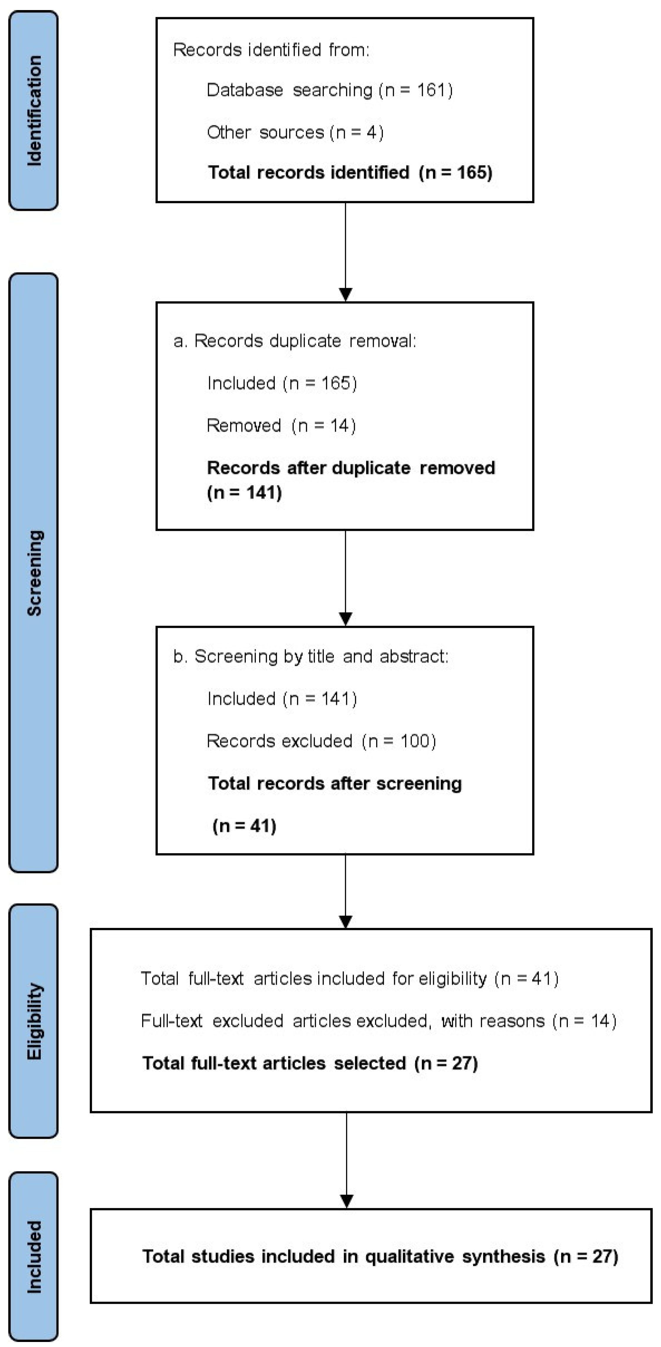
- All the implemented methodologies that use enlarged images, showing only one lesion per image, for example, dermatoscopy.
- Studies not published in English.
- Those publications which are neither open access nor available to the Polytechnic University of Valencia members.
- Studies that are a continuation of a previous one, in which the segmentation methodology is yet described. For instance, publications about acne lesions classification after image segmentation, when the applied segmentation method has been presented in a previous study and has not suffered substantial changes.
3. Results
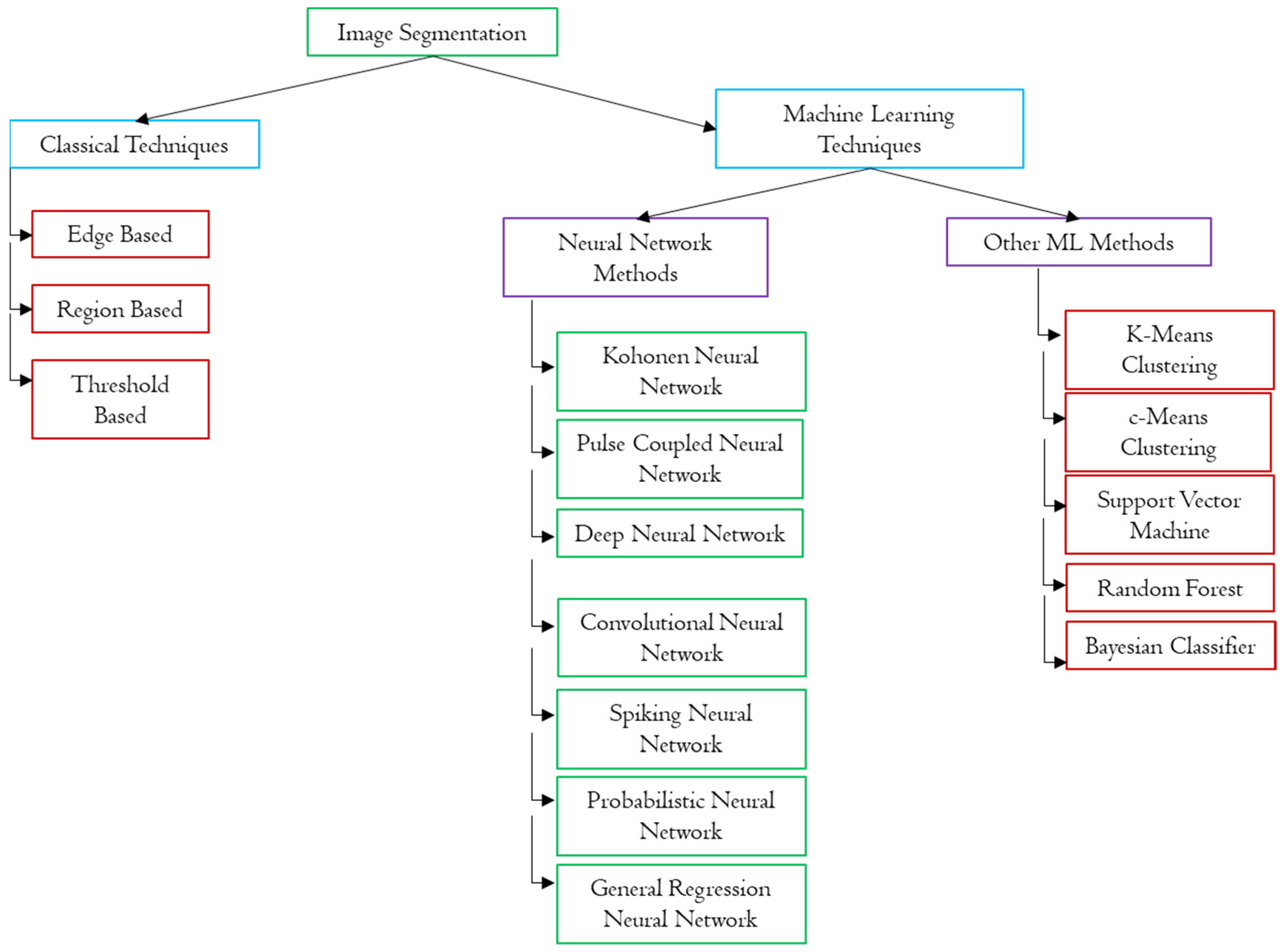
3.1. Segmentation Methods Based on Classical Image Processing Techniques
3.2. Segmentation Methods Based on Machine Learning Techniques Algorithms
4. Discussion
Summary of Limitations
5. Conclusions
Author Contributions
Funding
Data Availability Statement
Conflicts of Interest
References
- Williams, H.C.; Dellavalle, R.P.; Garner, S. Acne vulgaris. Lancet 2012, 379, 361–372. [Google Scholar] [CrossRef] [PubMed]
- Zouboulis, C.C. Acne as a chronic systemic disease. Clin. Dermatol. 2013, 32, 389–396. [Google Scholar] [CrossRef] [PubMed]
- Ramli, R.; Malik, A.S.; Hani, A.F.M.; Jamil, A. Acne analysis, grading and computational assessment methods: An overview. Skin Res. Technol. 2011, 18, 1–14. [Google Scholar] [CrossRef] [PubMed]
- Becker, M.; Wild, T.; Zouboulis, C.C. Objective assessment of acne. Clin. Dermatol. 2017, 35, 147–155. [Google Scholar] [CrossRef] [PubMed]
- Jena, M.; Mishra, S.P.; Mishra, D. A survey on applications of machine learning techniques for medical image segmentation. Int. J. Eng. Technol. 2018, 7, 4489–4495. [Google Scholar]
- Ramli, R.; Malik, A.S.; Hani, A.F.M.; Yap, F.B.-B. Identification of acne lesions, scars and normal skin for acne vulgaris cases. In Proceedings of the 2011 National Postgraduate Conference, Seri Iskanda, Malaysia, 19–20 September 2011; pp. 1–4. [Google Scholar] [CrossRef]
- Chantharaphaichi, T.; Uyyanonvara, B.; Sinthanayothin, C.; Nishihara, A. Automatic acne detection for medical treatment. In Proceedings of the 2015 6th International Conference of Information and Communication Technology for Embedded Systems (IC-ICTES), Hua Hin, Thailand, 22–24 March 2015; pp. 1–6. [Google Scholar]
- Malik, A.S.; Humayun, J.; Kamel, N.; Yap, F.B.-B. Novel techniques for enhancement and segmentation of acne vulgaris lesions. Skin Res. Technol. 2013, 20, 322–331. [Google Scholar] [CrossRef] [PubMed]
- Humayun, J.; Malik, A.S.; Belhaouari, S.B.; Kamel, N.; Yap, F.B.B. Localization of acne lesion through template matching. In Proceedings of the 2012 4th International Conference on Intelligent and Advanced Systems (ICIAS2012), Kuala Lumpur, Malaysia, 12–14 June 2012; pp. 91–94. [Google Scholar]
- Chen, D.H.; Chang, T.R.; Cao, R.J. The development of a skin inspection imaging system on an Android device. In Proceedings of the 7th International Conference on Communications and Networking, Kunming, China, 8–10 August 2012; pp. 653–658. [Google Scholar]
- Min, S.; Kong, H.-J.; Yoon, C.; Kim, H.C.; Suh, D.H. Development and evaluation of an automatic acne lesion detection program using digital image processing. Skin Res. Technol. 2012, 19, e423–e432. [Google Scholar] [CrossRef] [PubMed]
- Liu, Z.; Zerubia, J. Towards automatic acne detection using a MRF model with chomophore descriptos. In Proceedings of the 2013 EUSIPCO, Marrakech, Morocco, 9–13 September 2013; pp. 1–5. [Google Scholar] [CrossRef]
- Kittigul, N.; Uyyanonvara, B. Automatic acne detection system for medical treatment progress report. In Proceedings of the 2016 7th International Conference of Information and Communication Technology for Embedded Systems (IC-ICTES), Bangkok, Thailand, 20–22 March 2016; pp. 41–44. [Google Scholar]
- Budhi, G.S.; Adipranata, R.; Gunawan, A. Acne Segmentation and Classification using Region Growing and Self-Organizing Map. In Proceedings of the 2017 International Conference on Soft Computing, Intelligent System and Information Technology (ICSIIT), Denpasar, Indonesia, 26–29 September 2017; pp. 78–83. [Google Scholar] [CrossRef]
- Maroni, G.; Ermidoro, M.; Previdi, F.; Bigini, G. Automated detection, extraction and counting of acne lesions for automatic evaluation and tracking of acne severity. In Proceedings of the 2017 IEEE Symposium Series on Computational Intelligence (SSCI), Honolulu, HI, USA, 27 November–1 December 2017; pp. 1–6. [Google Scholar] [CrossRef]
- Khongsuwan, M.; Kiattisin, S.; Wongseree, W.; Leelasantitham, A. Counting number of points for acne vulgaris using UV Fluorescence and image processing. In Proceedings of the 4th 2011 Biomedical Engineering International Conference, Shanghai, China, 15–17 October 2011; pp. 142–146. [Google Scholar] [CrossRef]
- Son, T.; Han, B.; Jung, B.; Nelson, J.S. Fluorescent image analysis for evaluating the condition of facial sebaceous follicles. Skin Res. Technol. 2007, 14, 201–207. [Google Scholar] [CrossRef] [PubMed]
- Abu Zaki, A.I.; Ibrahim, N.; Sari, S.; Mahadi, L.F. Applied of image processing technique on semi-auto count of skin spots. Bioeng. Princ. Technol. Appl. 2019, 2, 51–62. [Google Scholar]
- Wu, Y.; Akimoto, M.; Igarashi, H.; Shibagaki, Y.; Tanaka, T. Quantitative Assessment of Age-dependent changes in Porphyrins from Fluorescence Images of Ultraviolet Photography by Image Processing. Photodiagnosis Photodyn. Ther. 2021, 35, 102388. [Google Scholar] [CrossRef] [PubMed]
- Fujii, H.; Yanagisawa, T.; Mitsui, M.; Murakami, Y.; Yamaguchi, M.; Ohyama, N.; Abe, T.; Yokoi, I.; Matsuoka, Y.; Kubota, Y. Extraction of acne lesion in acne patients from multispectral images. In Proceedings of the 2008 30th Annual International Conference of the IEEE Engineering in Medicine and Biology Society, Vancouver, BC, Canada, 21–22 August 2008; pp. 4078–4081. [Google Scholar]
- Ramli, R.; Malik, A.S.; Hani, A.F.M.; Yap, F.B.B. Segmentation of acne vulgaris lesions. In Proceedings of the 2011 International Conference on Digital Image Computing: Techniques and Applications, Noosa, Australia, 6–8 December 2011; pp. 335–339. [Google Scholar]
- Malik, A.S.; Ramli, R.; Hani, A.F.M.; Salih, Y.; Yap, F.B.B.; Nisar, H. Digital assessment of facial acne vulgaris. In Proceedings of the 2014 IEEE International Instrumentation and Measurement Technology Conference (I2MTC) Proceedings, Montevideo, Uruguay, 12–15 May 2014; pp. 546–550. [Google Scholar]
- Khan, J.; Malik, A.S.; Kamel, N.; Dass, S.C.; Affandi, A.M. Segmentation of acne lesion using fuzzy C-means technique with intelligent selection of the desired cluster. In Proceedings of the 2015 37th Annual International Conference of the IEEE Engineering in Medicine and Biology Society (EMBC), Milan, Italy, 25–29 August 2015; pp. 3077–3080. [Google Scholar]
- Madan, S.K.; Dana, K.J.; Cula, O. Learning-based detection of acne-like regions using time-lapse features. In Proceedings of the 2011 IEEE Signal Processing in Medicine and Biology Symposium (SPMB), Brooklyn, NY, USA, 10 December 2011; pp. 1–6. [Google Scholar]
- Arifin, M.S.; Kibria, M.G.; Firoze, A.; Amini, M.A.; Yan, H. Dermatological disease diagnosis using color-skin images. In Proceedings of the 2012 International Conference on Machine Learning and Cybernetics, Xian, China, 15–17 July 2012; pp. 1675–1680. [Google Scholar]
- Chang, C.-Y.; Liao, H.-Y. Automatic Facial Spots and Acnes Detection System. J. Cosmet. Dermatol. Sci. Appl. 2013, 3, 28–35. [Google Scholar] [CrossRef]
- Zhao, T.; Zhang, H.; Spoelstra, J. A computer vision application for assessing facial acne severity from selfie images. arXiv 2019, arXiv:1907.07901. [Google Scholar]
- Junayed, M.S.; Jeny, A.A.; Atik, S.T.; Neehal, N.; Karim, A.; Azam, S.; Shanmugam, B. AcneNet-a deep CNN based classification approach for acne classes. In Proceedings of the 2019 12th International Conference on Information & Communication Technology and System (ICTS), Surabaya, Indonesia, 18 July 2019; pp. 203–208. [Google Scholar]
- Lim, Z.V.; Akram, F.; Ngo, C.P.; Winarto, A.A.; Lee, W.Q.; Liang, K.; Oon, H.H.; Thng, S.T.G.; Lee, H.K. Automated grading of acne vulgaris by deep learning with convolutional neural networks. Skin Res. Technol. 2019, 26, 187–192. [Google Scholar] [CrossRef] [PubMed]
- Yadav, N.; Alfayeed, S.M.; Khamparia, A.; Pandey, B.; Thanh, D.N.H.; Pande, S. HSV model-based segmentation driven facial acne detection using deep learning. Expert Syst. 2021, 39, e12760. [Google Scholar] [CrossRef]
- Wang, Y.; Li, A.; Li, C.; Cui, Y. Automatic Acne Classification using VISIA. In Proceedings of the 4th International Conference on Control and Computer Vision, Macau, China, 13–15 August 2021; pp. 107–111. [Google Scholar]
- Hasanah, R.L.; Rianto, Y.; Riana, D. Identification of Acne Vulgaris Type in Facial Acne Images Using GLCM Feature Extraction and Extreme Learning Machine Algorithm. Rekayasa 2022, 15, 204–214. [Google Scholar] [CrossRef]
- Alamdari, N.; Tavakolian, K.; Alhashim, M.; Fazel-Rezai, R. Detection and classification of acne lesions in acne patients: A mobile application. In Proceedings of the 2016 IEEE International Conference on Electro Information Technology (EIT), Grand Forks, ND, USA, 19–21 May 2016; pp. 739–743. [Google Scholar] [CrossRef]
- Chantharaphaichit, T.; Uyyanonvara, B.; Sinthanayothin, C.; Nishihara, A. Automatic acne detection with featured Bayesian classifier for medical treatment. In Proceedings of the 3rd International Conference on Robotics, Informatics and Intelligence Control Technology, Bangkok, Thailand, 30 April 2015; pp. 10–16. [Google Scholar]
- The MathWorks Inc. Segmentación Basada En Color Mediante Clustering K-means. 2019. Available online: https://es.mathworks.com/help/images/examples/color-based-segmentation-using-k-means-clustering.html (accessed on 5 February 2022).
- The MathWorks Inc. ColorBolb Utility with Automatic Thresholding and Tolerance Calculations. 2019. Available online: https://es.mathworks.com/matlabcentral/fileexchange/45605-color-bolb-utility-with-authomatic-thresholding-and-tolerance-calculations (accessed on 5 February 2022).
- Phillips, S.B.; Kollias, N.; Gillies, R.; Muccini, J.A.; Drake, L.A. Polarized light photography enhances visualization of inflammatory lesions of acne vulgaris. J. Am. Acad. Dermatol. 1997, 37, 948–952. [Google Scholar] [CrossRef] [PubMed]
- Rizova, E.; Kligman, A. New photographic techniques for clinical evaluation of acne. J. Eur. Acad. Dermatol. Venereol. 2001, 15, 13–18. [Google Scholar] [PubMed]
- Lucchina, L.C.; Kollias, N.; Gillies, R.; Phillips, S.B.; Muccini, J.A.; Stiller, M.J.; Trancik, R.J.; Drake, L.A. Fluorescence photography in the evaluation of acne. J. Am. Acad. Dermatol. 1996, 35, 58–63. [Google Scholar] [CrossRef] [PubMed]


| Population | Humans |
| Intervention | Segmentation of face images of acne vulgaris patients |
| Comparison | Manual segmentation |
| Outcome | Diverse results |
| Study | Image Modality | Images Database | Proposed Method | Ground Truth | Samples for the Validation | Relevant Limitations | Outcomes |
|---|---|---|---|---|---|---|---|
| [14] | Conventional photography | Not indicated | Region growing | Not indicated | Not indicated | The user must place the seed manually. Each lesion needs a different threshold value. | Authors only report that results are satisfactory, without providing any data. |
| [15] | Conventional photography | DermNet, DermQuest | Heat map + thresholding | Not indicated | Not indicated | The difficulty of thresholding makes the manual selection of threshold value necessary in some of the images. | No details of segmentation results are provided. |
| [13] | Conventional photography | Own images | Adaptive thresholding | Method has not been validated yet | Method has not been validated yet | No limitations indicated yet. | Results not evaluated yet. |
| [7] | Conventional photography | Not indicated | Enhancement of ROI (normalized gray image − V component from HSV space) + thresholding | Manual lesion counting | Ten images were used in the validation | High probability of obtaining false positives because of similarity with the characteristics of acne lesions. | Precision = 80% Sensitivity = 86.37% Accuracy = 70% |
| [8] | Conventional photography | Own images | Increase in dynamic range + mapping of each pixel to a band of the visible spectrum | Severity of acne estimated by experts | Number of images not indicated | The authors do not indicate any limitations. | Evaluation of the method’s ability to grade acne: Specificity > 80% Sensitivity > 60% |
| [12] | Conventional photography | DermNet NZ | Color-based segmentation using an iterative method based on energy minimization | Human visual inspection | 50 images were used in the validation | No limitations indicated. | Quantitative analysis of results has not been performed yet. |
| [11] | Conventional photography | Own images | Processing techniques vary depending on acne lesion type | Manual lesion counting and lesion type classification | Number of images not indicated | No limitations indicated. | Correlation between manual and automatic counting, for each lesion type: Pearson correlation coefficient > 0.93 (0.54 for open comedos) Detection of each lesion type: Sensitivity > 66% Specificity > 74% except for open comedos (21%). |
| [10] | Conventional photography | Own images | Thresholding | Skin inspection by experts | 99 images were used in the validation | No limitations indicated. | Distinction between acne and healthy skin: Accuracy = 82.82% |
| [9] | Conventional photography | Kuala Lumpur hospital database | Template matching | Not indicated | Ten images were used in the validation | Optimization of filtering is needed in order to decrease the errors caused by illumination changes and texture of images. | Results are good, but they cannot be considered, since the described method is still under development: it is not robust, there are considerable variations in sensitivity and precision among the images. |
| [16] | Fluorescence images | Own images | Histogram equalization + extended maxima transform | Manual lesion counting | Ten images were used in the validation | No limitations indicated. | Automatic counting: Accuracy = 83.75% Sensitivity = 98.22% Precision = 85.04% |
| [21] | Conventional photography | Own images | Thresholding by Otsu’s method | Segmentation by experts | 185 images were used in the validation | No limitations indicated. | Sensitivity > 80% Specificity > 80% |
| [17] | Fluorescence images | Own images | Thresholding + color-based segmentation | Not indicated for the calibrated system | 29 images were used in the validation | Readjustment of parameters of the color-based segmentation is needed. | Results for the calibrated system are not quantitatively evaluated. |
| [18] | LED microscopy color images | Own Images | Thresholding by color + morphological operators segmentation | Manual counting | 15 images | No limitations indicated | Do not provide good results. |
| [19] | Fluorescence images | Own Images | Thresholding by color and contour detection | Manual counting | 3595 images | No limitations indicated | Accuracy 71% Sensitivity 72% Precision 88% |
| Study | Image Modality | Images Database | Proposed Method | Ground Truth | Samples for the Validation | Relevant Limitations | Outcomes |
|---|---|---|---|---|---|---|---|
| [33] | Conventional photography | Own images | Nested k-means | Not indicated | 35 images were used in the validation | It is necessary to develop a new concept of relative color, in order to prevent differences in segmentation results due to the differences among individuals’ skin tone. | Accuracy = 70% |
| [23] | Conventional photography | Own images | Fuzzy c-means | Not indicated | 50 images were used in the validation | No limitations indicated. | With Q component: Sensitivity = 89.67% Specificity = 93.19% Accuracy = 92.63% With I3 component: Sensitivity = 89.54% Specificity = 91.62% Accuracy = 91.05% |
| [22] | Conventional photography | Own images | K-means + SVM | Segmentation by experts | 50 images were used in the validation | No limitations indicated. | After postprocessing: Sensitivity = 90% Specificity = 97.2% Accuracy = 93.6% |
| [26] | Conventional photography | Own images + database images | Thresholding in YCbCr space + SVM | Not indicated | 97 images were used in the validation | No limitations indicated. | Accuracy = 99.40% Sensitivity = 80.91% Specificity = 99.42% |
| [25] | Conventional photography | Own images | Color gradient + thresholding + k-means | Not indicated | 405 samples were used in the validation | No limitations indicated. | In acne detection: Accuracy = 96.66% |
| [24] | Polarised light photography | Own images | Classifier based on logistic regression | Not indicated | 39 images were used in the validation | Big precision needed in images registration. | 89.2% of regions correctly classified |
| [6] | Conventional photography | Own images | K-means | Segmentation by experts | Not indicated | No limitations indicated. | Sensitivity > 81%, specificity > 81% |
| [20] | Multispectral imaging | Own images | Linear discriminant function | Not indicated | Not indicated | Authors manifest the need of a more complex classifier. Finding spectral features that don not depend on image conditions is also needed. | Results are diverse and details are not reported. |
| [27] | Conventional selfie images | Own images | Convolutional Neural Networks | 230 images selected by dermatologist | 4000 images | No limitations indicated. | Good results distinguishing acne and health skin |
| [28] | Color Images | Own images | Deep Residual Neural Networks | Auto trained validation model | 1800 images | No limitations indicated. | 94% accuracy classifying 5 acne classes |
| [29] | Conventional Photographs | Own images | Convolutional Neural Networks | Classification by experts | 472 images | No limitations indicated. | Three acne groups with a Pearson correlation accuracy 67% |
| [30] | Conventional Photographs | Own images | K-means + HSV segmentation + Texture Analysis + CNN classification | Experts Labelling | 120 images | No limitations indicated. | K-means + CNN allow detect 70% lesions |
| [32] | Conventional Photographs | Own images | K-means segmentation + ELMA classification | N/A | 100 images | No limitations indicated. | 80% effectivity classifying acne lesions in three categories. |
Disclaimer/Publisher’s Note: The statements, opinions and data contained in all publications are solely those of the individual author(s) and contributor(s) and not of MDPI and/or the editor(s). MDPI and/or the editor(s) disclaim responsibility for any injury to people or property resulting from any ideas, methods, instructions or products referred to in the content. |
© 2023 by the authors. Licensee MDPI, Basel, Switzerland. This article is an open access article distributed under the terms and conditions of the Creative Commons Attribution (CC BY) license (https://creativecommons.org/licenses/by/4.0/).
Share and Cite
Moncho-Santonja, M.; Aparisi-Navarro, S.; Defez, B.; Peris-Fajarnés, G. Segmentation of Acne Vulgaris Images Techniques: A Comparative and Technical Study. Appl. Sci. 2023, 13, 6157. https://doi.org/10.3390/app13106157
Moncho-Santonja M, Aparisi-Navarro S, Defez B, Peris-Fajarnés G. Segmentation of Acne Vulgaris Images Techniques: A Comparative and Technical Study. Applied Sciences. 2023; 13(10):6157. https://doi.org/10.3390/app13106157
Chicago/Turabian StyleMoncho-Santonja, María, Silvia Aparisi-Navarro, Beatriz Defez, and Guillermo Peris-Fajarnés. 2023. "Segmentation of Acne Vulgaris Images Techniques: A Comparative and Technical Study" Applied Sciences 13, no. 10: 6157. https://doi.org/10.3390/app13106157
APA StyleMoncho-Santonja, M., Aparisi-Navarro, S., Defez, B., & Peris-Fajarnés, G. (2023). Segmentation of Acne Vulgaris Images Techniques: A Comparative and Technical Study. Applied Sciences, 13(10), 6157. https://doi.org/10.3390/app13106157


