Article

Assessment of Antidiabetic and Anti-Inflammatory Activities of Carissa carandas Linn Extract: In Vitro and In Vivo Study

by
Manaschanok Lailerd
1,
Thiri Wai Linn
2,
Narissara Lailerd
2,
Duangporn Amornlerdpison
3 and
Arisa Imsumran
1,*
1
Department of Biochemistry, Faculty of Medicine, Chiang Mai University, Chiang Mai 50200, Thailand
2
Department of Physiology, Faculty of Medicine, Chiang Mai University, Chiang Mai 50200, Thailand
3
Faculty of Fisheries Technology and Aquatic Resources, Maejo University, Chiang Mai 50290, Thailand
*
Author to whom correspondence should be addressed.
Appl. Sci. 2023, 13(11), 6454; https://doi.org/10.3390/app13116454
Submission received: 26 April 2023 / Revised: 21 May 2023 / Accepted: 23 May 2023 / Published: 25 May 2023

Abstract

:
This study investigated the effects of aqueous fruit extracts of Carissa carandas (CCA) on inflammation and insulin resistance using an in vitro cellular model, in vivo high-fat diets, and a streptozotocin-induced type 2 diabetic (T2DM) rat model. CCA significantly ameliorated inflammation by decreasing nitric oxide production in lipopolysaccharide (LPS)-induced RAW 264.7 macrophages. Interestingly, CCA showed anti-insulin resistance activities, as it significantly improved glucose uptake and decreased glycerol release in LPS-induced 3T3-L1 adipocytes. In vivo studies showed that a high dose of 12-week oral supplementation of CCA (400 mg/kg BW/day) significantly reduced visceral fat, triglycerides, and cholesterol level in the blood of diabetic rats. Importantly, the metabolic parameters in both fasting and postprandial states, including fasting plasma glucose, HOMA-IR, and glucose intolerance, significantly improved, indicating its antihyperglycemic benefit in diabetic rats. Moreover, the results of the HOMA-β and histological examination suggested that pancreatic β-cell function and pancreatic morphological changes of the CCA and metformin treatments appeared to be better than those in non-treated diabetes, indicating the protective effect of CCA against pancreatic damage caused by hyperglycemia. In conclusion, the present study first reported that the C. carandas fruit extract has anti-inflammation and anti-insulin resistance, and subsequently improved glycemic control in the T2DM rat model.

1. Introduction

The incidence of type 2 diabetes mellitus (T2DM) has increased and is continuously rising worldwide. T2DM is characterized by hyperglycemia resulting from insulin insufficiency, insulin resistance, or both. Diabetic condition is likely accompanied by obesity due to excessive food consumption and less daily activity. As a consequence, fat accumulation in adipose tissue leads to chronic inflammation and insulin resistance of these cells [1]. Adipocytes and their activated macrophages produce various pro-inflammatory cytokine factors, including tumor necrosis factor-alpha (TNF-α), interleukin-6 (IL-6), and interleukin-1β (IL-1β). These cytokines have paracrine effects to activate inflammatory pathways within insulin target cells, including the liver and muscle, causing systemic insulin resistance and hyperglycemia [2,3]. Chronic hyperglycemia has been reported to increase levels of reactive oxygen species in β-cells and leads to toxic effects on the β-cell. A prolonged and excess cell workload induces β-cell dysfunction and failure, resulting in inadequate insulin compensation and finally leading to the development of T2DM [4]. The obese adipocytes or high-fat diet induces chronic inflammation, which contribute to insulin resistance and leads to T2DM [2]. Anti-inflammation and anti-insulin resistance have become the main targets for treatment of T2DM.
At present, about 40 medications are available to treat T2DM. However, adverse effects and safety concerns of available hypoglycemic drugs are severe hypoglycemia, weight gain, infections, cancer, pancreatitis, and cardiovascular events. This complicates the treatment plan that usually lasts throughout the lifetime of diabetic patients who are also at high risk of cardiovascular disease [5]. Therefore, a new approach is needed to identify safe, effective, affordable, and low side effect agents, especially natural products for T2DM treatment. Currently, medicinal plants are possible ways of treating diabetes. These include Momordica charantia L., Gymnema inodorum, and Carissa carandas [6,7,8,9]. C. carandas fruits are rich in iron, vitamin C, antioxidants, and anthocyanins and are widely consumed as fresh or food-processing products [10]. Several anthocyanins are found in C. carandas fruits, such as cyanidin-3-O-rhamnoside, cyanidin-3-O-glucoside, cyanidin-3-O-galactoside, pelargonidin-3-O-glucoside, peonidin-3-O-glucoside, and malvidin-3-O-glucoside [10,11,12]. Previous studies suggest that the C. carandas fruit has antioxidant, anti-inflammatory, and antidiabetic activities in rat models. A methanolic extract from C. carandas fruits reduced oxidative stress in gentamicin-induced diabetic nephrotoxicity in rats [13]. In addition, a dried fruit methanolic extract showed significant inhibition of carrageenan-induced hind paw edema in rats [14]. Unripe C. carandas fruits have been reported to effectively prevent T1DM in rat models induced by alloxan [8]. Furthermore, C. carandas fruit extracts demonstrate hypoglycemic activity in streptozotocin (STZ)-induced diabetic rats. It also promotes insulin secretion by closing the KATP channels in β-cells of pancreatic islets [9]. However, the antidiabetic property of C. carandas, especially in T2DM, remains to be clarified.
This study aimed to investigate the treatment ability of C. carandas extracts on inflammation in macrophages and insulin resistance in adipocytes and T2DM rats. Lipopolysaccharide (LPS)-induced inflammation of RAW 264.7 macrophages and LPS-induced insulin resistance of 3T3-L1 adipocytes were studied by treatment with a C. carandas extract as in the in vitro model. Furthermore, the in vivo model investigated C. carandas extract treatment on T2DM rats that had been fed a high-fat diet with streptozotocin (STZ).

2. Materials and Methods

2.1. Chemicals and Reagents

Phenolic standards (gallic acid, ferulic acid, and rutin), anthocyanin standards (delphinidin-3-O-glucoside, cyanidin-3-O-glucoside, cyanidin-3-O-galactoside, petunidin-3-O-glucoside, malvidin-3-O-glucoside, and cyanidin), glycerol standard, ABTS reagent, Griess reagent, acetic acid, acetonitrile, aluminum-chloride, citrate buffer, dexametasone, formic acid, 3-isobutyl-1-metylxanthine (IBMX), insulin, naphthylenediamine dihydrochloride, and potassium-persulfate were purchased from Sigma Aldrich (St. Louis, MO, USA). The antibiotics 2-NBD-glucose and penicillin-streptomycin were purchased from Invitrogen (Carlsbad, CA, USA). Fetal bovine serum (FBS), Folin–Ciocalteu reagent, and Dulbecco’s Modified Eagle Medium (DMEM) were obtained from HyClone (Logan, UT, USA), BDH (East Yorkshire, England), and Gibco (Langley, OK, USA), respectively.

2.2. Plant Materials and Preparation of CCA Extract

Ripe fruits of C. carandas were collected from the Nakhon Chaisri District, Nakhon Pathom Province, Thailand. The plant was authenticated and a voucher specimen (Number: 123875) was deposited at the Botanical Garden Organization, Ministry of Natural Resources and Environment, Queen Sirikit Botanic Garden Herbarium, Chiang Mai, Thailand.
After washing the fruits, the seeds were removed and discarded. Then, the edible part was dried in an oven at 60 °C for 24 h. The dried fruits were ground into a fine powder (particle size ≤ 200 µm) and soaked in deionized water for 24 h. After filtration, the aqueous extract of C. carandas (CCA) was centrifuged at 5000× g for ten minutes to remove any residual materials and lyophilized using a freeze dryer under −90 °C and 0.1 mbar for 24 h (Thermo Scientific, Waltham, MA, USA). The lyophilized powder was stored in the dark at −20 °C for further study.

2.3. Chemical Profiling of CCA

The total phenolic content of the CCA extract was determined using the Folin–Ciocalteu reagent according to a method described in the study of Gorinstein et al. [15]. The total phenolic content was calculated as milligrams of gallic acid equivalents per gram of the crude extract (mg GAE/g extract), using a standard curve of different concentrations of gallic acid.
The total flavonoid content was measured using an aluminum chloride colorimetric assay by Zhishen et al. [16]. The standard curve was plotted using different concentrations of rutin. The results were expressed as milligrams of rutin equivalent per gram of the crude extract (mg RU/g extract).
To quantify the major anthocyanins in the CCA extract, high-performance liquid chromatography, HPLC (Agilent Technologies, Santa Clara, CA, USA), was performed using a reversed-phase Brownlee C18 column (4.6 mm, 250 mm, 5 m, 5 µm, PerkinElmer, Waltham, MA, USA). The mobile phase was 10% formic acid in water (A) and 10% formic acid in acetonitrile (B) with gradient conditions with gradient condition from 5 to 95% B in 60 min. The flow rate was 1 mL/min. A UV-Vis diode array detector was used as a detector to monitor anthocyanins at 520 nm. Peak identification was accomplished by comparing with standard compound retention times (delphinidin-3-O-glucoside, cyanidin-3-O-glucoside, cyanidin-3-O-galactoside, petunidin-3-O-glucoside, malvidin-3-O-glucoside, and cyanidin). The amount of each anthocyanin in the CCA extract was calculated and expressed as mg/g extract.

2.4. Antioxidant Properties

The 2,2′-azino-bis (3-ethylbenzthiazoline-6-sulphonic acid) (ABTS) radical scavenging assay was examined using the method of Dhar et al. [17] with slight alterations. The ABTS radical cation solution was prepared by mixing a seven mM ABTS stock solution with 2.45 mM potassium persulfate and incubated for 12–16 h in the dark at room temperature. The assay was conducted on 190 µL of diluted ABTS solution (A734 = 0.70 ± 0.02) and 10 µL of the CCA extract. The absorbance of the mixture was measured by a spectrophotometer at 734 nm. The ABTS radical scavenging activities were calculated using Equation (1). The IC50 values were calculated using linear regression analysis.
% of ABTS radical scavenging activity = [1 − (A/B)] × 100
where A is the absorbance of ABTS with sample and B is the absorbance of ABTS without sample.

2.5. Cell Culture and Viability

RAW 264.7 macrophages and 3T3-L1 preadipose cells were obtained from American Type Culture Collection (ATCC, VA, USA). RAW 264.7 macrophages were maintained as a suspension culture in an ultralow attachment culture dish (Corning, NY, USA) in Dulbecco’s Modified Eagle Medium (DMEM) with L-glutamine supplemented with 10% fetal bovine serum and 1% antibiotics, at 37 °C under a 5% CO2 atmosphere. The 3T3-L1 preadipocytes were cultured in DMEM containing 10% calf serum and 1% antibiotics at 37 °C under a 5% CO2 atmosphere. Differentiation of 3T3-L1 was induced by incubating the cells in DMEM containing 0.5 µg/mL dexamethasone, 0.5 mM 3-isobutyl-1-metylxanthine (IBMX), 0.5 µg/mL insulin, and 10% FBS for 72 h. Then, the cells were replaced by the same medium without IBMX and dexamethasone for another 72 h. Differentiation was completed by culturing the cell in DMEM with 10% FBS for seven to nine days.

2.6. Nitric Oxide Production Assay in RAW 264.7 Macrophages

RAW 264.7 macrophage cells were seeded in 96-well plates at 2.5 × 10⁴ cells/well and cultured overnight. The cells were pretreated for two hours with the CCA extract prepared in DMEM at the concentrations of 100, 200, and 400 µg/mL, then stimulated with lipopolysaccharide (LPS) at 100 ng/mL for an additional 22 h in standard culture conditions. The culture medium was collected to measure the accumulation of nitrite using the Griess reagent (1% sulfanilamide and 0.1% naphthylethylenediamine dihydrochloride in 2.5% phosphoric acid). In short, 100 µL of culture medium and 100 µL of Griess reagent were mixed and incubated at room temperature for 10 min. The absorbance was determined at 540 nm. The amount of nitric oxide production was determined in comparison with a sodium nitrite standard curve.
Measurement of cell viability was performed by sulforhodamine B assay. RAW 264.7 macrophages and 3T3-L1 adipocytes were seeded at 2.5 × 10⁴ cells/well and 3 × 10³ cells/well in 96-well plates, respectively. Both cell lines were treated with the CCA extract at 3.125–800 µg/mL under 37 °C and 5% CO2 for 24 h. Then, cells were fixed with 10% (w/v) TCA at 4 °C for 1 h. After washing with water and drying, fixed cells were dyed at room temperature for 30 min with 0.057% SRB and washed repeatedly with 1% (v/v) acetic acid to remove the excess stains. The protein-bound dye is dissolved in a 10 mM Tris base solution. The color intensity of protein-bound dye that related to cell number was measured at 510 nm using a microplate reader. The percentage of viable cells was calculated relative to the viability of untreated cells, which was set to 100%.

2.7. Glucose Uptake and Lipolysis Assay in 3T3-L1 Adipocytes

The 3T3-L1 adipocytes were seeded in a 24-well plate at 2 × 10⁴ cells/well and grown to maturation. The matured adipose cells were incubated with the CCA extracts (100–400 µg/mL) in the presence of 1 µg/mL LPS for 24 h. Then, the cells and the culture media were collected to determine glucose uptake and lipolysis activities.
To determine the glucose uptake assay, the cells were washed with phosphate-buffered saline (PBS) and then replaced with 1 mg/mL of BSA in PBS containing 80 µM 2-NBD-glucose and 1 µM insulin at 37 °C for 30 min. Fluorescence was measured using a microplate reader. After incubation for 30 min at 37 °C, the cells were washed with PBS, and fluorescence was measured at λex = 485 nm and λem = 535 nm using a fluorescence microplate reader.
To determine the lipolysis activity of the adipose cell, the concentration of free glycerol released into the culture media during the 24 h of LPS incubation was measured using a free glycerol determination kit (Sigma Aldrich, St. Louis, MO, USA) following the manufacturer’s instructions. The collected culture media or standard glycerol were mixed with the free glycerol assay reagent and incubated at room temperature for 15 min. A microplate reader was used to measure the absorbance at 540 nm. The glycerol standard was used to determine the glycerol concentration of the samples.

2.8. Animals and Diets

A total of forty-eight male Wistar rats, weighing 150–200 g, were obtained from Nomura Siam International, Thailand. All animals were housed under controlled temperature at 25 ± 2 °C and 12 h of light/dark cycle. T2DM rats were induced according to the method described by Srinivasan et al. [18]. The rats were assigned to two dietary regimens, the feeding of a normal chow diet (11% energy from fat) or high-fat diet (58% energy from fat) ad libitum. After two weeks of the initial dietary period, rats that consumed the high-fat diet were intraperitoneally (i.p.) injected with a dose of 35 mg/kg BW of STZ dissolved in citrate buffer (pH 4.5), while the normal-diet-fed group was given the vehicle, citrate buffer (pH 4.5), i.p., at the same volume. After two weeks of STZ induction, rats with a fasting plasma glucose level of ≥250 mg/dL without hypoinsulinemia were used as the diabetic rats in this study.

2.9. Experimental Design

The experimental rats were randomly divided into six groups (n = 8 per group). The rats fed with the normal diet were separated into two groups. Group 1 continued receiving the normal diet and was fed with distilled water (NDC). Group 2 received a normal diet and was fed with the CCA extract at 400 mg/kg BW (NDH). The diabetic rats were separated into four groups (Groups 3–6), which continued receiving the high-fat diet and water ad libitum. Rats in Group 3, the diabetic control (DMC), were fed with distilled water. Diabetic rats in Groups 4–6 were orally fed with 200 mg/kg BW of CCA (DML), 400 mg/kg BW of CCA (DMH), and 30 mg/kg BW of metformin (DMM), respectively. The dose of C. carandas L. extract used in this study is referred from the previous study which showed effective decreases in blood glucose levels [8,13]. Each condition was administered with oral administration once a day in the evening for 12 weeks. Body weight, food consumption, and water consumption were recorded weekly, and the oral glucose tolerance test (OGTT) was conducted at the 11th week before being sacrificed. All animal protocols and procedures were carried out in accordance with the rules and regulations of the Animal Research Committee at the Faculty of Medicine, Chiang Mai University, Thailand (ethics approval no. 40/2562).

2.10. Oral Glucose Tolerance Test

An oral glucose tolerance test (OGTT) was performed after overnight fasting. At 0 min, a blood sample (0.5 mL) was taken by cutting the tail tip as a baseline value, and then a glucose solution (2 g/kg BW) was administered by gavage feeding. Blood samples (0.5 mL) were collected at 15, 30, 60, and 120 min after glucose loading. The plasma was separated by centrifugation, and aliquots were stored at −80 °C for further analysis of plasma glucose concentrations. The area under the curve (AUC) for glucose was calculated to assess glucose tolerance using the trapezoidal rule.

2.11. Biochemical Analysis and Determination of the HOMA Index

The plasma glucose, triglyceride, and total cholesterol concentrations were determined by the enzymatic colorimetric method using a commercial kit. The plasma insulin was determined using the commercial ELISA kit (LINCO, Research, St. Charles, MO, USA).
The insulin resistance and beta-cell function were assessed by the homeostasis model assessment (HOMA) index [19]. Insulin resistance was assessed by the homeostasis model assessment of insulin resistance (HOMA-IR), and the high value of the HOMA-IR indicated an increase in insulin resistance. The β-cell function was assessed by the homeostasis model assessment of HOMA-β, and the HOMA-β value directly correlates to the β-cell function. The HOMA-IR and HOMA-β are calculated as follows:
HOMA-IR = [Fasting plasma insulin (ng/mL) × Fasting plasma glucose (mg/dL)]/405.1
HOMA-β = [[Fasting plasma insulin level (ng/mL)] × 360]/[[Fasting plasma glucose level (mg/dL)] − 63]

2.12. Histology Determination of Pancreas Tissue

A splenic portion of pancreatic tissue was removed immediately after rats were sacrificed and was rinsed with ice-cold saline. The tissue samples were immediately placed in 10% formaldehyde in PBS. Specimens from each animal were paraffin-embedded and the 5 µm thick sections were stained by hematoxylin and eosin for light microscopic examination (40× and 100×) (Leica ICC50 W., Heerbrugg, Switzerland).

2.13. Statistical Analysis

In vitro data from triplicate samples of three independent experiments were expressed as mean (SD), whereas the statistical evaluation was conducted by a one-way analysis of variance (ANOVA) and Tukey’s multiple comparison tests using GraphPad Prism software (GraphPad Software Inc., San Diego, CA, USA). Values of p < 0.05 were considered significant.
In vivo, the results are presented as mean ± SEM. To detect the effects of supplementation among the six experimental groups, a one-way analysis of variance (ANOVA) followed by a Tukey post hoc analysis was used to determine significant differences between groups. The GraphPad Prism software (GraphPad Software Inc., USA) was used for statistical analysis. In all cases, a value of p < 0.05 was used and considered to be statistically significant.

3. Results

3.1. Extraction Yield and Total Phenolic and Flavonoid Contents of C. carandas Extract

Fresh ripe fruits of C. carandas were dried and then extracted in water. The percentage yield of the fruit extract was estimated concerning extracted weight and dry weight. As shown in Table 1, the extraction of C. carandas fruit in an aqueous solution (CCA) resulted in 38.82% yield. The total phenolic content of the CCA extract determined using the Folin–Ciocalteu reagent was 18.23 ± 0.37 mg GAE/g extract. It was measured from the linear regression equation of gallic acid (y = 9.5093x + 0.0686, r2 = 0.9993). In addition, the total flavonoid content of CCA was analyzed from the linear regression equation of rutin (y = 0.0074x − 0.0013, r2 = 0.9962) and resulted in 12.04 ± 2.74 mg RUE/g extract.

3.2. ABTS Radical Scavenging Activity In Vitro

When investigating the antioxidant ability of the CCA extract, the percentage of ABTS radical scavenging activity of the extract was compared with the ascorbic acid and is reported in Figure 1. The CCA extract was found to exhibit a significant reduction in ABTS radicals at the concentrations of 100 µg/mL (−23.76%), 200 µg/mL (−35.71%), and 300 µg/mL (−45.81%), decreasing progressively up to 600 µg/mL (−72.75%). The concentration of the CCA extract which could scavenge ABTS free radicals at 50 percent (SC50) was 315 ± 49 µg/mL.

3.3. HPLC Profiling

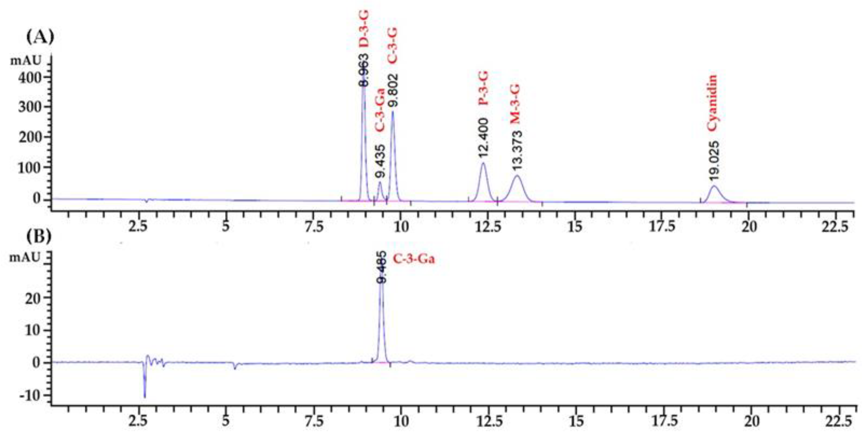
The analysis of specific anthocyanin constituents of the CCA extract was performed using a HPLC analysis. The identification and peak assignment of these compounds were primarily based on their retention time compared to that of the anthocyanin standard compounds. The results revealed the presence of cyanidin-3-O-galactoside with the amount of 36.25 mg/g extract, as shown in Figure 2.

3.4. Effect of CCA Extract on Cytotoxicity in RAW 264.7 Macrophages and 3T3-L1 Mature Adipocytes

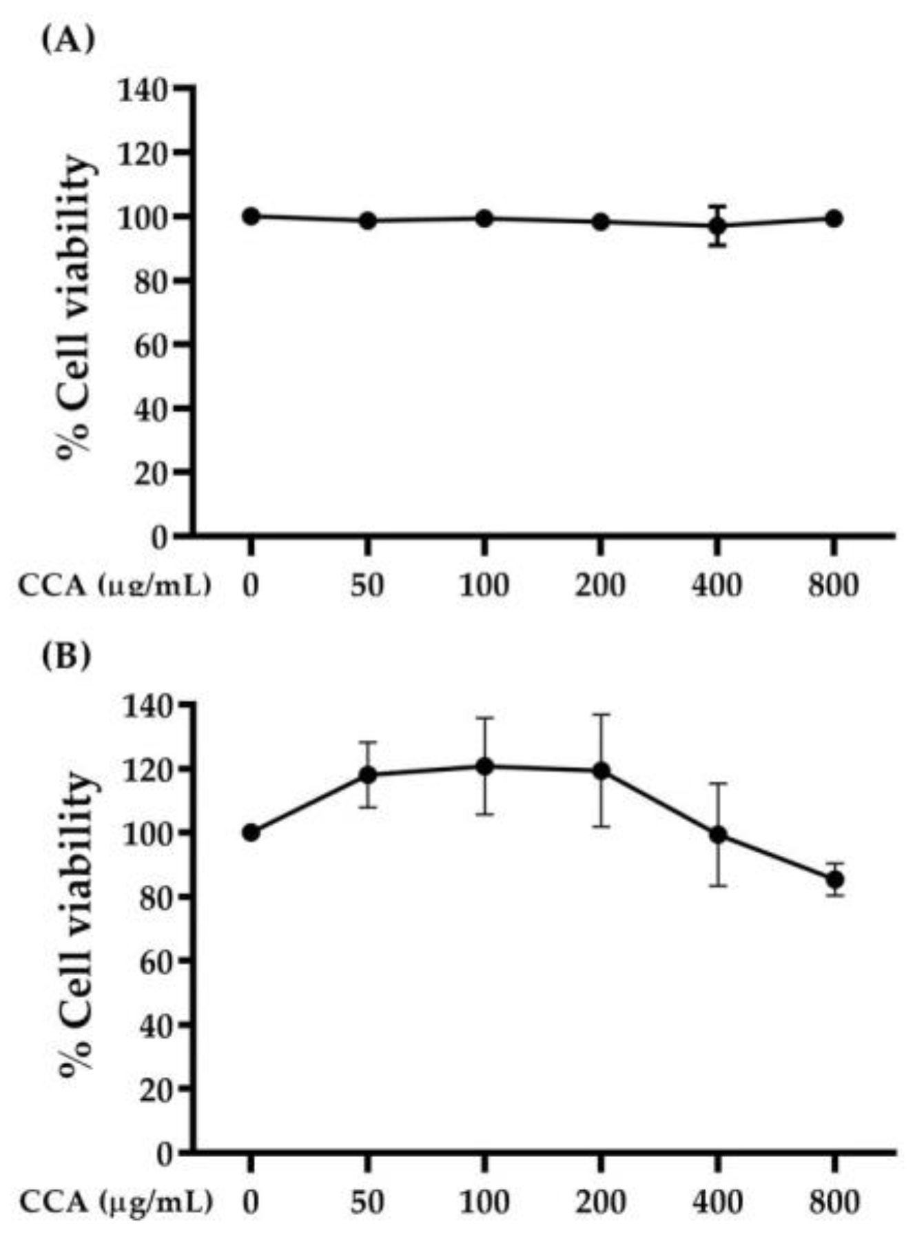
As shown in Figure 3, the CCA extract at up to 800 μg/mL did not demonstrate a harmful effect on either type of cells. Percentages of cell viability above 80% are considered to indicate non-cytotoxicity. Moreover, the CCA extract at a concentration range of 50–200 μg/mL was able to slightly stimulate the cell growth of RAW 264.7 macrophages.

3.5. Effect of CCA Extract on Nitric Oxide Production in LPS-Induced RAW 264.7 Macrophages

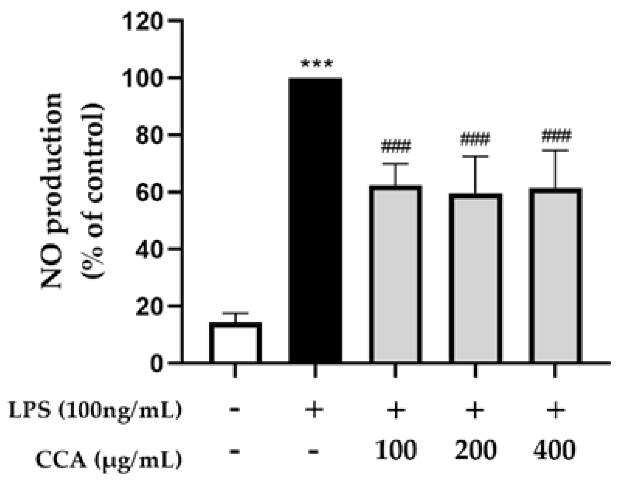
As illustrated in Figure 4, NO production exhibited a statistically significant increase (p < 0.001) following stimulation of the RAW 264.7 macrophages by LPS. Treatment with the CCA extract at concentrations of 100, 200, and 400 µg/mL achieved a statistically significant decrease (p < 0.001) in NO production in cells stimulated by LPS.

3.6. Effect of CCA Extract on Insulin Resistance in LPS-Induced 3T3-L1 Adipocytes

Similar to macrophages, LPS also activates the NF-κB and JNK pathways via Toll-like-receptors (TLR), resulting in the stimulation of cytokine production, which can impair insulin signaling in adipocytes. To evaluate the anti-insulin resistance effects of CCA, 3T3-L1 adipocytes were co-treated with the CCA extract and LPS. After that, glucose uptake and lipolysis were performed using a 2-NBDG uptake assay and glycerol content analysis, respectively.

3.6.1. Effect of CCA Extract on Glucose Uptake in LPS-Induced 3T3-L1 Adipocytes

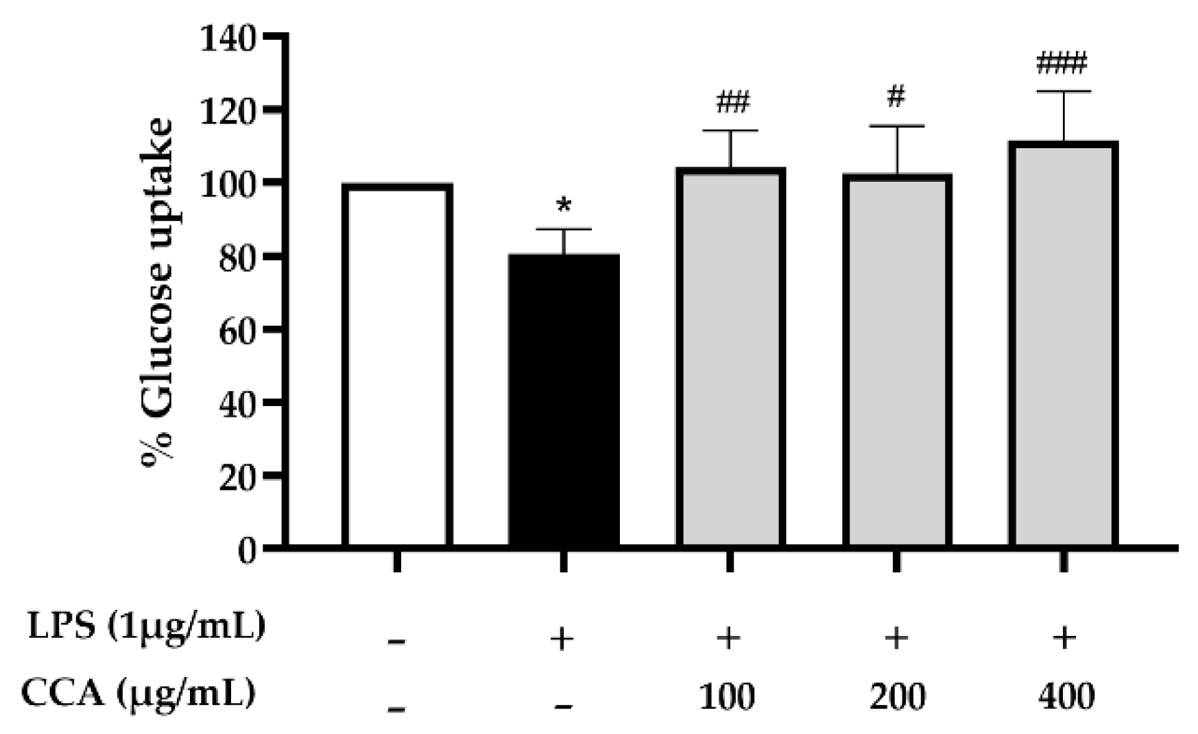
As shown in Figure 5, the cellular glucose uptake was decreased in LPS-treated 3T3-L1 adipocytes by 19.4% relative to the untreated control. Interestingly, the CCA extract significantly improved glucose uptake, indicating an anti-insulin resistance effect in LPS-treated adipocytes. The glucose uptakes of cells treated with the CCA extract at the concentrations of 100, 200, and 400 μg/mL were significantly increased by 29.3%, 27.3%, and 38.5%, respectively, when compared with LPS-treated cells.

3.6.2. Effect of CCA Extract on Lipolysis in LPS-Induced 3T3-L1 Adipocytes

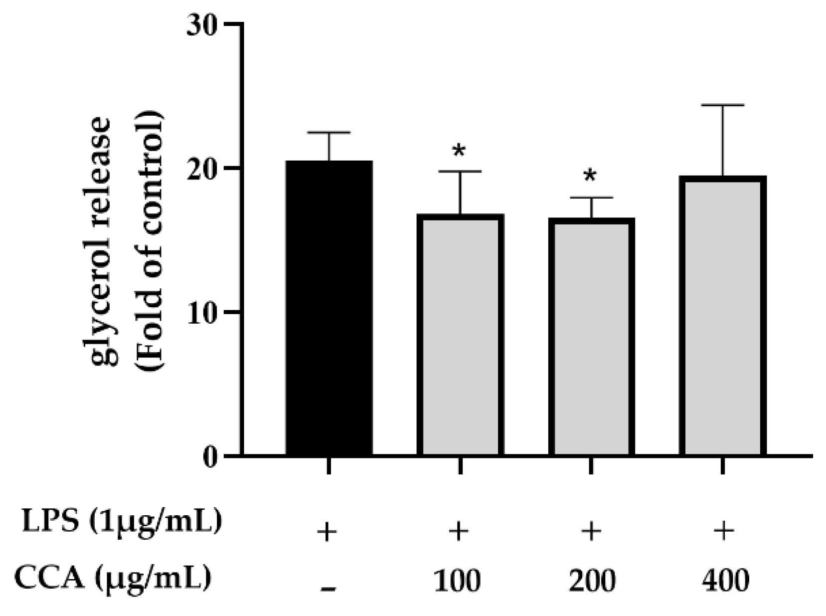
To further confirm the anti-insulin-resistant activity of the CCA extract, the conditioned media were collected to detect the free glycerol content. As shown in Figure 6, LPS 1 µg/mL could induce insulin resistance in the adipocytes, leading to free glycerol content, as observed in LPS-treated 3T3-L1 adipocytes. The inhibition of LPS-induced lipolysis was found in all the cells treated with the CCA extract, yet the significant reductions of 18.1% and 19.25% were found only in the dosages of 100 and 200 µg/mL, respectively (Figure 6). The effect of a higher dose of CCA (400 µg/mL) on inhibiting lipolysis was decreased. This finding suggested that the CCA extract at different doses induces biologically opposite effects. The anti-inflammatory activity of CCA is modulated by several different active compounds.

3.7. Effect of CCA Extract on Metabolic Variables in T2DM Rat Model

To verify the beneficial effects of the CCA extract on antidiabetic effects, an in vivo study in a high-fat diet with streptozotocin (STZ)-induced T2DM rats was performed. At the end of the experiment, the body weight of the normal diet with 400 mg/kg BW of the CCA (NDH) group was not significantly different compared with the normal diet control group (NDC). This suggested that supplementation with 400 mg/kg of CCA for 12 weeks did not affect body weight in normal rats. On the other hand, CCA tended to reduce visceral fat in the NDH group but did not significantly differ when compared with NDC group. (p > 0.05) (Table 2). The ratio of visceral fat and visceral fat/body weight did not increase significantly in the DMC group compared to the NDC group (p > 0.05). However, the visceral fat and the relative visceral fat mass in the DMH group significantly decreased when compared with the DMC group (p < 0.01). The average food intake and calorie consumption are shown in Table 2. There were no significant differences in food intake among the experimental groups, but the calorie intake significantly increased in the DMH and DMM groups compared to the DMC group (p < 0.001). These results suggested that supplementation with 400 mg/kg BW of CCA for 12 weeks effectively decreased the visceral fat and relative visceral fat mass in the T2DM rat model.

3.8. Effect of CCA Extract on the Fasting Plasma Metabolic Parameter in T2DM Rats at the End of the Experiment

The fasting plasma glucose levels of experimental rats are shown in Table 3. The fasting plasma glucose levels did not alter after 12 weeks of CCA extract supplementation in the normal rats (p > 0.05). The 12-week experiment period demonstrated a significant increase in the fasting plasma glucose level in the DMC group compared to the NDC group (−155%, p < 0.001), confirming the diabetes condition. Remarkably, administration of the CCA extract at doses of 200 and 400 mg/kg BW successfully decreased the fasting plasma glucose (−34% and −44%, respectively, p < 0.001). These results were similar to metformin treatment, which significantly reduced the fasting plasma glucose level by −43% concerning the DMC group (p < 0.001). To investigate whether the glucose-lowering effect of the CCA extract is involved in pancreatic insulin secretion, the fasting plasma insulin levels were measured. Notably, the fasting plasma insulin levels were not significantly different among the experimental groups (Table 3). These results suggest that an antihyperglycemic effect in the T2DM rat model of CCA extract supplementation may be similar to metformin treatment. Next, the whole-body insulin resistance and β-cell function were determined by the HOMA-IR and HOMA-β, respectively. As shown in Table 3, supplementation of the CCA extract did not alter the HOMA-IR and HOMA-β in the normal rats (p > 0.05). As expected, the HOMA-IR was markedly increased in the DMC group compared to the NDC group (p < 0.01), indicating that these T2DM rats had insulin resistance. Interestingly, the DML, DMH, and DMM groups had significant decreases in the HOMA-IR compared with the DMC group (p < 0.001, p < 0.01, and p < 0.01, respectively). Additionally, the HOMA-β of the DMC group significantly reduced when compared with the NDC group (p < 0.001). It implied that these T2DM rats not only exhibited insulin resistance but also represented impaired β-cell function. However, the HOMA-β of the DML and DMH groups had a trend to improve when compared to the DMC group, and the significant improvement of the HOMA-β was found only in the DMM group. All of these findings confirmed that the antihyperglycemic effect of CCA extract supplementation at the doses of 200 and 400 mg/kg BW was due to the amelioration of insulin resistance without affecting the pancreatic β-cell function in the T2DM rats.
The current study also examined the hypolipidemic effect of the CCA extract on T2DM rats, which are shown in Table 3. In normal rats, supplementation with the CCA extract did not change plasma triglycerides and total cholesterol levels. In DMC rats, plasma triglyceride and total cholesterol levels were significantly higher than those of NDC rats (p < 0.001). It is remarkable that CCA extracts administered to diabetic rats significantly reduced plasma triglyceride levels (p < 0.001). Plasma triglyceride levels also reduced significantly in DMM rats. In DML, DMH, and DMM rats, total cholesterol levels in plasma were reduced markedly (p < 0.001).

3.9. Effect of CCA Extract on Oral Glucose Tolerance Test in Type 2 Diabetes Rats

In order to determine whether CCA administration affected the insulin sensitivity in T2DM rats, OGTT was performed after 11 weeks of supplementation. The alteration of plasma glucose levels is presented in Figure 7A. In the NDC and NDH groups, there was no significant difference in plasma glucose levels at any time. Likewise, the incremental area under the curve for glucose (IAUC) values in the NDC and NDH groups were comparable (Figure 7B). As expected, the plasma glucose levels after glucose loading showed markedly higher values for the DMC group than those for the NDC group at all time points (p < 0.001). In comparison with the NDC group, the IAUC in the DMC group was increased significantly. This indicated that T2DM rats developed impairment of glucose tolerance. In particular, glucose levels in the DML, DMH, and DMM groups decreased markedly at all time points compared to the DMC group (p < 0.001). The IAUC values in the DMH and DMM groups tended to reduce when compared with the DMC group. These findings suggested that the CCA extract administered for 12 weeks with a dose of 400 mg/kg BW effectively attenuated glucose intolerance in T2DM rats, similar to the results obtained with metformin treatment in the DMC group.

3.10. Effect of CCA Extract on Pancreas Histology in Normal and T2DM Rats

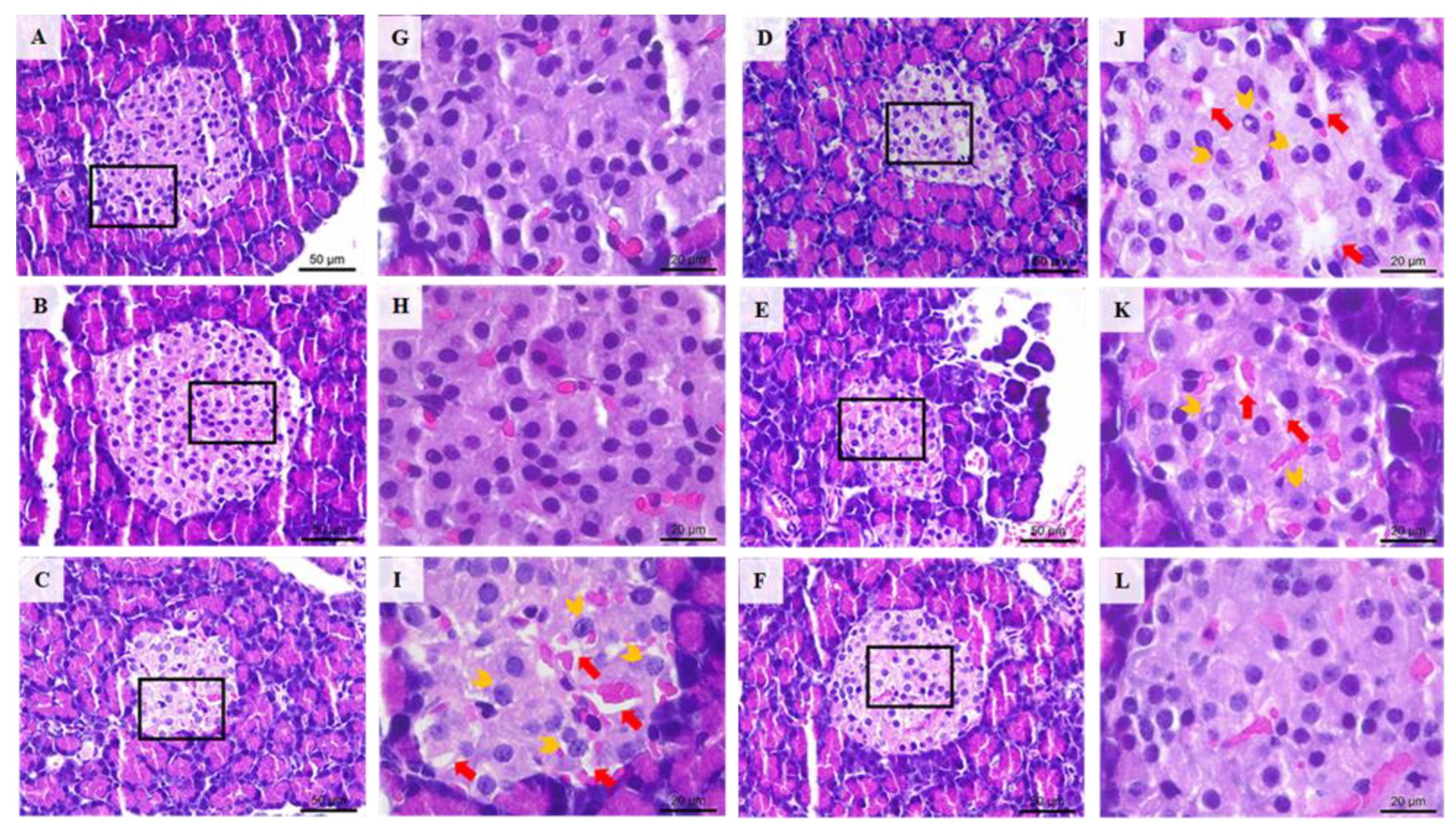
To evaluate the pancreatic injury, the pancreas was stained with H&E dye as shown in Figure 8. The pancreatic tissues of the DMC group showed changes in the morphology of islet. Some islets of the DMC groups were smaller than the NDC group, with irregular boundaries, and were also disorganized, with developed vacuolar degeneration and inflammation. Interestingly, histological investigation of pancreas in the DML, DMH, and DMM groups seemed to be much better than in the DMC group and appeared to be normal. These findings suggested that treatment with metformin or CCA extract, at 200 and 400 mg/kg BW, have effectively protected β-cell from progressive damage often associated with diabetes.

4. Discussion

A major issue considered in the present study was whether the aqueous extract of ripe Carissa carandas Linn. (CCA) could exert an antidiabetic effect or not. The outcomes of this study provide evidence to suggest that the antidiabetic effect of CCA is possibly mediated via reducing inflammation and insulin resistance using the in vitro model and in vivo model of T2DM rats. The percentage yield of C. carandas dried fruits extracted in an aqueous solution (CCA) was 38.8%, which is comparable to a previous study by Dhar et al. with a yield of 32.0% by using aqueous extraction [20]. The amount of total phenolic and flavonoid contents in the CCA extract (18.23 ± 0.4 mg GAE/g extract and 12.04 ± 2.7 mg RUE/g extract, respectively) were higher than the aqueous extract of C. carandas fruit reported by Dhar et al. (5.3 ± 3.8 mg GAE/g extract and 2.7 ± 1.5 mg CE/g extract, respectively) [20]. The ripe fruit of C. carandas is a rich source of the anthocyanin pigment. The major anthocyanin observed in this study was cyanidin-3-O-galactoside. This result confirms a previous report by Pewlong et al., who found cyanidin-3-O-galactoside to be the major anthocyanin compound in the ripe C. carandas fruit [10]. This finding was in contrast to the earlier reports. Previously, Iyer et al. reported that cyanidin-3-O-galactoside is a major pigment found in the ripe C. carandas fruit [11]. Moreover, the ripe C. carandas fruit has been reported by Weerawatanakorn et al. to have three anthocyanin pigments. The major anthocyanin of the C. carandas ripe fruit was cyanidin-3-O-glucoside, followed by peonidin-3-O-glucoside and malvidin-3-O-glucoside [12]. In addition, the C. carandas ripe fruit was previously reported to contain eight anthocyanins, which are cyanidin-3-O-glucoside, cyanidin-3-O-galactoside, delphinidin-3-O-galactoside, delphinidin-3-O-glucoside, delphinidin-3-O-rutinoside, 5-carboxypyranopelargonidin-3-beta-glucopyranoside, delphinidin, and peonidin-3-O-arabinoside, respectively [21]. The observed differences in phytochemical contents in the C. carandas fruit in this study might be due to several factors, including fruit ripeness, geographical locations, climatic conditions, and extraction methods [22,23,24,25].
An imbalance between the production of free radicals and the ability of the antioxidant system in the human body results in oxidative stress. Moreover, studies revealed that oxidative stress upregulates inflammatory mediators and also induces cellular or tissue damage, which leads to the pathogenesis of several diseases, such as insulin resistance, T2DM, and cardiovascular diseases [26]. The results from this study showed the abilities of the CCA extract to scavenge ABTS free radicals in a dose-dependent manner. Since the higher total antioxidant capacity of the diet has been found to decrease the risk of T2DM and metabolic diseases, the antioxidant activity of the CCA extract might be involved with its anti-inflammation and anti-insulin resistance [27,28].
A previous study found more macrophage migration into adipose tissue in obese mice than in lean mice [2]. Macrophages can activate the nitric oxide (NO) and the pro-inflammatory cytokines, including IL-1β, IL-6, and TNF-α production and secretion, resulting in a low-grade chronic inflammation condition in adipose tissue which can promote insulin resistance in adipocytes [3]. In this study, LPS was used to mimic the obesity-induced inflammation and insulin resistance in RAW 264.7 macrophages and 3T3-L1 adipocytes, respectively. LPS also stimulates the inflammation process via inducing IKK-NFκB signaling through Toll-like receptors [3,29]. In our study, when compared with untreated cells, LPS induced inflammation in RAW 264.7 macrophages by increasing the level of NO. Moreover, the CCA extract significantly inhibits the LPS-induced NO production in macrophages. Our findings confirm a previous study that described that the C. carandas fruit juice down-regulated the induction of inflammatory inducible nitric oxide synthase and COX-2 in LPS-stimulated macrophages [12].
Our in vitro study utilized LPS to stimulate insulin resistance. As mentioned above, LPS activates the expression and secretion of pro-inflammatory cytokines, such as TNF-α, from either macrophages or adipocytes [3,29]. These inflammatory cytokines stimulate lipolysis through the actions of adipose triglyceride lipase (ATGL) and hormone-sensitive lipase (HSL), which are responsible for the degradation of triglycerides into glycerol and free fatty acids [29]. It has been proven that LPS infusion causes increased lipolysis rates and insulin resistance [30]. The released FFAs and cytokines stimulate adipose tissue inflammation through IKK-NFκB signaling and impair insulin action in insulin-sensitive cells. Impairment in the insulin signaling cascade leads to impaired GLUT4 translocation in skeletal muscles and adipose cells [29,31].
The present study found that LPS induced insulin resistance in 3T3-L1 adipocytes by decreasing the cellular glucose uptake and increasing the free glycerol released content. This observation supports a previous study that LPS can block insulin signaling by disrupting the IRS-1 phosphorylation, resulting in the inhibition of the GLUT-4 translocation through the Akt pathway [29,32]. Importantly, the treatment of the CCA extract significantly inhibited insulin resistance in the LPS-treated adipocytes, as it induced insulin to stimulate glucose uptake by approximately 20% and inhibit lipolysis by approximately 20%. These indicated that the CCA extract could improve insulin sensitivity in LPS-treated adipocytes.
Rats with non-genetic type 2 diabetes induced by a combination of a high-fat diet followed by STZ injection are widely used in research because these rats show insulin resistance and hyperglycemia without hypoinsulinemia, which are the hallmarks of T2DM [33]. The low dose of the STZ injection (35 mg/kg BW) in the high-fat-diet-fed rats caused the absence of hypoinsulinemia, which normally appears with hyperglycemia. This is shown by no differences in circulating insulin levels between diabetic and normal rats, consistent with the partial destruction of pancreatic β-cells. Therefore, the combination of a high-fat diet and low-dose STZ injection was used to induce T2DM rats in this study. In the in vivo study, hyperglycemia, dyslipidemia, and insulin resistance were seen in rats that received an HFD plus STZ injection, suggesting that T2DM was successfully established as in previous studies [33].
Antihyperglycemic benefits of CCA extract supplements were found in diabetic rats. The metabolic parameters indicated that a 12-week treatment of the CCA extract at 200 and 400 mg/kg BW successfully reduced the fasting plasma glucose and improved glucose tolerance, showing its antihyperglycemic benefit in both fasting and post-prandial states. Consistently, the antihyperglycemic property of C. carandas unripe fruit was also demonstrated in previous reports using different diabetic models. This suggests that secondary metabolites, such as polyphenolics present in C. carandas fruit, may play an important role in its antidiabetic potential [8,9]. Hyperglycemia in T2DM results either from the inadequacy of insulin secretion by pancreatic β-cells or the inefficiency of cells to use insulin. Both conditions can affect glucose uptake or disposal by cells [34]. Since insulin plays an essential role in the entry of glucose into the cells, any disruptions in the transduction of insulin signals are associated with hyperglycemia due to cells’ inability to absorb glucose [35]. The binding of insulin to receptor and post-receptor events, such as activation of the IRS-PI3K-Akt-PKC pathway, cause GLUT4 translocation to the membrane and subsequent glucose uptake into cells.
Notably, a marked fall in body weight and a slight decline in food intake were observed in our experimental rats. A high-calorie diet might stimulate the release of hormones that promote satiety, such as cholecystokinin, glucagon-like peptide-1, and peptide YY [36], which may lead to a reduction in food intake. Moreover, the selective destruction of STZ to pancreatic β-cells diminishes insulin production [37]. Intense proteolysis and lipolysis due to insulin insufficiency make animals lose body weight. However, giving an HFD to T2DM rats throughout the experiment did not alter calorie intake and visceral fat mass. Previous studies also showed a reduction in body weight without variation in retroperitoneal, epidydimal, and perirenal lipid contents in animals given an HFD and STZ [38,39].
Supplementation with the CCA extract for 12 weeks at the dose of 400 mg/kg BW could effectively diminish visceral fat accumulation in diabetic rats, despite an increase in calorie intake. There are a few previous reports which directly described the effect of a C. carandas fruit extract on body weight and visceral fat weight. However, several studies demonstrate anthocyanin, a major flavonoid in the CCA extract, having such an effect. The reduction in body weight and white adipose tissue weight was observed in cyanidin-3-O-glucoside-treated diabetic mice, which was accompanied by a decline in the size of adipocytes [40]. Prior et al. reported that supplemental diets with an anthocyanin-rich blueberry extract markedly reduced body fat and body weight in obese HFD-fed C57BL/6 mice [41]. The previous study suggested that Aronia fruits rich in anthocyanin phytochemicals suppressed hyperglycemia and the accumulation of visceral fats by inhibiting pancreatic lipase activity and/or intestinal lipid absorption [42].
As with visceral fat, CCA could also diminish serum lipid levels in diabetic rats. Dyslipidemia, strongly correlated with obese T2DM individuals in several clinical reports, is characterized by the elevation of triglyceride, non-HDL cholesterol, and small dense LDL and HDL, as well as a reduction in HDL cholesterol [43]. Our study observed an anti-dyslipidemic effect of the CCA extract in the experimental diabetic animals after the 12-week intervention period. Researchers supposed that cyanidin-3-O-glucoside can inhibit intracellular de novo lipogenesis by inactivating the rate-limiting step of triglyceride synthesis, reducing hepatic steatosis in T2DM [44]. Similarly, Wang et al. found that a black rice anthocyanin extract normalized the increased body weight, serum cholesterol, and serum triglyceride in high-fat- and high-cholesterol-feeding mice [45].
Our in vitro study also confirmed the attenuation of insulin resistance observed in our in vivo study: the CCA extract treatment in mature adipocytes could alleviate the LPS-induced lipolysis, probably due to the presence of anthocyanin in CCA, which regulates FoxO1-mediated transcription of ATGL and thus inhibits adipocyte lipolysis [46]. In addition, decreased glucose uptake was also found in LPS-treated adipocytes. As mentioned earlier, incubation with the CCA extract exhibited improved glucose uptake into the mature adipocytes. Hence, CCA could probably upturn the LPS-induced inhibition of insulin signaling, consequently improving glucose disposal. Experiments in the 3T3-L1 cell line presented that cyanidin-3-O-glucoside-treated preadipocytes were differentiated into smaller, insulin-sensitive adipocytes, thus enhancing glucose uptake and insulin signaling [47]. Rutin, one of the flavonoid glycosides found in fruits and vegetables, has been discovered to possess a stimulatory effect on glucose uptake by enhancing GLUT4 and insulin receptor kinases [48]. Since C. carandas contains a high level of rutin, it is probable that it contributed to the antihyperglycemic effects of the CCA extract that we observed in our study.
To investigate whether the antihyperglycemic effect of CCA is due to an improvement in insulin secretion, we assessed fasting insulin level and employed a homeostatic model (HOMA-β). The HOMA model is used to estimate the sensitivity of basal insulin and the function of β-cells from fasting plasma glucose and insulin levels [19]. The untreated T2DM rats showed insignificantly lower plasma insulin levels and desperately worse HOMA-β, indicating that pancreatic β-cells were in severe dysfunction and probably unable to secrete adequate insulin. The CCA-extract-treated rats exhibited proper control in glycemia, but neither CCA nor metformin enhanced the fasting plasma insulin levels and HOMA-β, suggesting that the beneficial effect of the CCA extract on glycemic control is unrelated to the enhancement of insulin secretion. In contrast, Singh and fellows (2019) claimed that the antidiabetic effect of the C. carandas fruit extract hinged on the increase in insulin secretion; thereafter, they proved a possible mechanism of insulin secretagogue action with the closure of KATP channels in isolated islet β-cells [9]. The use of the HOMA to assess insulin sensitivity in subjects treated with insulin secretagogues has several drawbacks in result interpretation and needs further validation [49]. Nevertheless, the present study was unlikely to provide strong data to show that the CCA extract does not affect insulin secretion, since it did not investigate reliable parameters that reflect insulin production from the pancreas, such as C-peptide level, a marker of secretion [50], or parameters that reflect stimulated insulin secretion, such as insulin response curve and insulin sensitivity index (ISI) from an OGTT [51]. Remarkably, the histological study of the pancreas of diabetic rats treated with CCA showed that the severity of the degenerative changes in the islets of Langerhans was lower than that in the untreated diabetic rats. Hyperglycemia in diabetes was probably attenuated by supplementation with CCA, as a lower degree of hyperglycemia ameliorated β-cell damage progression. However, the antioxidative stress or anti-inflammation activity of the C. carandas extract might be partly involved in these findings [13,14].
Alternatively, the glucose-lowering effect of CCA supplements might be explained by other mechanisms. The results of this study revealed that improvement in insulin sensitivity might be a possible mechanism, and this conception was supported by the HOMA-IR index and the outcomes of the OGTT. Supplementation with the CCA extract showed an enhanced insulin sensitivity, the same as in the metformin-treated group. Metformin is a clinically common prescription in T2DM, which can reduce insulin resistance and liver glucose production and therefore effectively control blood glucose levels [52]. Kim and Abbasi reviewed studies of treatment with metformin that can positively affect insulin sensitivity by reducing lipolysis and free fatty acids, as well as increase the metabolic clearance rate of glucose [53]. As results show in the present study, the efficacy of the CCA supplement was comparable with that of metformin in terms of insulin sensitivity enhancement. Insulin resistance is a pathological condition in which insulin-dependent cells, such as skeletal muscle and adipocytes, do not respond appropriately to normal plasma insulin levels [43]. The mechanisms that promote insulin resistance include decreased glucose uptake into skeletal muscles and adipose tissues as well as increased liver glucose production. They are underlined by certain metabolic conditions, such as dyslipidemia, cellular oxidative stress, and chronic inflammation [54]. It has been proven that CCA extract supplementation improved serum lipids, adipocyte glucose uptake, and the scavenging of free radicals because of the manifestation of proper amounts of anthocyanins and phenolic compounds.
In conclusion, the present study evidenced the antidiabetic, anti-inflammation, and anti-insulin resistance of C. carandas ripe fruits in 3T3-L1 adipose cell lines and experimental T2DM rats. The performance of the CCA extract in the amelioration of insulin resistance by improving glucose uptake and by reducing lipolysis would be a possible explanation for its antihyperglycemic effect in the rat model. However, the precise mechanisms of these biological activities need further exploration.

Author Contributions

Conceptualization, A.I., N.L. and D.A.; methodology, A.I., M.L., T.W.L. and N.L.; investigation, M.L., T.W.L. and N.L.; formal analysis A.I., M.L., N.L. and D.A.; data curation, A.I. and N.L.; writing—original draft preparation, M.L.; writing—review and editing, A.I., N.L. and D.A.; supervision, A.I., N.L. and D.A.; project administration, A.I., N.L. and D.A.; funding acquisition, A.I. and M.L. All authors have read and agreed to the published version of the manuscript.

Funding

This research was partially supported by Thailand Science Research and Innovation (TSRI) and Ampol Food Processing Co., Ltd. under a Research and Researchers for Industries (RRi) Grant (MSD61I0061) to M.L. and A.I., and the Faculty of Medicine Research Fund (grant number: 005-2564), Chiang Mai University, Chiang Mai, Thailand to A.I.

Institutional Review Board Statement

The animal study protocol was approved by the Institutional Animal Care and Use Committee of the Faculty of Medicine, Chiang Mai University (protocol number: 40/2562).

Informed Consent Statement

Not applicable.

Data Availability Statement

The data presented in this study are available within the article.

Acknowledgments

This study was partially supported by Chiang Mai University, Thailand. This manuscript was proofread by Ruth Leatherman, RN Native English Proofreader Research Administration Section faculty of Medicine, Chiang Mai University, Chiang Mai, 50200, Thailand.

Conflicts of Interest

The authors declare no conflict of interest.

References

  1. McArdle, M.A.; Finucane, O.M.; Connaughton, R.M.; McMorrow, A.M.; Roche, H.M. Mechanisms of Obesity-Induced Inflammation and Insulin Resistance: Insights into the Emerging Role of Nutritional Strategies. Front. Endocrinol. 2013, 4, 52. [Google Scholar] [CrossRef] [PubMed]
  2. Gauthier, M.S.; Ruderman, N.B. Adipose Tissue Inflammation and Insulin Resistance: All Obese Humans Are Not Created Equal. Biochem. J. 2010, 430, e1–e4. [Google Scholar] [CrossRef] [PubMed]
  3. Yamashita, A.; Soga, Y.; Iwamoto, Y.; Yoshizawa, S.; Iwata, H.; Kokeguchi, S.; Takashiba, S.; Nishimura, F. Macrophage-Adipocyte Interaction: Marked Interleukin-6 Production by Lipopolysaccharide. Obesity 2007, 15, 2549–2552. [Google Scholar] [CrossRef] [PubMed]
  4. Robertson, R.P.; Harmon, J.; Tran, P.O.T.; Poitout, V. Β-Cell Glucose Toxicity, Lipotoxicity, and Chronic Oxidative Stress in Type 2 Diabetes. Diabetes 2004, 53 (Suppl. S1), S119–S124. [Google Scholar] [CrossRef]
  5. Razavi-Nematollahi, L.; Ismail-Beigi, F. Adverse Effects of Glycemia-Lowering Medications in Type 2 Diabetes. Curr. Diabetes Rep. 2019, 19, 132. [Google Scholar] [CrossRef] [PubMed]
  6. Tran, N.; Pham, B.; Le, L. Bioactive Compounds in Anti-Diabetic Plants: From Herbal Medicine to Modern Drug Discovery. Biology 2020, 9, 252. [Google Scholar] [CrossRef]
  7. Srinuanchai, W.; Nooin, R.; Pitchakarn, P.; Karinchai, J.; Suttisansanee, U.; Chansriniyom, C.; Jarussophon, S.; Temviriyanukul, P.; Nuchuchua, O. Inhibitory Effects of Gymnema Inodorum (Lour.) Decne Leaf Extracts and Its Triterpene Saponin on Carbohydrate Digestion and Intestinal Glucose Absorption. J. Ethnopharmacol. 2021, 266, 113398. [Google Scholar] [CrossRef] [PubMed]
  8. Itankar, P.R.; Lokhande, S.J.; Verma, P.R.; Arora, S.K.; Sahu, R.A.; Patil, A.T. Antidiabetic Potential of Unripe Carissa carandas Linn. Fruit Extract. J. Ethnopharmacol. 2011, 135, 430–433. [Google Scholar] [CrossRef]
  9. Singh, R.; Shrivastava, M.; Sharma, P. Antidiabetic Effect of Carissa carandas in Rats and the Possible Mechanism of Its Insulin Secretagogues Activity in Isolated Pancreatic Islets. J. Biomed. Ther. Sci. 2019, 6, 1–7. [Google Scholar]
  10. Pewlong, W.; Sajjabut, S.; Eamsiri, J.; Chookaew, S. Evaluation of Antioxidant Activities, Anthocyanins, Total Phenolic Content, Vitamin C Content and Cytotoxicity of Carissa carandas Linn. Chiang Mai Univ. J. Nat. Sci. 2014, 13, 509–517. [Google Scholar] [CrossRef]
  11. Iyer, C.M.; Dubash, P.J. Anthocyanin of Karwand (Carissa carandas) and Studies on Its Stability in Model Systems. J. Food Sci. Technol. Mysore 1993, 30, 246–248. [Google Scholar]
  12. Weerawatanakorn, M.; Pan, M.-H. Phytochemical Components of Carissa carandas and the Inhibitory Effects of Fruit Juice on Inducible Nitric Oxide Synthase and Cyclooxygenase-2. J. Food Biochem. 2017, 41, e12343. [Google Scholar] [CrossRef]
  13. Dhodi, J.B.; Thanekar, D.R.; Mestry, S.N.; Juvekar, A.R. Carissa carandas Linn. Fruit Extract Ameliorates Gentamicin-Induced Nephrotoxicity in Rats via Attenuation of Oxidative Stress. J. Acute Dis. 2015, 4, 135–140. [Google Scholar] [CrossRef]
  14. Anupama, N.; Madhumitha, G.; Rajesh, K.S. Role of Dried Fruits of Carissa carandas as Anti-Inflammatory Agents and the Analysis of Phytochemical Constituents by GC-MS. Biomed. Res. Int. 2014, 2014, 512369. [Google Scholar] [CrossRef] [PubMed]
  15. Gorinstein, S.; Zachwieja, Z.; Katrich, E.; Pawelzik, E.; Haruenkit, R.; Trakhtenberg, S.; Martin-Belloso, O. Comparison of the Contents of the Main Antioxidant Compounds and the Antioxidant Activity of White Grapefruit and His New Hybrid. Lebenson. Wiss. Technol. 2004, 37, 337–343. [Google Scholar] [CrossRef]
  16. Zhishen, J.; Mengcheng, T.; Jianming, W. The Determination of Flavonoid Contents in Mulberry and Their Scavenging Effects on Superoxide Radicals. Food Chem. 1999, 64, 555–559. [Google Scholar] [CrossRef]
  17. Dhar, P.; Bajpai, P.K.; Tayade, A.B.; Chaurasia, O.P.; Srivastava, R.B.; Singh, S.B. Chemical Composition and Antioxidant Capacities of Phytococktail Extracts from Trans-Himalayan Cold Desert. BMC Complement. Altern. Med. 2013, 13, 259. [Google Scholar] [CrossRef]
  18. Srinivasan, K.; Viswanad, B.; Asrat, L.; Kaul, C.L.; Ramarao, P. Combination of High-Fat Diet-Fed and Low-Dose Streptozoto-cin-Treated Rat: A Model for Type 2 Diabetes and Pharmacological Screening. Pharmacol. Res. 2005, 52, 313–320. [Google Scholar] [CrossRef]
  19. Matthews, D.R.; Hosker, J.P.; Rudenski, A.S.; Naylor, B.A.; Treacher, D.F.; Turner, R.C. Homeostasis Model Assessment: Insulin Re-Sistance and Beta-Cell Function from Fasting Plasma Glucose and Insulin Concentrations in Man. Diabetologia 1985, 28, 412–419. [Google Scholar] [CrossRef]
  20. Dhar, G.; Akther, S.; Sultana, A.; May, U.; Islam, M.M.; Dhali, M.; Sikdar, D. Effect of Extraction Solvents on Phenolic Contents and Antioxidant Capacities of Artocarpus chaplasha and Carissa carandas Fruits from Bangladesh. J. Appl. Biol. Biotechnol. 2017, 5, 039–044. [Google Scholar]
  21. Sarkar, R.; Kundu, A.; Banerjee, K.; Saha, S. Anthocyanin Composition and Potential Bioactivity of Karonda (Carissa carandas L.) Fruit: An Indian Source of Biocolorant. Lebenson. Wiss. Technol. 2018, 93, 673–678. [Google Scholar] [CrossRef]
  22. Le, X.T.; Huynh, M.T.; Pham, T.N.; Than, V.T.; Toan, T.Q.; Bach, L.G.; Trung, N.Q. Optimization of Total Anthocyanin Content, Stability and Antioxidant Evaluation of the Anthocyanin Extract from Vietnamese Carissa carandas L. Fruits. Processes 2019, 7, 468. [Google Scholar] [CrossRef]
  23. Shailajan, S.; Menon, S.; Sayed, N.; Tiwari, B. Simultaneous Estimation of Three Triterpenoids from Carissa Carandas Using Validated High Performance Liquid Chromatography. Int. J. Green Pharm. 2012, 6, 241. [Google Scholar] [CrossRef]
  24. Shailajan, S.; Sayed, N.; Tiwari, B. Impact of Regional Variation on Lupeol Content in Carissa Carandas Linn. Fruits: Evaluation Using Validated High Performance Thin Layer Chromatography. J. Adv. Sci. Res. 2013, 4, 21–24. [Google Scholar]
  25. Patra, P.A.; Basak, U. Nutritional and Antinutritional Properties of Carissa carandas and Cordia dichotoma, Two Medicinally Important Wild Edible Fruits of Odisha. J. Basic Appl. Sci. Res. 2017, 7, 1–12. [Google Scholar]
  26. Phaniendra, A.; Jestadi, D.B.; Periyasamy, L. Free Radicals: Properties, Sources, Targets, and Their Implication in Various Diseases. Indian J. Clin. Biochem. 2015, 30, 11–26. [Google Scholar] [CrossRef] [PubMed]
  27. Nascimento-Souza, M.A.; Paiva, P.G.; Martino, H.S.D.; Ribeiro, A.Q. Dietary Total Antioxidant Capacity as a Tool in Health Outcomes in Middle-Aged and Older Adults: A Systematic Review. Crit. Rev. Food Sci. Nutr. 2018, 58, 905–912. [Google Scholar] [CrossRef]
  28. Bahadoran, Z.; Golzarand, M.; Mirmiran, P.; Shiva, N.; Azizi, F. Dietary Total Antioxidant Capacity and the Occurrence of Metabolic Syndrome and Its Components after a 3-Year Follow-up in Adults: Tehran Lipid and Glucose Study. Nutr. Metab. 2012, 9, 70. [Google Scholar] [CrossRef]
  29. Liang, H.; Hussey, S.E.; Sanchez-Avila, A.; Tantiwong, P.; Musi, N. Effect of Lipopolysaccharide on Inflammation and Insulin Action in Human Muscle. PLoS ONE 2013, 8, e63983. [Google Scholar] [CrossRef]
  30. Rittig, N.; Bach, E.; Thomsen, H.H.; Pedersen, S.B.; Nielsen, T.S.; Jørgensen, J.O.; Jessen, N.; Møller, N. Regulation of Lipolysis and Adipose Tissue Signaling during Acute Endotoxin-Induced Inflammation: A Human Randomized Crossover Trial. PLoS ONE 2016, 11, e0162167. [Google Scholar] [CrossRef]
  31. Cullberg, K.B.; Larsen, J.Ø.; Pedersen, S.B.; Richelsen, B. Effects of LPS and Dietary Free Fatty Acids on MCP-1 in 3T3-L1 Adipocytes and Macrophages in Vitro. Nutr. Diabetes 2014, 4, e113. [Google Scholar] [CrossRef]
  32. Toda, G.; Soeda, K.; Okazaki, Y.; Kobayashi, N.; Masuda, Y.; Arakawa, N.; Suwanai, H.; Masamoto, Y.; Izumida, Y.; Kamei, N.; et al. Insulin- and Lipopolysaccharide-Mediated Signaling in Adipose Tissue Macrophages Regulates Postprandial Glycemia through Akt-mTOR Activation. Mol. Cell 2020, 79, 43–53. [Google Scholar] [CrossRef] [PubMed]
  33. Khat-Udomkiri, N.; Toejing, P.; Sirilun, S.; Chaiyasut, C.; Lailerd, N. Antihyperglycemic Effect of Rice Husk Derived Xylooligosac-Charides in High-Fat Diet and Low-Dose Streptozotocin-Induced Type 2 Diabetic Rat Model. Food Sci. Nutr. 2020, 8, 428–444. [Google Scholar] [CrossRef] [PubMed]
  34. Gowd, V.; Gurukar, A.; Chilkunda, N.D. Glycosaminoglycan Remodeling during Diabetes and the Role of Dietary Factors in Their Modulation. World J. Diabetes 2016, 7, 67–73. [Google Scholar] [CrossRef] [PubMed]
  35. Samuel, V.T.; Shulman, G.I. The Pathogenesis of Insulin Resistance: Integrating Signaling Pathways and Substrate Flux. J. Clin. Investig. 2016, 126, 12–22. [Google Scholar] [CrossRef]
  36. Ohlsson, L.; Rosenquist, A.; Rehfeld, J.F.; Härröd, M. Postprandial Effects on Plasma Lipids and Satiety Hormones from Intake of Liposomes Made from Fractionated Oat Oil: Two Randomized Crossover Studies. Food Nutr. Res. 2014, 58, 24465. [Google Scholar] [CrossRef] [PubMed]
  37. Al-Awar, A.; Kupai, K.; Veszelka, M.; Szűcs, G.; Attieh, Z.; Murlasits, Z.; Török, S.; Pósa, A.; Varga, C. Experimental Diabetes Mellitus in Different Animal Models. J. Diabetes Res. 2016, 2016, 9051426. [Google Scholar] [CrossRef] [PubMed]
  38. Ji, J.; Zhang, C.; Luo, X.; Wang, L.; Zhang, R.; Wang, Z.; Fan, D.; Yang, H.; Deng, J. Effect of Stay-Green Wheat, a Novel Variety of Wheat in China, on Glucose and Lipid Metabolism in High-Fat Diet Induced Type 2 Diabetic Rats. Nutrients 2015, 7, 5143–5155. [Google Scholar] [CrossRef] [PubMed]
  39. Magalhaes, D.A.; Kume, W.T.; Correia, F.S.; Queiroz, T.S.; Neto, A.; Santos, E.W. High-Fat Diet and Streptozotocin in the Induction of Type 2 Diabetes Mellitus: A New Proposal. An. Da Acad. Bras. De Cienc. 2019, 91, E20180314. [Google Scholar] [CrossRef]
  40. Liu, Y.; Li, D.; Zhang, Y.; Sun, R.; Xia, M. Anthocyanin Increases Adiponectin Secretion and Protects against Diabetes-Related Endothelial Dysfunction. Am. J. Physiol. Endocrinol. Metab. 2014, 306, E975–E988. [Google Scholar] [CrossRef]
  41. Prior, R.L.; Wu, X.; Gu, L.; Hager, T.J.; Hager, A.; Howard, L.R. Whole Berries versus Berry Anthocyanins: Interactions with Dietary Fat Levels in the C57BL/6J Mouse Model of Obesity. J. Agric. Food Chem. 2008, 56, 647–653. [Google Scholar] [CrossRef] [PubMed]
  42. Takahashi, A.; Shimizu, H.; Okazaki, Y.; Sakaguchi, H.; Taira, T.; Suzuki, T.; Chiji, H. Anthocyanin-Rich Phytochemicals from Aronia Fruits Inhibit Visceral Fat Accumulation and Hyperglycemia in High-Fat Diet-Induced Dietary Obese Rats. J. Oleo Sci. 2015, 64, 1243–1250. [Google Scholar] [CrossRef] [PubMed]
  43. Bjornstad, P.; Eckel, R.H. Pathogenesis of Lipid Disorders in Insulin Resistance: A Brief Review. Curr. Diabetes Rep. 2018, 18, 127. [Google Scholar] [CrossRef] [PubMed]
  44. Guo, H.; Guo, J.; Jiang, X.; Li, Z.; Ling, W. Cyanidin-3-O-Beta-Glucoside, a Typical Anthocyanin, Exhibits Antilipolytic Effects in 3T3-L1 Adipocytes during Hyperglycemia: Involvement of FoxO1-Mediated Transcription of Adipose Triglyceride Lipase. Food Chem. Toxicol. 2012, 50, 3040–3047. [Google Scholar] [CrossRef]
  45. Wang, H.; Liu, D.; Ji, Y.; Liu, Y.; Xu, L.; Guo, Y. Dietary Supplementation of Black Rice Anthocyanin Extract Regulates Cholesterol Metabolism and Improves Gut Microbiota Dysbiosis in C57BL/6J Mice Fed a High-Fat and Cholesterol Diet. Mol. Nutr. Food Res. 2020, 64, 1900876. [Google Scholar] [CrossRef]
  46. Guo, H.; Li, D.; Ling, W.; Feng, X.; Xia, M. Anthocyanin Inhibits High Glucose-Induced Hepatic MtGPAT1 Activation and Prevents Fatty Acid Synthesis through PKCzeta. J. Lipid Res. 2011, 52, 908–922. [Google Scholar] [CrossRef]
  47. Matsukawa, T.; Inaguma, T.; Han, J.; Villareal, M.O.; Isoda, H. Cyanidin-3-Glucoside Derived from Black Soybeans Ameliorate Type 2 Diabetes through the Induction of Differentiation of Preadipocytes into Smaller and Insulin-Sensitive Adipocytes. J. Nutr. Biochem. 2015, 26, 860–867. [Google Scholar] [CrossRef] [PubMed]
  48. Salehi, B.; Ata, A.; Anil Kumar, N.V.; Sharopov, F.; Ramírez-Alarcón, K.; Ruiz-Ortega, A.; Abdulmajid Ayatollahi, S.; Tsouh Fokou, P.V.; Kobarfard, F.; Amiruddin Zakaria, Z.; et al. Antidiabetic Potential of Medicinal Plants and Their Active Components. Biomolecules 2019, 9, 551. [Google Scholar] [CrossRef]
  49. Wallace, T.M.; Levy, J.C.; Matthews, D.R. Use and Abuse of HOMA Modeling. Diabetes Care 2004, 27, 1487–1495. [Google Scholar] [CrossRef]
  50. Gonzalez-Mejia, M.E.; Porchia, L.M.; Torres-Rasgado, E.; Ruiz-Vivanco, G.; Pulido-Pérez, P.; Báez-Duarte, B.G.; Pérez-Fuentes, R. C-Peptide Is a Sensitive Indicator for the Diagnosis of Metabolic Syndrome in Subjects from Central Mexico. Metab. Syndr. Relat. Disord. 2016, 14, 210–216. [Google Scholar] [CrossRef]
  51. Stumvoll, M.; Mitrakou, A.; Pimenta, W.; Jenssen, T.; Yki-Järvinen, H.; Van Haeften, T.; Renn, W.; Gerich, J. Use of the Oral Glucose Tolerance Test to Assess Insulin Release and Insulin Sensitivity. Diabetes Care 2000, 23, 295–301. [Google Scholar] [CrossRef] [PubMed]
  52. Zhao, M.; Li, X.W.; Chen, D.Z.; Hao, F.; Tao, S.X.; Yu, H.Y.; Cheng, R.; Liu, H. Neuro-Protective Role of Metformin in Patients with Acute Stroke and Type 2 Diabetes Mellitus via AMPK/Mammalian Target of Rapamycin (MTOR) Signaling Pathway and Oxidative Stress. Med. Sci. Monit. 2019, 25, 2186–2194. [Google Scholar] [CrossRef] [PubMed]
  53. Kim, S.H.; Abbasi, F. Myths about Insulin Resistance: Tribute to Gerald Reaven. Endocrinol. Metab. 2019, 34, 47–52. [Google Scholar] [CrossRef] [PubMed]
  54. Yaribeygi, H.; Farrokhi, F.R.; Butler, A.E.; Sahebkar, A. Insulin Resistance: Review of the Underlying Molecular Mecha-Nisms. J. Cell. Physiol. 2019, 234, 8152–8161. [Google Scholar] [CrossRef] [PubMed]
Figure 1. The percentage of ABTS scavenging activity of CCA extract. The results represent a mean (SD) of a triplicate independent experiment; *** p < 0.001 vs. control treated without the extract.
Applsci 13 06454 g001
Figure 2. A Representative HPLC chromatogram of (A) standards of anthocyanin compounds delphinidin-3-O-glucoside (D-3-G), cyanidin-3-O-galactoside (C-3-Ga), cyanidin-3-O-glucoside (C-3-G), petunidin-3-O-glucoside (P-3-G), malvidin-3-O-glucoside (M-3-G), and cyanidin; and (B) C-3-Ga contained in HPLC chromatogram of C. carandas extract.
Applsci 13 06454 g002
Figure 3. Effect of CCA extract on cell viability in (A) RAW 264.7 macrophages and (B) 3T3-L1 adipocytes. The cells were treated with the extract for 24 h. The cell survival rate was determined by SRB assay. The values are expressed as mean (SD) (n = 3).
Applsci 13 06454 g003
Figure 4. Effect of CCA extract on nitric oxide production in LPS-induced RAW 264.7 macrophages. The cells were pretreated with CCA extract at concentrations of 100, 200, and 400 µg/mL for two hours, and then co-incubated with LPS 100 ng/mL for 24 h. The values are expressed as mean (SD) (n = 3); *** p < 0.01 vs. control group, ### p < 0.001 vs. LPS-treated group.
Applsci 13 06454 g004
Figure 5. Effect of CCA extract on glucose uptake in LPS-induced 3T3-L1 adipocytes. The cells were co-treated with 1 µg/mL LPS and CCA extract at concentrations of 100, 200, and 400 µg/mL and incubated for 24 h. The cellular glucose uptake was determined using 2-NBDG. The values are expressed as mean (SD) (n = 3); * p < 0.05 vs. control group, # p < 0.05, ## p < 0.01, and ### p < 0.001 vs. LPS-treated group.
Applsci 13 06454 g005
Figure 6. Effect of CCA extract on glycerol release in LPS-treated 3T3-L1 adipocytes. The cells were co-treated with 1 µg/mL LPS and CCA extract at concentrations of 100, 200, and 400 µg/mL and incubated for 24 h. The values are expressed as mean (SD) (n = 3); * p < 0.05 vs. LPS-treated group.
Applsci 13 06454 g006
Figure 7. Effect of CCA extract on plasma glucose responses (A) and incremental area under the curve for glucose (B) at 12 weeks of experiment. Results are presented as mean ± SEM and incremental area under the curve for glucose; *** p < 0.001 vs. NDC, ## p < 0.01, ### p < 0.001 vs. DMC.
Applsci 13 06454 g007
Figure 8. Photomicrographs of pancreatic tissues: normal control group (A,G); normal rats treated with 400 mg/kg BW of CCA (B,H); diabetic control group (C,I); 200 mg/kg BW of CCA (D,J); 400 mg/kg BW of CCA (E,K) and metformin (F,L) given to diabetic rats. The black-boxed areas in A-F are magnified in G–L AC: acinar cells; IL: islets of Langerhans cells. Cytoplasmic vacuolar degeneration (red arrow); Pyknotic nuclei (yellow arrow head); hematoxylin and eosin; (AF) (40×), (GL) (100×).
Applsci 13 06454 g008
Table 1. Extract yield (%) and total phenolic and flavonoid contents of C. carandas extract.
C. carandas Extract
Extract yield (%) 38.82 (1.19)
Phenolic contents (mg GAE/g extract)18.23 (0.37)
Flavonoid contents (mg RUE/g extract)12.04 (2.74)
The results are represented as mean (SD) of the triplicate independent experiment.
Table 2. Effect of CCA extract on body weight, visceral fat, visceral fat/body weight (VF/BW) ratio and food intake in rats at end of experiment.
GroupsParameters
BW (g)VF (g)VF/BWFood Intake
(g)
Calorie Intake
(Kcal)
NDC538.13 ± 13.9540.49 ± 4.397.08 ± 0.5724.06 ± 0.6891.58 ± 4.88
NDH547.50 ± 16.4838.31 ± 2.906.41 ± 0.3125.07 ± 0.3893.77 ± 1.42
DMC491.25 ± 15.52 ***38.85 ± 5.486.94 ± 0.6918.41 ± 0.4786.18 ± 2.21
DML487.50 ± 16.93 ***34.76 ± 4.577.17 ± 0.4319.88 ± 1.4093.02 ± 6.57
DMH484.38 ± 16.97 ***27.80 ± 3.33 *** ##5.20 ± 0.50 *** ###21.21 ± 1.0499.25 ± 4.86 * ###
DMM499.29 ± 20.94 ***38.19 ± 8.537.84 ± 0.9421.30 ± 2.3299.68 ± 10.87 ** ###
Results are presented as mean ± SEM (n = 7–8 for each group); * p < 0.05, ** p < 0.01, *** p < 0.001 vs. NDC; ## p < 0.01, ### p < 0.001 vs. DMC.
Table 3. Effect of CCA. Extract on fasting plasma metabolic parameter at end of experiment.
GroupsParameters
Glucose
(mg/dL)
Insulin
(ng/mL)
HOMA-IRHOMA-βTriglyceride
(mg/dL)
Total Cholesterol
(mg/dL)
NDC143.61 ± 5.613.74 ± 0.841.49 ± 0.3019.34 ± 3.8673.14 ± 3.8567.60 ± 2.99
NDH135.30 ± 6.023.44 ± 0.721.16 ± 0.3117.93 ± 2.3267.50 ± 2.4864.18 ± 6.99
DMC366.24 ± 30.0 ***2.53 ± 0.612.38 ± 0.66 ***3.11 ± 0.75 ***126.42 ± 11.16 ***139.56 ± 5.79 ***
DML241.70 ± 14.69 *** ###2.12 ± 0.231.04 ± 0.23 ###4.08 ± 0.45 ***67.05 ± 6.00 ###105.77 ± 9.09 *** ###
DMH205.41 ± 12.49 *** ###3.18 ± 0.871.46 ± 0.35 ##7.03 ± 2.54 ***75.02 ± 4.64 ###104.51 ± 8.81 *** ###
DMM210.17 ± 16.76 *** ###3.35 ± 0.661.58 ± 0.20 #8.90 ± 2.32 *** ##67.28 ± 3.55 ###78.19 ± 7.29 ###
Results are presented as mean ± SEM (n = 7–8 for each group); *** p < 0.001 vs. NDC; # p < 0.05, ## p < 0.01, ### p < 0.001 vs. DMC.
Disclaimer/Publisher’s Note: The statements, opinions and data contained in all publications are solely those of the individual author(s) and contributor(s) and not of MDPI and/or the editor(s). MDPI and/or the editor(s) disclaim responsibility for any injury to people or property resulting from any ideas, methods, instructions or products referred to in the content.