Article

The Interplay of Exogenous and Endogenous Hydrogen Sulfide (H2S) in Maintaining Redox Homeostasis in Individuals with Low Ferritin Levels

1
Department of Basic Sciences, Faculty of Medicine, Yarmouk University, Irbid 21163, Jordan
2
Department of Biological Sciences, Faculty of Science, Yarmouk University, Irbid 21163, Jordan
3
Clinical Pharmacy and Pharmacy Practice Department, Faculty of Pharmacy, Yarmouk University, Irbid 21163, Jordan
4
Department of Internal Medicine, Jordanian Royal Medical Services, Amman 11855, Jordan
*
Author to whom correspondence should be addressed.
Appl. Sci. 2023, 13(11), 6621; https://doi.org/10.3390/app13116621
Submission received: 11 December 2022 / Revised: 22 May 2023 / Accepted: 28 May 2023 / Published: 30 May 2023
(This article belongs to the Section Applied Biosciences and Bioengineering)

Abstract

:
The primary objective of this study was to investigate the influence of living near a thermal spring and being exposed to hydrogen sulfide (H2S) gas, which lingers in the surrounding atmosphere, on the redox and lipid peroxidation status of individuals with low ferritin levels. Participants in the study were divided into two groups based on their H2S exposure frequency and ferritin levels. Within Group A, individuals who did not experience occasional H2S exposure (2–3 h, 2–3 times per year) and had an average of normal ferritin levels of 87.5 ng/mL exhibited lower levels of oxidative stress and lipid peroxidation compared to those with low ferritin levels (average of 6.57 ng/mL) in the same group. Additionally, individuals with normal ferritin levels showed higher levels of thiol, H2S, and antioxidant capacity. However, it was observed that individuals with low ferritin levels had significantly lower levels of endogenous H2S and thiol and higher levels of oxidative stress and lipid peroxidation. In Group B, individuals who experienced occasional H2S exposure (2–3 h, 10–12 times per year) and had an average of normal ferritin levels of 91.65 ng/mL showed lower oxidative stress and lipid peroxidation, as well as higher thiol, H2S, and antioxidant capacity. Surprisingly, individuals in Group B with low ferritin levels (average of 6.18 ng/mL) showed increased endogenous levels of H2S, which resulted in lower oxidative stress, and lipid peroxidation. These findings suggest that inhaling environmental H2S emitted from thermal springs may serve as an adaptive and protective mechanism for individuals with low ferritin levels by enhancing antioxidant capacity and reducing oxidative stress and lipid peroxidation modification, thus mitigating the harmful effects of reactive oxygen species.

1. Introduction

Hydrogen sulfide (H2S) is a flammable, colorless gas that is toxic at extremely low concentrations [1,2]. Mammalian cells produce biologically active H2S gas as a byproduct of cysteine metabolism via the enzymatic actions of three major enzymes: cystathionine β-synthase (CBS), cystathionine γ-lyase (CSE), and 3-mercaptopyruvate sulfurtransferase (MST) [3]. H2S has been identified as a highly active signaling molecule that plays many functional roles in a variety of physiological and pathological processes [4]. H2S is involved in a variety of physiological activities, such as signal transduction, controlling vascular tone, protecting cells against oxidative stress, and ischemia-reperfusion damage in a number of tissues [5].
H2S is generated naturally in the atmosphere through the anaerobic degradation of sulfate by bacteria as well as from the contact of magma from volcanoes with groundwater. Additionally, H2S can be produced through a variety of human and industrial activities [6]. H2S has the propriety of being soluble in water, and as a result, it is commonly found in the water of thermal springs. When the water containing H2S is exposed to the atmosphere, the H2S is released into the surrounding environment [6]. This release of H2S into the air from thermal springs can have implications for air quality and potentially impact the health and well-being of individuals in the vicinity. However, the rapid and accurate measurement of H2S remains challenging due to its high biological reactivity and short half-life of H2S [3]. Despite its very low concentration, endogenous H2S in human tissue has been reported to act in many physiological activities [3]. A small portion of H2S can undergo conversion into low-toxicity compounds through the cytosolic detoxification pathway, while the majority of these low-toxicity H2S compounds are oxidized and metabolized into sulfate and thiosulfate in mitochondria [3]. The H2S metabolites are excreted rapidly within 24 h through the kidneys, intestines, and lungs, serving to maintain the balance of physiological H2S levels in the body [3]. In recent years, there has been significant research focused on the beneficial effects of antioxidants on maintaining human health [7].
The antioxidant proprieties of H2S have been extensively investigated, highlighting its potential role in combating oxidative cell damage associated with conditions such as Alzheimer’s disease, cancer, heart disease, and many other diseases [8]. While the human body naturally produces endogenous antioxidants to neutralize free radicals, it relies on exogenous sources, primarily from the diet, to obtain the remaining antioxidant needs [9]. Various mechanisms have been suggested to explain the anti-oxidative action of H2S, including direct scavenging of reactive oxygen species (ROS) and upregulating the antioxidant defense system [10,11].
The accumulation of toxic quantities of iron leads to the formation of ROS, which induces oxidative stress [12]. In addition, ferritin, which is a cytoplasmic protein that stores iron as ferric, plays a crucial role in modulating cellular sensitivity to oxidant insults, as intracellular iron catalyzes the generation of ROS [13]. Beyond their function in iron storage, ferritins are also involved in protection against oxidative stress through their potential detoxification properties of excess iron, dioxygen, and hydrogen peroxide [14,15]. The storage and metabolism of iron are tightly controlled under normal physiological conditions [16]. However, the disruption of normal iron homeostasis may increase reactive iron levels and the generation of oxygen-free radicals’ [16]. Furthermore, disturbances in iron levels or iron deficiency not only affects the production of hemoglobin but also impair the synthesis of iron-containing proteins, such as cytochromes, myoglobin, catalase, and peroxidase [17].
However, there is limited data and often contradictory information on oxidative and antioxidant defenses in IDA [18]. The primary pathophysiology of IDA is reduced hemoglobin production, which impairs the ability of blood cells to carry oxygen [19]. Furthermore, in ID states enzymes of the antioxidant defense system may be functionally defective, leading to an imbalance of free radicals and increase oxidative stress [19]. It has been reported that IDA is associated with increased oxidative stress, and that treatment of IDA can enhance antioxidant capacity [20]. The inhalation of H2S gas or injection of aqueous H2S donor solutions has been shown to have a variety of biological effects in humans [3].
Various studies have explored the antioxidant properties of H2S and its impact on oxidative stress in different contexts [3]. However, there is a scarcity of research specifically investigating the interaction between H2S and low ferritin levels concerning oxidative stress. Therefore, the main objective of this study was to evaluate the oxidative status of individuals with low ferritin levels and investigate the influence of prolonged exposure to naturally occurring H2S on their oxidative status. The study aimed to assess several oxidative stress parameters, including ferritin levels, total oxidative stress (TOS), total thiol, endogenous plasma H2S, lipid hydroperoxides (MDA), and total antioxidant capacity. The investigation involved both low-ferritin levels and normal individuals who had been exposed to H2S emissions over an extended period of time.

2. Results

2.1. Ferritin Levels and the Frequency of Spring Visits

Table 1 displays the characteristics of two study groups. Group A represents individuals who were not exposed to environmental H2S from thermal springs. Within Group A, the average ferritin concentration was 87.5 ng/mL for individuals with normal ferritin levels and 6.57 ng/mL for individuals with low ferritin levels. Group B, on the other hand, consisted of individuals who were exposed to environmental H2S and had normal and low ferritin levels. The average ferritin concentration in Group B was 91.65 ng/mL for individuals with normal ferritin levels and 6.18 ng/mL for individuals with low ferritin levels. Additionally, the study investigated the visitation patterns to thermal springs, specifically examining the frequency and duration of visits among the study participants. Specifically, individuals in Group A reported an average visitation frequency of 2–3 times per year to the thermal springs, while individuals in Group B reported an average visitation frequency of 10–12 per year. Remarkably, both groups reported spending an average of 2–3 h per visit at the thermal springs. These visitation patterns were assessed to understand the frequency and duration of exposure to the thermal springs among the study participant, providing valuable information on their level of engagement with the H2S thermal spring environment.

2.2. Sulfide and Thiol Measurement in Plasma

Based on the data presented in Figure 1, individuals characterized by low ferritin levels exhibited significantly lower levels of endogenous sulphide-H2S in their plasma in comparison to individuals with normal ferritin levels (* p < 0.05). Furthermore, individuals belonging to Group B, who reported frequent exposure to environmental H2S, demonstrated significantly higher levels of sulfide-H2S in their plasma compared to individuals in Group A, who reported sporadic exposure to H2S. Notably, it was observed that individuals with low ferritin levels residing in the surrounding area, who were frequently exposed to environmental H2S, exhibited significantly higher levels of sulfide-H2S in their plasma when compared to individuals with low ferritin levels in Group A, who had sporadic H2S exposure (# p < 0.05).
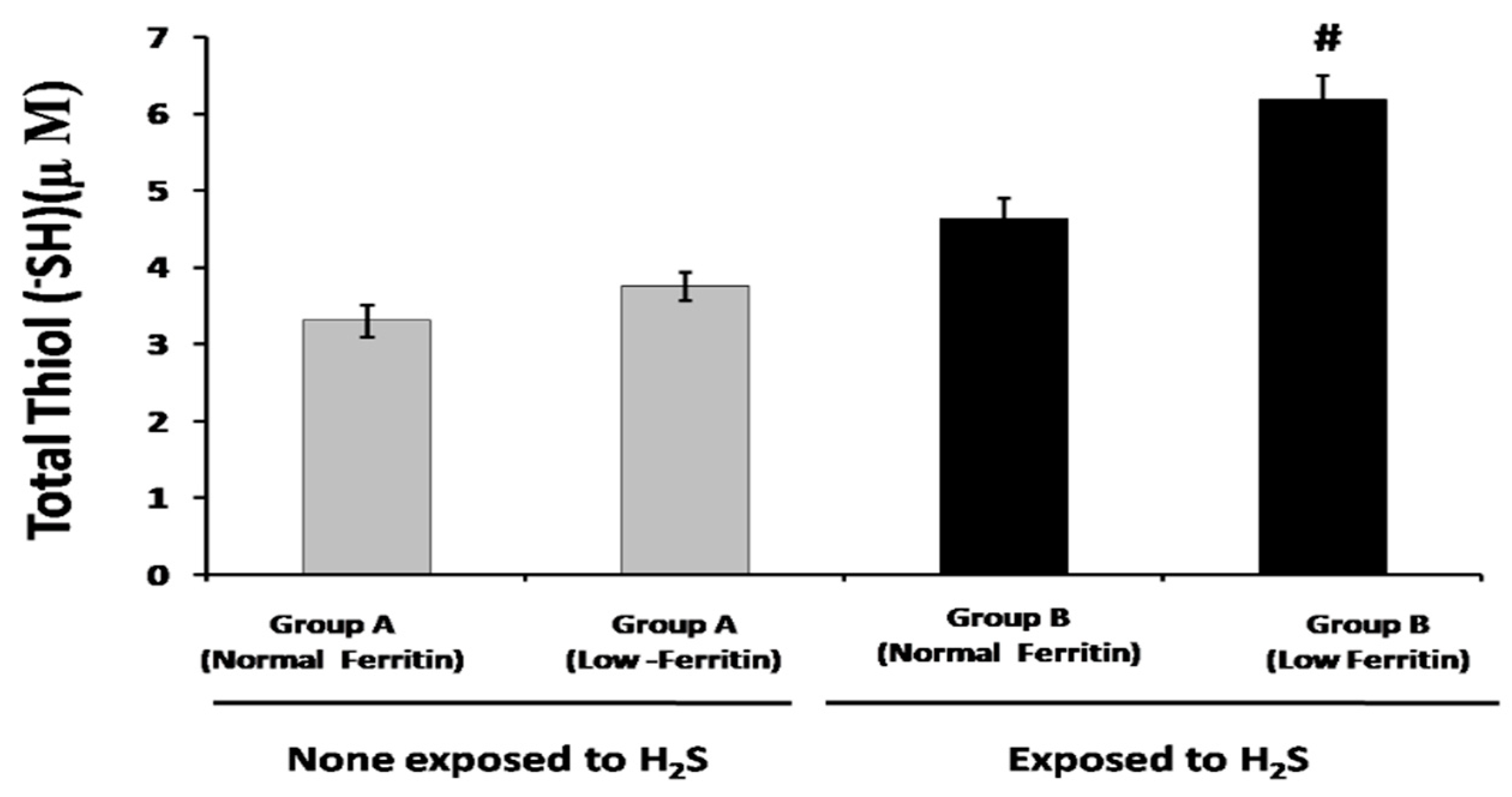
According to the data shown in Figure 2, no significant difference in total thiol levels was observed between individuals with low and normal ferritin in group A, who had sporadic exposure to H2S. However, individuals with low-ferritin levels displayed significantly higher total thiol levels compared to those with normal ferritin levels (* p < 0.05). Additionally, a significant decrease in total thiol levels was observed when comparing Group B, characterized by frequent exposure to environmental H2S, to Group A (* p < 0.05).

2.3. Lipid Peroxidation and Total Oxidative Stress Measurement

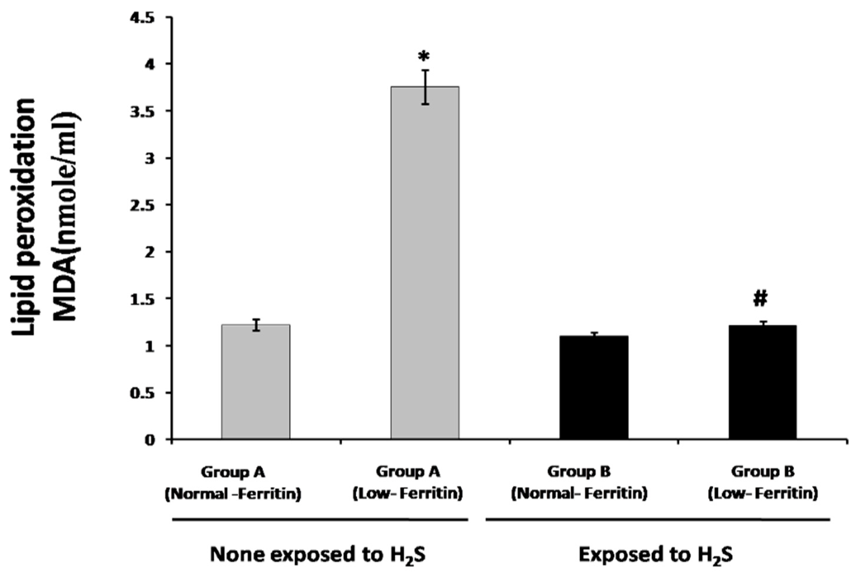
According to the data depicted in Figure 3, it was observed that individuals with low ferritin levels exhibited significantly higher mean serum lipid peroxidation (MDA) values compared to individuals with normal ferritin levels in group A, who were not exposed to H2S. Notably, a significant decrease in lipid peroxidation levels was observed among low-ferritin individuals who were exposed to environmental H2S in Group B when compared to low-ferritin individuals who were not exposed to H2S in Group A (# p < 0.05), as illustrated in Figure 3 (# p < 0.05).
Based on the data presented in Figure 4, the total oxidative stress levels were significantly higher in individuals with low-ferritin levels compared to individuals with normal ferritin levels in both groups A and B. Furthermore, Figure 4 demonstrates that exposure to H2S had a significant impact on oxidative stress, particularly among individuals with low-ferritin levels. The data clearly indicated a significant reduction in the total oxidative stress in low-ferritin individuals who were exposed to environmental H2S emitted from thermal springs compared to low-ferritin individuals who were not exposed to environmental H2S emission (Group B).

2.4. Total Antioxidant Capacity

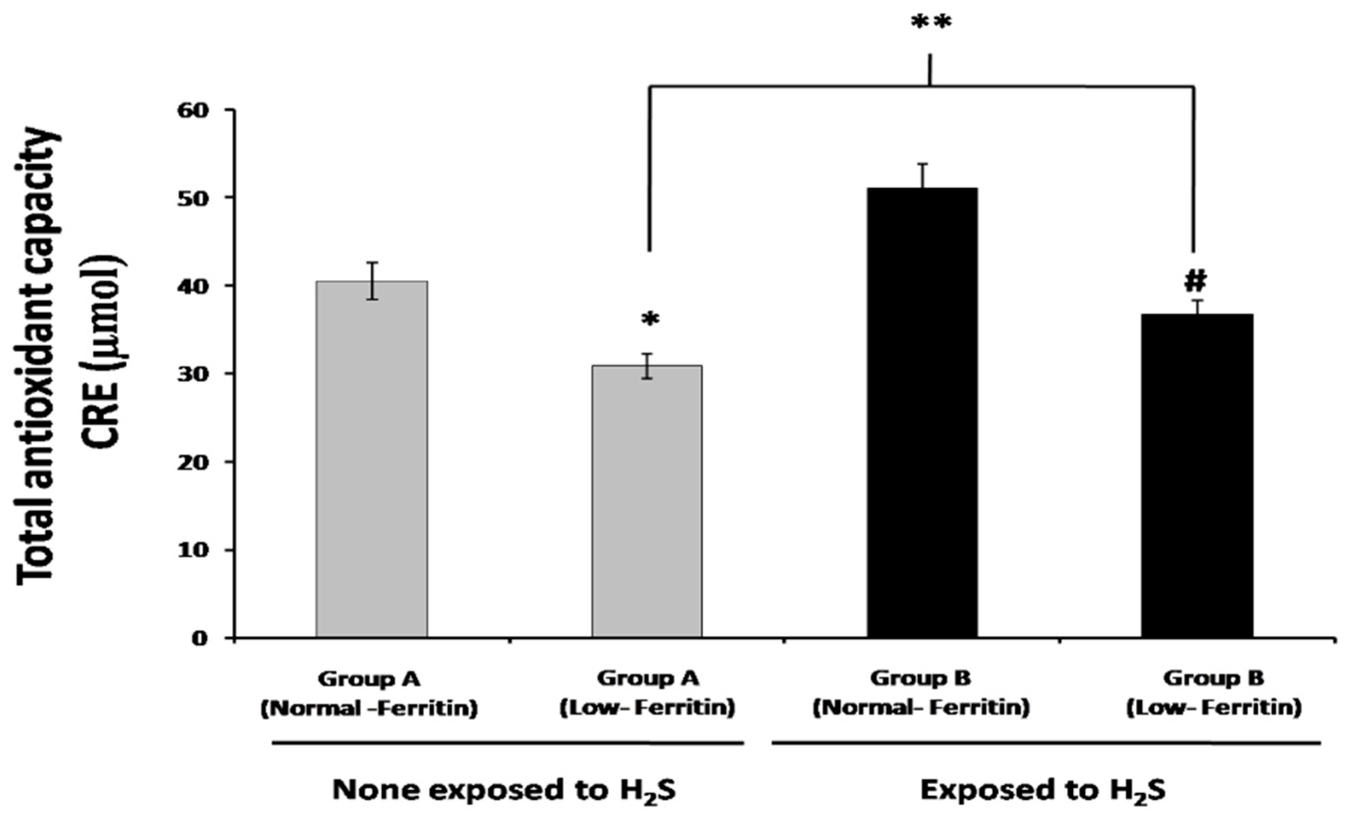
Figure 5 illustrates the measurement of total antioxidant capacity in the plasma. Based on the data presented in Figure 5, evident that individuals with low-ferritin had significantly lower levels of total antioxidants compared to individuals with normal ferritin levels, regardless of their exposure to H2S (Groups A and B). Moreover, Figure 5 highlights that when individuals with low-ferritin levels were exposed to environmental H2S, their antioxidant capacity levels were significantly higher compared to low-ferritin individuals who were not exposed to environmental (Group A).

3. Discussion

In this study, we propose that exogenous administration of H2S can effectively elevate endogenous H2S levels, although the precise underling mechanism remains unknown, and is yet to be determined. It is hypothesized that this elevation in H2S levels may directly influence the expression and activity of antioxidant and detoxification genes/mechanisms in individuals with low ferritin levels, as well as potentially in other conditions characterized by oxidative stress imbalance. The endogenous cellular signaling and potent reducing properties of H2S make it a highly promising candidate for protective applications. H2S can easily interact with disulfide bonds (RSSSR) and effectively scavenge ROS [3,21]. Furthermore, H2S has been shown to have the ability to directly scavenge peroxynitrite and reduce peroxynitrite toxicity in various cellular modules [21]. The effect of H2S on antioxidant profile and cellular redox has already been highlighted [7,22]. Notably, H2S directly modifies thioredoxin and enhances its activity, thereby causing a protective effect to the cells against oxidative damage [23]. These innovative strategies hold promising potential for the development of therapies approaches targeting oxidative stress-related pathologies associated with IDA. Additionally, it is important to acknowledge that iron-related oxidative stress can catalyze the conversion of superoxide and hydrogen peroxide into more potent oxidants through Fenton-type reactions [24]. However, the failure to effectively neutralize these ROS, which are produced during normal metabolic processes, can lead to the damage of cellular proteins, lipids and nucleic acids [25] and thereby the development of various chronic diseases [25]. Endogenous antioxidant plays a major role in protecting the human body against damage caused by oxidative stress and maintaining redox homeostasis [26]. Therefore, enhancing the capacity of endogenous antioxidant capacity presents a valuable approach for managing diseases associated with oxidative stress. H2S is a well-known endogenous antioxidant modulator.
In this study, H2S exposure from sulfur springs were employed to examine the potential correlation between H2S levels and antioxidant effects in individuals with low ferritin levels who exhibit elevated levels of ROS. The results demonstrated a significant elevation in oxidative stress and a reduction in antioxidant-H2S levels among individuals with low ferritin status. However, following exposure to environmental H2S, both endogenous H2S and antioxidant levels exhibited an increase. A previous study reported that exogenous H2S administration increased the expression of all three H2S producing enzymes in vivo [27]. Similarly, pretreatment with the H2S chemical donor NaHS, exhibited a concentration-dependent increase in CBS levels. Notably, a concentration of 50 μM NaHS treatment was found to increase the activity of CSE relative to CBS in vitro experiment [28]. The production of antioxidants in the body encompasses both enzymatic and non-enzymatic mechanisms. Enzymatic antioxidants include superoxide dismutase, catalase, and glutathione peroxidase [26]. The non-enzymatic antioxidant includes glutathione, uric acid, and bilirubin [26]. However, under certain pathological conditions or in the presence of IDA, the endogenous antioxidant defense system can become overwhelmed. This disruption leads to an imbalance between the production of ROS and antioxidant defense system, resulting in the accumulation of ROS and increased oxidative stress [20]. The depletion of endogenous cellular antioxidant systems has been associated to the damage caused by oxidized cellular components [29]. Antioxidants act as scavengers of excess ROS in human system and play a major role in detoxifying excess ROS [30]. Furthermore, antioxidants assist and maintain the body’s delicate oxidant/antioxidant balance or hemostasis [30]. Given the significance balances of antioxidant system, there is a considerable interest in developing different strategies to enhance endogenous antioxidant capacity. One promising approach involves targeting H2S producing enzymes, CSE, CBS, 3MST, which can increase H2S endogenous levels, and thus enhance antioxidant activity.
The emission of thermal spring components can have varying impacts on the environment and human health, which are influenced by the levels and duration of exposure. These impacts can be both positive and negative. It is important to consider the specific composition of thermal spring emissions and the concentration of different components, including H2S, as they can determine the potential effects on the surrounding ecosystem and individuals exposed to them. In our study, we focused on investigating the visitation patterns to thermal springs, and specifically examining the frequency and duration of visits among the study participants. These visitation patterns were assessed to understand the frequency and duration of exposure to the thermal springs among the study participants and gather valuable insight into their level of engagement with the H2S thermal spring environment. Through our analysis, we observed that visitation is related to the enhancement of the endogenous activity of H2S-releasing enzymes, as well as a substantial increase in the antioxidant capacity among IDA patients. These findings highlight the potential benefits of regular visits to thermal springs, where a visit duration of 2–3 h was found to be particularly beneficial for individuals with IDA, as it can contribute to the improvement of their overall health and well-being. Based on our results, we highly recommend considering regular visits to thermal springs as a means of obtaining these beneficial effects. However, it is important to note that further research and clinical studies are necessary to validate these findings and provide more comprehensive evidence regarding the therapeutic potential of thermal spring visitation in the management of IDA and associated conditions. For a deeper understanding of the specific mechanisms and underlying physiological changes associated with thermal spring visitation and its impact on endogenous activity, we encourage referencing scientific studies and research articles in the fields of hydrotherapy, balneology, biochemistry, and related disciplines.
Typically, the biological effects of H2S exhibit a biphasic response, with low-to-mid levels typically evoking regulatory, protective, or stimulatory effects [31]. Conversely, high toxic levels of H2S tend to produce the opposite effect, which is frequently related to the inhibition of mitochondrial Complex IV [31]. Inhalation of high levels of H2S can cause acute respiratory symptoms, including irritation of the respiratory tract, coughing, shortness of breath, and in severe cases, pulmonary edema [32]. Additionally, H2S can exert systemic effects on the central nervous system, manifesting as symptoms like headache, dizziness, confusion, and even loss of consciousness [32]. Prolonged or repeated exposure to H2S may contribute to chronic effects, including neurological symptoms, memory impartment, and respiratory issues. However, in the context of our study, we have observed that repeated exposure to environmental H2S emitted from thermal spring surroundings has demonstrated a beneficial effect without any apparent negative or toxic consequences. It is important to note that exposure to H2S can indeed lead to various negative effects, which can vary depending on the concentration and duration of exposure.
Another noteworthy finding indicates that the levels of lipid peroxidation were lower in all groups exposed to H2S compared to non-exposed groups. This effect was particularly pronounced in the low-ferritin group, demonstrating the crucial role of H2S in conferring a protective effect against lipid peroxidation. These findings align with the previous research where administration of H2S donor NaHS at concentration of 10 and 100 M have a protective effect against lipid peroxidation in plasma samples obtained from healthy individuals pre-incubated with 1 mM homocysteine [33]. This correspondence between our findings and prior studies further supports the notion that H2S possesses significant potential in preventing lipid peroxidation and preserving cellular integrity through its antioxidant properties.
The use of various H2S donor systems has shown therapeutic potential [34]. For instance, in a phase II clinical trial (NCT01989208), sodium polythionate (SG1002) is being investigated for its ability to improve plasma H2S levels and lower oxidative stress biomarkers in patients with heart failure [35]. Building upon this knowledge, we explored the possibility that inhalation of H2S released by thermal springs can augment the human body’s antioxidant defense system and mitigate oxidative stress. Inhalation of H2S offers a controlled method of delivering H2S over time, bypassing challenges associated with intravenous delivery, such as H2S oxidation and volatilization [36]. Previous studies have established a link between increased levels of lipid peroxides and a decrease in the antioxidant defense system in erythrocytes of anemic patients [37,38]. Specifically, microcytic patients red blood cells exhibited higher production of malonyl dialdehyde and increase the susceptible to oxidants [39]. In a murine model of IDA, oxidative stress was induced by feeding the animals an iron-deficient diet, further supporting the association between IDA and oxidative stress [26]. H2S has been shown to mitigate plasma lipid peroxidation induced by homocysteine and its thiolactone [20]. By accepting excess electrons, H2S facilitates the formation of disulfide bonds from ROS-initiated oxidation reactions, thereby maintaining thiol-disulfide homeostasis, which is crucial for proper enzymatic antioxidant function [27]. We here highlighted that H2S increase thiol levels endogenously as our study results indicate that individuals with low ferritin levels who experienced periodic exposure to environmental H2S exhibited higher levels of thiol, a crucial antioxidant molecule, and lower levels of oxidative stress. H2S mainly can modulate the activity of many enzymes through S-sulfhydration modification [2]. Furthermore, H2S acts as a potent protector against carbonyl stress and reduces oxidative stress by S-sulfhydration of numerous protective proteins [28]. These findings highlight the potential of H2S as a therapeutic agent in mitigating oxidative stress-related pathologies associated with IDA and other conditions.
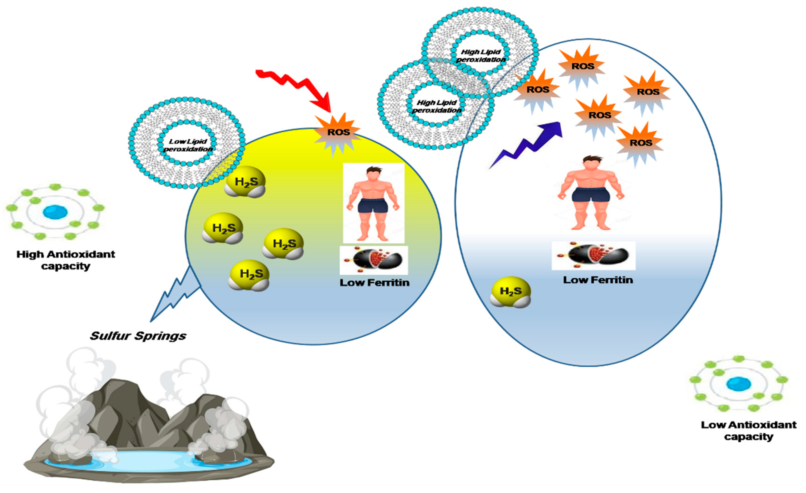
In conclusion, our study demonstrates that inhalation of H2S has a differential effect on oxidative homeostasis depending of the ferritin levels on individuals as illustrated in Figure 6. In individuals with normal ferritin levels, the impact of H2S on oxidative balance is minimal, resulting in a slight increase in thiol concentration. However, in individuals with reduced ferritin levels, H2S exhibits a more pronounced influence. Both H2S and thiol levels show a significant increase, indicating a modulation of the oxidative imbalance. These findings suggest that ferritin may serve as a regulatory factor in the biological mechanism of H2S action, potentially acting as a “counterweight” to maintain oxidative hemostasis. Further research is warranted to elucidate the underlying molecular mechanisms and unravel the intricate interplay between ferritin and H2S in the context of oxidative homeostasis. Investigating the specific pathways and signaling cascades involved in H2S-mediated modulation of oxidative stress, as well as the potential involvement of ferritin in these processes, will contribute to a comprehensive understanding of the complex interactions between H2S and ferritin in cellular redox regulation. Such knowledge will provide insights into the therapeutic potential of targeting H2S and ferritin in oxidative stress-related conditions and may pave the way for the development of novel therapeutic strategies.

Limitation and Perspective of the Study

The present study has several limitations that should be considered. Firstly, the study did not assess the actual levels of H2S exposure experienced by the participants, limiting our understanding of the relationship between H2S exposure and health outcomes. Additionally, the study did not investigate the potential adverse effects or toxicity of H2S exposure at high levels. Future research should focus on quantifying H2S exposure levels and exploring the dose-response relationship to determine optimal and safe levels of exposure. Furthermore, comprehensive investigations into the potential adverse effects and mechanisms of H2S toxicity are warranted to ensure the safe and effective utilization of H2S-based therapies. Moreover, the findings of the study may have limited generalizability due to the specific characteristics of the study participants, highlighting the need for inclusion of diverse populations in future research. Addressing these limitations and pursuing the suggested research perspectives will contribute to a more thorough understanding of H2S exposure and its implications for health outcomes and oxidative homeostasis, guiding the development of safe and effective therapeutic approaches.

4. Materials and Methods

4.1. Study Participants

The participants of this study were recruited from two distinct geographical areas in Jordan. Group A consisted of individuals from the Jordan Rift Valley, an area known for its naturally hot springs. Thus group was directly exposed to H2S emitted from the hot spring. Group B, on the other hand, comprised individuals from Irbid, which was not close proximity to hot spring and therefore, had no direct H2S exposure. Both geographical areas chosen are located in the Middle East and have similar mean sea levels. All participants from Group A resided in close proximity to the water source, which was conveniently accessible within a short 5-min drive.
Group A consisted of 100 individuals who were exposed to ambient H2S emitted from hot springs and had either low or normal ferritin levels. Group B consisted of of 100 individuals who had low or normal ferritin levels but had not been directly exposed to environmental-H2S. The study exclusion criteria included having a febrile illness within the previous four weeks, a chronic illness, liver disease or inflammation, a thyroid disorder, rare inherited defects of iron metabolism, and the use of iron supplements or other medications. Plasma samples were collected by taking 5 mL of blood in tubes containing ethylenediamine tetraacetic acid (EDTA), and properly separated and stored in a sterile tube after centrifuging for 15 s at 5000 rpm at 4 °C. Ferritin levels were categorized as follows, a normal ferritin concentration limit of >30 ng/mL and a low-ferritin concentration cutoff of 10 ng/mL for females.

4.2. Plasma H2S-Sulphide Measurement

A total 75 µL mL of plasma was combined with 425 µL of water, 250 µL of 1% (wt/vol) zinc acetate, and 250 µL of trichloroacetic acid to remove proteins. A 133 µL of 20 mM N-dimethyl-p-phenylenediamine sulfate in 7.2 M HCl and 133 µL of 30 mM FeCl3 in 1.2 M HCl were added to the mixture. The reaction mixture was centrifuged at 12,000× g for 2 min after being incubated at room temperature for 10 min. A UV spectrophotometer was used to test the samples’ absorbance at 670 nm in a 96-well plate. Finally, the concentration of H2S in the solution was determined using a calibration curve for NaHS ranging from 3.125–200 M [4].

4.3. Antioxidant Capacity Total Oxidative Stress Measurement

The antioxidant status of samples was determined using the OxiSelect-TM total antioxidant capacity (TAC) assay kit from Cell Biolabs Inc. (San Diego, CA, USA). following the manufacturer’s instructions. Antioxidant capacity was calculated by comparing the net OD 490 nm values of samples to the uric acid standard curve. The results are given in terms of micromolar “μM Copper Reducing Equivalents” (CRE) for samples, multiply the uric acid equivalence (UAE) concentration by 2189 μM Cu++/mM uric acid. For measuring total oxidative stress, 100 μL of plasma diluted 20 times in phosphate-buffered saline (PBS) was dissolved in one ml of acetate buffer, and 20 μL of working chromagen solution was added. A 100 mM of freshly prepared chromagen solution (N,N-dimethyl-p-phenylenediamine sulphate, Sigma-Aldrich, St. Louis, MO, USA) was used in the assay, which was prepared by dissolving 23.5 mg in 10 mL of 20 mM PBS (pH 7.4). The absorbance values obtained at 4 to 6 min for each sample were compared to the curve obtained using H2O2.

4.4. Total Sulfhydryl Group/Total Thiol (-SH) Measurement

Total thiol or sulfhydryl (-SH) groups in serum samples were measured using a colorimetric method provided by Cell Biolabs Inc. (San Diego, CA, USA). The assay involved the addition of a colorimetric probe to the samples or standards in a 96-well plate. The probe covalently interacted with the sulfhydryl groups, leading to the release of a chromophore. The absorbance of the samples was then measured at 450 nm. Samples were either analyzed immediately or stored at −80 °C for a maximum of 1–2 months before the analysis. By comparing the unknown samples thiol content to a predetermined reduced glutathione standard curve, the amount of thiol in each sample is identified. The content of thiol in the unknown samples is determined by comparison with a predetermined reduced glutathione standard curve. Based on a standard curve created using reduced glutathione as the sulfhydryl group standard and concentrations of 0 to 1 mM, the amount of total thiol in serum samples was calculated.

4.5. Lipid Peroxidation Determination

The malondialdehyde (MDA) reactive products and the overall amount of lipid peroxidation products in plasma were measured using the thiobarbituric acid reactive substances (TBARS) method [40]. In this method, a controlled reaction between the plasma sample and 2-thiobarbituric acid was performed. The resulting TBARS values, representing the extent of lipid peroxidation, were quantified as nmol/mL of plasma using a commercial kit from Cayman Chemical Company, (Ann Arbor, Michigan, USA).

4.6. Statistical Analysis

Statistical analysis of the data was performed using SPSS 14 version for windows software (SPSS, Inc., Chicago, IL, USA). The results were presented as mean ± standard deviation (SD). A significance level of p < 0.05 was used to determine statistical significance, indicating that differences with a p-value of less than 0.05 were considered to be significant.

Author Contributions

Methodology, Z.A., A.A., O.T. and M.K.; Validation, Z.A., A.A. and O.T.; Formal analysis, A.A., O.A.A. and M.K.; Investigation, Z.A. and O.A.A.; Resources, Z.A.; Data curation, A.A. and M.K.; Writing—original draft, Z.A. and O.T.; Visualization, O.A.A.; Supervision, Z.A.; Funding acquisition, Z.A. All authors have read and agreed to the published version of the manuscript.

Funding

This research was funded by the Deanship of Graduate Studies and Scientific Research at Yarmouk University- Irbid- Jordan, Grant # 48/2019.

Institutional Review Board Statement

The study was conducted according to the guidelines of the Declaration of Helsinki, and approved by the Institutional Review Board of Jordan University of Science and Technology (IRB # 36/111/2017).

Informed Consent Statement

Informed consent was obtained from all participants, and their confidentiality and privacy were strictly maintained throughout the study. The study procedures adhered to the principles outlined in the Declaration of Helsinki and other relevant international guidelines for research involving human subjects.

Data Availability Statement

The data can be accessed by professionals for research purposes by contacting the corresponding author directly.

Conflicts of Interest

The authors declare no conflict of interest.

References

  1. Wang, R.U.I. Two’s company, three’s a crowd: Can H2S be the third endogenous gaseous transmitter? FASEB J. 2002, 16, 1792–1798. [Google Scholar] [CrossRef] [PubMed]
  2. Altaany, Z.; Moccia, F.; Munaron, L.; Mancardi, D.; Wang, R. Hydrogen sulfide and endothelial dysfunction: Relationship with nitric oxide. Curr. Med. Chem. 2014, 21, 3646–3661. [Google Scholar] [CrossRef] [PubMed]
  3. Wang, R. Physiological implications of hydrogen sulfide: A whiff exploration that blossomed. Physiol. Rev. 2012, 92, 791–896. [Google Scholar] [CrossRef] [PubMed]
  4. Altaany, Z.; Yang, G.; Wang, R. Crosstalk between hydrogen sulfide and nitric oxide in endothelial cells. J. Cell. Mol. Med. 2013, 17, 879–888. [Google Scholar] [CrossRef] [PubMed]
  5. Kimura, H. Signaling molecules: Hydrogen sulfide and polysulfide. Antioxid. Redox Signal. 2015, 22, 362–376. [Google Scholar] [CrossRef]
  6. Rubright, S.L.M.; Pearce, L.L.; Peterson, J. Environmental toxicology of hydrogen sulfide. Nitric Oxide 2017, 71, 1–13. [Google Scholar] [CrossRef]
  7. Xie, Z.Z.; Liu, Y.; Bian, J.S. Hydrogen Sulfide and Cellular Redox Homeostasis. Oxid. Med. Cell. Longev. 2016, 2016, 6043038. [Google Scholar] [CrossRef]
  8. Rajendran, P.; Nandakumar, N.; Rengarajan, T.; Palaniswami, R.; Gnanadhas, E.N.; Lakshminarasaiah, U.; Gopas, J.; Nishigaki, I. Antioxidants and human diseases. Clin. Chim. Acta 2014, 436, 332–347. [Google Scholar] [CrossRef]
  9. Bouayed, J.; Bohn, T. Exogenous antioxidants—Double-edged swords in cellular redox state: Health beneficial effects at physiologic doses versus deleterious effects at high doses. Oxid. Med. Cell. Longev. 2010, 3, 228–237. [Google Scholar] [CrossRef]
  10. Xiao, Q.; Ying, J.; Xiang, L.; Zhang, C. The biologic effect of hydrogen sulfide and its function in various diseases. Medicine 2018, 97, e13065. [Google Scholar] [CrossRef]
  11. Corsello, T.; Komaravelli, N.; Casola, A. Role of Hydrogen Sulfide in NRF2- and Sirtuin-Dependent Maintenance of Cellular Redox Balance. Antioxidants 2018, 7, 129. [Google Scholar] [CrossRef] [PubMed]
  12. Asif, M.; Manzoor, Z.; Farooq, M.S.; Munawar, S.H.; Aziz, A.; Khan, I.A. Status of oxidant, antioxidantand serum enzymes in thalassaemic children receiving multiple blood transfusions. J. Pak. Med. Assoc. 2015, 65, 838–843. [Google Scholar] [PubMed]
  13. Aust, S.D.; Morehouse, L.A.; Thomas, C.E. Role of metals in oxygen radical reactions. J. Free. Radic. Biol. Med. 1985, 1, 3–25. [Google Scholar] [CrossRef]
  14. Zhao, G.; Ceci, P.; Ilari, A.; Giangiacomo, L.; Laue, T.M.; Chiancone, E.; Chasteen, N.D. Iron and hydrogen peroxide detoxification properties of DNA-binding protein from starved cells. A ferritin-like DNA-binding protein of Escherichia coli. J. Biol. Chem. 2002, 277, 27689–27696. [Google Scholar] [CrossRef] [PubMed]
  15. Theil, E.C.; Matzapetakis, M.; Liu, X. Ferritins: Iron/oxygen biominerals in protein nanocages. J. Biol. Inorg. Chem. 2006, 11, 803–810. [Google Scholar] [CrossRef]
  16. Aisen, P.; Listowsky, I. Iron transport and storage proteins. Annu. Rev. Biochem. 1980, 49, 357–393. [Google Scholar] [CrossRef]
  17. Rockey, D.C.; Cello, J.P. Evaluation of the gastrointestinal tract in patients with iron-deficiency anemia. N. Engl. J. Med. 1993, 329, 1691–1695. [Google Scholar] [CrossRef]
  18. Zaka-Ur-Rab, Z. Effect of Oral Iron on Markers of Oxidative Stress and Antioxidant Status in Children with Iron Deficiency Anaemia. J. Clin. Diagn. Res. 2016, 10, sc13–sc19. [Google Scholar] [CrossRef]
  19. Isler, M.; Delibas, N.; Guclu, M.; Gultekin, F.; Sutcu, R.; Bahceci, M.; Kosar, A. Superoxide dismutase and glutathione peroxidase in erythrocytes of patients with iron deficiency anemia: Effects of different treatment modalities. Croat. Med. J. 2002, 43, 16–19. [Google Scholar]
  20. Kurtoğlu, E.; Ugur, A.; Baltaci, A.K.; Undar, L. Effect of iron supplementation on oxidative stress and antioxidant status in iron-deficiency anemia. Biol. Trace Elem. Res. 2003, 96, 117–123. [Google Scholar] [CrossRef]
  21. Whiteman, M.; Cheung, N.S.; Zhu, Y.Z.; Chu, S.H.; Siau, J.L.; Wong, B.S.; Armstrong, J.S.; Moore, P.K. Hydrogen sulphide: A novel inhibitor of hypochlorous acid-mediated oxidative damage in the brain? Biochem. Biophys. Res. Commun. 2005, 326, 794–798. [Google Scholar] [CrossRef] [PubMed]
  22. Pal, V.K.; Bandyopadhyay, P.; Singh, A. Hydrogen sulfide in physiology and pathogenesis of bacteria and viruses. IUBMB Life 2018, 70, 393–410. [Google Scholar] [CrossRef]
  23. Wedmann, R.; Onderka, C.; Wei, S.; Szijártó, I.A.; Miljkovic, J.L.; Mitrovic, A.; Lange, M.; Savitsky, S.; Yadav, P.K.; Torregrossa, R.; et al. Improved tag-switch method reveals that thioredoxin acts as depersulfidase and controls the intracellular levels of protein persulfidation. Chem. Sci. 2016, 7, 3414–3426. [Google Scholar] [CrossRef] [PubMed]
  24. Halliwell, B.; Gutteridge, J.M. Role of iron in oxygen radical reactions. Methods Enzymol. 1984, 105, 47–56. [Google Scholar] [PubMed]
  25. Imam, M.U.; Zhang, S.; Ma, J.; Wang, H.; Wang, F. Antioxidants Mediate Both Iron Homeostasis and Oxidative Stress. Nutrients 2017, 9, 671. [Google Scholar] [CrossRef]
  26. Sies, H. Oxidative stress: A concept in redox biology and medicine. Redox Biol. 2015, 4, 180–183. [Google Scholar] [CrossRef]
  27. Kimura, H. Hydrogen sulfide: Its production and functions. Exp. Physiol. 2011, 96, 833–835. [Google Scholar] [CrossRef]
  28. Abe, K.; Kimura, H. The possible role of hydrogen sulfide as an endogenous neuromodulator. J. Neurosci. 1996, 16, 1066–1071. [Google Scholar] [CrossRef]
  29. Valko, M.; Leibfritz, D.; Moncol, J.; Cronin, M.T.D.; Mazur, M.; Telser, J. Free radicals and antioxidants in normal physiological functions and human disease. Int. J. Biochem. Cell Biol. 2007, 39, 44–84. [Google Scholar] [CrossRef]
  30. Agarwal, A.; Aponte-Mellado, A.; Premkumar, B.J.; Shaman, A.; Gupta, S. The effects of oxidative stress on female reproduction: A review. Reprod. Biol. Endocrinol. 2012, 10, 49. [Google Scholar] [CrossRef]
  31. Szabo, C.; Papapetropoulos, A. International Union. of Basic. and Clinical Pharmacology. CII: Pharmacological Modulation of H2S Levels: H2S Donors and H2S Biosynthesis Inhibitors. Pharmacol. Rev. 2017, 69, 497–564. [Google Scholar] [CrossRef] [PubMed]
  32. Lim, E.; Mbowe, O.; Lee, A.S.W.; Davis, J. Effect of environmental exposure to hydrogen sulfide on central nervous system and respiratory function: A systematic review of human studies. Int. J. Occup. Environ. Health 2016, 22, 80–90. [Google Scholar] [CrossRef] [PubMed]
  33. Olas, B.; Kontek, B. Hydrogen sulfide decreases the plasma lipid peroxidation induced by homocysteine and its thiolactone. Mol. Cell. Biochem. 2015, 404, 39–43. [Google Scholar] [CrossRef] [PubMed]
  34. Papapetropoulos, A.; Pyriochou, A.; Altaany, Z.; Yang, G.; Marazioti, A.; Zhou, Z.; Jeschke, M.G.; Branski, L.K.; Herndon, D.N.; Wang, R.; et al. Hydrogen sulfide is an endogenous stimulator of angiogenesis. Proc. Natl. Acad. Sci. USA 2009, 106, 21972–21977. [Google Scholar] [CrossRef]
  35. Marutani, E.; Ichinose, F. Emerging pharmacological tools to control hydrogen sulfide signaling in critical illness. Intensive Care Med. Exp. 2020, 8, 5. [Google Scholar] [CrossRef] [PubMed]
  36. Powell, C.R.; Dillon, K.M.; Matson, J.B. A review of hydrogen sulfide (H2S) donors: Chemistry and potential therapeutic applications. Biochem. Pharmacol. 2018, 149, 110–123. [Google Scholar] [CrossRef]
  37. Usman, S.S.; Dahiru, M.; Abdullahi, B.; Abdullahi, S.B.; Maigari, U.M.; Uba, A.I. Status of malondialdehyde, catalase and superoxide dismutase levels/activities in schoolchildren with iron deficiency and iron-deficiency anemia of Kashere and its environs in Gombe State, Nigeria. Heliyon 2019, 5, e02214. [Google Scholar] [CrossRef]
  38. Kumerova, A.; Lece, A.; Skesters, A.; Silova, A.; Petuhovs, V. Anaemia and antioxidant defence of the red blood cells. Mater. Med. Pol. 1998, 30, 12–15. [Google Scholar]
  39. Chang, J.H.; Kim, D.K.; Kim, H.W.; Park, S.Y.; Yoo, T.-H.; Kim, B.S.; Kang, S.-W.; Choi, K.H.; Han, D.-S.; Jeong, H.J.; et al. Changing prevalence of glomerular diseases in Korean adults: A review of 20 years of experience. Nephrol. Dial. Transplant. 2009, 24, 2406–2410. [Google Scholar] [CrossRef]
  40. Ohkawa, H.; Ohishi, N.; Yagi, K. Assay for lipid peroxides in animal tissues by thiobarbituric acid reaction. Anal. Biochem. 1979, 95, 351–358. [Google Scholar] [CrossRef]
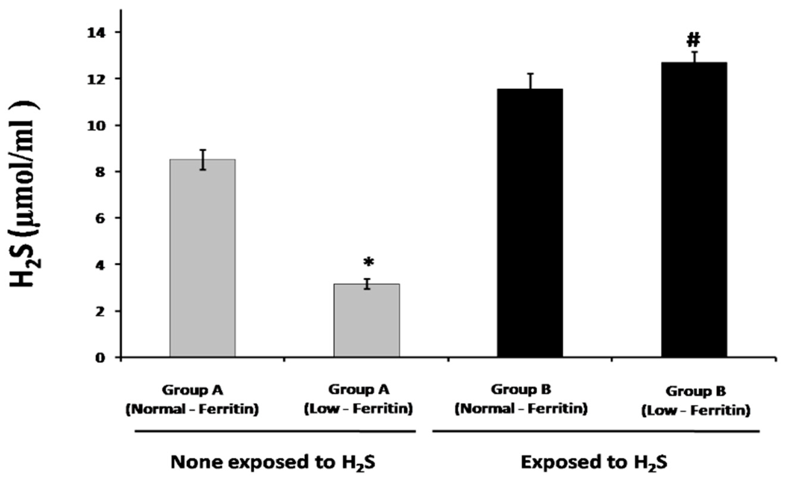
Figure 1. The impact of environmental H2S exposure on endogenous H2S plasma levels in individuals with low and normal ferritin status. The values are reported as the mean ± standard deviation. The symbol “#” indicates a significant difference at p < 0.05 when comparing the low-ferritin group who were not exposed to H2S. The symbol “*” indicates a significant difference at p < 0.05 when comparing the normal ferritin group to the low-ferritin group in the group A, which had not been exposed to H2S. Group A refers to individuals who were not exposed to environmental H2S, while group B refers to individuals who were exposed to environmental H2S.
Applsci 13 06621 g001
Figure 2. The impact of environmental H2S exposure on plasma thiol (-SH) levels in individuals with low and normal ferritin status. The values are reported as the mean ± standard deviation. The symbol “#” indicates a significant difference at p < 0.05 when comparing the low-ferritin group who were not exposed to H2S. Group A refers to the individuals who are not exposed to environmental H2S, while Group B refers to individuals who are exposed to environmental H2S. Group A refers to individuals who were not exposed to environmental H2S, while Group B refers to individuals who were exposed to environmental H2S.
Applsci 13 06621 g002
Figure 3. The impact of environmental H2S exposure on plasma lipid peroxidation levels in individuals with low and normal ferritin status. The values are reported as the mean ± standard deviation. The symbol “#” indicates a significant difference at p < 0.05 when comparing the low-ferritin group who were not exposed to H2S. The symbol “*” indicates a significant difference at p < 0.05 when comparing the normal ferritin group to the low-ferritin group in group A, which had not been exposed to H2S. Group A refers to individuals who were not exposed to environmental H2S, while Group B refers to individuals who were exposed to environmental H2S.
Applsci 13 06621 g003
Figure 4. Total oxidative stress levels in individuals with low and normal ferritin levels, both exposed to environmental H2S and not exposed to H2S. The values are presented as the mean ± standard deviation; # indicates a significant difference at 0.05 when comparing the low-ferritin group who were not exposed to H2S; * indicates a significant difference at 0.05 when comparing the normal ferritin group to the low-ferritin group in group A that had not been exposed to H2S. Group A refers to individuals who were not exposed to environmental H2S, while group B refers to individuals who were exposed to environmental H2S.
Applsci 13 06621 g004
Figure 5. Total antioxidant capacity in individuals with low and normal ferritin status after environmental H2S exposure. The values are reported as the mean ± standard deviation. The symbol “#” indicates a significant difference at p < 0.05 when comparing the low-ferritin group who were not exposed to H2S. The symbol “*” indicates a significant difference at p < 0.05 when comparing the normal ferritin group to the low-ferritin group in group A, which had not been exposed to H2S. The symbol “**” indicates a significant difference at p < 0.05 when comparing the low-ferritin group B to the low-ferritin group A. Group A refers to individuals who were not exposed to environmental H2S, while group B refers to individuals who were exposed to environmental H2S.
Applsci 13 06621 g005
Figure 6. Illustration of the possible mechanism by which exposure to environmental H2S emitted from thermal springs leads to a reduction in ROS and lipid peroxidation levels in individuals with low ferritin status. Specifically, this effect is achieved by upregulating endogenous H2S levels.
Applsci 13 06621 g006
Table 1. Ferritin levels and visitation patterns to thermal springs for individuals who participated in this study.
Group A (Control Area)Group B (Exposed to H2S Area)
Age37384040
GenderFemale: 89
Male: 11
Female: 81
Male: 19
Female: 77
Male: 23
Female: 86
Male: 14
Ferritin levels (ng/mL)6.57 ± 0.2287.51 ± 4.996.18 ± 0.3291.65 ± 0.22
Number of visits (times/year) *~2–3~2–3~10–12~10–12
Duration of each visit (hrs) *~2–3~2–3~2–3~2–3
* Noting that exposed individuals live in the thermal spring’s surrounding area, the times represented in the table are the average number of visits to the spring spa to soak.
Disclaimer/Publisher’s Note: The statements, opinions and data contained in all publications are solely those of the individual author(s) and contributor(s) and not of MDPI and/or the editor(s). MDPI and/or the editor(s) disclaim responsibility for any injury to people or property resulting from any ideas, methods, instructions or products referred to in the content.