Article

Hamstring vs. All-Soft-Tissue Quadriceps Tendon Autograft for Anterior Cruciate Ligament Reconstruction in Adolescent Athletes: Early Follow-Up Results of a Prospective Study

by
Ritauras Rakauskas
1,*,
Laimonas Šiupšinskas
2,
Vytautas Streckis
3,4,
Justė Balevičiūtė
2,
Laurynas Galinskas
5,
Dalius Malcius
1 and
Emilis Čekanauskas
1
1
Department of Paediatric Surgery, Lithuanian University of Health Sciences, LT-44307 Kaunas, Lithuania
2
Department of Sports Medicine, Lithuanian University of Health Sciences, LT-44307 Kaunas, Lithuania
3
Institute of Sports Science and Innovations, Lithuanian Sports University, LT-44221 Kaunas, Lithuania
4
Department of Trainings Systems, Lithuanian Sports University, LT-44221 Kaunas, Lithuania
5
Department of Orthopaedics and Traumatology, Lithuanian University of Health Sciences, LT-44307 Kaunas, Lithuania
*
Author to whom correspondence should be addressed.
Appl. Sci. 2023, 13(11), 6715; https://doi.org/10.3390/app13116715
Submission received: 16 April 2023 / Revised: 25 May 2023 / Accepted: 29 May 2023 / Published: 31 May 2023
(This article belongs to the Special Issue Advances in Sport Injury Prevention)

Abstract

:
Background: The quadriceps tendon (QT) autograft has recently drawn attention for anterior cruciate ligament reconstruction (ACLR). Finding the best autograft option for adolescents after an ACL injury is essential to ensure them a high-quality active lifestyle. There are no studies comparing the all-soft-tissue QT autograft with the hamstring tendon (HT) autograft in such population. Methods: In this study, 68 patients younger than 18 years of age were assigned to the HT (38 patients) or the QT (30 patients) ACLR group. The groups were similar, allowing their comparison. The instrumented knee laxity was evaluated with a Genourob arthrometer. In total, 48 patients (27 HT and 21 QT) were tested 3 months post-op, and 45 patients (26 HT and 19 QT) were tested 6 months post-op. Results: We found that 3 months post-op, the side-to-side anterior tibial translation measurements (GNRB1) were worse in the HT than in the QT group (1.4 (0.2–5.2; 1.715) vs. 0.6 (0.1–2.1; 0.905) mm, p = 0.02). There was no difference in side-to-side anterior tibial translation at 6 months post-op (GNRB2) between the HT and the QT group (1 (0.2–5.3; 1.519) vs. 1.1 (0.3–3.4; 1.279) mm, p = 0.927). At 3 months post-op, the side-to-side anterior tibial translation (GNRB1) was worse in males than in females. irrespective of the graft choice (1.45 (0.1–5.2; 1.696) vs. 0.4 (0.1–3.4; 0.89) mm, p = 0.016). The displacement curve slopes 6 months post-op were better for females (3 (0–13.1; 3.335) vs. 5.3 (0–26.1; 7.848), p = 0.014). At 3 months post-op, the GNRB measurements showed that the side-to-side anterior tibial translation (GNRB1) was better for females when the HT autograft had been used (0.45 (0.2–3.4; 0.942) vs. 2.4 (0.3–5.2; 2.333) mm, p = 0.003). In general, both autografts provided excellent objective outcomes at early follow-up. Conclusions: The all-soft-tissue QT autograft should be considered as a reliable alternative for ACLR in adolescents. This autograft has at least the same properties as the HT autograft in the early stages after ACL reconstruction in adolescent athletes and, in some respects, seems superior to the HT autograft. Research should continue to find the best possible graft choice for the most active and willing-to-return-to-sport population.

1. Introduction

Previously, it used to be thought that anterior cruciate ligament (ACL) injuries are rare in pediatric patients. However, as the participation of children in high-impact sports has increased over the last 20 years, it is now agreed that ACL injuries are common in the pediatric population. Recently, it was estimated that sports-playing youth athletes sustain over 400/100,000 ACL injuries each year [1,2,3,4]. Furthermore, it seems that adolescents have complex knee injuries and higher rates of ACL graft re-rupture than adults [5].
For individuals with high physical activity levels, ACL reconstruction (ACLR) is strongly recommended. In the adult population, the hamstring tendon autograft remains the most popular choice for ACLR, and it is agreed that a decent rehabilitation is crucial for restoring the pre-injury activity levels [6,7].
The management of an ACL injury is quite a controversial subject in current pediatric orthopedics. The controversy is due to demanding surgical techniques, as there is a risk of growth disturbance complications. Today, various techniques of ACLR have been described in the literature [8]. Currently, in the scientific literature we can see an increased interest in quadriceps tendon graft adaptation. Articles with some catchy phrases can be found, e.g., stating that it is “the graft of the future” and that it should be used more in modern ACLR [9,10]. However, information about the use of quadriceps tendon in young and active patients is still lacking; therefore, more attention must be paid to it [11].
Very few studies have been published regarding an arthrometric side-to-side comparison of the quadriceps tendon autograft using a patellar bone block with the hamstring autograft [12,13]. Additionally, none of these studies examined the all-soft-tissue quadriceps autograft, which we promote as an excellent graft option for ACLR.
The graft maturation process is also a key factor in relation to the rehabilitation protocol. Magnetic resonance imaging studies showed a better maturity of the quadriceps tendon autograft compared to the hamstring autograft [14]. Furthermore, the arthrometric evaluation of the anterior side-to-side tibial translation can add essential information about graft performance after ACLR [13]. This prospective study evaluated the influence of autograft selection on knee stability and possible failure in young and active patients. The examined autografts included all-soft-tissue quadriceps tendon (QT) and hamstring tendons (HT) autografts. Our main hypothesis was that the QT autograft would provide as good knee stability after ACLR as the HT autograft. We believe that our results can enhance QT usage and popularity.

2. Materials and Methods

2.1. Participants

This prospective study was performed in the Lithuanian University of Health Sciences (LUHS) Kaunas Clinics and included a total of 68 (37 male, 31 female) patients from 12 to 17 years old. The autograft to be used for ACLR was chosen, and 38 HT and 30 QT grafts were used. All patients in this study underwent only primary ACLR, and it was assured that no concomitant soft tissue or bone injuries were present, except meniscus lesions. The diagnosis of ACL tear must was confirmed clinically and on MRI, while the decision for meniscal repair and the graft diameter measurement were taken intraoperatively. Post-operative radiographs were performed to assess the correct placement of the femoral and tibial buttons and to make sure that the repair was of the required quality. Before surgery, all patients and their parents provided their informed consent for inclusion in this study and filled Tegner activity score questionnaire to ensure that the patients were physically active. Six weeks after surgery, all patients underwent rehabilitation. To evaluate the post-operative knee stability and autograft maturation, the Genourob (GNRB) knee arthrometer was used 3 and 6 months after surgery at the 134 N force of anterior tibial translation. All procedures were conducted following the Declaration of Helsinki and were approved by the Kaunas Regional Biomedical Research Ethics Committee (protocol code BE-2-103).

2.2. Surgical Technique

All ACLRs for this study were performed by a single orthopedic surgeon.

2.2.1. Hamstring Tendon Autograft

For the hamstring tendon (HT) autografts, an anteromedial, 3-to-4 cm approach over the tibial insertion was used. The gracilis and semitendinosus tendons were harvested, and 8-strand, 70 mm-length and 8-to-11.5 mm-diameter grafts were obtained.

2.2.2. Quadriceps Tendon Autograft

The quadriceps tendon (QT) all-soft-tissue autografts were harvested using a 3-to-4 cm anterior approach on the distal thigh. The defect of the quadriceps tendon was closed with a running locking suture (Figure 1). The QT autografts were 65 to 80 mm in length and varied from 8.5 to 11 mm in diameter.
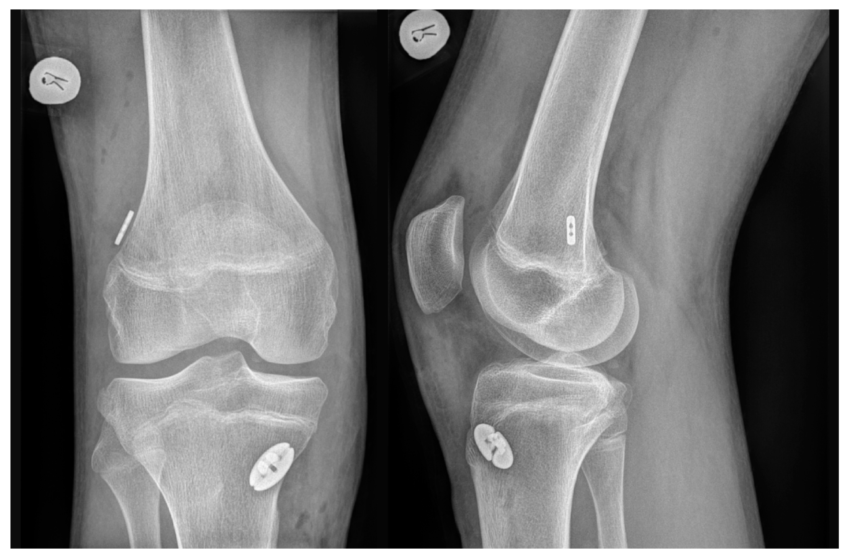
For all autografts, the same transepiphyseal tunnel drilling technique was performed. A 4 mm-diameter full femoral tunnel was drilled along with a 2-to-2.5 cm deep femoral socket, according to the graft diameter. Full tibial tunnel was drilled according to graft diameter. Both HT and QT autografts were fixed in the knee joint with cortical suspensory devices on both the femur and the tibia (TightRope RT and ABS TightRope, Arthrex (Naples, FL, USA)). A post-operative X-ray examination was performed after each ACLR to confirm the position of the implants (Figure 2).

2.3. Rehabilitation

All patients were examined by a surgeon on the first day after the ACLR surgery. Recommendations for the post-operative regime were provided by the surgeon according to the complexity of the injury and the surgical treatment method. The patients were provided with a special hinged knee brace locked in full extension. The angle of the knee flexion was increased only from the second week after the ACLR, and thereafter, the flexion angle was increased by 10–20 degrees every 3–4 days. It was recommended to gradually increase the angle of the knee flexion over 6 weeks. Patients who had received isolated ACLR or ACLR with meniscus resection were allowed to fully weight-bear the operated leg with the knee brace in order to prevent the development of kinesiophobia and excessive muscle weakening. If the ACLR was performed along with meniscus repair, the patients were not allowed to weight-bear the operated leg for 4 weeks and then gradually started putting weight on it until full weight-bear at 6 weeks to provide conditions for meniscus healing. The patients were taught to daily focus on reaching a full passive knee extension and on controlling the swelling of the knee joint by cryotherapy and the elevation of the leg above the heart level. All patients received a home-based exercise program for improving the mobility of the knee and increasing the strength of the lower extremity before they started the recovering process in the rehabilitation centers for 4 weeks.
Because most of our participants were athletes, we considered the evaluation of the GNRB results and the condition of the knee. If the results of the anterior tibial translation at 3 months after surgery had reached the normal values of ACL laxity, the operated knee did not have mobility limitation and was not swollen at the time, then we recommended to start a muscle strength training phase (exercises with weight lifts), while at the same time, continuing the stability- and balance function-improving exercises. If the anterior tibial translation was >1.5 mm, then it was considered that the ACL was still in the maturation process. In addition, if the operated knee showed a limited mobility and was still swollen, we recommended to continue the rehabilitation process under the supervision of a physiotherapist or to continue the mobility exercises, cryotherapy and muscle-strengthening exercises. The athletes were advised to start sport-specific tasks 6 months after the ACL reconstruction if the GNRB results reached normal values. Their eventual return to sport consisted of three phases: return to participation, return to training and return to playing.

2.4. Testing Procedure (Side-to-Side Anterior Tibial Translation)

The assessment of ligamentous laxity in both knees was evaluated by the computer-assisted Genourob®, (Laval, France (GNRB)) device at 3 and 6 months after ACLR by one of our trained researchers. GNRB is an objective computer-assisted system which is considered the gold standard for evaluating the anterior tibial translation and maturation of ACL transplants [15]. All subjects had both legs tested, and the non-injured leg was analyzed first. The maturation and dynamical function of the reconstructed ACL were compared with those of the uninjured leg. The differences of laxity between the uninjured and the operated side were measured in mm. The displacement curve slopes were also evaluated. This measurement illustrates the functional knee stability and could be have a prognostic value for ACL graft failure. The test was conducted in the supine position with the knee placed in 20° flexion in a molded support in order to reproduce the typical Lachman test position, according to the recommendations of the GNRB manufacturer [16]. The patellar pad was positioned as recommended by the manufacturer to immobilize the patella while allowing tibial translation [17]. The calf was supported by the belt of the device. The anterior tibial translation was measured by the posterior–anterior force of 134 N at 3 and 6 months after the ACL repair. The GNRB testing procedure was carried out in the same manner for the operated knee.

2.5. Statystical Analysis

Data analysis was performed using IBM SPSS Statistics version 29.0. Qualitative nominal data are presented using frequencies and relative frequencies (in percent). Quantitative data are presented using mean (with standard deviation) and median (with minimum and maximum values). Because of the small samples or the absence of normality in the data (which was tested by using the Shapiro–Wilk test), the quantitative data in two independent samples were compared by using the non-parametric Mann–Whitney test. Qualitative nominal data were compared by using the Chi-square test. Differences were considered statistically significant if p < 0.05.

3. Results

3.1. Sample Characteristics

Totally, 68 (37 male, 31 female) patients from 12 to 17 (avg. 15.63 (SD 1.359) years old participated in this study. A total of 61 (89.71%) patients in our study sustained an ACL injury playing some kind of sport. The sports activities varied as follows: 16 (23.53%) patients played some unspecific sport games, 11 (16.17%), basketball, 10 (14.7%), soccer, 9 (13.4%), handball, 5 (7.35%), volleyball, 4 (5.88%), martial arts, 2 (2.94%), rugby, 2 (2.94%), skiing, 1 (1.47%), athletics and 1 (1.47%), functional training. The remaining 7 (10.29%) patients were injured in some form of accident: 3 (4.41%) in a road accident, 2 (2.94%) in a fall from the stairs, 2 (2.94%) in an electric scooter accident.
Before inclusion in the study, all patients performed the Tegner activity test. The average result was 7.49 (median 8), with scores from 3 to 10, which showed that our patients were active before the injury. The patients were divided into two groups (HT and QT) according to the used autograft. The demographics between these groups are shown in Table 1. Both groups in most categories were similar; so, they could be compared. A concomitant meniscus injury had to be evaluated as a possible distorting factor.

3.2. Comparisons of the GNRB Results between the Groups

This is an ongoing study, and at the moment of writing, we had tested 48 patients (27 HT and 21 QT) 3 months post-surgery and 45 patients (26 HT and 19 QT) 6 months post-surgery (Figure 3). The dynamical differences between the autografts are presented in Table 2. From these results, we can see that the largest differences between the two groups were seen at 3 months post-surgery. We found that the side-to-side anterior tibial translation at a 134 N force 3 months post-op (GNRB1) was statistically significantly worse in the HT group (1.4 mm; 0.2–5.2; 1.715) than in the QT group (0.6 mm; 0.1–2.1; 0.905) (U = 172, p = 0.02). When the same measure was performed 6 months post-op, the statistical differences disappeared. There was no statistically significant difference in the side-to-side anterior tibial translation at a 134 N force 6 months post-op (GNRB2) between the HT and the QT group (1 mm; 0.2–5.3; 1.519, vs. 1.1 mm; 0.3–3.4; 1.279) (U = 243, p = 0.927). However, less variability was noticed in the QT group (Figure 4).
The possible graft failure was also evaluated. However, unlike for the anterior tibial displacement measures, no statistically significant difference was found in the displacement curve slopes 3 months (4.7 (0–23; 5.974) vs. 4.1 (1.8–9.4; 4.29)) (U = 277.5, p = 0.901)) and 6 months (4.7 (0.1–26.1; 6.3) vs. 4.1 (0–18.3; 5.216)) (U = 206, p = 0.345)) post-op between the HT and the QT group.
It was important to evaluate a concomitant meniscus injury as a possible distorting factor for our results. When we measured the side-to-side anterior tibial translation in each autograft in the sutured and non-sutured groups, no statistical difference was found. It is safe to say that concomitant injuries did not have a statistically significant influence on our main results.
In conclusion, these results showed that greater stability in the anterior tibial displacement was achieved faster when the QT autograft was used, but in a longer perspective, the results of both stability and possible failure between the grafts were similar.

3.3. GNRB Results According to the Patients’ Sex

One of the additional objectives of this study was to evaluate the sex influence on graft evolution in relation to the graft type. The results that we obtained regardless of the graft choice are presented in Table 3.
At 3 months post-op, the GNRB measurements showed that the side-to-side anterior tibial translation at a 134 N force (GNRB1) was statistically significantly worse in males than in females, irrespective of the graft choice (1.45 mm; 0.1–5.2; 1.696, vs. 0.4 mm; 0.1–3.4; 0.89) (U = 165.5, p = 0.016). However, if we tried to predict the graft failure, no statistically significant difference was found in the displacement curve slopes 3 months post-op between males and females, irrespective of the graft choice (4.7 mm; 0.1–23; 5.664, vs. 3.5 mm; 0–12.9; 4.64) (U = 245, p = 0.464).
However, 6 months post-op, the results of anterior tibial translation and the displacement curve slopes changed. No statistically significant difference was found in the side-to-side anterior tibial translation at a 134 N force 6 months post-op (GNRB2) between males and females, irrespective of the graft choice (1.4 mm; 0.2–5.3; 1.788, vs. 0.85 mm; 0.3–2.3; 0.955) (U = 185, p = 0.137). In contrast, the displacement curves were better for the female patients. The displacement curve slopes 6 months post-op for males and females, irrespective of the graft choice, were statistically significantly different (5.3; 0–26.1; 7.848, vs. 3; 0–13.1; 3.335) (U = 143, p = 0.014).
In conclusion, it seems that regardless of the graft choice, the results were slightly better for females. The females more quickly reached a greater knee stability in anterior tibial translation. Their displacement curve slope was also lower, although this achievement required more time.
The performance of the different autografts in males and females is presented in Table 4. Significant results were obtained only for the HT group. At 3 months post-op, the GNRB measurements showed that the side-to-side anterior tibial translation at a 134 N force (GNRB1) was statistically significant better for females than for males when the HT autograft had been used (0.45 mm; 0.2–3.4; 0.942, vs. 2.4 mm; 0.3–5.2; 2.333) (U = 31.5, p = 0.003). On the other hand, at 6 months post-op, the difference in side-to-side anterior tibial translation at a 134 N force (GNRB2) between males and females in the HT group disappeared (2.3 mm; 0.2–5.3; 2.036, vs. 0.7 mm; 0.3–2.3; 0.917) (U = 54.5, p = 0.133). The curve slopes 3 and 6 months post-op were similar in the HT group for males and females. The displacement curve slopes at 3 months post-op were 6.5 (0.1–23; 7.227) vs. 2.9 (0–12.9; 4.408)) (U = 69, p = 0.316) and at 6 months, they were 6.55 (1.7–26.1; 8.629) vs. 3.85 (0.1–7.6; 3.583) (U = 49.5, p = 0.078) for males and females in the HT group, respectively.
In the QT autograft group, no significant differences were measured. No statistically significant difference was seen in GNRB1 (0.7 mm; 0.1–2.0; 0.962, vs. 0.35 mm; 0.1–2.1; 0.812) (U = 43.5, p = 0.556), GNRB2 (0.8 mm; 0.3–3.4; 1.473, vs. 1.15 mm; 0.3–1.7; 1.013) (U = 39.5, p = 0.731) and curve slopes 3 months (3.6; 1.8–7.6; 3.862, vs. 4.15; 3–9.4; 4.988) (U = 37.5, p = 0.309) and 6 months post-op (5.3; 0–18.3; 6.855, vs. 0.9; 0–13.1; 2.963) (U = 21.5, p = 0.063) between males and females in the QT group.
In conclusion, when we evaluated the instrumented laxity measurements for different autograft options according to sex, we found that the female patients reached greater knee stability faster than the male ones when the HT autograft had been used. However, this did not have a considerable influence on the results in a longer perspective, as the measurements of both stability and curve slopes for the two types of graft were the same.

4. Discussion

This prospective study demonstrated that the QT autograft has at least the same properties as the HT autograft in the early stages after ACL reconstruction in adolescent athletes. However, in some respects it seems superior to the HT autograft. At the very first assessment of operated knee laxity (side-to-side anterior tibial translation) 3 months after the ACLR, the QT autograft performed twice better than the HT autograft.
The QT is the least studied and used autograft, with its use just expected to increase [18]. Mouarbes et al. [19] in their meta-analysis published in 2019, reported only five articles that compared the HT and QT autografts for ACLR. Moreover, in all those studies, the QT autograft was harvested with a patellar bone block. In general, only few studies have analyzed QT autografts with a patellar bone block [13,20,21,22]. Crum et al. [23], in their systematic review, compared only 181 all-soft-tissue QT patients (5 studies) with 1534 patellar bone QT patients (20 studies). There is limited information on the all-soft-tissue QT autograft for ACLR, and comparative studies are lacking, but the results are promising, as researchers reported no difference in graft rupture between all-soft-tissue QT and patellar-bone-block QT autografts [23], great functional outcomes at short and intermediate follow-up [24] and decreased donor site morbidity [25]. Our study mainly concentrated on two poorly studied fields: the all-soft-tissue QT autograft and its use in young and active patients [11,18]. That makes this study interesting and of current relevance for today’s orthopedics.
Our results regarding the stability of the knee are quite interesting. Comparing the HT and QT autograft performances during Genourob testing regardless of the sex, we found that the QT results were statistically significantly better at the 3-month follow-up. In contrast, at the 6-month follow-up, each autograft option appeared to perform statistically equally. There are no studies comparing such early knee laxity follow-up results in adolescents. However, some studies reported similar results. For example, Cavaignac et al. [13] reported a significant variation in side-to-side laxity in patients after ACLR with an HT autograft after 3 years from surgery. Less variability was seen when the QT autograft was chosen. Our measurements were performed 3 and 6 months post-surgery, but our results are similar to these previous ones (HT: 1.7 mm (SD 1.4) at 3 months and 1.5 mm (SD 1.4) at 6 months in our study vs. 3.1 mm (SD 1.3) in Cavaignac et al.; QT: 0.9 mm (SD 0.7) at 3 months and 1.3 mm (SD 1.0) at 6 months for in study vs. 1.1 mm (SD 0.9) in Cavaignac et al.). We presented our results as a median (minimum-maximum; mean (SD)) because non-parametric tests were used, but in this case, for a better understanding, we showed the mean (SD). Akoto et al. [26], in their study where all patients were older than 18 years, measured the side-to-side knee stability with a Rolimeter. After a little more than a year from surgery, no statistical difference was found between the QT and HT autografts. Kim et al. [21] also studied the QT as a possible autograft. Although they compared it with the bone–patellar tendon–bone (BPTB) autograft, there were no statistical side-to-side differences between the grafts, using a KT-2000 (MEDmetric Corp, San Diego, CA, USA) arthrometer. They concluded that the QT graft has a comparable performance to that of the BPTB graft for ACL reconstruction, with less donor site morbidity. Lund et al. [27] obtained the same results as Kim et al. They made it clear that the QT is a viable option for ACL reconstruction. Runer et al. [28] in their study obtained even more significant results. They concluded that, even though functional testing, knee laxity and re-rupture rates of the HT and QT grafts were similar, there was a tendency for a higher graft failure in highly active patients treated with the HT autograft.
From reviewed studies and our results, we can clearly conclude that the QT is an excellent choice for ACL repair. It would be interesting to investigate the biological or mechanical properties that may influence the QT graft performance. It is known that tenocytes are the predominant cells in tendons and ligaments. They produce collagen and have an important role in donor site healing, regeneration after graft harvesting and ligamentization of the graft. It is assumed that they may promote a better knee functional outcome after QT harvesting due to the higher abundance of tenocytes and collagen in the QT than in other tendons. [29,30]. Another reason may be hamstrings sparing. Hamstrings are important for knee stability. They are involved in the rotational, translational and varus/valgus stability of the knee. Their loss during HT harvesting before regeneration significantly alters the knee kinematics and stability [31].
The latest evidence of early rehabilitation after ACLR showed that there was no difference between the unsupervised and the supervised programs for laxity, subjective function, functional outcomes, strength and atrophy, as well as that the duration of the rehabilitation did not play a leading role in the laxity and subjective function [32].
However, the laxity measurement 3 months post-op with an acceptable side-to-side difference could allow the physiotherapist to adjust the home-based exercise program, putting more focus on limb strength symmetry and delaying the running-based exercises. The laxity measures 6 months after the ACLR with an acceptable laxity difference up to 1.5 mm could allow starting sports-based tasks and returning to sports activities.
It has been reported that the outcomes after ACLR, in general, are not different according to sex [33]. Postoperative ligamentous laxity was reported to be similar in males and females after ACLR using the HT autograft [34,35]. In our study, we found a statistically significant difference in knee laxity 3 months post ACLR with the HT autograft in favor of the female group. However, Tan et al., in their systematic review and meta-analysis, stated that females had inferior outcomes in instrumented laxity [36]. We suggest that these results could help to understand and plan the rehabilitation process. Theoretically, for adolescent female patients, a wider range of rehabilitation methods could be used earlier after ACLR.
Overall, we strongly believe that our study is the first that objectively evaluated graft performance in adolescent patients after ACLR. First, our patient population consisted only of physically active adolescents (the median of the Tegner preoperative score was 8 (3–10; 7.49), and 89.71% of our patients suffered an injury while playing sports). Second, all ACLRs were performed by a single orthopedic surgeon, and all knee laxity evaluations using an arthrometer were conducted by a single trained senior physiotherapist. Lastly, the QT autograft was an all-soft-tissue graft, which makes the data of our study very valuable because of the shortage of reliable information about this graft choice.
In a period of 2 years, we offered the possibility to all the adolescent patients with primary ACL tear who were presented to the Hospital of Lithuanian University of Health Sciences Kaunas Clinics to participate in our study. Therefore, we believe that the population of our study wi representative in the sense that the patients had an equal chance of being selected. The high physical activity level of the participants of this study illustrates the type of patient who is at risk of suffering an ACL injury. The minimum sample size was achieved, but with a larger sample, in our opinion, we would see more statistically significant differences.
The present study has some limitations. The small sample size was due to the loss of some patients in the follow-up and in the ongoing research process. As for the patients, it was simply too early to perform the instrumented laxity testing. This study focused on objective measurements after ACLR. Therefore, another limitation is the lack of preoperative data and the subjective evaluation. As we mentioned, here we presented early results of our work, and subjective patient improvement regarding IKDC and Lysholm scores will be available after the final follow-up. Lastly, the examiners during the follow-ups were not blinded. Therefore, an assessment bias is possible.

5. Conclusions

The all-soft-tissue QT autograft for ACLR should be considered as a reliable alternative for ACLR in adolescents. We found that this autograft demonstrated less variability in side-to-side anterior laxity compared to the HT autograft. At 3 months post-surgery, the QT autograft was shown to perform particularly well, and this allowed tuning up the rehabilitation process after ACLR. We also concluded that the female patients achieved knee stability more quickly than the male patients when the HT autograft was used. While there are very few high-quality prospective comparative studies regarding the all-soft-tissue QT autograft usage, we are certain that our research will help to promote this autograft option for primary ACLR. Research should continue to find the best possible graft choice for the most active and willing-to-return-to-sport population.

Author Contributions

Conceptualization, L.Š. and E.Č.; methodology, R.R., L.Š., E.Č. and V.S.; statistical analysis, R.R. and L.G.; formal analysis, R.R., L.Š., E.Č. and V.S.; investigation, R.R., L.Š., E.Č. and V.S.; data curation, R.R.; writing—original draft preparation, R.R., J.B., L.G.; writing—review and editing, L.Š., E.Č., D.M.; supervision, D.M. All authors have read and agreed to the published version of the manuscript.

Funding

This research received no external funding.

Institutional Review Board Statement

All procedures were conducted following the Declaration of Helsinki and were approved by the Kaunas Regional Biomedical Research Ethics Committee (protocol code BE-2-103; date of approval 30 August 2021).

Informed Consent Statement

Informed consent was obtained from all subjects involved in the study and their legal guardians.

Data Availability Statement

Supporting data and results not included in the publication are available upon reasonable request from the authors of this study.

Conflicts of Interest

The authors declare no conflict of interest.

References

  1. Weitz, F.K.; Sillanpää, P.J.; Mattila, V.M. The incidence of paediatric ACL injury is increasing in Finland. Knee Surg. Sports Traumatol. Arthrosc. 2019, 28, 363–368. [Google Scholar] [CrossRef] [PubMed]
  2. Werner, B.C.; Yang, S.; Looney, A.M.; Gwathmey, F.W. Trends in Pediatric and Adolescent Anterior Cruciate Ligament Injury and Reconstruction. J. Pediatr. Orthop. 2016, 36, 447–452. [Google Scholar] [CrossRef]
  3. Bram, J.T.; Magee, L.C.; Mehta, N.N.; Patel, N.M.; Ganley, T.J. Anterior Cruciate Ligament Injury Incidence in Adolescent Athletes: A Systematic Review and Meta-analysis. Am. J. Sports Med. 2020, 49, 1962–1972. [Google Scholar] [CrossRef]
  4. Beck, N.A.; Lawrence, J.T.R.; Nordin, J.D.; DeFor, T.A.; Tompkins, M. ACL Tears in School-Aged Children and Adolescents Over 20 Years. Pediatrics 2017, 139, e20161877. [Google Scholar] [CrossRef] [PubMed]
  5. Lee, D.W.; Lee, J.K.; Kwon, S.H.; Moon, S.G.; Cho, S.I.; Chung, S.H.; Kim, J.G. Adolescents show a lower healing rate of anterolateral ligament injury and a higher rotational laxity than adults after anterior cruciate ligament reconstruction. Knee 2021, 30, 113–124. [Google Scholar] [CrossRef] [PubMed]
  6. Güzel, N.; Yılmaz, A.K.; Genç, A.S.; Karaduman, E.; Kehribar, L. Pre- and Post-Operative Hamstring Autograft ACL Reconstruction Isokinetic Knee Strength Assessments of Recreational Athletes. J. Clin. Med. 2022, 12, 63. [Google Scholar] [CrossRef] [PubMed]
  7. Güzel, N.; Genç, A.S.; Yılmaz, A.K.; Kehribar, L. The Relationship between Lower Extremity Functional Performance and Balance after Anterior Cruciate Ligament Reconstruction: Results of Patients Treated with the Modified All-Inside Technique. J. Pers. Med. 2023, 13, 466. [Google Scholar] [CrossRef]
  8. Pierce, T.P.; Issa, K.; Festa, A.; Scillia, A.J.; McInerney, V.K. Pediatric Anterior Cruciate Ligament Reconstruction: A Systematic Review of Transphyseal Versus Physeal-Sparing Techniques. Am. J. Sports Med. 2016, 45, 488–494. [Google Scholar] [CrossRef]
  9. Xerogeanes, J.W. Quadriceps Tendon Graft for Anterior Cruciate Ligament Reconstruction: The Graft of the Future! Arthrosc. J. Arthrosc. Relat. Surg. 2019, 35, 696–697. [Google Scholar] [CrossRef]
  10. Sheean, A.J.; Musahl, V.; Slone, H.S.; Xerogeanes, J.W.; Milinkovic, D.; Fink, C.; Hoser, C. Quadriceps tendon autograft for arthroscopic knee ligament reconstruction: Use it now, use it often. Br. J. Sports Med. 2018, 52, 698–701. [Google Scholar] [CrossRef]
  11. De Petrillo, G.; Pauyo, T.; Franklin, C.C.; Chafetz, R.S.; Nault, M.-L.; Veilleux, L.-N. Limited evidence for graft selection in pediatric ACL reconstruction: A narrative review. J. Exp. Orthop. 2022, 9, 9. [Google Scholar] [CrossRef]
  12. Lee, J.K.; Lee, S.; Lee, M.C. Outcomes of Anatomic Anterior Cruciate Ligament Reconstruction. Am. J. Sports Med. 2016, 44, 2323–2329. [Google Scholar] [CrossRef]
  13. Cavaignac, E.; Coulin, B.; Tscholl, P.; Fatmy, N.N.M.; Duthon, V.; Menetrey, J. Is Quadriceps Tendon Autograft a Better Choice Than Hamstring Autograft for Anterior Cruciate Ligament Reconstruction? A Comparative Study with a Mean Follow-up of 3.6 Years. Am. J. Sports Med. 2017, 45, 1326–1332. [Google Scholar] [CrossRef]
  14. Ma, Y.; Murawski, C.D.; Rahnemai-Azar, A.A.; Maldjian, C.; Lynch, A.; Fu, F.H. Graft maturity of the reconstructed anterior cruciate ligament 6 months postoperatively: A magnetic resonance imaging evaluation of quadriceps tendon with bone block and hamstring tendon autografts. Knee Surg. Sports Traumatol. Arthrosc. 2014, 23, 661–668. [Google Scholar] [CrossRef] [PubMed]
  15. Jenny, J.-Y.; Puliero, B.; Schockmel, G.; Harnoist, S.; Clavert, P. Experimental validation of the GNRB® for measuring anterior tibial translation. Orthop. Traumatol. Surg. Res. 2017, 103, 363–366. [Google Scholar] [CrossRef] [PubMed]
  16. Robert, H.; Nouveau, S.; Gageot, S.; Gagnière, B. A new knee arthrometer, the GNRB®: Experience in ACL Complete and Partial Tears. Orthop. Traumatol. Surg. Res. 2009, 95, 171–176. [Google Scholar] [CrossRef] [PubMed]
  17. Bouguennec, N.; Odri, G.; Graveleau, N.; Colombet, P. Comparative reproducibility of TELOS™ and GNRB® for instrumental measurement of anterior tibial translation in normal knees. Orthop. Traumatol. Surg. Res. 2015, 101, 301–305. [Google Scholar] [CrossRef]
  18. Lubowitz, J.H. Editorial Commentary: Quadriceps Tendon Autograft Use for Anterior Cruciate Ligament Reconstruction Predicted to Increase. Arthrosc. J. Arthrosc. Relat. Surg. 2016, 32, 76–77. [Google Scholar] [CrossRef]
  19. Mouarbes, D.; Menetrey, J.; Marot, V.; Courtot, L.; Berard, E.; Cavaignac, E. Anterior Cruciate Ligament Reconstruction: A systematic review and meta-analysis of outcomes for quadriceps tendon autograft versus bone–patellar tendon–bone and ham-string-tendon autografts. Am. J. Sports Med. 2019, 47, 3531–3540. [Google Scholar] [CrossRef]
  20. Galan, H.; Escalante, M.; Della Vedova, F.; Slullitel, D. All inside full thickness quadriceps tendon ACL reconstruction: Long term follow up results. J. Exp. Orthop. 2020, 7, 13. [Google Scholar] [CrossRef]
  21. Kim, S.-J.; Kumar, P.; Oh, K.-S. Anterior Cruciate Ligament Reconstruction: Autogenous Quadriceps Tendon–Bone Compared with Bone–Patellar Tendon–Bone Grafts at 2-Year Follow-up. Arthrosc. J. Arthrosc. Relat. Surg. 2009, 25, 137–144. [Google Scholar] [CrossRef]
  22. Garofalo, R.; Djahangiri, A.; Siegrist, O. Revision Anterior Cruciate Ligament Reconstruction with Quadriceps Tendon-Patellar Bone Autograft. Arthrosc. J. Arthrosc. Relat. Surg. 2006, 22, 205–214. [Google Scholar] [CrossRef] [PubMed]
  23. Crum, R.J.; Kay, J.; Lesniak, B.P.; Getgood, A.; Musahl, V.; de Sa, D. Bone Versus All Soft Tissue Quadriceps Tendon Autografts for Anterior Cruciate Ligament Reconstruction: A Systematic Review. Arthrosc. J. Arthrosc. Relat. Surg. 2021, 37, 1040–1052. [Google Scholar] [CrossRef] [PubMed]
  24. Pichler, L.; Pichler, L.; Liu, M.; Payr, S.; Binder, H.; Kaiser, G.; Hofbauer, M.; Tiefenboeck, T. Functional Outcome of All-Soft-Tissue Quadriceps Tendon Autograft in ACL Reconstruction in Young and Athletic Patients at a Minimum Follow-Up of 1 Year. J. Clin. Med. 2022, 11, 6706. [Google Scholar] [CrossRef]
  25. DeAngelis, J.P.; Fulkerson, J.P. Quadriceps Tendon—A Reliable Alternative for Reconstruction of the Anterior Cruciate Ligament. Clin. Sports Med. 2007, 26, 587–596. [Google Scholar] [CrossRef]
  26. Akoto, R.; Hoeher, J. Anterior cruciate ligament (ACL) reconstruction with quadriceps tendon autograft and press-fit fixation using an anteromedial portal technique. BMC Musculoskelet. Disord. 2012, 13, 161. [Google Scholar] [CrossRef]
  27. Lund, B.; Nielsen, T.; Faunø, P.; Christiansen, S.E.; Lind, M. Is quadriceps tendon a better graft choice than patellar tendon? A prospective randomized study. Arthrosc. J. Arthrosc. Relat. Surg. 2014, 30, 593–598. [Google Scholar] [CrossRef]
  28. Runer, A.; Suter, A.; di Sarsina, T.R.; Jucho, L.; Gföller, P.; Csapo, R.; Hoser, C.; Fink, C. Quadriceps tendon autograft for primary anterior cruciate ligament reconstruction show comparable clinical, functional, and patient-reported outcome measures, but lower donor-site morbidity compared with hamstring tendon autograft: A matched-pairs study with a mean follow-up of 6.5 years. J. ISAKOS 2022, 8, 60–67. [Google Scholar] [CrossRef]
  29. Hadjicostas, P.T.; Soucacos, P.N.; Berger, I.; Koleganova, N.; Paessler, H.H. Comparative Analysis of the Morphologic Structure of Quadriceps and Patellar Tendon: A Descriptive Laboratory Study. Arthrosc. J. Arthrosc. Relat. Surg. 2007, 23, 744–750. [Google Scholar] [CrossRef]
  30. Hadjicostas, P.T.; Soucacos, P.N.; Paessler, H.H.; Koleganova, N.; Berger, I. Morphologic and Histologic Comparison Between the Patella and Hamstring Tendons Grafts: A Descriptive and Anatomic Study. Arthrosc. J. Arthrosc. Relat. Surg. 2007, 23, 751–756. [Google Scholar] [CrossRef]
  31. Toor, A.S.; Limpisvasti, O.; Ihn, H.E.; McGarry, M.H.; Banffy, M.; Lee, T.Q. The significant effect of the medial hamstrings on dynamic knee stability. Knee Surg. Sports Traumatol. Arthrosc. 2018, 27, 2608–2616. [Google Scholar] [CrossRef]
  32. Kotsifaki, R.; Korakakis, V.; King, E.; Barbosa, O.; Maree, D.; Pantouveris, M.; Bjerregaard, A.; Luomajoki, J.; Wilhelmsen, J.; Whiteley, R. ASPETAR clinical practice guideline on rehabilitation after Anterior Cruciate Ligament Reconstruction. Br. J. Sports Med. 2023, 57, 500–514. [Google Scholar] [CrossRef] [PubMed]
  33. Ryan, J.; Magnussen, R.A.; Cox, C.L.; Hurbanek, J.G.; Flanigan, D.C.; Kaeding, C.C. ACL reconstruction: Do outcomes differ by sex? J. Bone Jt. Surg. 2014, 96, 507–512. [Google Scholar] [CrossRef]
  34. Tohyama, H.; Kondo, E.; Hayashi, R.; Kitamura, N.; Yasuda, K. Gender-Based Differences in Outcome After Anatomic Double-Bundle Anterior Cruciate Ligament Reconstruction with Hamstring Tendon Autografts. Am. J. Sports Med. 2011, 39, 1849–1857. [Google Scholar] [CrossRef]
  35. Ahldén, M.; Sernert, N.; Karlsson, J.; Kartus, J. Outcome of anterior cruciate ligament reconstruction with emphasis on sex-related differences. Scand. J. Med. Sci. Sports 2011, 22, 618–626. [Google Scholar] [CrossRef] [PubMed]
  36. Tan, S.H.; Lau, B.P.; Khin, L.W.; Lingaraj, K. The importance of patient sex in the outcomes of anterior cruciate ligament recon-structions. Am. J. Sports Med. 2015, 44, 242–254. [Google Scholar] [CrossRef] [PubMed]
Figure 1. Graft harvesting and fixation: (a) QT graft harvesting; (b) quadriceps tendon defect closed; (c) QT autograft fixed in the knee joint; (d) HT autograft fixed in the knee joint.
Applsci 13 06715 g001
Figure 2. Post-operative X-ray examination confirming the position of the implants.
Applsci 13 06715 g002
Figure 3. Study flowchart describing the follow-up process for the two groups.
Applsci 13 06715 g003
Figure 4. Diagrams showing the variation in the anterior tibial translation values in the HT and QT groups. HT: hamstring tendon autograft, QT: quadriceps tendon autograft. Min-max, mean and quartiles 1 to 3 represented.
Applsci 13 06715 g004
Table 1. Demographic characteristics of the patients in each group.
HTQTp Value
Number of patients38 30 n.s.
Age in years (median (min-max; mean)) 15 (12–17; 15.55)16 (13–17; 15.73) n.s.
Sex (male/female)19/19 18/12 n.s.
Height in cm (median (min-max; mean))175 (157–198; 175.16) 179,5 (158–187; 176.53) n.s.
Weight in kg (median (min-max; mean))71.5 (49–124; 72.21) 71.5 (51–113; 72.57) n.s.
Graft diameter in mm (median (min-max; mean))9.25 (8–11.5; 9.513) 9.5 (8.5–11; 9.55)n.s.
Tegner (median (min-max; mean))7.5 (3–10; 7.32)8.5 (3–10; 7.7) n.s.
Concomitant meniscus
injury (sutured/not sutured)
23/15 10/20 p = 0.047
HT: hamstring tendon autograft, QT: quadriceps tendon autograft, n.s.: not significant.
Table 2. Side-to-side anterior tibial translation (GNRB) results of each autograft group.
HTQTp Value
GNRB1 134 N (3 months post-op) p = 0.02
N2721
Median (min-max; mean)1.4 (0.2–5.2; 1.715)0.6 (0.1–2.1; 0.905)
GNRB1 134 N (6 months post-op) n.s.
N2619
Median (min-max; mean)1 (0.2–5.3; 1.519)1.1 (0.3–3.4; 1.279)
Curve slope 134 N (3 months post-op) n.s.
N2721
Median (min-max; mean)4.7 (0–23; 5.974)4.1 (1.8–9.4; 4.29)
Curve slope 134 N (6 months post-op) n.s.
N2619
Median (min-max; mean)4.7 (0.1–26,1; 6.3)4.1 (0–18.3; 5.216)
HT: hamstring tendon autograft, QT: quadriceps tendon autograft, N: number of patients, n.s.: not significant.
Table 3. Side-to-side anterior tibial translation (GNRB) results for the two sexes irrespective of the graft choice.
MalesFemalesp Value
GNRB1 134 N (3 months post-op) p = 0.016
N2820
Median (min-max; mean)1.45 (0.1–5.2; 1.696)0.4 (0.1–3.4; 0.89)
GNRB1 134 N (6 months post-op) n.s.
N2520
Median (min-max; mean)1.4 (0.2–5.3; 1.788)0.85 (0.3–2.3; 0.955)
Curve slope 134 N (3 months post-op) n.s.
N2820
Median (min-max; mean)4.7 (0.1–23; 5.664) 3.5 (0–12.9; 4.64)
Curve slope 134 N (6 months post-op) p = 0.014
N2520
Median (min-max; mean)5.3 (0–26,1; 7.848)3 (0–13.1; 3.335)
N: number of patients, n.s.: not significant.
Table 4. Performance of the different autografts in males and females.
MalesFemalesp Value
GNRB1 134 N (3 months post-op) p = 0.003
HTN1512
Median (min-max; mean)2.4 (0.3–5.2; 2.333)0.45 (0.2–3.4; 0.942)
GNRB1 134 N (6 months post-op) n.s.
N1412
Median (min-max; mean)2.3 (0.2–5.3; 2.036)0.7 (0.3–2.3; 0.917)
Curve slope 134 N (3 months post-op) n.s.
N1512
Median (min-max; mean)6.5 (0.1–23; 7.227)2.9 (0–12.9; 4.408)
Curve slope 134 N (6 months post-op) n.s.
N1412
Median (min-max; mean)6.55 (1.7–26.1; 8.629)3.85 (0.1–7.6; 3.583)
GNRB1 134 N (3 months post-op) n.s.
QTN138
Median (min-max; mean)0.7 (0.1–2; 0.962)0.35 (0.1–2.1; 0.812)
GNRB1 134 N (6 months post-op) n.s.
N118
Median (min-max; mean)0.8 (0.3–3.4; 1.473)1.15 (0.3–1.7; 1.013)
Curve slope (3 months post-op) n.s.
N138
Median (min-max; mean)3.6 (1.8–7.6; 3.862)4.15 (3–9.4; 4.988)
Curve slope (6 months post-op) n.s.
N118
Median (min-max; mean)5.3 (0–18.3; 6.855)0.9 (0–13.1; 2.963)
HT: hamstring tendon autograft, QT: quadriceps tendon autograft, N: number of patients, n.s.: not significant.
Disclaimer/Publisher’s Note: The statements, opinions and data contained in all publications are solely those of the individual author(s) and contributor(s) and not of MDPI and/or the editor(s). MDPI and/or the editor(s) disclaim responsibility for any injury to people or property resulting from any ideas, methods, instructions or products referred to in the content.