Abstract
This study presents data on the antioxidant and antibacterial activities of honey-based topical formulations incorporating four Western Australian (WA) honeys along with New Zealand Manuka honey as a comparator honey. The antioxidant activity of the pure honeys and the various honey-loaded topical formulations were assessed by the ferric reducing antioxidant power (FRAP) assay and high-performance thin-layer chromatography (HPTLC) coupled with 2,2-diphenyl-1-picrylhydrazyl (DPPH) derivatization. An optimised agar overlay assay was employed to determine the antibacterial activity of the pure honeys and honey-loaded topical formulations with a Trimethoprim antibiotic disc acting as a positive control. It was found that the antioxidant activity was retained in all formulation types irrespective of the honey that was utilized. WA Manuka honey 2 and its formulations showed the highest antioxidant activity in the FRAP assay with a recorded activity of 6.56, 6.54, 6.53 and 18.14 mmol Fe2+ equivalent/kg honey, its pre-gel solution, and its corresponding wet and dry sheets, respectively. Additionally, the band activity of WA Manuka honey 2 and its formulations was also found to be the highest activity with values equivalent to 29.30, 29.28, 29.27 and 81.30 µg of gallic acid/g honey, its pre-gel solution, and also its corresponding wet and dry sheets, respectively. In the overlay assay, the antibacterial activity of honey-loaded formulations was recorded to be comparable to that of their respective pure honeys. The findings of this study suggest that WA honeys and the investigated semi-solid topical formulations that were loaded with these honeys exert antibacterial and antioxidant activities that at times exceeded that of the NZ Manuka honey, which was used as a comparator in this study.
1. Introduction
Honey, a sweet, aromatic and highly thick natural agent, is normally made by bees from the nectar of flowers or plant exudates [1,2,3]. Honeys primarily contain sugars, which constitute approximately 70–80% of the total solids. Next to monosaccharides, particularly glucose and fructose, which are the key sugars found in honeys, honeys also contain smaller quantities of other sugars, such as the disaccharides maltose, sucrose, isomaltose, turanose, nigerose, melibiose, panose, maltotriose and melezitose [3,4,5,6]. Furthermore, honeys contain 13–20% water and about 3% other minor constituents [2,3,4,5,6]. These minor, non-sugar constituents include minerals, amino acids, vitamins, enzymes, proteins, simple phenolics and flavonoids [5,6,7,8,9,10]. To date, more than 400 bioactive compounds have been isolated from honeys, which make them a complex but also interesting natural product for medicinal purposes along with their widespread use as a foodstuff and tasting agent [7,8,9].
Given its high sugar content, sugar-induced osmosis is one of the key antibacterial contributors of honey [10]. On the other hand, the amount of water existing in a honey plays a significant role in influencing its physicochemical properties (taste, color, solubility, specific gravity, viscosity, solubility and crystallization propensity) and has also an impact on potential honey fermentation and microbial spoilage [4,5,6]. Despite their relatively low concentrations, ‘minor constituents’ are considered important honey components as they not only influence the organoleptic characteristics of a honey but potentially also impact its bioactivities and medicinal properties. These ‘minor constituents’ include flavonoids (e.g., kaempferol, quercetin, isorhamnetin myricetin, pinobanksin, rutin, galangin, genkwanin, luteolin, apigenin, tricetin, chrysin, pinocembrin, pinostrobin), simple phenolic and other organic acids (e.g., gallic acid, benzoic acid, ellagic acid, vanillic acid, protocatechuic acid, methyl syringate, syringic acid, 4-hydroxybenzoic acid, chlorogenic acid, caffeic acid, p-coumaric acid, ferulic acid), proteins (i.e., enzymes), amino acids, minerals (Cu, Ca, Fe, Zn, Mg, P, K, Zn, Na), vitamins (specifically vitamin C, vitamin B6, thiamine, niacin, riboflavin and pantothenic acid) and pigments alongside various other compounds [2,3,4,5,6,7,8,9,10]. Numerous in vitro and in vivo studies have revealed antimicrobial [2,10,11,12], antiviral [11,12,13,14], antifungal [2,10,11,12], anti-cancer [11,12,13,14,15] and antidiabetic [2,10,13] activities, which have often been related to these minor constituents. Furthermore, the preventive role of honey on the human cardiovascular, nervous, respiratory and gastrointestinal systems have also been reported [11,12,13,14,15,16]. The reported phenolic compounds in Coastal Peppermint honey are kojic acid, ellagic acid, epicatechin, gallic acid, lumichrome, luteolin, syringic acid and m-coumaric acid [6,7,8,9,10]. Jarrah honey contains a variety of phenolic compounds, such as taxifolin, kojic acid, gallic acid, lumichrome, o-anisic acid, chlorogenic acid, tricetin, vanillic acid, m-coumaric acid and hesperitin [7,8,9,10]. The reported phenolic compounds in Manuka honey are chrysin, caffeic acid, galangin, ferulic acid, isorhamnetin, gallic acid, kaempferol, syringic acid, luteolin, pinobanksin, pinocembrin and quercetin [5,6,7,8,9,10]. Of particular interest with respect to their antimicrobial activity is the existence of methylglyoxal (MGO) in many honeys obtained from various Leptospermum species, amongst them Manuka honey from Leptospermum scoparium [5,6,7,8,9].
In vivo, reactive oxygen species (ROS) and free radicals are formed in various cellular bio-chemical reactions, such as in mitochondria (during the aerobic generation of oxygen [17,18], during the metabolism of fatty acids [19] and during the metabolism of drugs [20,21,22]). Antioxidants have the capability to provide an electron, which ultimately binds with the free radicals, and thereby, the detrimental effects of ROS and free radicals in vital biomolecules such as nucleic acids, lipids and proteins are eliminated [23]. The antioxidant activity of honey is mostly related with its phenolic constituents, but enzymes, amino acids and carotenoids may also contribute to this effect. Honey’s radical scavenging activity and its protective effects against lipid peroxidation can have a positive impact on oxidative stress, and thus, honey’s antioxidant capacity (AOC) may prevent certain diseases caused by oxidative stress [24].
Since the earliest times, the usage of honey is well documented as an antimicrobial agent. The antibacterial action of honeys is mainly linked to various physicochemical aspects of its sugar matrix, such as its low water activity (Aw) and high osmotic pressure but also its low pH and low protein content, which collectively inhibit bacterial growth. As honeys are a super-saturated natural agent, it is strongly hypertonic in its undiluted state and can inhibit bacterial growth completely. Bacterial cell death occurs due to the transport of water out of the cell because of osmotic pressure [10]. Furthermore, the antimicrobial activity of honeys is also related to the presence of glucose oxidase and the associated enzymatic generation of the antibacterial agent, H2O2, on contact with water. The antibacterial activity of honeys is further in part also attributable to the presence of some phenolic compounds, for example, pinocembrin and syringic acid, as well as some bee-related peptides (e.g., bee defensin-1) [25,26,27]. Bee defensin-1, also known as royalisin, is found in Revamil® source (RS) honey that is characterized by strong antibacterial activity, particularly against Gram-positive bacteria, such as B. subtilis, S. aureus and P. larvae [10]. Apart from these, recent studies have confirmed the antimicrobial role of MGO (CH3-CO-CH=O), which is claimed to be the paramount antibacterial honey constituent in so-called non-peroxide honeys [28,29].
Due to its documented antioxidant and antimicrobial activities, honeys have been proclaimed to play a role in the four phases of wound healing (hemostasis, inflammation, proliferation and remodelling) and thus favorably influences the usual environment required for wound recovery, in particular decreasing oedema and wound exudate [30]. The wound healing potential of honeys is evident from several studies [1,2,30,31,32,33,34,35]. Due to its antioxidant activity, it can counteract oxidative stress and its detrimental effect on wounds. Moreover, the progression of bacterial infections can be reduced due to honey’s capability to inhibit bacterial growth, which in turn also inhibits inflammation associated with infection [36,37]. The anti-inflammatory outcome of honeys is also associated with the inhibition of the 5-LOX enzyme, which is accountable for the synthesis of pro-inflammatory mediators [38]. Honeys also inhibit the overproduction of inflammatory mediators, such as nitric oxide, tumour necrosis factor alpha and prostaglandin E2 [38,39].
Commercial honey-based products have been designed to promote wound healing, with the majority of these products being prepared using Manuka honeys sourced from the tree genus Leptospermum (inherent to Australia and New Zealand) [27,40]. There are no studies to date that report on the bioactivity (i.e., antioxidant and antibacterial activity) of honey-loaded topical formulation incorporating WA honeys. WA is home to rich and often endemic flora, which gives rise to many unique honeys. There is scope to incorporate these honeys into novel wound dressing formulations. Three such preparations, here referred to as a pre-gel solution, wet sheet and dry sheet, were each prepared with a high honey content. The honeys incorporated were four Western Australian honeys (a Jarrah honey, two Manuka honeys, and a Coastal Peppermint honey), and the formulations were evaluated alongside those prepared using a New Zealand Manuka honey as a comparator [41]. The manufacturing process for these formulations did not lead to a significant loss of the honey constituents, and the physicochemical characteristics of the formulations were maintained over a storage period of six months at ambient conditions [41]. The goal of this research was to assess the antioxidant and antibacterial activities of these three formulation prototypes incorporating Western Australian honeys.
2. Materials and Methods
2.1. Chemicals and Reagents
DPPH was procured from Fluka AG (Fluka AG, Buchs, St. Gallen, Switzerland), gallic acid from Ajax Chemicals Ltd. (Sydney, New South Wales, Australia), 4,5,7-trihydroxyflavanone from Alfa Aesar (UK), methanol from Scharlau (Barcelona, Catalonia, Spain), dichloromethane and anhydrous sodium sulfate from Merck KGaA (Darmstadt, Germany), toluene from Advance Performance Specialty Chemicals (Sydney, Australia) and ethyl acetate and formic acid from Ajax Finechem (Pvt Ltd., Cheltenham, Australia). Mueller Hinton Agar and Mueller Hinton Broth were sourced from Merck KGaA (Darmstadt, Germany).
2.2. Microorganisms
Eight bacterial strains viz., Staphylococcus aureus ATCC 29213, Enterococcus faecalis ATCC 29212, Staphylococcus epidermidis ATCC 12228, Micrococcus luteus ATCC 15307, Escherichia coli ATCC 25922, Pseudomonas aeruginosa ATCC 27853, Acinetobacter baumannii ATCC 19606 and Klebsiella pneumoniae ATCC 29665, were used in this study.
2.3. Honey-Based Formulations
Honey-based formulations were prepared according to a previously published protocol [34]. Briefly, honey-loaded pre-gel solutions were prepared by incorporating 70% honey (w/v) into sodium alginate solution in a concentration of 3% for Coastal Peppermint honey and 2% for the remaining honeys. The pre-gel solutions were transformed into ‘wet sheets’ via cross-linking with aqueous CaCl2 solution (200 mM) at room temperature. The prepared wet sheets were transformed into dry formulations (referred to as ‘dry sheets’) by freeze-drying. The honeys used in this study were two Western Australian (WA) Manuka Honeys (Leptospermum spp.), a WA Coastal Peppermint (Agonis flexuosa) and a WA Jarrah honey (Eucalyptus marginata) as well as a New Zealand Manuka honey (Leptospermum scoparium), which was included as a comparator honey (Table 1). All pure honeys were stored at ambient temperature, and honey-loaded formulations at 4 °C. All samples were protected from light to avoid any possible degradation. Artificial honey was prepared by dissolving 1.5 g sucrose, 7.5 g maltose, 40.5 g fructose and 33.5 g glucose in 17 mL of sterile distilled water [41].

Table 1.
Honey samples including botanical origin.
2.4. Assessment of Antioxidant Activity
The in vitro antioxidant activity of pure honeys and their corresponding formulations were determined via the FRAP and HPTLC-DPPH assay as described below:
2.4.1. Ferric Reducing Antioxidant Power (FRAP) Assay
The FRAP assay was accomplished following the methodology presented by Almeida et al. [42] with minor modifications. In this assay, the reduction of ferric 2,4,6-tris(2-pyridyl)-1,3,5-triazine [Fe(III)-TPTZ] to ferrous complex is carried out at low pH via a spectrophotometric analysis.
Sample and Reagent Preparation
A 20% (w/v) solutions of pure honeys and their formulations were prepared. The FRAP reagent was prepared through the mixing of 10 mM TPTZ (prepared in 40 mM HCL), 20 mM FeCl3 (aqueous) and 300 mM acetate buffer (aqueous at pH3.6) in a ratio of 1:1:10 (v/v/v). The standard curve was constructed with Ferrous sulphate (FeSO4 7H2O); standards (200 µM to 1200 µM) were used to construct the standard curve for the quantification of antioxidant activity.
Working Procedure
An amount of 20 µL of the honey solutions, their corresponding formulations or the ferrous sulphate standards were mixed with 180 µL of FRAP reagent in a 96-well microplate (Greiner Bio-One 96-well Microplate Flat Bottom, Frickenhausen, Germany), and after 30 min incubation, the absorbance of the solution was measured at 620 nm (BMG Labtech POLARstar Optima Microplate Reader, Männedorf, Switzerland) at room temperature. The recorded FRAP antioxidant activity was expressed as mmol Fe2+ equivalent (FE)/kg of the honey or its formulations.
2.4.2. High-Performance Thin-Layer Chromatography (HPTLC) Coupled with 2,2-Diphenyl-1-Picrylhydrazyl (DPPH)
The HPTLC-DPPH antioxidant activity of the five pure honeys and their corresponding formulations were determined using the method narrated by Islam et al. [43] with minor modifications. The employed HPTLC–DPPH analysis enabled the visualization of individual components present in the honeys’ non-sugar part and expressed their individual antioxidant activity as gallic acid equivalents.
Sample Preparation
- Standard Solution and Reagent Preparations
The gallic acid standard solution (20 µg/mL) and of 4,5,7-trihydroxyflavanone reference solution (0.5 mg/mL) in methanol were prepared. The mobile phase was prepared through the mixing of toluene: ethyl acetate: formic acid at the ratio of 1:6:1 (v/v/v). The DPPH derivatization solution was prepared in an amber glass bottle by dissolving 40 mg DPPH in 10 mL of a mixture of 50% methanol and 50% ethanol. Being light and temperature sensitive, the prepared derivatization solution was sorted at 4 °C in a dark place [44].
- Sample Preparation
An amount of 1 g of each pure honey and their corresponding formulations were mixed with 2 mL of phosphate buffer solution (PBS) to get a homogeneous solution through vortexing. The resulting solutions were each extracted three times with 5 mL of a mixture of dichloromethane and acetonitrile (50:50 v/v). The collective organic extracts were dried with anhydrous MgSO4 at room temperature. The extracts were kept at 4 °C until further analysis. During the HPTLC analysis, the samples were reconstituted in 100 µL methanol.
HPTLC Analysis
The reference solution (4 µL) as well as 3, 5, 7, 9 and 11 µL of the gallic acid solution along with particular pure honey and formulation extracts (5 µL) were placed on silica gel 60 F254 HPTLC plates as 8 mm bands of 8 mm at 10 mm from the lower edge of the HPTLC plate via a semi-automated application device called Linomat 5 (CAMAG, Muttenz, Switzerland). After that, the plates were developed in the developing chamber, which is maintained saturated with 33% relative humidity. As part of the chromatographic development, the plates were pre-saturated with the mobile phase for 5 min, spontaneously developed at room temperature to a distance of 70 mm from the application baseline and dried for 5 min. The results were processed by means of the imaging device (TLC Visualizer 2, CAMAG, Muttenz, Basel-Landschaft, Switzerland) under 254, 366 and white light. Afterward primary processing, each plate was derivatised with 2 mL of 0.4% DPPH reagent using an automated derivatiser (CAMAG derivatiser). At 60 min after derivatisation, the plates were again documented once more under white light. Individual bands were then transformed into corresponding chromatograms, which were utilised to draw calibration curves of absorbance area vs concentration (using gallic acid standards) and to quantify the antioxidant activity of honey constituents as gallic acid equivalents. The antioxidant activity of various bands was expressed as gallic acid equivalent per gram of pure honey or per sheet in the case of wet and dry sheets.
2.5. Assessment of Antibacterial Activity
An optimised agar overlay assay described by Hossain et al. [45] was employed to assess the antibacterial activity of the five honeys and their respective formulations against a range of bacterial strains, namely Staphylococcus aureus ATCC 29213, Enterococcus faecalis ATCC 29212, Staphylococcus epidermidis ATCC 12228, Micrococcus luteus ATCC 15307, Escherichia coli ATCC 25922, Pseudomonas aeruginosa ATCC 27853, Acinetobacter baumannii ATCC 19606 and Klebsiella pneumoniae ATCC 29665. The optimised method enables the determination of antimicrobial activity of samples instead of destroying their integrity, which means that samples can be analysed in the same form as they would be used clinically. The overlay assay comprises a uniform lawn of bacteria seeded in soft agar, which is overspread onto a base agar layer with the sample already in place. The inhibition zone was determined after the incubation period of 24 h at 37 °C with 95% humidity to ensure sufficient time for bacterial growth, replication and toxin excretion.
2.5.1. Preparation of Agar Medium
Soft agar (Mueller Hinton Broth (MHB) with 0.5% agar) and standard Mueller Hinton Agar (MHA) were prepared in distilled water, followed by sterilization and then cooling in a water bath (Thermoline Scientific, Wetherill Park, Sydney, NSW, Australia) at 50 °C before use.
2.5.2. Sample Preparation
The dressing sheets (i.e., wet and dry sheets) were cut into circles of 19 mm diameter using sterile cork borers. Other samples were used without further manipulation. The weight of the respective pure honey, pre-gel solution and wet sheet was 450 mg whereas the dry sheet weighed 350 mg.
2.5.3. Working Procedure
Suspensions of bacterial strains were prepared by suspending the colonies in 0.85% saline, which were streaked onto blood agar and incubated overnight at 37 °C, adjusting the cell density to 0.5 McFarland. The basal agar layer was prepared by pouring 15 mL of freshly prepared standard Mueller Hinton Agar (MHA) into a 90 mm petri dish. Once solidified, a sterile cork borer (19 mm) was used to cut wells into the agar. The samples were applied with sterile popsticks (in the case of pure honeys and honey-loaded pre-gel solutions) or sterile forceps (in the case of dressing sheets) into the pre-cut wells. After that, inocula were made with an ultimate concentration of 1 × 10⁶ CFU/mL by adding a proper volume of the 0.5 McFarland suspensions of test organisms to the soft agar. Next, 10 mL of freshly prepared soft agar was overlaid onto the base layer with the product previously in place. The plates were incubated at 37 °C for 24 h after the solidification of the top layer. Once solidified, zones of inhibition were recorded by means of a ruler. All tests were performed in triplicate.
2.6. Statistical Analysis
Analysis of variance (ANOVA) was carried out using GraphPad Prism 9.4.1 (GraphPad Software, San Diego, CA, USA). Tukey’s honestly significant difference (TukeyHSD) test was performed, where the level of significance was set at 0.05 and a p-value of less than 0.05 was reasoned statistically significant.
3. Results
3.1. FRAP Antioxidant Activity
Table 1 displays the average FRAP antioxidant activity of the studied pure honeys and their corresponding formulations. The normalized antioxidant data suggest that the recorded activity of pre-gel solutions and wet sheets was similar to that of the corresponding pure honeys (Table 2). For example, in the case of Jarrah honey, no significant difference (p = 0.994) was recorded in terms of the antioxidant activity of the pure honey and its pre-gel solution and corresponding wet sheet with the F value of 0.00619. It appears that the formulated dry sheets have a much higher antioxidant activity (Table 2); however, it should be noted that the activity is expressed in relation to the sample’s weight, and since water was removed from the dry sheets with freeze-drying, this impacts the weight-based expression. In fact, when considering the antioxidant activity in wet and dry sheets on a ‘per sheet’ basis rather than per kg of formulation (Table 2), almost identical values were obtained for both formulation types for all honeys, indicating that the transformation of wet sheets into dry sheets with freeze-drying did not lead to a loss in antioxidant activity.

Table 2.
Antioxidant activity of pure honeys and their corresponding formulations (n = 3, data represents mean ± SD).
3.2. HPTLC-DPPH Antioxidant Activity
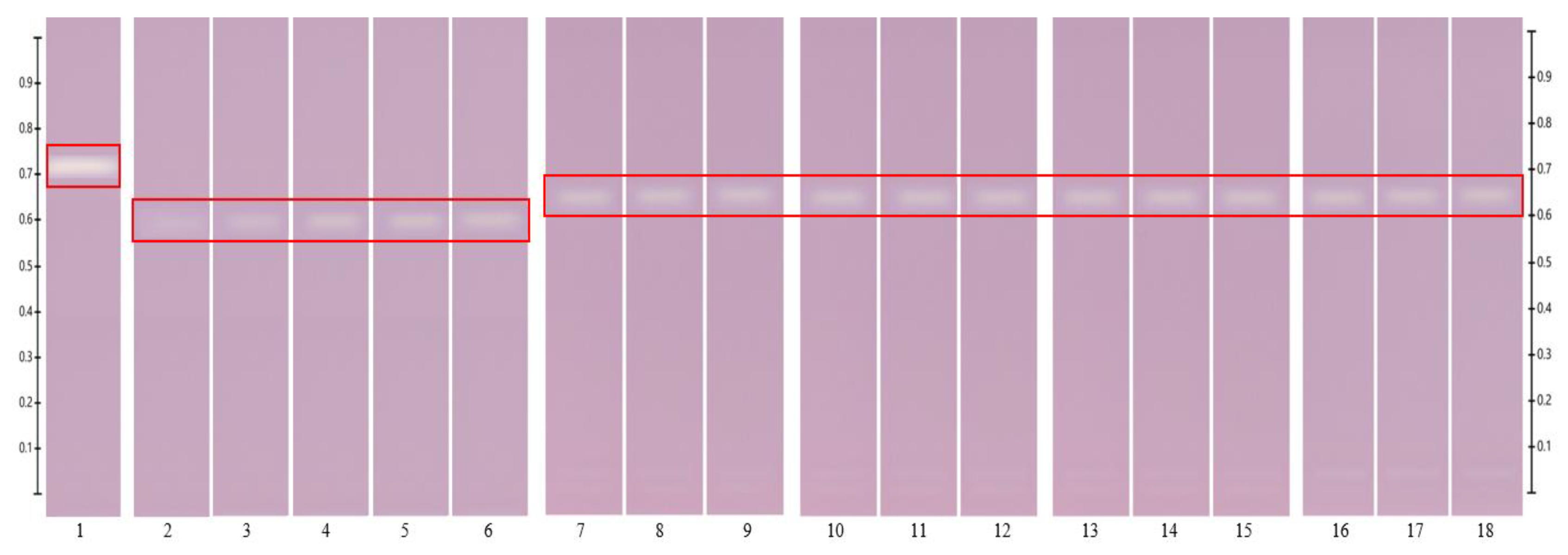
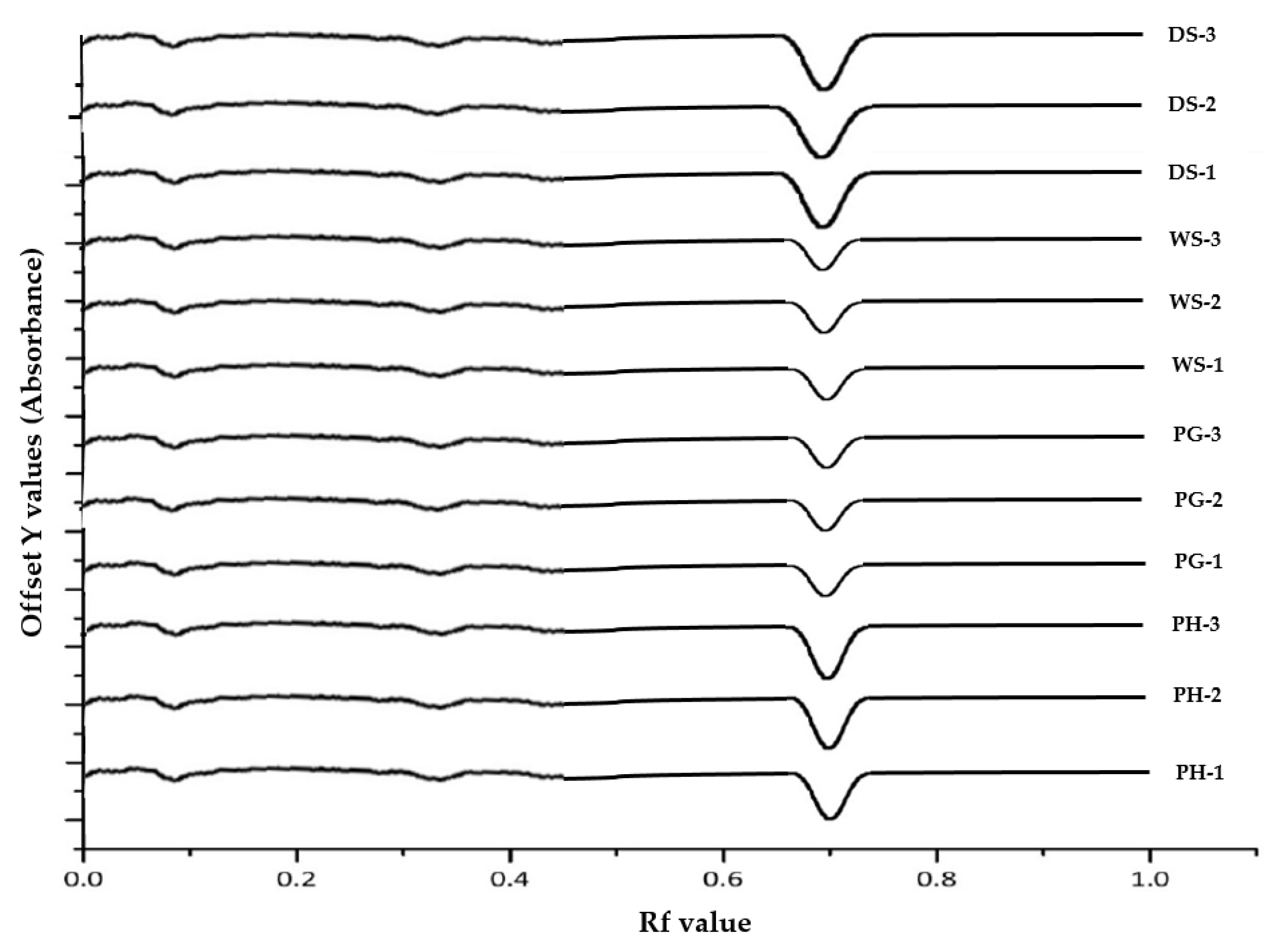
The antioxidant activity of various bands in the samples was visualized by HPTLC-DPPH analysis. The fingerprints of Jarrah (JAR) pure honey and its formulations at the 254 and 366 development stage (prior derivatisation with DPPH reagent) are shown in Figure 1 and Figure 2, respectively, to allow for a direct comparison between the extracts’ fingerprints, which feature a number of bands, and the few compound(s) amongst them that contribute to this activity. Antioxidant bands for the five honeys and their respective formulations were detected at Rf 0.650 (WA Jarrah honey and NZ Manuka honey) and Rf 0.630 (Coastal Peppermint honey, WA Manuka honey 1 and WA Manuka honey 2). For illustrative purposes, the HPTLC-DPPH fingerprint and chromatogram of antioxidant components in Jarrah honey are shown in Figure 3 and Figure 4, respectively. The fingerprints and chromatograms representing the antioxidant compounds in NZ Manuka, Coastal Peppermint, WA Manuka 1 and WA Manuka 2 honey extracts are shown in the Supplementary File (Supplementary Figures S1–S16).

Figure 1.
HPTLC fingerprints of Jarrah (JAR) honey and its corresponding formulations. Images taken at 254 at development stage (prior derivatization with DPPH reagent); Track 1—4,5,7-trihydroxyflavone (internal standard), Tracks 2–6—Gallic acid at different concentrations (60–200 µg/spot), Tracks 7–9—pure JAR honey, Tracks 10–12—JAR honey pre-gel solution, Tracks 13–15—JAR honey wet sheet, Tracks 16–18—JAR honey dry sheet.

Figure 2.
HPTLC fingerprints of Jarrah (JAR) honey and its corresponding formulations. Images taken at 366 at development stage (prior derivatization with DPPH reagent); Track 1—4,5,7-trihydroxyflavone (internal standard), Tracks 2–6—Gallic acid at different concentrations (60–200 µg/spot), Tracks 7–9—pure JAR honey, Tracks 10–12—JAR honey pre-gel solution, Tracks 13–15—JAR honey wet sheet, Tracks 16–18—JAR honey dry sheet.
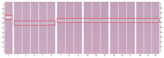
Figure 3.
Antioxidant activity of Jarrah (JAR) honey and its corresponding formulations—red box indicates the antioxidant band at Rf 0.650. Images taken at white light after derivatization with DPPH reagent; Track 1—4,5,7-trihydroxyflavone (internal standard), Tracks 2–6—Gallic acid at different concentrations (60–200 µg/spot), Tracks 7–9—pure JAR honey, Tracks 10–12—JAR honey pre-gel solution, Tracks 13–15—JAR honey wet sheet, Tracks 16–18—JAR honey dry sheet.
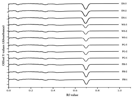
Figure 4.
Chromatograms of Jarrah (JAR) honey and its corresponding formulations. Images taken at white light after derivatization with DPPH reagent; (PH-1–PH-3)—pure JAR honey, (PG-1–PG-3)—JAR honey pre-gel solution, (WS-1–WS-3)—JAR honey wet sheet, (DS-1–DS-3)—JAR honey dry sheet.
The normalized antioxidant band activity suggests that the activity detected in the extracts of the respective pre-gels and wet sheets was identical to that of the corresponding pure honeys (Table 3). For example, the antioxidant band activity of the pre-gel solution and wet sheet containing Jarrah honey were the same (p = 0.813) as that of pure Jarrah honey (F value was 0.2143). In the case of the dry sheets, it may seem like the recorded antioxidant band activities were much higher than those of the pure honeys and the corresponding pre-gels and wet sheets (Table 3). However, it should be noted that water had been removed from the dry sheets by freeze-drying, which would impact the weight-based expression of antioxidant band activity. Indeed, when considering the antioxidant band activity in wet and dry sheets on a ‘per sheet’ basis rather than per g of formulation (Table 3), almost identical values were obtained for both formulation types, indicating that the transformation of wet sheets into dry sheets did not lead to a loss in the antioxidant band activity.

Table 3.
Antioxidant band activity of pure honeys and their corresponding formulations (n = 3, data represents mean ± SD).
3.3. Antibacterial Activity
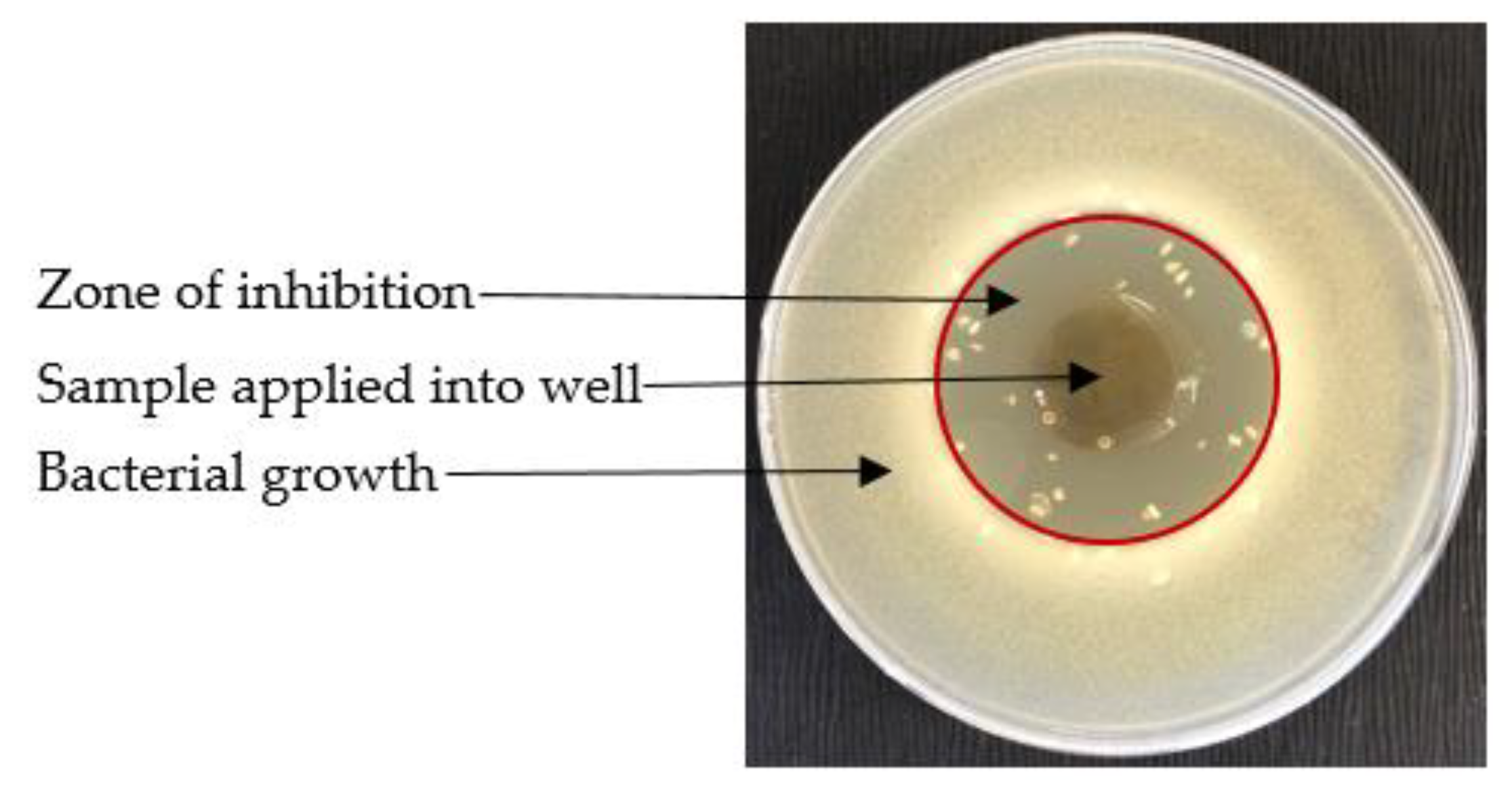
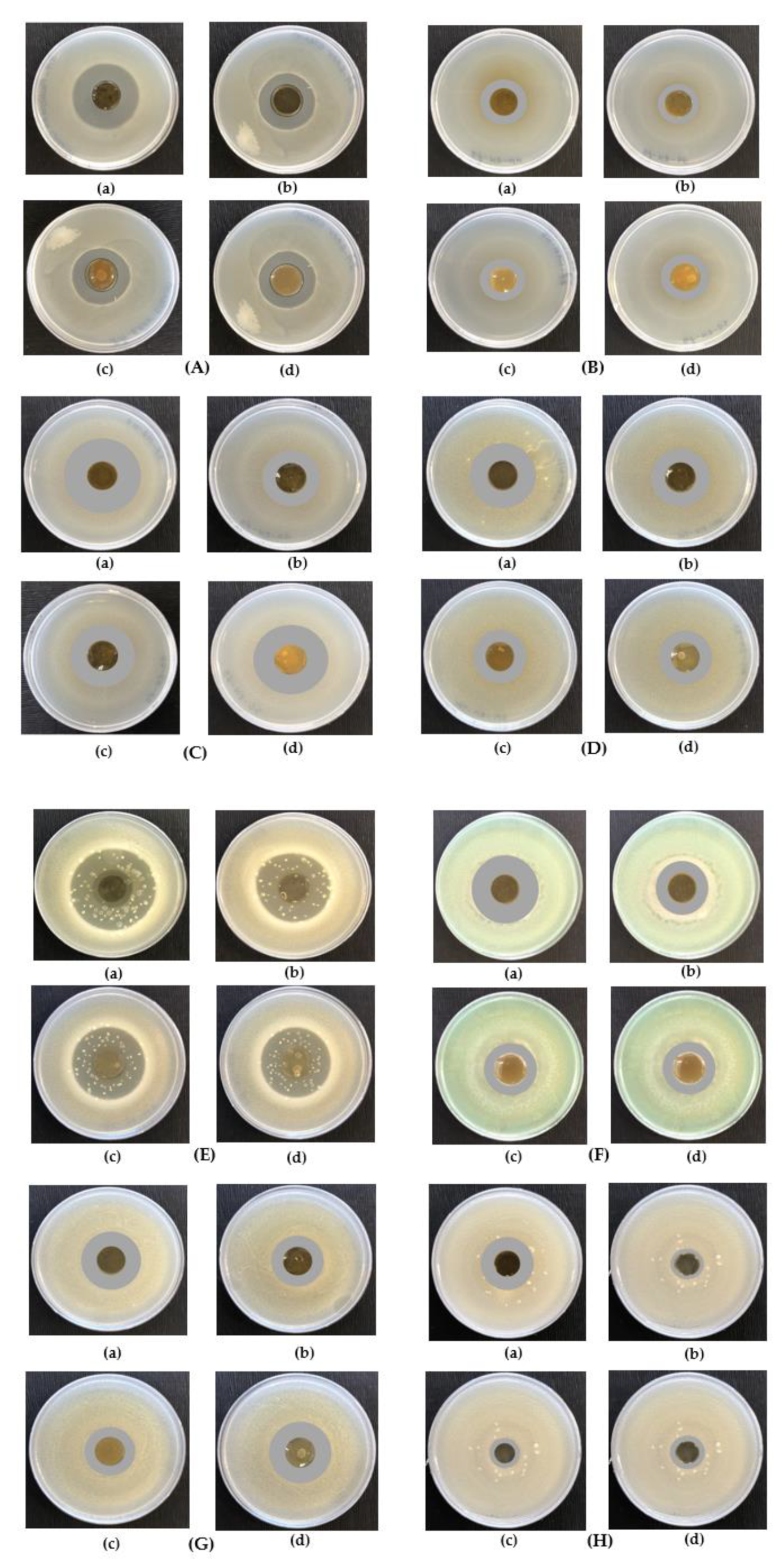
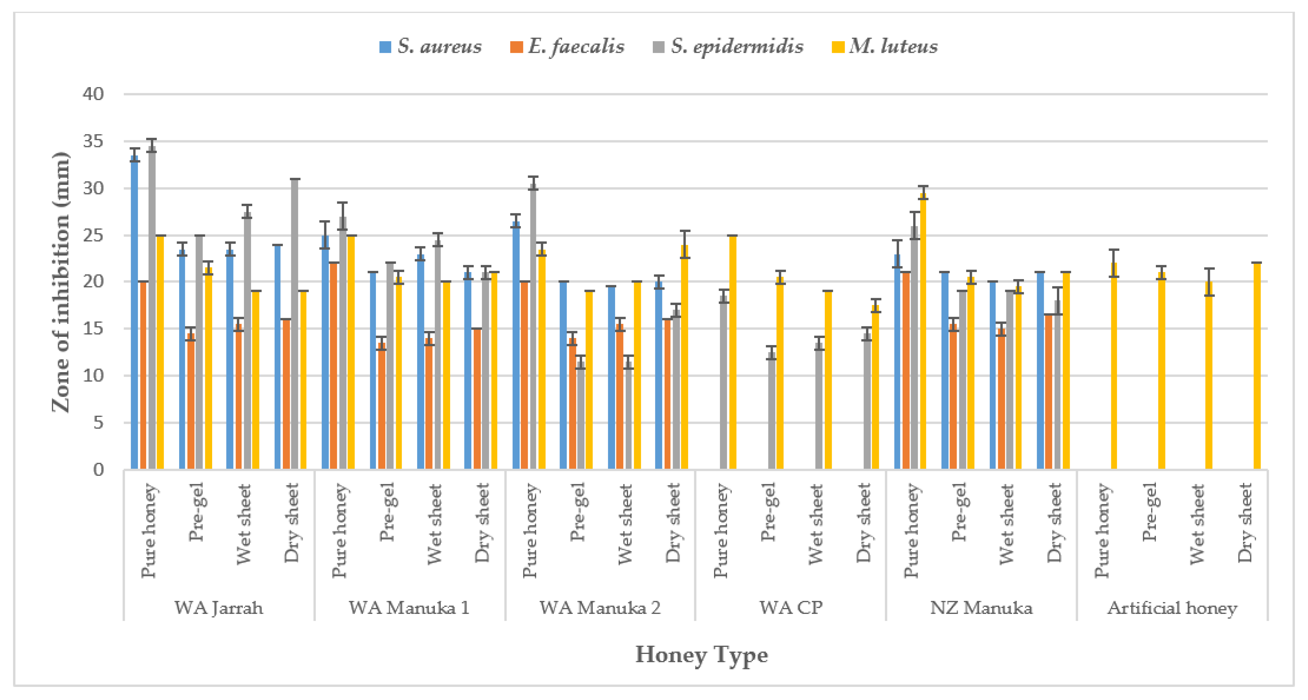
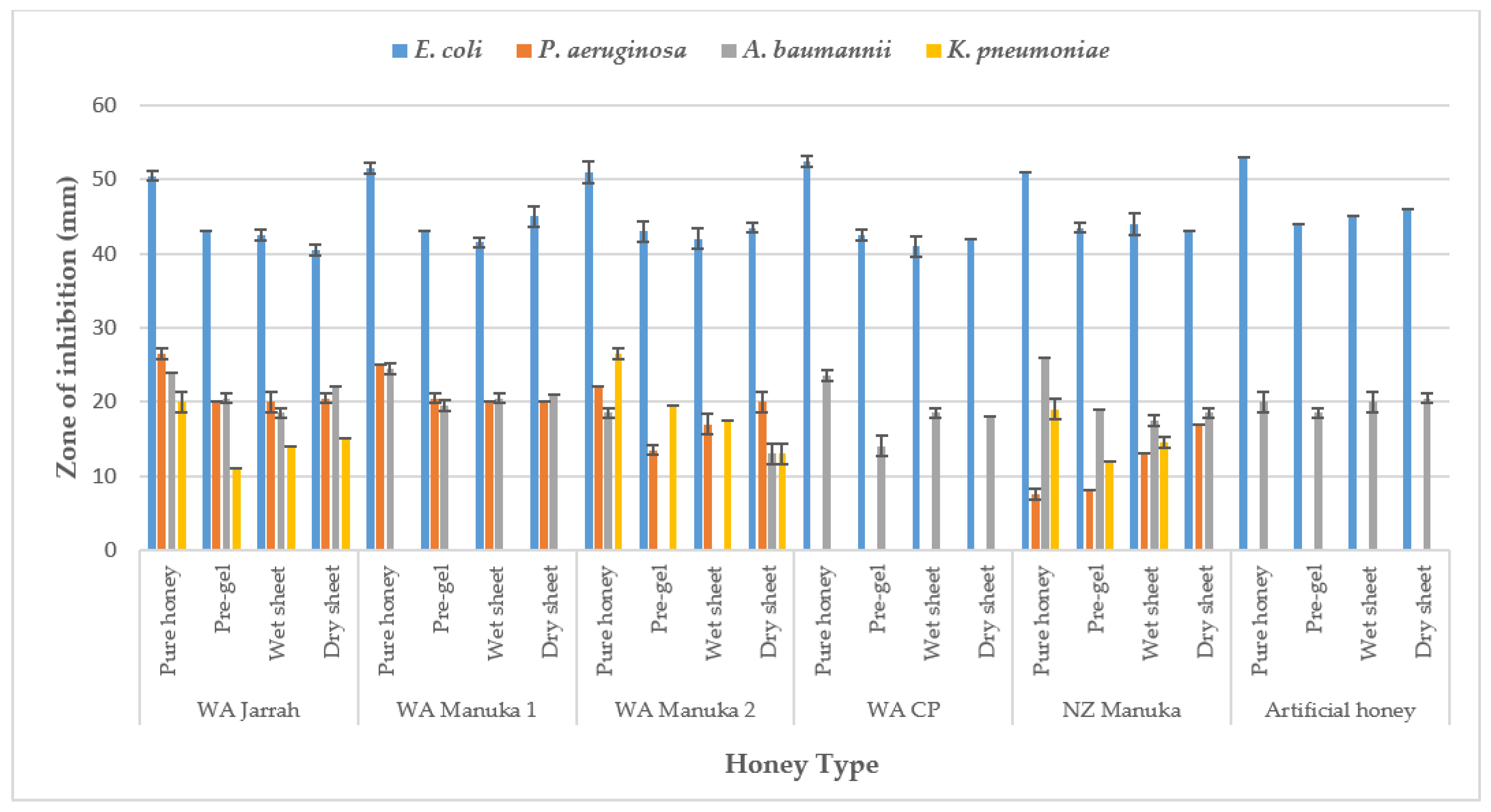
For illustrative purposes, a representative inhibition zone observed in the agar overlay assay for WA Manuka honey 2 pre-gel solutions is shown in Figure 5. Few bubbles were observed, which might be owing to the production of CO2 as a consequence of the metabolism of the glucose and fructose in honeys by E. coli. Figure 6 displays the zone of inhibition recorded for WA Jarrah honey and its respective formulations (i.e., pre-gel solution, wet sheet, dry sheet) against all tested bacterial strains. The comparative antibacterial activity of all samples against the eight bacterial strains that were tested as part of this study are presented as bar diagram in Figure 7 and Figure 8. The inhibition zone exhibited by honey-loaded formulations are also shown in Supplementary Tables S1 and S2. In most of the pure honeys and honey-loaded formulations, the pure honey showed the highest activity followed by the dry sheet, pre-gel solution and wet sheet (Figure 7 and Figure 8) against a particular bacterial strain. However, this trend was not consistent in a few samples, such as the NZ Manuka honey and its formulations against S. epidermidis (Figure 7).
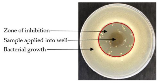
Figure 5.
Zone of inhibition recorded for WA Manuka honey 2 pre-gel solution against E. coli.
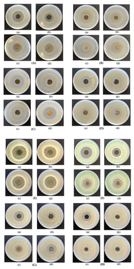
Figure 6.
Zone of inhibitions recorded for WA Jarrah (JAR) honey—(a) Pure honey, (b) Pre-gel solution, (c) Wet sheet, (d) Dry sheet against (A) S. aureus, (B) E. faecalis, (C) S. epidermidis, (D) M. luteus, (E) E. coli, (F) P. aeruginosa, (G) A. baumannii, (H) K. pneumoniae.
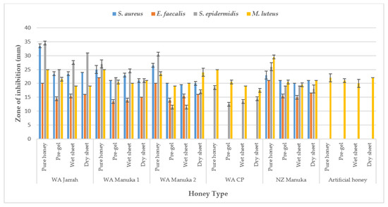
Figure 7.
Comparison of antibacterial activity of honey-loaded products and pure honeys against S. aureus, E. facaelis, S. epidermidis, M. luteus.
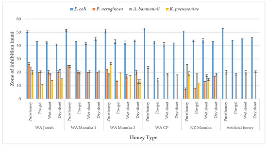
Figure 8.
Comparison of antibacterial activity of honey-loaded products and pure honeys against E. coli, P. aureginosa, A. baumannii, K. pneumoniae.
4. Discussion
Amongst the many reported bioactivities, the antioxidant and antibacterial activities of honeys are the most explored [27,28,36,37,38] to provide supportive evidence for some honeys to be used as natural medicines [33]. Several in vitro and in vivo studies have demonstrated honey’s antioxidant and antibacterial activity [13,46,47,48]. It has been shown that the non-sugar honey constituents, including phenolic acids and flavonoids [49], enzymes (e.g., glucose oxidase and catalase) [47], ascorbic acid, proteins and carotenoids [50] all contribute to the antioxidant features of honey. Correlation between a honey’s floral origin and its antioxidant capacity has also been demonstrated [49,51,52]. With regards to the antibacterial activity, next to high levels of sugar and a low pH that are found across all honeys, the antibacterial properties of so-called peroxide honeys are related to their hydrogen peroxide content, whereas the antibacterial potential of non-peroxide honeys (derived mainly from Leptospermum spp.) correlates to levels of MGO. Other non-peroxide factors, such as lysozymes, phenolic acids and flavonoids, have also been found to contribute to the antibacterial activity of a honey [53]. Taken as a whole, these antioxidant and antibacterial effects offer a unique potential for honeys to be used in wound dressings to assist with the rapid healing of infections, debridement of wounds, vanquishing of inflammation, reduction of scarring tissues and acceleration of angiogenesis along with tissue granulation and epithelial growth [49].
Western Australia is home to 8 of Australia’s 15 biodiversity hotspots [41] and has a diverse and often endemic melliferous flora that leads to the production of a wide variety of honeys. WA has, for example, more than 80 different Leptospermum species [41] that are not yet thoroughly investigated as potential sources of non-peroxide honeys, nor are any data available on their bioactivity. As the bioactivities of honeys are often attributable to their floral source, geographical origin and phytochemical profile, at present, the potential of WA honeys as possible future medicinal honeys appears underexplored. However, applying pure honey as a topical agent for the management of wounds can be challenging. Honey’s inherent stickiness and liquefaction is inconvenient and hinders its application at an adequate and reproducible dose [40]. Although some honey-based formulations are already commercially available that address these challenges, to date, very few incorporate WA honeys. Furthermore, to our knowledge, no study has yet been conducted to determine the bioactivity (i.e., antioxidant and antibacterial activity) of honey-loaded topical formulations, including those incorporating WA honeys. A prior study presented the manufacturing process and the physicochemical characterization and storage stability of the three types of alginate-based honey formulations prepared using two WA Manuka, one WA Jarrah and one WA Coastal Peppermint honeys along with NZ Manuka honey as a comparator [41]. The current study focuses on their antioxidant and antibacterial activity to support potential future clinical applications. Moreover, the focus of this study is extended to investigate if the manufacturing process had any negative impact on a honey’s bioactivity.
The antioxidant capacity of the honey-loaded products was found to be unchanged compared to that of the corresponding pure honeys. This indicates that the formulation process and also the employed carrier (i.e., sodium alginate) and cross-linking agent (i.e., Calcium chloride) does not affect the antioxidant activity of these honeys. Lawag et al. 2023 [54] utilized a comprehensive HPTLC-derived database to identify antioxidant compounds in these honeys via an HPTLC-DPPH assay. Hesperitin was found to contribute to the antioxidant activity of WA Jarrah and NZ Manuka honeys, and m-coumaric acid was identified as an antioxidant compound in Coastal Peppermint honeys as well as in WA Manuka honey 1 and WA Manuka honey 2. WA Manuka honey 2 and its formulations showed the highest antioxidant as found in both the FRAP assay (6.56 mmol Fe2+ equivalent/kg of pure honey) and HPTLC-DPPH assay (band activity equivalent to 29.30 µg gallic acid/g pure honey) among all analyzed WA honeys (Table 2 and Table 3). In the FRAP assay, WA Manuka honey 2 and its formulations (i.e., pre-gel and wet sheet) showed higher activity (p = 0.029) compared to the NZ Manuka honey and its pre-gel solution and wet sheet (Table 2). Moreover, WA Manuka honey 2 and its pre-gel solution and wet sheet formulations showed higher band activities (p < 0.0001) compared to the NZ Manuka comparator honey and its formulations (Table 3).
The antibacterial activity of the honeys and the honey-loaded formulations was measured via the optimised agar overlay assay. It was found that WA Jarrah, WA Manuka 2 and NZ Manuka honeys and their corresponding formulations showed growth inhibition zones against all Gram-positive and Gram-negative bacteria. In the case of Jarrah honey, the activity might be related to its hydrogen peroxide component and osmotic pressure, while this activity recorded in WA Manuka 2 and NZ Manuka honey might be due to their MGO content as a non-peroxide antibacterial factor along with osmotic pressure [10,12,25]. The MGO level of WA Jarrah, WA Manuka 2 and NZ Manuka honey was reported in previous study [29]. Coastal Peppermint honey and its corresponding formulations did not show any zones of growth inhibition against S. aureus, E. facecalis, S. epidermidis and A. baumannii (Figure 7 and Figure 8). The carrier, sodium alginatedissolved in deionised water, was investigated as a blank, and it was found that the alginate solution itself had no antibacterial activity. Interestingly, it could be demonstrated that an artificial honey and its formulations produced zones of growth inhibition against E. coli, A. baumannii and M. luteus (Figure 7 and Figure 8). The osmotic pressure exerted by its high sugar content might be responsible for the antibacterial activity seen [10,27]. However, the hypothesised osmotic pressure-induced antibacterial activity appears selective to particular bacterial strains as it was only seen for M. luteus, E. coli and A. baumannii but not for S. aureus, E. faecalis, S. epidermidis, P. aeruginosa, K. pneumoniae. The findings of the overlay assay allow us to draw conclusions on the antibacterial activity of the prepared formulations in the actual form they would be applied in an in vivo setting. Thus, this study generated clinically relevant data as the formulations did not undergo any further processing, such as dissolution, during the antibacterial analysis.
5. Conclusions
It is important to ensure that the bioactivities of honeys incorporated into honey-loaded formulations as the active pharmaceutical ingredient are retained. In this study, the antioxidant and antibacterial activity of WA honey-loaded formulations were therefore investigated and found to be comparable to that of the respective pure honeys. The generated data suggest that the formulations retain the honeys’ antioxidant and antibacterial activities throughout the preparation process and might therefore be suitable for future therapeutic application as topical agents for wound healing. This study adds novel information on the investigated Western Australian honey-based formulations. Its findings warrant further in vivo investigations to fully assess the potential clinical use of these honey-loaded formulations.
Supplementary Materials
The following supporting information can be downloaded at: https://www.mdpi.com/article/10.3390/app13137440/s1, Table S1: Zones of inhibition (mm) of pure honeys and prepared formulations against four Gram positive species of bacteria (n = 3, data represents mean ± SD); Table S2: Zones of inhibition (mm) of pure honeys and prepared formulations against four Gram negative species of bacteria (n = 3, data represents mean ± SD). Figure S1: HPTLC fingerprints of NZ Manuka (NZM) honey and its corresponding formulations. Images taken at 254 development stage (prior derivatization with DPPH reagent); Track 1—4,5,7-trihydroxyflavone (internal standard), Tracks 2–6—Gallic acid at different concentrations (60–200 µg/spot), Tracks 7–9—pure NZM honey, Tracks 10–12—NZM honey pre-gel solution, Tracks 13–15—NZM honey wet sheet, Tracks 16–18—NZM honey dry sheet. Figure S2: HPTLC fingerprints of NZ Manuka (NZM) honey and its corresponding formulations. Images taken at 366 development stage (prior derivatization with DPPH reagent); Track 1—4,5,7-trihydroxyflavone (internal standard), Tracks 2–6—Gallic acid at different concentrations (60–200 µg/spot), Tracks 7–9—pure NZM honey, Tracks 10–12—NZM honey pre-gel solution, Tracks 13–15—NZM honey wet sheet, Tracks 16–18—NZM honey dry sheet. Figure S3: Antioxidant activity of NZ Manuka (NZM) honey and its corresponding formulations—red box indicates the antioxidant band at Rf 0.650. Images taken at white light after derivatisation with DPPH reagent; Track 1—4,5,7-trihydroxyflavone (internal standard), Tracks 2–6—Gallic acid at different concentrations (60–200 µg/spot), Tracks 7–9—pure NZM honey, Tracks 10–12—NZM honey pre-gel solution, Tracks 13–15—NZM honey wet sheet, Tracks 16–18—NZM honey dry sheet. Figure S4: Chromatograms of NZ Manuka (NZM) honey and its corresponding formulations. Images taken at white light after derivatisation with DPPH reagent; (PH-1–PH-3)—pure NZM honey, (PG-1–PG-3)—NZM honey pre-gel solution, (WS-1–WS-3)—NZM honey wet sheet, (DS-1–DS-3)—NZM honey dry sheet. Figure S5: HPTLC fingerprints of Jarrah (JAR) honey and its corresponding formulations. Images taken at 254 development stage (prior derivatization with DPPH reagent); Track 1—4,5,7-trihydroxyflavone (internal standard), Tracks 2–6—Gallic acid at different concentrations (60–200 µg/spot), Tracks 7–9—pure JAR honey, Tracks 10–12—JAR honey pre-gel solution, Tracks 13–15—JAR honey wet sheet, Tracks 16–18—JAR honey dry sheet. Figure S6: HPTLC fingerprints of Coastal Peppermint (CP) honey and its corresponding formulations. Images taken at 366 development stage (prior derivatization with DPPH reagent); Track 1—4,5,7-trihydroxyflavone (internal standard), Tracks 2–6—Gallic acid at different concentrations (60–200 µg/spot), Tracks 7–9—pure CP honey, Tracks 10–12—CP honey pre-gel solution, Tracks 13–15—CP honey wet sheet, Tracks 16–18—CP honey dry sheet. Figure S7: Antioxidant activity of Coastal Peppermint (CP) honey and its corresponding formulations—red box indicates the antioxidant band at Rf 0.630. Images taken at white light after derivatisation with DPPH reagent; Track 1—4,5,7-trihydroxyflavone (internal standard), Tracks 2–6—Gallic acid at different concentrations (60–200 µg/spot), Tracks 7–9—pure CP honey, Tracks 10–12—CP honey pre-gel solution, Tracks 13–15—CP honey wet sheet, Tracks 16–18—CP honey dry sheet. Figure S8: Chromatograms of Coastal Peppermint (CP) honey and its corresponding formulations. Images taken at white light after derivatisation with DPPH reagent; (PH-1–PH-3)—pure CP honey, (PG-1–PG-3)—CP honey pre-gel solution, (WS-1–WS-3)—CP honey wet sheet, (DS-1–DS-3)—CP honey dry sheet. Figure S9: HPTLC fingerprints of WA Manuka honey 1 (WAM1) honey and its corresponding formulations. Images taken at 254 development stage (prior derivatization with DPPH reagent); Track 1—4,5,7-trihydroxyflavone (internal standard), Tracks 2–6—Gallic acid at different concentrations (60–200 µg/spot), Tracks 7–9—pure WAM1 honey, Tracks 10–12—WAM1 honey pre-gel solution, Tracks 13–15—WAM1 honey wet sheet, Tracks 16–18—WAM1 honey dry sheet. Figure S10: HPTLC fingerprints of WA Manuka honey 1 (WAM1) honey and its corresponding formulations. Images taken at 366 development stage (prior derivatization with DPPH reagent); Track 1—4,5,7-trihydroxyflavone (internal standard), Tracks 2–6—Gallic acid at different concentrations (60–200 µg/spot), Tracks 7–9—pure WAM1 honey, Tracks 10–12—WAM1 honey pre-gel solution, Tracks 13–15—WAM1 honey wet sheet, Tracks 16–18—WAM1 honey dry sheet. Figure S11: Antioxidant activity of WA Manuka honey 1 (WAM1) and its corresponding formulations—red box indicates the antioxidant band at Rf 0.630. Images taken at white light after derivatisation with DPPH reagent; Track 1—4,5,7-trihydroxyflavone (internal standard), Tracks 2–6—Gallic acid at different concentrations (60–200 µg/spot), Tracks 7–9—pure WAM1 honey, Tracks 10–12—WAM1 honey pre-gel solution, Tracks 13–15—WAM1 honey wet sheet, Tracks 16–18—WAM1 honey dry sheet. Figure S12: Chromatograms of WA Manuka honey 1 (WAM1) and its corresponding formulations. Images taken at white light after derivatisation with DPPH reagent; (PH-1–PH-3)—pure WAM1 honey, (PG-1–PG-3)—WAM1 honey pre-gel solution, (WS-1–WS-3)—WAM1 honey wet sheet, (DS-1–DS-3)—WAM1 honey dry sheet. Figure S13: HPTLC fingerprints of WA Manuka honey 2 (WAM2) honey and its corresponding formulations. Images taken at 254 development stage (prior derivatization with DPPH reagent); Track 1—4,5,7-trihydroxyflavone (internal standard), Tracks 2–6—Gallic acid at different concentrations (60–200 µg/spot), Tracks 7–9—pure WAM2 honey, Tracks 10–12—WAM2 honey pre-gel solution, Tracks 13–15—WAM2 honey wet sheet, Tracks 16–18—WAM2 honey dry sheet. Figure S14: HPTLC fingerprints of WA Manuka honey 2 (WAM2) honey and its corresponding formulations. Images taken at 366 development stage (prior derivatization with DPPH reagent); Track 1—4,5,7-trihydroxyflavone (internal standard), Tracks 2–6—Gallic acid at different concentrations (60–200 µg/spot), Tracks 7–9—pure WAM2 honey, Tracks 10–12—WAM2 honey pre-gel solution, Tracks 13–15—WAM2 honey wet sheet, Tracks 16–18—WAM2 honey dry sheet. Figure S15: Antioxidant activity of WA Manuka honey 2 (WAM2) and its corresponding formulations—red box indicates the antioxidant band at Rf 0.630. Images taken at white light after derivatisation with DPPH reagent; Track 1—4,5,7-trihydroxyflavone (internal standard), Tracks 2–6—Gallic acid at different concentrations (60–200 µg/spot), Tracks 7–9—pure WAM2 honey, Tracks 10–12—WAM2 honey pre-gel solution, Tracks 13–15—WAM2 honey wet sheet, Tracks 16–18—WAM2 honey dry sheet. Figure S16: Chromatograms of WA Manuka honey 2 (WAM2) and its corresponding formulations. Images taken at white light after derivatisation with DPPH reagent; (PH-1–PH-3)—pure WAM2 honey, (PG-1–PG-3)—WAM2 honey pre-gel solution, (WS-1–WS-3)—WAM2 honey wet sheet, (DS-1–DS-3)—WAM2 honey dry sheet.
Author Contributions
Conceptualization, M.L.H. and C.L.; methodology, M.L.H., C.L., K.H. and L.Y.L.; formal analysis, M.L.H.; writing—original draft preparation, M.L.H.; writing—review and editing, C.L., L.Y.L., K.H. and D.H.; supervision, C.L., L.Y.L., K.H. and D.H.; project administration, C.L.; funding acquisition, C.L. All authors have read and agreed to the published version of the manuscript.
Funding
This research was funded by the University of Western Australia.
Institutional Review Board Statement
Not applicable.
Informed Consent Statement
Not applicable.
Data Availability Statement
Not applicable.
Conflicts of Interest
The authors declare no conflict of interest. The funders had no role in the design of the study; in the collection, analyses, or interpretation of data; in the writing of the manuscript, or in the decision to publish the results.
References
- Martinotti, S.; Ranzato, E. Honey, Wound Repair and Regenerative Medicine. J. Funct. Biomater. 2018, 8, 34. [Google Scholar]
- Saranraj, P.; Sivasakthi, S.; Feliciano, G. Pharmacology of Honey: A Review. Biol. Res. 2016, 10, 271–289. [Google Scholar]
- Sultana, S.; Foster, K.; Lim, L.Y.; Hammer, K.; Locher, C. A Review of the Phytochemistry and Bioactivity of Clover Honeys (Trifolium spp.). Foods 2022, 11, 1901. [Google Scholar]
- Cianciosi, D.; Forbes-Hernández, T.Y.; Afrin, S.; Gasparrini, M.; Reboredo-Rodriguez, P.; Manna, P.P.; Zhang, J.; Lamas, L.B.; Flórez, S.M.; Toyos, P.A.; et al. Phenolic Compounds in Honey and Their Associated Health Benefits: A Review. Molecules. 2018, 11, 2322. [Google Scholar]
- Cornara, L.; Biagi, M.; Xiao, J.; Burlando, B. Therapeutic Properties of Bioactive Compounds from Different Honeybee Products. Front. Pharm. 2017, 8, 412. [Google Scholar]
- Libonatti, C.; Varela, S.; Basualdo, M. Antibacterial activity of honey: A review of honey around the world. J. Microbiol. Antimicrob. 2014, 6, 51–56. [Google Scholar]
- White, R. The benefits of honey in wound management. Nurs. Stand. 2005, 20, 57–64. [Google Scholar] [CrossRef]
- da Silva, P.M.; Gauche, C.; Gonzaga, L.V.; Costa, A.C.; Fett, R. Honey: Chemical composition, stability and authenticity. Food Chem. 2016, 196, 309–923. [Google Scholar]
- Minden-Birkenmaier, B.A.; Bowlin, G.L. Honey-Based Templates in Wound Healing and Tissue Engineering. Bioengineering 2018, 5, 46. [Google Scholar] [PubMed]
- Hossain, M.L.; Lim, L.Y.; Hammer, K.; Hettiarachchi, D.; Locher, C. A Review of Commonly Used Methodologies for Assessing the Antibacterial Activity of Honey and Honey Products. Antibiotics 2022, 11, 975. [Google Scholar] [PubMed]
- Rana, S.; Mishra, M.; Yadav, D.; Subramani, S.K.; Katare, C.; Prasad, G. Medicinal uses of honey: A review on its benefits to human health. Prog. Nutr. 2018, 20, 5–14. [Google Scholar]
- Lee, D.S.; Sinno, S.; Khachemoune, A. Honey and Wound Healing. Am. J. Clin. Dermatol. 2011, 12, 181–190. [Google Scholar]
- Alvarez-Suarez, J.M.; Giampieri, F.; Battino, M. Honey as a source of dietary antioxidants: Structures, bioavailability and evidence of protective effects against human chronic diseases. Curr. Med. Chem. 2013, 20, 621–638. [Google Scholar]
- Shahzad, A.; Cohrs, R.J. In vitro antiviral activity of honey against varicella zoster virus (VZV): A translational medicine study for potential remedy for shingles. Transl. Biomed. 2012, 3, 2. [Google Scholar]
- Abubakar, M.B.; Abdullah, W.Z.; Sulaiman, S.A.; Suen, A.B. A Review of Molecular Mechanisms of the Anti-Leukemic Effects of Phenolic Compounds in Honey. Int. J. Mol. Sci. 2012, 13, 15054–15073. [Google Scholar]
- Alvarez-Suarez, J.M.; Giampieri, F.; Cordero, M.; Gasparrini, M.; Forbes-Hernández, T.Y.; Mazzoni, L.; Afrin, S.; Beltrán-Ayala, P.; González-Paramás, A.M.; Santos-Buelga, C.; et al. Activation of AMPK/Nrf2 signalling by Manuka honey protects human dermal fibroblasts against oxidative damage by improving antioxidant response and mitochondrial function promoting wound healing. J. Funct. Foods. 2016, 25, 38–49. [Google Scholar]
- Rahal, A.; Kumar, A.; Singh, V.; Yadav, B.; Tiwari, R.; Chakraborty, S.; Dhama, K. Oxidative stress, prooxidants, and antioxidants: The interplay. BioMed Res. Int. 2014, 2014, 761264. [Google Scholar]
- Hussain, T.; Tan, B.; Yin, Y.; Blachier, F.; Tossou, M.C.; Rahu, N. Oxidative stress and inflammation: What polyphenols can do for us? Oxid. Med. Cell. Longev. 2016, 2016, 7432797. [Google Scholar]
- North, J.A.; Spector, A.A.; Buettner, G.R. Cell fatty acid composition affects free radical formation during lipid peroxidation. Am. J. Physiol. 1994, 267, 177–188. [Google Scholar]
- Banerjee, S.; Ghosh, J.; Sil, P.C. Drug metabolism and oxidative stress: Cellular mechanism and new therapeutic insights. Biochem. Anal. Biochem. 2016, 5, 255. [Google Scholar] [CrossRef]
- Yang, Y.; Bazhin, A.V.; Werner, J.; Karakhanova, S. Reactive oxygen species in the immune system. Int. Rev. Immunol. 2013, 32, 249–270. [Google Scholar]
- Pham-Huy, L.A.; He, H.; Pham-Huy, C. Free radicals, antioxidants in disease and health. Int. J. Biomed. Sci. 2008, 4, 89–96. [Google Scholar] [PubMed]
- Lobo, V.; Patil, A.; Phatak, A.; Chandra, N. Free radicals, antioxidants and functional foods: Impact on human health. Pharmacogn. Rev. 2010, 4, 118–126. [Google Scholar] [PubMed]
- Bouayed, J.; Bohn, T. Exogenous antioxidants-Double-edged swords in cellular redox state. Oxid. Med. Cell. Longev. 2010, 3, 228–237. [Google Scholar] [CrossRef] [PubMed]
- Agbaje, E.O.; Ogunsanya, T.; Aiwerioba, O.I.R. Conventional use of honey as antibacterial agent. Ann. Afr. Med. 2006, 5, 79–81. [Google Scholar]
- Tan, H.T.; Rahman, R.A.; Gan, S.H.; Halim, A.S.; Hassan, S.A.; Sulaiman, S.A.; Kirnpal-Kaur, B. The antibacterial properties of Malaysian tualang honey against wound and enteric microorganisms in comparison to manuka honey. BMC Complement. Altern. Med. 2009, 9, 34. [Google Scholar]
- Mandal, M.D.; Mandal, S. Honey: Its medicinal property and antibacterial activity. Asian Pac. J. Trop. Biomed. 2011, 1, 154–160. [Google Scholar]
- Mavric, E.; Wittmann, S.; Barth, G.; Henle, T. Identification and quantification of methylglyoxal as the dominant antibacterial constituent of Manuka (Leptospermum scoparium) honeys from New Zealand. Mol. Nutr. Food Res. 2008, 52, 483–489. [Google Scholar]
- Hossain, M.L.; Lim, L.Y.; Hammer, K.; Hettiarachchi, D.; Locher, C. Monitoring the Release of Methylglyoxal (MGO) from Honey and Honey-Based Formulations. Molecules 2023, 28, 2858. [Google Scholar]
- Saikaly, S.; Khachemoune, A. Honey and Wound Healing: An Update. Am. J. Clin. Dermatol. 2017, 18, 237–251. [Google Scholar]
- Angioi, R.; Morrin, A.; White, B. The Rediscovery of Honey for Skin Repair: Recent Advances in Mechanisms for Honey-Mediated Wound Healing and Scaffolded Application Techniques. Appl. Sci. 2021, 11, 5192. [Google Scholar]
- Oryan, A.; Alemzadeh, E.; Moshiri, A. Biological properties and therapeutic activities of honey in wound healing: A narrative review and meta-analysis. J. Tissue Viability 2016, 25, 98–118. [Google Scholar]
- Molan, P.C. The evidence and the rationale for the use of honey as wound dressing. Wound Pract. Res. J. Aust. Wound Manag. Assoc. 2011, 19, 204–220. [Google Scholar]
- Robson, V.; Dodd, S.; Thomas, S. Standardized antibacterial honey (Medihoney) with standard therapy in wound care: Randomized clinical trial. J. Adv. Nurs. 2009, 65, 565–575. [Google Scholar]
- Simon, A.; Sofka, K.; Wiszniewsky, G.; Blaser, G.; Bode, U.; Fleischhack, G. Wound care with antibacterial honey (Medihoney) in pediatric hematology-oncology. Support Care Cancer 2006, 14, 91–97. [Google Scholar]
- Hadagali, M.D.; Chua, L.S. The anti-inflammatory and wound healing properties of honey. Eur Food Res. Technol. 2014, 239, 1003–1014. [Google Scholar]
- Kazlowska, K.; Hsu, T.; Hou, C.C.; Yang, W.C.; Tsai, G.J. Anti-inflammatory properties of phenolic compounds and crude extract from Porphyra dentata. J. Ethnopharmacol. 2010, 128, 123–130. [Google Scholar]
- Kassim, M.; Achoui, M.; Mustafa, M.R.; Mohd, M.A.; Yusoff, K.M. Ellagic acid, phenolic acids, and flavonoids in Malaysian honey extracts demonstrate in vitro anti-inflammatory activity. Nutr. Res. 2010, 30, 650–659. [Google Scholar]
- Hussein, S.Z.; Mohd Yusoff, K.; Makpol, S.; Mohd Yusof, Y.A. Gelam Honey Inhibits the Production of Proinflammatory, Mediators NO, PGE(2), TNF-alpha, and IL-6 in Carrageenan-Induced Acute Paw Edema in Rats. Evid. Based Complement. Alternat. Med. 2012, 2012, 109636. [Google Scholar] [CrossRef]
- Hossain, M.L.; Lim, L.Y.; Hammer, K.; Hettiarachchi, D.; Locher, C. Honey-Based Medicinal Formulations: A Critical Review. Appl. Sci. 2021, 11, 5159. [Google Scholar]
- Hossain, M.L.; Lim, L.Y.; Hammer, K.; Hettiarachchi, D.; Locher, C. Design, Preparation and Physicochemical Characterisation of Alginate Based Honey-Loaded Topical Formulations. Pharmaceutics 2023, 15, 1483. [Google Scholar] [PubMed]
- Almeida, A.M.M.d.; Oliveira, M.B.S.; Costa, J.G.d.; Valentim, I.B.; Goulart, M.O.F. Antioxidant Capacity, Physicochemical and Floral Characterization of Honeys from the Northeast of Brazil. Rev. Virtual Quím. 2016, 8, 57–77. [Google Scholar]
- Islam, M.K.; Sostaric, T.; Lim, L.Y.; Hammer, K.; Locher, C. Development and validation of an HPTLC–DPPH assay and its application to the analysis of honey. J. Planar Chromatogr. Mod. TLC 2020, 33, 301–311. [Google Scholar]
- Ibrahim, R.S.; Khairy, A.; Zaatout, H.H.; Hammoda, H.M.; Metwally, A.M. Digitally-optimized HPTLC coupled with image analysis for pursuing polyphenolic and antioxidant profile during alfalfa sprouting. J. Chromatogr. B Analyt. Technol. Biomed. Life Sci. 2018, 1099, 92–96. [Google Scholar] [PubMed]
- Hossain, M.L.; Hammer, K.; Lim, L.Y.; Hettiarachchi, D.; Locher, C. Optimisation of an agar overlay assay for the assessment of the antimicrobial activity of topically applied semi-solid antiseptic products including honey-based formulations. J. Microbiol. Methods 2022, 202, 106596. [Google Scholar]
- Alvarez-Suarez, J.M.; Tulipani, S.; Romandini, S.; Vidal, A.; Battino, M. Methodological aspects about determination of phenolic compounds and in vitro evaluation of antioxidant capacity in the honey: A review. Curr. Anal. Chem. 2009, 5, 292–302. [Google Scholar]
- Molan, P.C.; Betts, J.A. Clinical usage of honey as a wound dressing: An update. J. Wound Care 2004, 13, 353–356. [Google Scholar]
- Swellam, T.; Miyanaga, N.; Onozawa, M.; Hattori, K.; Kawai, K.; Shimazui, T.; Akaza, H. Antineoplastic activity of honey in an experimental bladder cancer implantation model: In vivo and in vitro studies. Int. J. Urol. 2003, 10, 213–219. [Google Scholar]
- Meda, A.; Lamien, C.E.; Romito, M.; Millogo, J.; Nacoulma, O.G. Determination of the total phenolic, flavonoid and proline contents in Burkina Fasan honey, as well as their radical scavenging activity. Food Chem. 2005, 91, 571–577. [Google Scholar]
- Alvarez-Suarez, J.M.; Tulipani, S.; Romandini, S.; Bertoli, E.; Battino, M. Contribution of honey in nutrition and human health: A review. Mediterr. J. Nutr. Metab. 2009, 3, 15–23. [Google Scholar]
- Tomas-Barberán, F.A.; Martos, I.; Ferreres, F.; Radovic, B.S.; Anklam, E. HPLC flavonoid profiles as markers for the botanical origin of European unifloral honeys. J. Sci. Food Agric. 2001, 81, 485–496. [Google Scholar]
- Gheldof, N.; Xiao-Hong, W.; Engeseth, N. Identification and quantification of antioxidant components of honeys from various floral sources. J. Agric. Food Chem. 2002, 50, 5870–5877. [Google Scholar]
- Snowdon, J.A.; Cliver, D.O. Microorganisms in honey. Int. J. Food Microbiol. 1996, 31, 1–26. [Google Scholar] [PubMed]
- Lawag, I.L.; Islam, M.K.; Sostaric, T.; Lim, L.Y.; Hammer, K.; Locher, C. Antioxidant Activity and Phenolic Compound Identification and Quantification in Western Australian Honeys. Antioxidants 2023, 12, 189. [Google Scholar] [PubMed]
Disclaimer/Publisher’s Note: The statements, opinions and data contained in all publications are solely those of the individual author(s) and contributor(s) and not of MDPI and/or the editor(s). MDPI and/or the editor(s) disclaim responsibility for any injury to people or property resulting from any ideas, methods, instructions or products referred to in the content. |
© 2023 by the authors. Licensee MDPI, Basel, Switzerland. This article is an open access article distributed under the terms and conditions of the Creative Commons Attribution (CC BY) license (https://creativecommons.org/licenses/by/4.0/).


