Article

Development of a Method for Assessing the Resistance of Building Coatings to Phoatoautotrophic Biofouling

1
Interdisciplinary Doctoral School, Lodz University of Technology, Żeromskiego 116, 90-924 Łódź, Poland
2
Department of Environmental Biotechnology, Faculty of Biotechnology and Food Sciences, Lodz University of Technology, Wólczańska 171/173, 90-530 Łódź, Poland
3
Bolix, S.A. (Inc.), Stolarska 8, 34-300 Żywiec, Poland
*
Authors to whom correspondence should be addressed.
Appl. Sci. 2023, 13(14), 8009; https://doi.org/10.3390/app13148009
Submission received: 10 May 2023 / Revised: 3 July 2023 / Accepted: 4 July 2023 / Published: 8 July 2023

Abstract

:
The aim of this study was to develop a method for assessing the growth of photoautotrophs on plaster coatings, which will be used to reliably assess the resistance of these materials to photoautotrophic growth in the simulation of long-term exposure. In the course of the study, mineral and silicone plaster substrates were inoculated with a mixture of Stichococcus bacillaris, Nostoc commune, Pseudochlorella signiensis, and Coenochloris signiensis, and incubated for 28 days in model conditions. At 14 and 28 days after inoculation, the degree of photoautotrophic growth was determined using hemocytometer cell counting, a HY-LiTE 2 ATP measuring system, chlorophyll a concentration quantification, CIE L*a*b spectrophotometric color change evaluation, and visual assessment. The acquired results allowed us to select visual assessment and spectrophotometric color change evaluation as quick-to-perform and reliable techniques for further laboratory studies. The impact of minor changes introduced in the inoculation and incubation procedures on the rate of biofilm formation and severity of microbial fouling was studied. Differences in inoculation and incubation procedures strongly affected the results of the performed tests. Both methods have shown high potential and should be further expanded upon in environmental studies.

1. Introduction

The natural degradation of building materials is a complex process that incorporates several co-occurring abiotic factors as well as biotic components, mainly microorganisms such as bacteria, fungi, and algae. The impact of living organisms on the destruction of building materials and facades, i.e., biodeterioration [1], is currently considered as significant as atmospheric factors. This problem is now recognized for cultural heritage objects and new buildings alike.
Photoautotrophs, able to fix atmospheric carbon, are usually the first organisms to colonize building facades, affecting inorganic materials. These organisms, by conducting active metabolic processes, contribute to substrate decay. While the influence of photoautotrophic organisms on colonized materials might be greater than previously believed, it is the so-called aesthetic changes that are easiest and fastest to observe [2,3]. Simultaneously, they are the most responsible for economic losses affecting both homeowners and cultural heritage.
In terrestrial environments, algal cells are usually scattered by air. As a result, the probability and rate of migration will be dependent on the presence of algal biofilms near exposed facades. Assuming that algae cells are in contact with new material, its colonization will be mostly determined by favorable conditions. Factors determining the resistance of a material against microbial growth, including those associated with environmental conditions and intrinsic properties of the substrate, are called bioreceptivity [3]. Academic studies on the biodeterioration of building materials have allowed us to determine that for terrestrial algae the most important factors are temperature, water availability and relative air humidity, insolation, as well as the porosity and roughness of the substrate. However, it should be noted that the adaptation of algae to different environmental conditions is not yet fully understood [4]. Several studies have already expanded on the bioreceptivity of inorganic materials in response to, inter alia, photoautotrophic growth [5,6,7,8,9,10]. Some of them point to the urgent need for further laboratory and field studies [5,6,7] performed in various climatic regions. This is especially true considering recent climate changes influencing the weathering of building facades [7].
Unfortunately, the laboratory procedures used to study biofilm communities on building substrates have not yet been standardized, especially for resistance tests [5,7,11]. Furthermore, the majority of research relates to the bioreceptivity of cultural heritage buildings and not newly manufactured façade coatings usually more resistant to biofouling. One of the most known methods against green biofilm colonization is the use of hydrophobic protective layers (e.g., silicone-based coatings) aimed at the dependence of algal growth on water availability. The effectiveness of the abovementioned solution is now also supported by the common use of antimicrobial agents, i.e., biocides [12]. These can be applied to either remove biofilms already existing on building facades or be incorporated into the coating during the production process. As typical biocides, those based on quaternary ammonium salts [13], photoactive compounds including TiO2 and ZnO2 [3,14], metal salts (Ag, Cu, and Zn), and nanoparticles (AgNPs, AuNPs, and ZnONPs) [3,15] are predominantly used. In recent years, polymer coatings with TiO2 have been rapidly gaining popularity and are now commonly implemented. Such solutions can be applied to ceramic materials [3,16,17], mortars [18], and other materials, combining the benefits of hydrophobic coatings and photoactive biocides. For the abovementioned systems, the main antagonistic factor in relation to photoautotrophic biofilms is the photo-dependent oxidation that causes ruptures in the bonds forming between microbial cells and the colonized substrate. The effectiveness of protective coating is further enhanced by the creation of a super-hydrophobic layer hindering the accumulation of pollutants or cells and above all facilitating the evaporation of water [3].
Dybowska-Józefiak and Wesołowska [19] as well as Stanaszek-Tomal [11] have described the problem of facade biocorrosion, including the bioreceptivity of external coatings and external thermal insulation composite systems (ETICS) used in architecture typical for Central Europe. Stanaszek-Tomal [11] also noted the previously mentioned lack of standardization of procedures. The deficiencies of the currently recommended method for testing the efficacy of manufactured coatings against algae found in the European standards [20] have also been noted. Unfortunately, the abovementioned norms do not include the conditioning of the protective coating itself (only the material to which the coating will be applied), especially against the influence of UV radiation and leaching. The importance of water leaching in relation to the antimicrobial resistance of protective coatings has already been pointed out in previous studies [11,21]. The significance of simulating the environmental conditions is particularly important in the case of research conducted over several successive research cycles determining the long-term warranty of studied coatings. On the other hand, many commercially produced coatings are tested exclusively according to the manufacturer’s internal guidelines, and the existing norms currently serve only as recommendations, with producers not obliged to test for biological corrosion [11]. Although many methods for the determination of photoautotrophic growth on inorganic materials and cultural objects have already been evaluated [8,9,10,22,23,24], these have not yet been widely incorporated for testing manufactured plaster-based coatings. Additionally, some techniques are inadequate for testing a large number of highly differentiated sets of samples, or require costly, highly specialized equipment.
The complexity of colonization processes and the degradation of building materials, the lack of standardized test methods and simulations of environmental influence on the substrate, and, above all, technological development necessitate the development of new research methods. These should be characterized by high accuracy and repeatability, include necessary imitations of environmental conditions, be relatively quick to conduct, enable simultaneous testing of many samples, and at the same time allow for reliable representation of the coating resistance even over decades. The key in this aspect is to determine the growth/lack of growth of photoautotrophs on the surface of building materials, which determines the resistance/lack of resistance.
Therefore, the aim of this study was to develop a method for assessing the growth of photoautotrophs on plaster coatings, which will be used to reliably assess the resistance of these materials to photoautotrophic growth in the simulation of long-term exposure with the goal of fulfilling all the abovementioned requirements.

2. Materials and Methods

2.1. Experiment Design

The conducted research was divided into 2 stages. In the initial stage of the experiment, previously prepared modified and non-modified (see Section 2.2) mineral and silicone plaster samples were inoculated with algal suspension and incubated for 28 days. After inoculation, the severity of photoautotrophic growth occurring on material samples was tested at 14 and 28 days of incubation using cell enumeration with counting chambers, luminometric ATP tests, chlorophyll a concentration measurements, spectrophotometric CIE L*a*b color change evaluation, and visual assessment. For each different measuring method, at least 2 repetitions of test samples were prepared. The acquired results were used to select a reliable, fast, and convenient method of assessing the resistance of protective coatings against photoautotrophic growth.
In the second stage of the experiment, aiming to simulate the influencing environmental factors, the tested material samples were examined. For this purpose, methods for assessing photoautotrophic growth, selected in the previous stage of the experiment, were used to test material samples incubated and inoculated with two different methods (see Section 2.5) after 28 days of incubation. The gathered results and observations allowed us to propose significant changes in the tested methods as well as point to the areas that should be further tested and resolved in future studies.

2.2. Technical Material

As the technical material, facade plaster samples prepared in the form of discs with a diameter of 50 mm were used. Samples were subjected to 3 aging cycles, performed in accordance with the norms described by the European Organization for Technical Assessment [25]. A total of 4 different coating types were prepared for the study:
  • MP—Mineral plaster without biocide additives;
  • MPGS—Mineral plaster with primer and silicone paint, without biocide additives;
  • S—Silicone plaster without biocide additives;
  • SGS—Silicone plaster with primer and silicone paint, without biocide additives.
Prior to testing, samples were soaked in 3 L of distilled water for 24 h to simulate the effect of leaching. The samples were dried and placed individually in plastic Petri dishes. Open plates with samples were placed in a laminar chamber and irradiated with a UV lamp for 3 h. Afterward, the samples were placed upside down using sterile tweezers and irradiated again for another 3 h. Irradiation allowed us to sterilize the tested surfaces without otherwise potentially degrading the biocides (not used in this study) and at the same time imitating the effect of sunlight on the tested substrates.

2.3. Biological Material

Bold’s Basal Medium and its modifications were used as microbial media for the cultivation of biological material, incubation of tested samples, and simulation of dirt accumulating on the surface of material samples. BBM and 3N-BBM media were prepared fully in accordance with Andersen [26]. For the solidified medium, 1.5% agar (BTL, Łódź, Poland) was added prior to sterilization.
As the biological material, the following 4 species of aerophytic algae and cyanobacteria were used:
  • Stichococcus bacilliaris (CCAP, Culture Collection of Algae and Protozoa, Dunbeg, Scotland, UK);
  • Nostoc commune (CCAP, Culture Collection of Algae and Protozoa, Dunbeg, Scotland, UK);
  • Pseudochlorella signiensis (Environmental isolate);
  • Coenochloris signiensis (Environmental isolate).
Biological isolates were cultivated on solid Bold’s Basal Medium, incubated at 20 ± 1 °C, with relative air humidity equal to 50%, and under 1200 Lux from artificial fluorescence light tubes (Osram FLUORA T8 L 36W/77 Osram, Munich, Germany) with a 16 h/8 h day/night cycle.

2.4. Experiment 1: Selection of Methods Used for Assessing Photoautotrophic Growth on Plaster Coatings

The first stage of the study was performed in accordance with the experimental design described in Section 2.1. The method of conducting the research cycle was developed on the basis of literature data, the factors determining the resistance of protective coatings, and their bioreceptivity.

2.4.1. Inoculation Mixture

For the preparation of the inoculation mixture, the biological material of each actively growing species (see Section 2.3) was collected using a sterile loop and suspended in Bold’s Basal Medium (BBM). The density of each suspension was examined using a hemocytometer (Thom counting chamber) and calculated with Formula (1).
Ld = a × b × 4000 × 1000
where Ld is the average cell density in 1 mL of suspension (cfu/mL), a is the average cell count in the small square of the hemocytometer, and b is the dilution factor
The final density of each suspension was corrected to 4 × 10⁶ cfu/mL. The suspensions of all four strains were mixed in equal proportions to give an inoculum with a density of 4 × 10⁶ cfu/mL. The density of the inoculation mixture was confirmed by re-counting the cell density in the prepared inoculum.

2.4.2. Inoculation and Incubation Procedure

Samples of each tested coating were placed individually in sterile Petri dishes. An amount of 1 mL of sterile, non-solidified BBM medium with 1.5% agar additive was applied to the surface of each sample and spread with a sterile pad. Afterward, 1 mL of the algal inoculum prepared in accordance with Section 2.4.1 was introduced onto the surface of each sample.
Prepared and inoculated samples were incubated for 28 days under artificial lightning of 1200 Lux in a 16 h/8 h day/night cycle with a temperature of 22.0 °C ± 0.2 °C and air humidity of 50% ± 5. After inoculation, the degree of photoautotrophic growth was examined at 14 days and 28 days of incubation using biofilm cell enumeration, luminometric ATP measurement, chlorophyll a determination, visual assessment, and spectrophotometric color change evaluation.

2.4.3. Biofilm Cell Enumeration

The biofilm growth intensity was estimated using cell enumeration under a microscope with a hemocytometer following the methodology described previously in Komar et al. [27].

2.4.4. Luminometric ATP Measurement

ATP measurement was performed using the HY-LiTE 2® system (Merck, Darmstadt, Germany) in accordance with the manufacturer’s instructions.
For this purpose, biological material was collected using sterile, ATP-free swabs from the surface of 4 cm2 of technical material. The manufacturer’s instructions were followed in using reaction tubes compatible with the HY-LiTE 2® system. Three replicates were used for each type of technical material. The results were expressed in comparative relative light units (RLU) per cm2 of the technical material surface.

2.4.5. Chlorophyll a Determination

The concentration of chlorophyll a (chl-a) was determined based on the method described previously by Komar et al. [27] using cold extraction with 90% methanol as an extractant.

2.4.6. Visual Assessment

In order to visually assess the intensity of algae growth, a method based on the [20,28] European standards was used. For this purpose, technical material samples (see Section 2.2) were inoculated according to a previously described methodology. At 14 and 28 days after inoculation, the degree of microbial growth occurring on the substrate surface was assessed macroscopically. Due to its higher precision, the scale of material fouling degree described in the European standards PN-EN 15457 [28] was used. The acquired results were also compared against control samples. The following scale was adapted:
0—no visible growth on the surface of tested samples;
1—growth visible on less than 10% of the sample surface;
2—growth visible on 10—30% of the sample surface;
3—growth visible on 30—50% of the sample surface;
4—growth visible on more than 50% of the sample surface.

2.4.7. Spectrophotometric Color Change Evaluation

Color changes were determined in a CIE L*a*b trichromatic color model using a portable spectrophotometer CM-700d (Konica Minolta, Warsaw, Poland) and CM-S100w SpectraMagicTM NXv.2.0 software following the procedure described in Komar et al. [27].

2.4.8. Correlation Analysis

The acquired data were subjected to correlation analysis. To determine the degree of the linear relationship between the results, the correlation coefficient of determination (R2) was used. Based on calculated factors, the correlations between the tested methods for assessing photoautotrophic growth on plaster substrates were determined according to the following scale, based on Evans [29]:
R2 = 0.00–0.19 = very weak correlation;
R2 = 0.20–0.39 = weak correlation;
R2 = 0.40–0.59 = moderate correlation;
R2 = 0.60–0.79 = strong correlation;
R2 = 0.80–1.00 = very strong correlation.

2.5. Experiment 2: Assessment of the Inoculation and Incubation Conditions of Samples Tested for Resistance against Photoautotrophic Growth

In accordance with the experimental design of the study, described in Section 2.1, the main goal of Experiment 2 was to develop a sample preparation, inoculation, and incubation procedure aimed at simulating the use of building materials, considering the simulation of environmental conditions, inter alia precipitation, temperature, the presence of dust particles, and UV radiation impacting tested substrates. Trials were performed based on the results acquired from previous stages of the described studies. During the experiment, two different methods of sample inoculation and incubation were tested.
For the first method, hereinafter referred to as method 1 (M1), 6 samples of each coating type (see Section 2.2) were placed individually in the sterile Petri dishes containing previously poured and solidified 3N-BBM medium. An amount of 1 mL of inoculation mixture prepared in accordance with the methodology described in Section 2.4.1, substituting liquid BBM with the 3N-BBM modified medium, was added onto the surface of each of 4 samples and spread evenly with a sterile plastic spreader. Two samples were left as control samples, without inoculating suspension and only 1 mL of sterile, 3N-BBM medium was applied to the surface. The use of a solid 3N-BBM substrate poured onto the surface of a Petri dish was intended to protect the tested material from excessive drying and to increase the availability of water in the microenvironment, a key parameter determining the growth of algae on the materials.
As a reference, a method hereinafter referred to as method 2 (M2) was used. An amount of 1 mL of sterile, non-solidified BBM medium with 1.5% agar additive was applied to the surface of each sample and spread with a sterile pad. Afterward, 1 mL of the algal inoculum prepared in accordance with Section 2.3 was introduced onto the surface of each of 4 samples. For two control samples, an inoculation mixture was not applied. A microbial medium solidified with 1.5% agar, applied to the surface of technical samples, was used to simulate the effect of dirt and fouling potentially influencing photoautotrophic growth.
Prepared and inoculated samples were placed in a climatic chamber and incubated for 28 days under artificial lightning of 1200 Lux in a 16 h/8 h day/night cycle with a temperature of 22.0 °C ± 0.2 °C and air humidity of 50% ± 5%. After 28 days of incubation, the degree of photoautotrophic growth was examined using visual assessment and the spectrophotometric method (Section 2.4.6 and Section 2.4.7 respectively) selected as the most suitable in the previous experiment.

3. Results

3.1. Experiment 1: Selection of Methods Used for Assessing Photoautotrophic Growth on Plaster Coatings

The results of biofilm cell enumeration, luminometric ATP measurement, chlorophyll a determination, and spectrophotometric color change evaluation performed on plaster coating samples after inoculation, at 14 and 28 days of incubation, are presented in Table 1.
During a 28-day culture of microalgae on building materials, an increase in cell density was observed for all tested samples. At the beginning of the analysis (t = 0 days), no cells were observed in the measurement field of the hemocytometer. After 14 days of incubation, a significant increase in cell density was observed, with the highest value for the technical material MPGS (3.67 × 10⁷ cfu/cm2) and the lowest for S (2.00 × 10⁷ cfu/cm2). After 28 days of incubation, a further increase in the density of photoautotrophic cells per 1 cm2 of all tested technical materials was observed. At the end of the analysis, the lowest density of cells on building materials was obtained for S (4.00 × 10⁷ cfu/cm2), and the highest density for MP (6.33 × 10⁷ cfu/cm2). For all times and all types of technical material, no cells were observed in the control samples.
No chlorophyll a (chl-a) was detected for any of the control samples of the tested technical materials, regardless of the incubation time. During 28 days of the analysis, an increase in the concentration of chlorophyll a, produced by active microalgal cells, was observed on all tested building materials. After inoculation, the concentration of chlorophyll a in most of the analyzed samples was close to 0. After 14 days of cultivation, the chl-a value slightly increased for three out of four tested plates, reaching the highest value for MPGS (0.28 mg/cm2). No growth in the chl-a indicator was observed for the S coating type. After 28 days of incubation, a significant increase in the examined index was observed for all materials except for SGS. The highest chl-a value was recorded for MP (0.44 mg/cm2), and the lowest for SGS (0.05 mg/cm2).
Simultaneously, a semi-linear increase in ATP was observed for the MP, S, and SGS samples during the experiment. This trend was not observed for the tested MPGS coating type, showing the highest RLU values after 14 days of incubation. After the inoculation process, the mean ATP value per 1 cm2 of the surface of the technical material was in the range of 1.19 × 101–1.41 × 101 RLU. After 14 days of incubation, the highest value of the index, 1.13×10³, was recorded for MPGS, and the lowest for S (7.15 × 101 RLU). It should be noted that the values recorded for the MPGS coating were disproportionally higher than for other types. After another 2 weeks of cultivation, the amount of ATP in the MPGS decreased to 2.82 × 10² RLU, which was still the highest value among the tested materials on the 28th day of the analysis. A similar level of ATP was noted for the material marked MP. The lowest value of the examined indicator was recorded for sample S, reaching 7.67 × 101 RLU.
For all tested materials, a trend of a significant increase in color change over time was observed. The mean color change was also visible after the inoculation process and was in the range of ΔE = 1.61 × 100 (S) ± 1.91 × 10⁻¹–2.93 × 100 ± 1.03 × 100 (MP) depending on the technical material type. After 14 days of incubation, the mean color change for all materials was significant and in the range of 5.97 × 100 ± 1.46 × 100 (S)–1.32 × 101 ± 7.50 × 10⁻¹ (MP). The largest average color change after 14 days was observed for the sample of material marked MP and the smallest for material S. After 28 days of incubation, depending on the tested material, the color change compared to the control samples was in the range of ΔE = 1.20 × 101 ± 2.93 × 100 (SSS)–2.65 × 101 ± 3.08 × 100 (MP). Finally, after 28 days of incubation, a greater average color change was observed for the MP and MPGS substrates and lower overall discoloration was affecting S and SGS. Relatively high standard deviation values, visible especially for measurements performed after 14 days of incubation, may result from the intrinsic properties of tested materials, mainly low homogeneity of surface and high porosity.
The change in the color components is illustrated in Figure 1 and Figure 2. Overall, the surface of the tested samples became progressively darker, greener, and more yellow. Slight differences were observed between inoculated material types. After 28 days of incubation, samples representing S and SGS coatings showed discoloration towards more yellow (average a = −0.19 ± 0.66, b = 10.88 ± 0.68, and a = −0.80 ± 0.35, b = 16.48 ± 1.99, respectively) while MP (average a = −3.50 ± 0.88, b = 12.04 ± 2.33) and MPGS (average a = −6.20 ± 1.05, b = 7.71 ± 4.74) showed discoloration towards greener shades.
The results of the abovementioned tests were subjected to correlation analysis and the indicators (R2) are presented in Table 2.
With a minor exception of the test performed for the MPGS substrate, all tested methods showed a strong (R2 = 0.60–0.79) or very strong (R2 = 0.80–1.00) linear relationship to the change in cell density during the 28-day incubation. For the method of luminometric measurement of ATP, a significantly lower correlation coefficient, differing from the other substrates, was observed for the material designated as MPGS (R2 = 0.342; weak linear dependence). This phenomenon corresponds to a much higher ATP value determined in the MPGS material after 14 days of incubation.
For the correlation of the spectrophotometric color change determination method and the cell density evaluation, lower dependence was found for the S-type substrate than for other materials. Additionally, the most significantly low dependence (R2 = 0.006), indicating a very weak correlation (close to no correlation), was found for the MPGS substrate, occurring between results acquired with the spectrophotometric color change assay and luminometric ATP measurement. Such occurrence is most probably connected with the high RLU values noted for MPGS samples tested after 14 days of incubation with simultaneously low values calculated for chlorophyll a concentration. Spectrophotometric color change determination showed a mostly strong or very strong correlation to other tested methods. The results of the visual evaluation, performed in accordance with Section 2.4.6, are presented in Table 3.
The degree of fouling for all tested technical materials increased linearly over time. After inoculation, small single colonies could be observed on the surface of the material. Their growth did not exceed 1% of the sample surface area. After 14 days of incubation, the degree of overgrowth of all tested materials increased rapidly and was in the range of 40–50%. This trend continued for the next 14 days, and after 28 days of incubation, all tested samples showed a degree of sprouting in the range of 6–80%. All changes, including those induced by the inoculation process and subsequent biofilm growth, were easily observable.

3.2. Experiment 2: Assessment of the Inoculation and Incubation Conditions of Samples Tested for Resistance against Photoautotrophic Growth

The results of visual assessment and spectrophotometric color change evaluation, performed after 28 days of incubation on samples inoculated and incubated with two different methods M1 and M2 (see Section 2.5), are presented in Table 4.
For the M1 method, the first signs of biofilm formation were observed macroscopically after 14 days of incubation. Simultaneously, samples inoculated and incubated in accordance with the M2 method did not show any signs of photoautotrophic growth almost until the 28th day of incubation. Regardless of the method used for incubation, all samples exhibited colonization after 28 days of incubation. However, samples incubated with the M1 method manifested a significantly higher degree of biofilm growth both in terms of covered surface and mean values of discoloration. Additionally, for less hydrophobic substrates, i.e., MP and MPGS, the overall growth was higher than for S and SGS types of substrates. Macroscopic observations also revealed that for the S and SGS samples, the forming biofilm was localized on the outer areas of the surface without a colonized center, while for MP and MPGS, the growth occurred throughout the whole surface area. For samples inoculated with the M1 method, green biofilm could also be visible on the solid 3N-BBM medium layer outside of the technical materials’ surface. For the M2 method, higher variability between sample repetitions was noted. For both methods used, a high correlation was observed between the results acquired by visual assessment and spectrophotometric ΔE determination.

4. Discussion

4.1. Selection of Methods Used for Assessing Photoautotrophic Growth on Plaster

Among the tested methods, visual assessment, spectrophotometric color change evaluation, and luminometric ATP measurements showed the highest sensitivity, allowing the detection of changes occurring on tested samples just after inoculation. Unlike for field surveys and studies performed on cultural heritage, for warranty tests conducted on newly manufactured plaster samples, designed to resist microbial biofouling, the ability to detect even small changes is crucial.
To address the previously raised lack of homogeneity among methods used to assess the biofouling of building materials [5,7,11], the techniques employed should be independently reliable even against a large variety of coating types. Due to the technological progress and high diversity of solutions against microbial growth [3,30], plaster coatings can show vast differences in their intrinsic properties, such as porosity and roughness. To obtain the most accurate results, all biological material should be carefully collected from the designated surface area of tested samples. In the case of highly porous materials, biofilm acquisition might be difficult using standard equipment such as inoculation loops or laboratory swabs that will not allow for all of the biological material to be detached from the tested surfaces. In the performed studies, this was visible for the biofilm cell enumeration and chlorophyll a determination methods. Similarly to cell counting, for ATP determination, proper biofilm acquisition from a controlled surface area is necessary. With most commercially available kits, such as the HY-LiTE 2® (Merck, Darmstadt, Germany) system, the biological material is collected using specially designed swabs. These are characterized by high softness and are often not able to collect all biological material from a specific surface, especially from materials with high porosity and uneven surface. This could be the main factor leading to the high standard deviation of results acquired during the experiment. Despite its low complexity and short execution time, the use of the luminometric ATP measurement method might be insufficient for the determination of photoautotrophic growth on highly porous materials without further improvements.
The acquired results and observations suggest the need to develop and modify the laboratory techniques used to collect biological material during tests. To circumvent the abovementioned issue, carefully selected brushes or methods based on sample grinding and biofilm extraction [22] could be used. On the other hand, non-invasive methods, such as visual assessment and CIE L*a*b color change, were less reliant on biofilm collection procedures and at the same time still produced reliable results. These were also quicker to perform and therefore are better suited for large sets of samples.
It should be noted that visual assessment is a highly subjective method, and to notice macroscopic changes occurring at a low density of cells, the assessment of a qualified observer may be necessary. The subjective nature of the tested method may be circumvented with the parallel use of other evaluation methods, e.g., spectrophotometric ΔE measurements. Such a solution can also reduce the risk of not observing changes on the surface of the material despite the actual presence and cellular activity of microorganisms. For the performed analysis, a 5-point scale for the fouling degree assessment was incorporated from European standards PN-EN 15457 [28], originally dedicated to the effectiveness of protective coatings against the growth of fungi. In the European standard PN-EN 15458 [20] normative method applicable to the assessment of the effectiveness of protective coatings against algae growth, only a 3-point scale is used. Given the low complexity and the speed of the analysis, the visual assessment method remains especially suitable for large sets of samples tested and at the same time can be easily reinforced with other analytical methods. Additionally, macroscopic visual changes affecting colonized building substrates are most directly related to functional biodeterioration [3]. To improve on the visual growth assessment method, a more precise scale determining both the microbial growth area and the intensity of biofilm formation should be further examined.
The results acquired with spectrophotometric color change measurements strongly correlated with the observations made during the visual assessment. Additionally, the mean scope of the changes corresponded to the mean ΔE values acquired during other laboratory and field studies [8,27,31].
The effectiveness of the method used depends to a large extent on the number of measurements for each of the tested samples, which should be as large as possible, especially in the case of porous materials that show differences in color between different areas of the surface of the technical material. Differences in the color parameters will also depend on the species of microorganisms used in the inoculation mixtures and the conditions of incubation. In order to maintain the high accuracy of the method, it is recommended to use and compare changes occurring under the influence of an inoculum mixture developed always with a consistent taxonomic composition and in constant incubation conditions. Regardless of the differences in specific color components, the numerical representation of the overall color change (ΔE) allows the assessment of the intensity of algae growth and the degree of fouling of the material. The threshold levels determining the interpretation of numerical values should be determined experimentally for different coating types. With a proper assessment model and the technological advancements of spectrophotometric tools used by manufacturers, further automatization of the analysis is possible and should facilitate the testing of large numbers of samples and their variants.
Based on the analysis above, the visual assessment technique in conjunction with spectrophotometric CIE Lab color change were selected as the methods most suitable for further experiments.

4.2. Assessment of the Inoculation and Incubation Conditions of Samples Tested for Resistance against Photoautotrophic Growth

The conducted studies confirmed that both the visual evaluation and spectrophotometric determination of color change (performed with the CIE L*a*b color system) can be successfully used for the assessment of microbial growth on plaster samples tested for resistance against photoautotrophic fouling. At the same time, the acquired results prove that even relatively small changes introduced in the sample inoculation and incubation procedure might strongly affect the results of the performed tests. Differences visible in the acquired values suggest that the M1 method allowed for easier surface colonization and biofilm formation than the M2 method. Given the same species used for the inoculation procedure, the equal cell density between inoculation mixtures, and identical incubation conditions (i.e., temperature, relative air humidity, irradiation time, and intensity as well as incubation time), the factors differentiating the two methods tested were the composition of the medium used during inoculation and the presence (M1) or lack of (M2) a solid microbial medium poured into Petri dishes.
For the M1 inoculation and incubation procedure, 3N-BBM medium was used instead of standard Bold’s Basal Medium, containing 3-fold nitrogen concentration. Higher nitrogen levels might contribute to increased microalgal growth. Rindi [32] draws attention to the dependence of mycrosporine-like amino acid (MAA) synthesis on increased nitrogen bioavailability. For photoautotrophic organisms such as terrestrial algae, MAAs are a crucial component for counteracting UV radiation. Therefore, a sufficient nitrogen concentration might be necessary for the physiological performance of terrestrial algae irradiated at regular intervals. For studies conducted mostly in aquatic environments, nitrogen concentration is proven to affect algal cell density and biomass composition [33]. For the M1 method, an additional layer of solidified 3N-BBM medium on top of which technical material samples were placed could also supply higher levels of humidity inside the Petri dish, circumvent possible sample desiccation, and allow for higher nutrient availability. As a result, a much more intense growth occurred on samples inoculated and incubated with the M1 method. For the M2 method, the application of a microbial medium layer on top of the material surface can be implemented to simulate especially exposed façade areas, where dirt and dust particles start to accumulate. Furthermore, this technique visibly facilitated the process of inoculation and prevented the inoculum from spilling from the targeted surface which can be a noteworthy advantage when dealing with large sample sets. The fact that, for long-term warranty studies, repeated cycles are usually necessary, the abovementioned method can further improve the cleaning of tested surfaces between experimental cycles and lower the risk of damaging the substrates. To minimize the impact of medium layer application on the artificially changing porosity of tested substrates, alternative, superior medium layer application techniques should be explored.
For all methods used, the silicone coatings exhibited on average higher resistance to microalgal growth than mineral plasters. This was visible during both research stages and is probably associated with higher hydrophobicity [3,17,19]. Furthermore, the color change components determined with the spectrophotometric method showed that for silicone plasters, discoloration shifted strongly toward yellow, which could be related to biofilm desiccation (increased production of auxiliary pigments) [3,34].
Both inoculation and incubation methods could be potentially used to determine the longevity of protective coatings; however, the period of effectiveness should be further correlated with environmental results. Variables controlled in the laboratory studies will determine the warranty period of tested coatings and should faithfully represent environmental conditions typical for the geographical region where the coating will be used [7]. In order to allow for the creation of the most reliable research method enabling faithful representation of the plaster coating resistance, further research accurately correlating laboratory scale methodology with environmental tests should be performed.
The performed research strongly suggests that the spectrophotometric color determination and visual assessment methods can be easily used to reliably determine the resistance of plaster coatings against photoautotrophic growth, with environmental conditions being simulated in the laboratory. With simple solutions such as water leeching, UV irradiation, and medium manipulation, the environmental conditions characteristic for different geographical regions can be imitated. However, the amount of water used, the composition of the microbial medium, as well as the time of soaking and irradiation used should be further determined in accordance with environmental studies as even small changes introduced can strongly influence the rate and severity of photoautotrophic colonization. Apart from warranty tests aimed at determining the resistance of newly produced plaster coatings, the same methods can be applied to examine new solutions for cultural heritage protection without interference with highly valuable objects.

5. Conclusions

Hemocytometer cell counting, luminometric ATP tests, chlorophyll a concentration measurements, spectrophotometric CIE L*a*b color change evaluation, and visual assessment were tested as potential methods for the determination of photoautotrophic growth on plaster substrates. Spectrophotometric color change measurement and visual assay methods were selected as the most reliable for tests determining the resistance of plaster coatings against photoautotrophic fouling. For all tested methods, improvements aimed at increasing the technique’s accuracy were proposed. Using previously determined techniques, two different inoculation and incubation conditions were examined. The performed studies allowed us to indicate areas especially significant for the further development of laboratory methods replicating the resistance of plaster coatings against photoautotrophic fouling with a 10-year simulation time. The conducted research will be used as a basis for further studies incorporating environmental analysis.

Author Contributions

Conceptualization, M.K., J.S., I.K., K.S. and B.G.; investigation, M.K. and J.S.; writing—original draft preparation, M.K.; writing—review and editing, M.K. and B.G.; supervision, B.G., I.K. and K.S. All authors have read and agreed to the published version of the manuscript.

Funding

This research received no external funding.

Institutional Review Board Statement

Not applicable.

Informed Consent Statement

Not applicable.

Data Availability Statement

The data presented in this study are available on request from the corresponding author.

Conflicts of Interest

The authors declare no conflict of interest.

References

  1. Hueck, H.J. The biodeterioration of materials as a part of hylobiology. Mater. Org. 1965, 1, 5–34. [Google Scholar]
  2. Allsopp, D.; Seal, K.J.; Gaylarde, C.C. Introduction to Biodeterioration, 2nd ed.; Cambridge University Press: Cambridge, UK, 2004; ISBN 9780521821353. [Google Scholar]
  3. Nowicka-Krawczyk, P.; Komar, M.; Gutarowska, B. Towards understanding the link between the deterioration of building materials and the nature of aerophytic green algae. Sci. Total Environ. 2022, 802, 149856. [Google Scholar] [CrossRef] [PubMed]
  4. Häubner, N.; Schumann, R.; Karsten, U. Aeroterrestrial microalgae growing in biofilms on facades—Response to temperature and water stress. Microb. Ecol. 2006, 51, 285–293. [Google Scholar] [CrossRef]
  5. Miller, A.Z.; Sanmartín, P.; Pereira-Pardo, L.; Dionísio, A.; Saiz-Jimenez, C.; Macedo, M.F.; Prieto, B. Bioreceptivity of building stones: A review. Sci. Total Environ. 2012, 426, 1–12. [Google Scholar] [CrossRef] [PubMed]
  6. Sanmartín, P.; Miller, A.Z.; Prieto, B.; Viles, H.A. Revisiting and reanalysing the concept of bioreceptivity 25 years on. Sci. Total Environ. 2021, 770, 145314. [Google Scholar] [CrossRef] [PubMed]
  7. Fuentes, E.; Vázquez-Nion, D.; Prieto, B. Laboratory development of subaerial biofilms commonly found on buildings. A methodological review. Build. Environ. 2022, 223, 109451. [Google Scholar] [CrossRef]
  8. Manso, S.; De Muynck, W.; Segura, I.; Aguado, A.; Steppe, K.; Boon, N.; De Belie, N. Bioreceptivity evaluation of cementitious materials designed to stimulate biological growth. Sci. Total Environ. 2014, 481, 232–241. [Google Scholar] [CrossRef]
  9. Sanmartín, P.; Grove, R.; Carballeira, R.; Viles, H. Impact of colour on the bioreceptivity of granite to the green alga Apatococcus lobatus: Laboratory and field testing. Sci. Total Environ. 2020, 745, 141179. [Google Scholar] [CrossRef]
  10. Gambino, M.; Sanmartín, P.; Longoni, M.; Villa, F.; Mitchell, R.; Cappitelli, F. Surface colour: An overlooked aspect in the study of cyanobacterial biofilm formation. Sci. Total Environ. 2019, 659, 342–353. [Google Scholar] [CrossRef]
  11. Stanaszek-Tomal, E. The problem of biological destruction of façades of insulated buildings—Causes and effects. IOP Conf. Ser. Mater. Sci. Eng. 2017, 245, 32012. [Google Scholar] [CrossRef]
  12. Nugari, M.P.; Pietrini, A.M.; Caneva, G.; Imperi, F.; Visca, P. Biodeterioration of mural paintings in a rocky habitat: The Crypt of the Original Sin (Matera, Italy). Int. Biodeter. Biodegr. 2009, 63, 705–711. [Google Scholar] [CrossRef]
  13. Moreau, C.; Vergès-Belmin, V.; Leroux, L.; Orial, G.; Fronteau, G.; Barbin, V. Water-repellent and biocide treatments: Assessment of the potential combinations. J. Cult. Herit. 2008, 9, 394–400. [Google Scholar] [CrossRef]
  14. Munafò, P.; Goffredo, G.B.; Quagliarini, E. TiO2-based nanocoatings for preserving architectural stone surfaces: An overview. Constr. Build. Mater. 2015, 84, 201–218. [Google Scholar] [CrossRef]
  15. Carrillo-González, R.; Martínez-Gómez, M.A.; González-Chávez, M.D.C.A.; Mendoza Hernández, J.C. Inhibition of microorganisms involved in deterioration of an archaeological site by silver nanoparticles produced by a green synthesis method. Sci. Total Environ. 2016, 565, 872–881. [Google Scholar] [CrossRef] [PubMed]
  16. Tobaldi, D.M.; Tucci, A.; Camera-Roda, G.; Baldi, G.; Esposito, L. Photocatalytic activity for exposed building materials. J. Eur. Ceram. Soc. 2008, 28, 2645–2652. [Google Scholar] [CrossRef]
  17. Bondioli, F.; Taurino, R.; Ferrari, A.M. Functionalization of ceramic tile surface by sol–gel technique. J. Colloid Interface Sci. 2009, 334, 195–201. [Google Scholar] [CrossRef] [PubMed]
  18. Folli, A.; Pade, C.; Hansen, T.B.; De Marco, T.; Macphee, D.E. TiO2 photocatalysis in cementitious systems: Insights into self-cleaning and depollution chemistry. Cem. Concr. Res. 2012, 42, 539–548. [Google Scholar] [CrossRef]
  19. Dybowska-Józefiak, M.; Wesołowska, M. Internal abiotic components that influence the development of biocorrosion on ETICS plasters. Materials 2022, 15, 127. [Google Scholar] [CrossRef]
  20. PN-EN 15458; Paints and Varnishes—Laboratory Method for Testing the Efficacy of Film Preservatives in a Coating against Algae. European Standards: Brussels, Belgium, 2014.
  21. Wiejak, A. Ocena skuteczności działania środków ochrony powłok elewacyjnych przed grzybami pleśniowymi i glonami. Pr. Inst. Tech. Bud. 2011, 40, 15–25. [Google Scholar]
  22. Miller, A.Z.; Rogerio-Candelera, M.A.; Laiz, L.; Wierzchos, J.; Ascaso, C.; Sequeira Braga, M.A.; Hernández-Mariné, M.; Maurício, A.; Dionísio, A.; Macedo, M.F.; et al. Laboratory-induced endolithic growth in calcarenites: Biodeteriorating potential assessment. Microb. Ecol. 2010, 60, 55–68. [Google Scholar] [CrossRef] [Green Version]
  23. Prieto, B.; Vázquez-Nion, D.; Silva, B.; Sanmartín, P. Shaping colour changes in a biofilm-forming cyanobacterium by modifying the culture conditions. Algal Res. 2018, 33, 173–181. [Google Scholar] [CrossRef]
  24. Ramil, A.; Vázquez-Nion, D.; Pozo-Antonio, J.S.; Sanmartín, P.; Prieto, B. Using hyperspectral imaging to quantify phototrophic biofilms on granite. J. Environ. Inform. 2018, 35, 34–44. [Google Scholar] [CrossRef]
  25. European Organization for Technical Assessment (EOTA). External Thermal Insulation Composite Systems (ETICS) with Renderings; EAD (European Assessment Document) 040083-00-0404; EOTA: Brussels, Belgium, 2020. [Google Scholar]
  26. Andersen, R. Algal Culturing Techniques; Elsevier: Amsterdam, The Netherlands, 2005. [Google Scholar]
  27. Komar, M.; Nowicka-Krawczyk, P.; Ruman, T.; Nizioł, J.; Dudek, M.; Gutarowska, B. Biodeterioration potential of algae on building materials—Model study. Int. Biodeter. Biodegr. 2023, 180, 105593. [Google Scholar] [CrossRef]
  28. PN-EN 15457; Paints and Varnishes—Laboratory Method for Testing the Efficacy of Film Preservatives in a Coating against Fungi. European Standards: Brussels, Belgium, 2014.
  29. Evans, J.D. Straightforward Statistics for the Behavioral Sciences; Thomson Brooks/Cole Publishing Co.: Belmont, CA, USA, 1996; ISBN 0-534-23100-4. [Google Scholar]
  30. Negi, A.; Sarethy, I.P. Microbial biodeterioration of cultural heritage: Events, colonization, and analyses. Microb. Ecol. 2019, 78, 1014–1029. [Google Scholar] [CrossRef]
  31. Komar, M.; Nowicka-Krawczyk, P.; Ruman, T.; Nizioł, J.; Konca, P.; Gutarowska, B. Metabolomic analysis of photosynthetic biofilms on building façades in temperate climate zones. Int. Biodeter. Biodegr. 2022, 169, 105374. [Google Scholar] [CrossRef]
  32. Rindi, F. Terrestrial green algae: Systematics, biogeography and expected responses to climate change. In Climate Change, Ecology and Systematics; Cambridge University Press: Cambridge, UK, 2011; pp. 201–227. ISBN 9780511974540. [Google Scholar]
  33. Zarrinmehr, M.J.; Farhadian, O.; Heyrati, F.P.; Keramat, J.; Koutra, E.; Kornaros, M.; Daneshvar, E. Effect of nitrogen concentration on the growth rate and biochemical composition of the microalga, Isochrysis galbana. Egypt. J. Aquat. Res. 2020, 46, 153–158. [Google Scholar] [CrossRef]
  34. Choudhury, N.K.; Behera, R.K. Photoinhibition of photosynthesis: Role of carotenoids in photoprotection of chloroplast constituents. Photosynthetica 2001, 39, 481–488. [Google Scholar] [CrossRef]
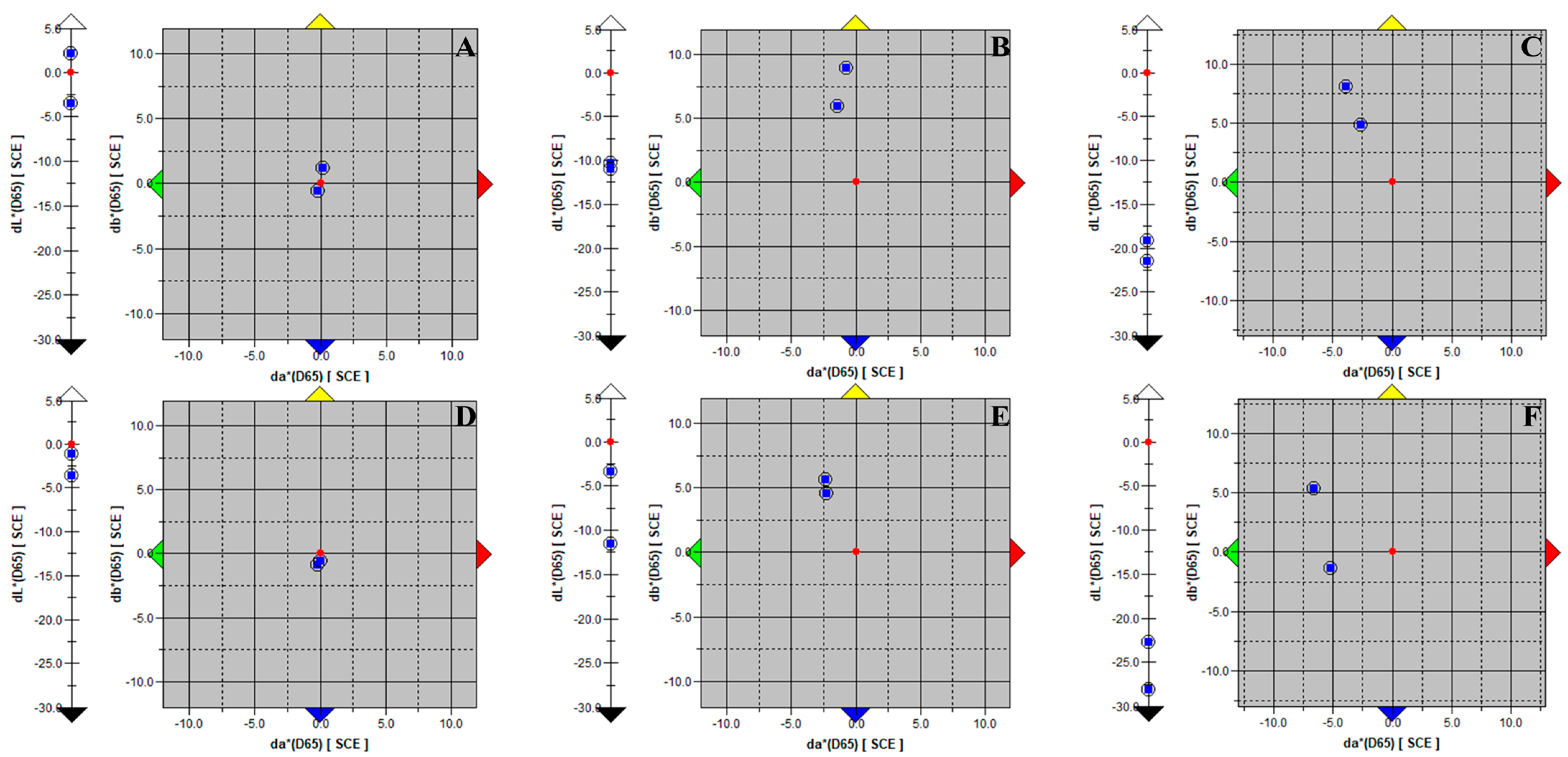
Figure 1. Color change measured for mineral plasters. (A)—mineral plaster (MP) measured after inoculation; (B)—mineral plaster (MP) measured after 14 days of incubation; (C)—mineral plaster (MP) measured after 28 days of incubation; (D)—mineral plaster with primer and silicone paint (MPGS) after inoculation; (E)—mineral plaster with primer and silicone paint after 14 days of incubation; (F)—mineral plaster with primer and silicone paint after 28 days of incubation. The red dot represents the control sample, the blue dots represent the tested samples; arrows represent shifts in color components. dL* scale represents the achromatic component (lightness of the object); da* axis (green–red) and db* axis (blue–yellow) represent the respective color components.
Applsci 13 08009 g001
Figure 2. Color change measured for silicone plasters. (A)—silicone plaster (S) measured after inoculation; (B)—silicone plaster (S) measured after 14 days of incubation; (C)—silicone plaster (S) measured after 28 days of incubation; (D)—silicone plaster with primer and silicone paint (MPGS) after inoculation; (E)—silicone plaster with primer and silicone paint after 14 days of incubation; (F)—silicone plaster with primer and silicone paint after 28 days of incubation. The red dot represents the control sample, the blue dots represent the tested samples; arrows represent shifts in color components. dL* scale represents the achromatic component (lightness of the object); da* axis (green–red) and db* axis (blue–yellow) represent the respective color components.
Applsci 13 08009 g002
Table 1. Factors describing the severity of biofilm growth occurring with incubation time on tested material samples. MP—mineral plaster; MPGS—mineral plaster with primer and silicone paint; S—silicone plaster; SGS—silicone plaster with primer and silicone paint.
Time (d)MPMPGSSSGS
Cell density (cfu/cm2)00.00 × 100
± 0.00 × 100
0.00 × 100
± 0.00 × 100
0.00 × 100
± 0.00 × 100
0.00 × 100
± 0.00 × 100
143.33 × 10⁷
± 9.43 × 10⁶
3.67 × 10⁷
± 4.71 × 10⁶
2.00 × 10⁷
± 9.43 × 10⁶
2.67 × 10⁷
± 9.43 × 10⁶
286.33 × 10⁷
± 4.71 × 10⁶
4.67 × 10⁷
± 1.89 × 10⁷
4.00 × 10⁷
± 9.43 × 10⁶
4.67 × 10⁷
± 9.43 × 10⁶
Chl-a (mg/cm2)01.00 × 10−2
± 1.00 × 10⁻²
0.00 × 100
± 0.00 × 100
0.00 × 100
± 0.00 × 100
0.00 × 100
± 0.00 × 100
144.00 × 10⁻²
± 0.00 × 100
2.80 × 10⁻¹
± 8.00 × 10⁻²
0.00 × 100
± 0.00 × 100
4.00 × 10⁻²
± 0.00 × 10⁻¹
284.40 × 10⁻¹
± 4.00 × 10⁻²
3.30 × 10⁻¹
± 3.00 × 10⁻²
2.10 × 10⁻¹
± 2.00 × 10⁻²
5.00 × 10⁻²
± 1.00 × 10⁻²
ATP (RLU)01.41 × 101
± 4.22 × 100
1.56 × 101
± 2.53 × 100
1.47 × 101
± 1.28 × 100
1.19 × 101
± 5.24 × 100
141.09 × 101
± 7.42 × 101
8.79 × 10²
± 4.60 × 10²
7.15 × 101
± 4.23 × 101
9.33 × 101
± 4.00 × 101
282.58 × 10²
± 9.02 × 101
2.82 × 10²
± 4.91 × 101
7.67 × 101
± 5.76 × 101
1.65 × 10²
± 1.23 × 10²
ΔE (-)02.93 × 100
± 1.03 × 100
2.51 × 100
± 1.67 × 100
1.61 × 100
± 1.91 × 10⁻¹
2.84 × 100
± 1.50 × 100
141.32 × 101
± 7.50 × 10⁻¹
9.60 × 100
± 4.87 × 100
5.97 × 100
± 1.46 × 100
1.02 × 101
± 1.41 × 100
282.16 × 101
± 2.33 × 100
2.65 × 101
± 3.08 × 100
1.21 × 101
± 3.96 × 100
1.20 × 101
± 2.93 × 100
Table 2. Correlation indicators (R2) calculated between results acquired with the tested methods.
Cell Density Chl-aΔEATP
MPCell density 1.0000.7900.9990.9750.00
Chl-a0.7901.0000.7700.9020.20
ΔE0.9990.7701.0000.9670.40
ATP0.9750.9020.9671.0000.60
MPGSCell density 1.0000.9970.7270.3420.80
Chl-a0.9971.0000.6740.9951.00
ΔE0.7270.6741.0000.006
ATP0.3420.9950.0061.000
SCell density 1.0000.7500.6600.812
Chl-a0.7501.0000.6600.318
ΔE0.6600.8301.0000.729
ATP0.8120.3180.7291.000
SSSCell density 1.0000.7460.9330.998
Chl-a0.7461.0000.9970.939
ΔE0.9330.9971.0000.909
ATP0.9980.9390.9091.000
Table 3. Results of the visual evaluation of biofilm growth. 0—no visible photoautotrophic growth; 1—growth visible for less than 10% of the surface; 2—growth occurring on 10 to 30% of the surface; 3—microbial fouling covering from 30 to 50% of the sample surface; 4—more than 50% of surface area covered with biofilm.
Substrate Type0 Days14 Days28 Days
MP111333444
MPGS111333444
S111333444
SGS111333444
Table 4. Visual assessment and spectrophotometric color change results of test samples incubated for 28 days with the M1 and M2 incubation methods.
M1 M2
Colonized AreaΔEColonized AreaΔE
MP185–90%15.46 ± 4.3615–20%8.55 ± 3.73
285–90%10–15%
380–85%80–85%
490–95%5–10%
MPGS170–75%8.87 ± 4.370–1%3.35 ± 2.85
265–70%1–5%
330–35%10–15%
455–60%5–10%
S130–35%5.36 ± 1.605–10%3.50 ± 1.05
220–25%1–5%
335–40%1–5%
425–30%5–10%
SGS115–20%6.81 ± 1.471–5%3.84 ± 1.32
25–10%0–1%
35–10%1–5%
410–15%5–10%
Disclaimer/Publisher’s Note: The statements, opinions and data contained in all publications are solely those of the individual author(s) and contributor(s) and not of MDPI and/or the editor(s). MDPI and/or the editor(s) disclaim responsibility for any injury to people or property resulting from any ideas, methods, instructions or products referred to in the content.