Article

Infrared Spectroscopy to Assess Manufacturing Procedures of Bone Artefacts from the Chalcolithic Settlement of Vila Nova de São Pedro (Portugal)

by
David Gonçalves
1,2,3,
Joana Rosa
2,3,4,5,
Ana L. Brandão
4,
Andrea Martins
6,7,8,
César Neves
7,8,
Mariana Diniz
6,8,
José M. Arnaud
8,
Maria Paula M. Marques
4,5,* and
Luís A. E. Batista de Carvalho
4
1
Archaeosciences Laboratory, Directorate General Cultural Heritage (LARC/CIBIO/InBIO), 1349-021 Lisbon, Portugal
2
Center for Functional Ecology, Department of Life Sciences, University of Coimbra, 3000-456 Coimbra, Portugal
3
Research Center for Anthropology and Health, Department of Life Sciences, University of Coimbra, 3000-456 Coimbra, Portugal
4
Molecular Physical-Chemistry R&D Unit, Department of Chemistry, University of Coimbra, 3004-535 Coimbra, Portugal
5
Department of Life Sciences, Faculty of Science and Technology, University of Coimbra, 3000-456 Coimbra, Portugal
6
UNIARQ Centre for Archaeology, School of Arts and Humanities, University of Lisbon, Alameda da Universidade, 1600-214 Lisboa, Portugal
7
FCT/Portuguese Foundation for Science and Technology, 1249-074 Lisboa, Portugal
8
AAP/Association of Portuguese Archaeologists, Largo do Carmo, 1200-092 Lisboa, Portugal
*
Author to whom correspondence should be addressed.
Appl. Sci. 2023, 13(14), 8280; https://doi.org/10.3390/app13148280
Submission received: 17 May 2023 / Revised: 21 June 2023 / Accepted: 11 July 2023 / Published: 18 July 2023

Abstract

:
Vibrational spectroscopy was applied to study cylindrical engraved bone boxes from the Chalcolithic settlement of Vila Nova de São Pedro (VNSP, Azambuja, Portugal) which has the largest and richest artefact assemblage of Copper Age Western Iberia. The objectives were to reconstitute manufacturing techniques, determine the role of pyrotechnology in the production of cylindrical engraved bone boxes and assess oxygen conditions during burning. Four fragments of cylindrical engraved bone “boxes” from VNSP were used in this research. Anaerobic experimental burn conditions were recreated by using a home-made steel airtight chamber under vacuum. Human bone fragments were burnt at 400–1000 °C for 120–211 min. Fourier-transform infrared spectroscopy analyses were performed on bone powder samples. The resulting spectra and chemometric indices were used as a reference to establish comparisons with the archaeological artefacts. None of these presented spectral features compatible with anaerobic burning. Therefore, aerobic burns were used to achieve the whitish look and were most probably used to attain the darker shade displayed by the artefacts. Artefact manufacturing appears to have relied on bone cutting, bone engraving and maybe polishing, followed by heat treatment. The population from VNSP appears to have been highly specialized in the use of fire to work different raw materials.

1. Introduction

Bone industries are a core item among 3rd millennium cal BC productions and integrate some of the most exquisite pieces of the period but are also simple and poorly transformed tools. However, opposite to lithic or pottery remains, bone artefacts are rarely preserved in the archaeological record since soil acidity tends to lixiviate organic materials. The walled chalcolithic site of Vila Nova de São Pedro (VNSP), settled on a hilltop in the Lower Tagus valley basin in a Miocene-Pontian limestone area, is an outstanding case–study due to environmental conditions that allow bone preservation but also because VNSP possesses the largest and richest artefact assemblage of Copper Age Western Iberia [1]. This settlement played a core role as a production, consumption, and redistribution centre during the third millennium cal BC which explains the largest concentration of bone, pottery, flaked/polished stone tools and copper artefacts retrieved at the site [2,3,4,5,6]. At VNSP, artefacts production depended on local (e.g., limestone, clay, flint) and non-local raw materials (e.g., copper, amphibolite) and the use of controlled fire was mandatory to transform clay into pottery, to melt copper and to heat flint to optimize the debitage process [7].
However, the production of bone utensils is not dependent on fire and Chalcolithic bone industries are usually made upon raw bones and, even in highly transformed pieces such as bone pins or idols/pendants, raw material curation usually does not go beyond the carving and polishing of the bone surfaces. Exceptionally, at VNSP, fire seems to have been involved in the bone industry to produce outstanding pieces from an aesthetic and manufacturing point of view. All required stages of the chaîne opératoire to produce bone boxes are recorded at the site and the sharp chromatic contrast between white and black shiny bone surfaces was intended to highlight some of the artefacts as the ones here described. Belonging to the typological class of small “cylindrical engraved bone/ivory boxes” [8]—even if both sides of the box are opened and require an organic material to be closed—the VNSP minimum number of 45 elements, cylindrical bones “boxes” (MNI), stand as the largest collection in the Iberian Peninsula. Examples of reconstructed bone boxes from VNSP are presented in Figure 1. The functionality of these small cylindrical engraved bone “boxes” measuring 4–7 cm in width, 4–8 cm in diameter and 0.5–1 cm in thickness is not clear although some considered it as “cosmetic boxes” [8].
Made after the long bones of large mammals (bovids or equids), cylindrical engraved bone “boxes” have been recorded for the third millennium cal BC in domestic and, more frequently, in funerary contexts from the Portuguese Lower Estremadura, being usually recovered in very small numbers. The VNSP assemblage is so far exceptional attending to its proportion, to the fact that all stages of production are recorded at the site but also due to crafts procedures. The potential use of fire to change bone colour and thus the final appearance of the finished product has not been recorded in other cylindrical engraved bone boxes from Lower Estremadura sites. According to published data, no significant colour changes are displayed by the latter artefacts.
Four fragments of cylindrical engraved bone “boxes” retrieved during the Afonso do Paço’s archaeological excavations were selected to attempt to understand how fire was potentially used to manufacture these prestigious artefacts.
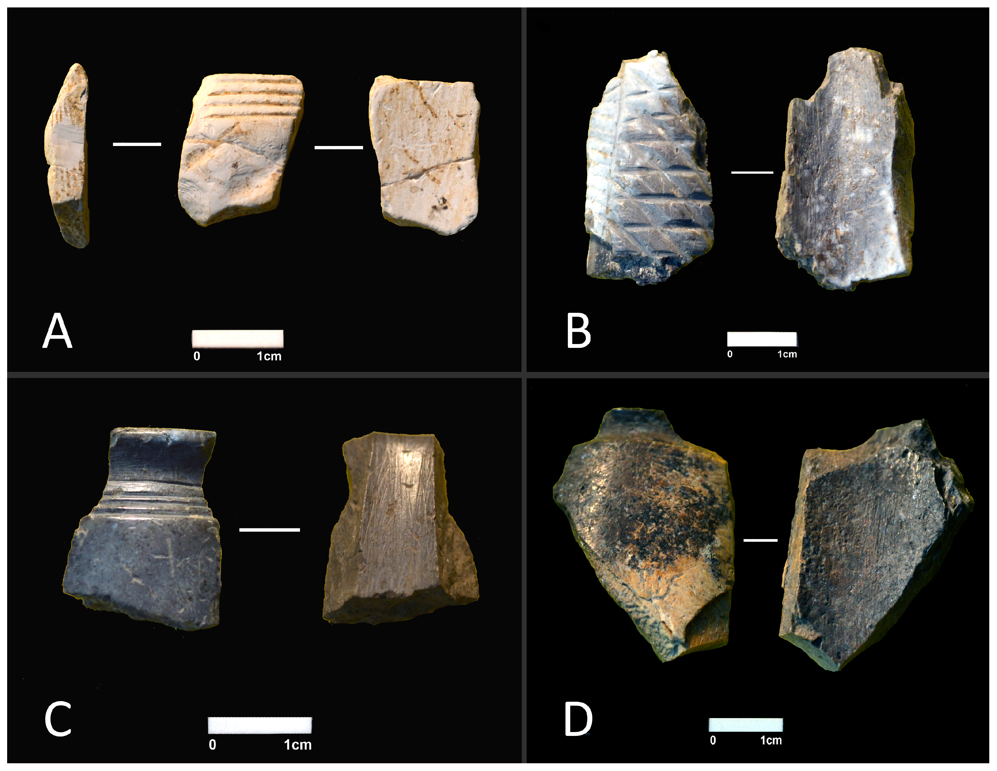
The manufacturing of these bone artefacts involved carving and, apparently, heat exposure. However, if fire was used, the order in which those tasks were accomplished is unknown. One of the bone pieces— [19-23 AP C16] —appears to be only partially burnt pointing to the possibility that the bone was initially carved and only then subjected to a heat treatment leading to a dark shade (Figure 2D). To validate this hypothesis, it is necessary to confirm that the bone is indeed burnt. However, determining whether a bone is burnt is not always easily achieved based on macroscopic observations since other taphonomic processes may mimic heat-induced changes. Weathering, soil staining, manganese oxide as well as other agents may imitate heat-induced colours displayed by charred and calcined bone [9,10,11,12,13]. Therefore, the confirmation of a bone’s burnt condition benefits from the application of other methods of analysis. In the case of specimen [19-23 AP C16], such validation would enlighten us about the technique used in the manufacturing of the bone artefact.
If heat exposure is confirmed, another important question associated with the VNSP artefacts regards the environmental conditions during the burning event. Some of the artefacts display a white colour typical of calcined bone under aerobic conditions. However, several other artefacts show a black colouration which can basically be achieved through two different heat exposure procedures: either by submitting bone to medium temperatures (ca. 300–500 °C) [14,15,16,17,18] or by reducing the oxygen available during the burn, as inferred from the work of Snoeck et al. [19], Reidsma et al. [20] and Marques et al. [21].
Snoeck et al. [19] argued that oxygen reducing conditions may explain the incorporation of cyanamide into burnt bone leading to incomplete oxidization of organic matter. According to Habelitz et al. [22], the formation of cyanamide results from NH3 released during the burn of organic matter under reducing conditions. Based on recent neutron spectroscopy data obtained for bones burnt under anaerobic conditions (sealed vs. unsealed containers), cyanamide was detected above 650 °C [21]. Therefore, this reaction must occur immediately, at the surface, and soon after the formation of NH3. Otherwise, the latter would have been removed through evacuation. More recently, Marques et al. [21,23] observed that cyanamide formation—in the presence of free ammonia formed during the burning process—is associated with incomplete oxidation of organic matter leading to incorporation of CN22− ions into bone’s inorganic matrix, substituting for OH and yielding cyanamidapatite (Ca10(PO4)6CN2). This is reflected by distinctive infrared features from CN22−: at 700 cm−1 (N-C≡N bending) and 2009 cm−1 (C≡N stretching). The presence of cyanamide in the infrared spectra can thus be used to identify burns under reducing conditions, possibly exchanging with hydroxyl groups in apatite.
The work of Reidsma et al. [20] with bones burnt under anaerobic conditions suggests another possible useful feature—OH groups. These were absent from their infrared spectra of bone burnt at 900 °C (bands 630 cm−1; 3572 cm−1; 3642 cm−1) although they are usually present in bones burnt under aerobic conditions [19,24,25].
In this paper, we used Fourier-transform infrared spectroscopy in attenuated total reflectance mode (FTIR-ATR) with the objective of investigating the issues described above. This technique has been routinely applied to the study of bone and is cost-effective, rapid, simple and non-destructive, requiring only a few mg of bone powder [26]. By contrast to solid-state NMR which yields overall information on bone, for each active nucleus probed [27,28,29], infrared spectroscopy is able to provide comprehensive data on bone’s chemical composition and structure simultaneously, in a straightforward and rapid manner, which renders it ideal for a routine analysis of the effect of heat on these types of samples. This investigation encompasses two different stages: (i) the first one documenting anaerobic conditions in experimentally burnt human samples at different maximum temperatures, following a previous work from Marques et al. [21]; (ii) the second one involving the FTIR-ATR analysis of selected archaeological artefacts from VNSP, to be compared against the experimental samples used here as a reference. The outcome of this research can be viewed as a contribution to the reconstitution of the manufacturing techniques of the population from the VNSP settlement as well as to a better understanding of their pyrotechnical skills.

2. Materials and Methods

Cortical bone sections (2 cm × 2 cm) from the femur and tibia of unidentified human skeleton CC_NI_42 were used for the experimental burns. This belongs to the collection of unidentified skeletons of the Laboratory of Forensic Anthropology of the University of Coimbra which were donated for research purposes. They come from the Capuchos cemetery of Santarém (Portugal). This is the same provenance of the identified skeletons comprising the 21st century identified skeletal collection [30,31]. The individuals were buried for at least three years, which is the minimum required period of inhumation time prior to exhumation according to Portuguese legislation (Decreto-Lei 411/98). Therefore, some degree of unspecified diagenetic impact on the skeletal remains is to be expected. The age at death and sex of individual CC_NI_42 are unknown. An investigation carried out on these Coimbra collections has been approved by the Ethics Committee of the Faculty of Medicine of the University of Coimbra (reference number: CE_026.2016).
It should be noted that the VNSP artefacts were most probably manufactured with non-human bones since, as far as it is known, human bones were not used as raw materials during the Chalcolithic. Nonetheless, no significant differences between infrared spectra of human and non-human burnt bone have been found so far. Furthermore, chronological differences do not appear to result in spectral differences as well [14,19,24,32,33,34,35].
Anaerobic conditions were implemented by placing each fragment of femur and tibia into a home-made steel airtight chamber, under vacuum (Figure 2A of Marques et al. [21]). This chamber was in turn placed inside an electric muffle (Barracha K3, three-phased) equipped with a type K probe (negative: nickel-aluminium, positive: nickel-chrome) following norm IEC 60584-2. The following maximum temperatures were implemented for the femoral samples: 400 °C (120′), 500 °C (120′), 550 °C (120′), 600 °C (120′), 650 °C (120′), 700 °C (120′), 750 °C (120′), 800 °C (120′), 900 °C (178′) and 1000 °C (211′). Due to insufficient availability of bone, the 550 °C, 600 °C, 650 °C, 750 °C and 800 °C experiments were not carried out for the tibia. Experiments at those temperatures had to be repeated due to the malfunction of the vacuum container. However, only femoral samples were still available for the second trial and are therefore presented in full. Results for the tibia are presented only to corroborate the results obtained on the femur. The burns took 2 to 3.5 h, after which the muffle furnace was switched off and samples were then allowed to cool down to room temperature.
Bone powder samples were collected with a scalpel from each experimentally burnt bone section. The most superficial layer of bone was discarded, and sampling targeted the subsurface level.
The FTIR-ATR spectra of the bone powder samples were acquired in the mid-IR interval (400–4000 cm−1), in a Bruker Optics Vertex 70 FTIR spectrometer purged by CO2-free dry air and equipped with a Bruker Platinum ATR single reflection diamond accessory. A Ge on KBr substrate beamsplitter and a liquid nitrogen-cooled wide band mercury cadmium telluride (MCT) detector were used. The frequency dependence of the penetration depth of the electric field in ATR was corrected through an algorithm from the Opus 8.1 software (using a mean refractive index of 1.25). A 2 cm−1 resolution, 128 scans per spectrum, and the 3-term Blackman–Harris apodization function were applied. Baseline correction and normalization (to the ν3(PO4) band at 1030 cm−1) were carried out. This allows reliable comparison of data despite possible differences regarding sample consistency.
Chemometric indices were calculated for all samples from experimental and archaeological materials. A description of those indices is given in Table 1. A description of the archaeological artefacts is given in Table 2.

3. Results and Discussion

3.1. Experimental Analysis

All experimentally burnt bone sections displayed a charred appearance regardless of the maximum temperature of exposure, as opposed to the usual heat-induced colour sequence documented for aerobically burnt bone—natural colour to darker colour to whitish colour (e.g., Walker et al. [18]).
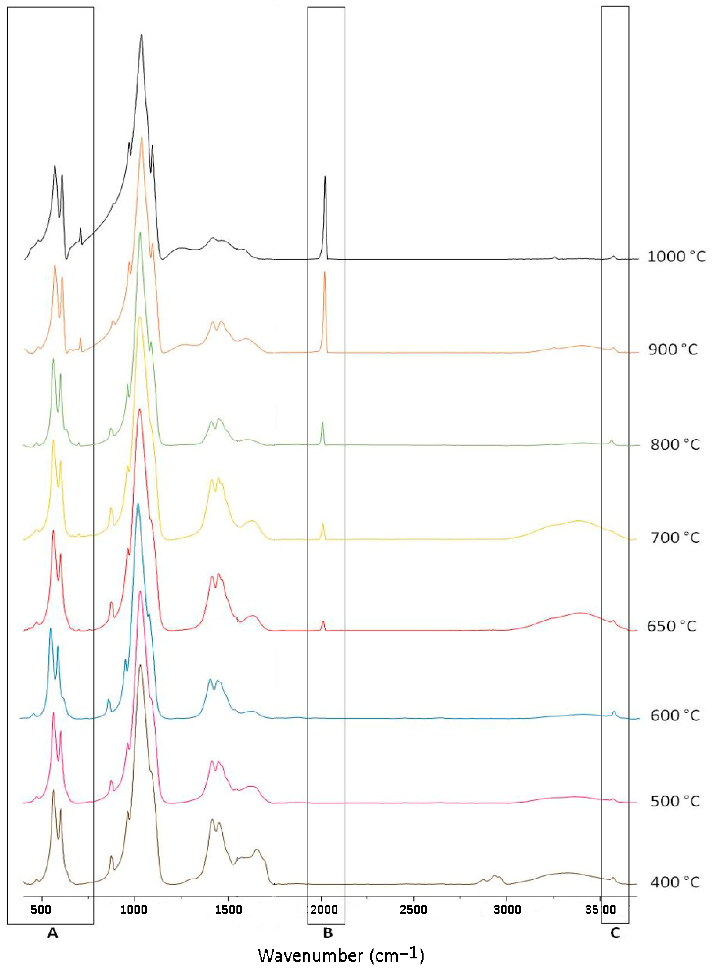
Infrared spectra for the experimentally burnt femur are given in Figure 3. Both presented similar basic features, the most noteworthy being:
  • the absence of bands for the OH libration at ~630 cm−1 (Figure 3A), in clear contrast to the sharp peak usually observed in bone burnt aerobically at temperatures above 600 °C [21,25,34,35,38]. Possibly, this is explained by the OH libration being a lattice mode which is visible only when the system has other neighbouring OH groups (within a network). When considerable CN22− for OH substitutions take place, only the OH stretching (at ca. 3500 cm−1) is visible.
  • the OH stretching band at ~3572 cm−1 was clearly present in spectra from bones burnt at all investigated temperature thresholds (Figure 3C). The spectra reported by Reidsma et al. [20], showed no OH stretching under anaerobic conditions which is, contrastingly, a frequent observation in aerobically burnt bones (Figure 4). This suggests that some variability may be expected as well for the intensity of this feature or, that it instead reflects burns at shorter durations (60 min against the 120–211 mins durations used in the present study);
  • progressively more intense bands from CN22− were present at ca. 700 cm−1 (N-C≡N bending, Figure 3A) and 2009 cm−1 (Figure 3B, C≡N stretching) in the spectra of bones burnt at temperatures above 650 °C. Cyanamide formation has been previously reported to be due to incomplete oxidation of organic matter [39], leading to the incorporation of CN22− ions into bone’s inorganic framework substituting for OH and yielding cyanamidapatite (Ca10(PO4)6CN2) [19,20,21,22,23];
  • additionally, between 600 and 700 °C, the formation of NH3/NH4+ was observed which resulted in broad signals at ca. 1700 and 3300 cm−1 due to NH deformation and stretching modes, respectively. As the temperature increased, more and more cyanamide was formed (from ammonia) and the characteristic NH signals gave way to those from cyanamide.
Results for the calculation of the spectroscopic indices measured for the bone samples are comprised in Table 3. The crystallinity index (CI) of bones burnt under both aerobic conditions—obtained from Gonçalves et al. [24] and Snoeck et al. [19]—and anaerobic settings (in a sealed chamber) presented somewhat similar values for temperature thresholds up to 600 °C. However, differences became much larger at higher temperatures, with anaerobically burnt bone presenting values considerably smaller than those obtained for aerobically burnt bone. As for the amount of B-type carbonates (BPI), it was always higher for anaerobically burnt bone, especially at the highest temperatures, with values considerably larger than the values obtained for aerobically burnt bone.
In the case of the C/C ratio, values were quite similar between the two settings for temperature thresholds up to 700 °C, revealing a similar intensity of the features allocated to the dividend (Carbonates A + Carbonates B & collagen + lipids) and to the divisor (B-type carbonates) thus leading the value to be approximate to one, on average. At higher temperatures (at 900 °C and 1000 °C), this remained to be the case for anaerobically burnt bones but, for aerobically burnt bones, B-type carbonates were proportionately less intense leading to higher C/C ratios. Apparently, B-type carbonates were not lost in anaerobically burnt bones as much as in the case of their aerobic counterparts [21,40]. This is coherent with the differential heat-related degradation of organic components in anaerobic and aerobic burns, which appears to take place at considerably lower temperatures for the latter [21]. However, interpretations must be carried out with caution, since a C/C of 1.22 was obtained at 800 °C which was an abnormal index given the much smaller values obtained for all other temperatures, both lower and higher. The appearance of a shoulder at 630 cm−1 may suggest that we failed to completely seal the chamber, and therefore the burning process may not have been totally anaerobic.
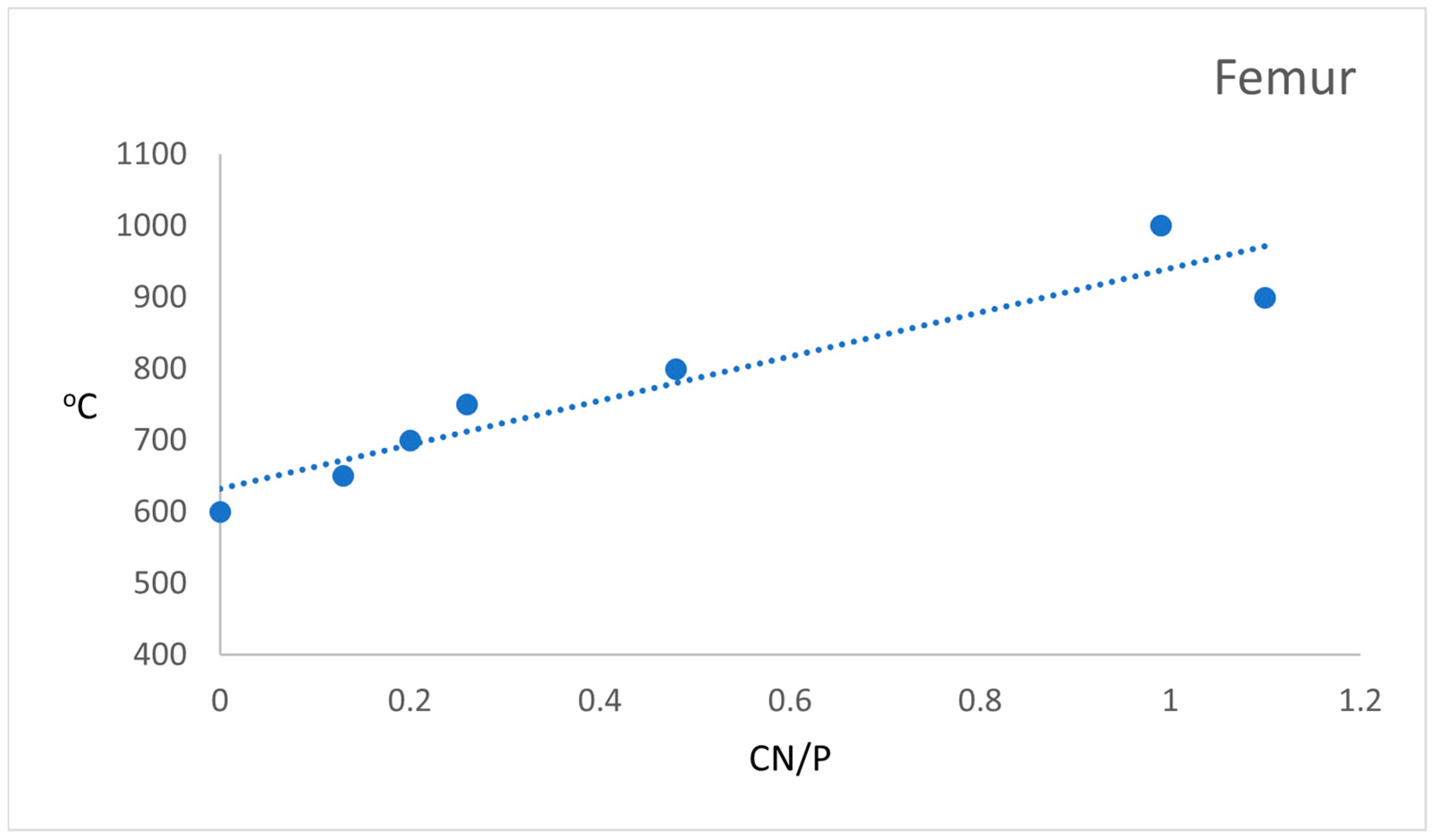
Regarding the amount of cyanamide, this finding has seldom been reported in aerobically burnt bones. Using the CN/P ratio, Snoeck et al. [19] reported the presence of this organic compound in bones burnt at 800 °C. To that purpose, these authors [19] took only into account values above 0.25. Therefore, it was not a consistent find during their experimental work, contrary to what was observed in our own anaerobically burnt bones experiments. This was especially clear for bones burnt at higher temperatures which corroborated the results of Marques et al. [21]. Cyanamide was present in all samples burnt at temperatures equal to or higher than 700 °C and this band became progressively more intense along with increasing temperature suggestive of a correlation between these two variables (Figure 5). At 900 °C and 1000 °C, it became even more intense than the 605 cm−1 phosphate band (Figure 3), which is reflected in the CN/P ratios higher than 1.
If the bone heat-induced changes under anaerobic conditions depicted here are demonstrated to be recurrent, spectral changes may then be able to discriminate between aerobically and anaerobically burnt bone. Summing up our results, the presence of anaerobically burnt bones can be suspected if bone presents a dark colouration, a high CI value (>4.00), the absence of an OH libration infrared band and intense bands associated with the presence of cyanamide (700 cm−1, 2010 cm−1). These four criteria are especially helpful in the case of bones burnt anaerobically (in a sealed environment) at relatively high temperatures (≥800 °C), since those burnt at lower temperatures may present characteristics somewhat similar to bones burnt aerobically in the same temperature range.
For bone samples subjected to low temperatures (<ca. 200 °C) the characteristic water stretching may explain the large band found at ca. 3400 cm−1, so caution is advisable when interpreting the infrared spectra. In fact, this may occur in bones burnt at higher temperatures that have been exposed to high moisture conditions, since water may then adsorb to the bone surface. Both hydration and dehydration are frequent occurrences in inhumed materials and are prone to affect the corresponding infrared profile [41,42]. The same can be said for B-type carbonates [43] thus rendering the usefulness of the BPI and C/C ratios arguable. Of course, it is not possible to state that taphonomy does not affect the four abovementioned criteria. For example, as previously discussed, bone discolouration can be caused by multiple taphonomic factors [9,10,11,12,13] and the CI is known to increase with post-depositional time [44]. Therefore, the interpretation of archaeological materials must have these factors in consideration.
Table 3. Values of the chemometric indices obtained for the experimental burnt femur samples and for the artefacts of the Vila Nova de São Pedro settlement. The aerobic values for the crystallinity index (CI), the amount of B-type carbonates (BPI), and the Carbonates A + Carbonates B to the highest peak of region 1445–1470 cm−1 (C/C) refer to the mean values recorded for the experimentally burnt samples from long bones mid-diaphyses used in Gonçalves et al. [24]. For the amount of cyanamide (CN/P), reference for the anaerobic burnt samples was collected from the work of Snoeck et al. [19] For the features referring to the libration (630 cm−1) and stretching (3572 cm−1) of hydroxyl groups, data from the dataset used in Gonçalves et al. [45] was used.
IndexBone
Type
400 °C500 °C550 °C600 °C650 °C700 °C750 °C800 °C900 °C1000 °C[19-27 AP C16][19-26 AP C16][19-11 AP C16][19-23 AP C16] Lighter Region[19-23 AP C16] Darker Region
CIAn F3.853.873.533.663.533.513.423.874.814.30-----
An T3.953.85---3.42--4.904.69
Ae3.533.68-3.75-4.33-5.525.695.34-----
Ar----------5.074.583.723.203.56
BPIAn F0.860.580.730.640.710.770.850.430.420.26-----
An T0.730.50---0.85--0.420.28
Ae0.670.50-0.45-0.35-0.240.140.11-----
Ar----------0.120.260.511.590.88
C/CAn F0.950.990.980.991.041.031.041.221.030.88-----
An T0.961.01---1.04--1.000.94
Ae0.991.04-1.03-1.03-1.261.271.27-----
Ar----------0.910.980.930.870.90
Cn/PAn F----0.130.200.260.451.100.99-----
An T-----0.20--1.160.96
Ae *-0.13
0.17
-0.10
0.17
-0.10
0.23
-0.09
0.54
0.10
0.18
------
Ar----------AbsAbsAbsAbsAbs
630 cm−1An FAbsAbsAbs.Abs.Abs.AbsAbsAbsAbsAbs-----
An TAbsAbsAbs.Abs.Abs.AbsAbsAbsAbsAbs-----
AeAbsAbs-Abs Pres PresPresPres-----
Ar----------PossibPresPossibAbsAbs
3572 cm−1An FPresPresPresPresPresPresPresPresPresPres-----
An TPresPresPresPresPresPresPresPresPresPres-----
AePossibPossib-Possib-Pres-PresPresPres-----
Ar----------AbsPresPresAbsPossib
Key: An = Anaerobic; Ae = Aerobic; F = Femur; T = Tibia; Ar = Artefact; Abs = Absent; Pres = Present; Possib = Possibly present. * Values were obtained from Snoeck et al. [19].

3.2. Analysis of Archaeological Artefacts

3.2.1. Artefact [19-27 AP C16]

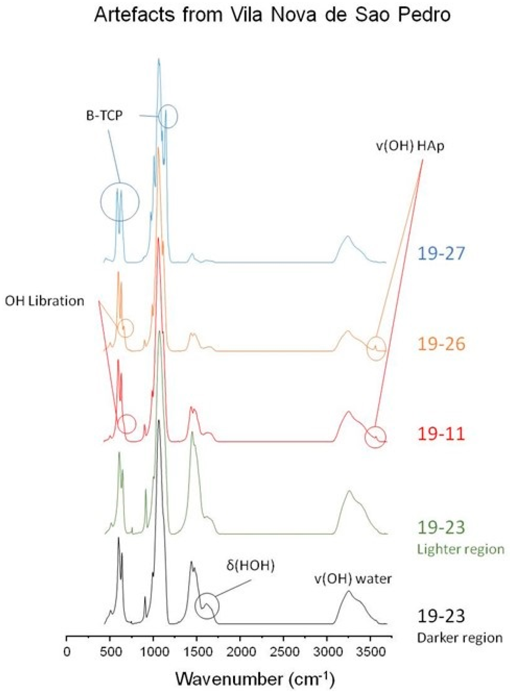
This artefact presented a white colouration typical of calcined bone (Figure 2A). Its infrared profile confirmed that the bone material was exposed to very high temperatures, as is also reflected by a high CI value (Figure 6). The OH libration and stretching bands are not evident but the spectrum revealed the presence of beta-tricalcium phosphate (β-TCP) which often affects the usual OH librational band at 630 cm−1 and results in an intense peak at ca. 1123 cm−1 [38]. This new bone mineral phase is usually produced at temperatures between 700 and 1100 °C [46]. In addition, no cyanamide was present. The absence of organic matter (no amide I and II bands were detected, at 1660 cm−1 and 1550 cm−1, respectively) was consistent with a high-temperature burn. Therefore, evidence points to an aerobic burning of artefact [19-27 AP C16] at temperatures above 700 °C, possibly even at a much higher temperature than this.

3.2.2. Artefact [19-26 AP C16]

This artefact presented a mixed colouration of black, grey and white shades (Figure 2B) which usually reflects a heat-induced transition stage between charred and calcined bone [18,47]. The infrared spectrum indicated this artefact has been burnt to a relatively high temperature given the presence of bands from hydroxyapatite’s OH libration and stretching (Figure 6). No clear signs of amide I and amide II were detectable. Again, no cyanamide was present. Evidence thus points to an aerobically burnt artefact at relatively high temperatures but lower than that corresponding to artefact [19-27 AP C16].

3.2.3. Artefact [19-11 AP C16]

This artefact showed a homogeneous black colouration (Figure 2C) which, when caused by heat, is typically associated with either an aerobic exposure to medium temperatures of 300–500 °C [18,47] or to an anaerobic exposure above 340 °C [20]. The infrared profile presented an almost undetectable shoulder (at 630 cm−1) probably ascribed to the hydroxyapatite’s OH libration, and a clear signal from the OH stretching at 3572 cm−1 (Figure 6). No clear signs of amide I, amide II or cyanamide were detected. The CI value was very low and therefore consistent with both aerobic burns at temperatures < 700 °C and anaerobic burns at temperatures < 600 °C. The infrared profile thus presented characteristics that can be found in bone burnt under either aerobic or anaerobic conditions, preventing an unequivocal identification of the burning environment.
Additionally, traces of adsorbed water were evident in the infrared profile of all the probed archaeological artefacts, at ca. 1620–1650 cm−1 (δ(HOH)) and 3300–3500 cm−1 (ν(OH)). Moreover, these samples showed features due to amine groups at ca. 1600 cm−1 (δ(NH2)) and 3200 cm−1 (ν(NH)), suggestive of contamination (e.g., from the soil or water at the archaeological site).

3.2.4. Artefact [19-23 AP C16]

As described above, this fragmented artefact presented a heterogeneous colouration composed of a light brownish/yellowish area and of a darker scorched area (Figure 2D). As mentioned above, the latter can result from both aerobic and anaerobic burns. The heat-induced brownish lighter shade can be the result of low intensity aerobic burns up to 200 °C [18,47]. The infrared profiles of samples from both regions revealed the minor heat-prompted effects on bone’s composition—small CI values (substantially smaller for the lighter region which can suggest a low intensity heat exposure), preservation of amide II (not clearly seen for the lighter region probably due to the considerable amount of carbonates that yield IR bands in nearby wavelengths [25], and apparent absence of bands from OH groups (although a small peak in the OH stretching region may be present). In addition, no presence of cyanamide was detected. Based on the features of the bone, this was most probably exposed to an aerobic heat exposure of low intensity.

4. Conclusions

This study is an example of how infrared spectroscopy analyses may contribute to the study of the chaîne opératoire of artefacts pyrotechnologically manufactured. Our results confirmed that the people from Vila Nova de São Pedro used fire during the manufacturing of engraved artefacts thus revealing a comprehensive knowledge of the effect of heat on bone. It can be confidently stated that aerobic burns were used to achieve the whitish look of some of the artefacts. Therefore, furnaces yielding aerobic burns were carefully monitored and artefacts were masterly handled to give the desired appearance to bone artefacts.
The use of anaerobic burns to provide a darker shade to artefacts cannot be discarded entirely but the absence of clear cyanamide bands leads us to suggest that, at least for the assemblage studied here, reducing conditions were not used. However, it is important to note that it is difficult to know how similar the conditions under which our experiments took place are to those that would have been implemented by the pyrotechnicians from Vila Nova de São Pedro. Admittedly, our anaerobic conditions were more hermetically sealed than those experienced in the third millennium cal BC. Nonetheless, the differences in the two anaerobic burning environments—experimental versus real scenario—would possibly not have led to two radically contrasting spectra given that Marques et al. [21] observed similar profiles in spectra from samples burnt anaerobically in both sealed-tight and partially sealed (i.e., allowing volatiles venting partially) chambers.
The chaîne opératoire of the artefact manufacturing appears to have relied on bone cutting, bone engraving and maybe polishing, followed by heat treatment. The finishing touch will also have been polishing. This sequence is testified by artefact [19-23 AP C16] which appears to refer to the result of incomplete work. Apparently, the artefact underwent fracture during heat exposure and was immediately discarded, thus demonstrating that the burning process was carefully monitored. The procedure thus clearly incorporated some risk—as shown by the above mentioned artefact—since heat could potentially cause the fracturing of the artefacts which meant that the time invested in the engraving could be utterly lost. The choosing of this sequence is probably related to the fact that manufacturing a bone artefact using the inverse sequence, i.e., carving after heat exposure, would be much more difficult to achieve, as bone loses mechanical strength when submitted to heat [48] thus rendering carving and polishing an even riskier affair.
This study confirms the hypothesis of white or black and brown colours in the engraved bone boxes being intentionally attained by a clearly expert craftsperson, thus contributing to its exceptional social value. If large mammal long bones from where bones “boxes” were cut were a common material at Chalcolithic sites, the rarity of these engraved bone “boxes” and the unique chromatic appearance of the VNSP objects should be highlighted.
The [19-23 AP C16] fragment supports the previous assumption that VNSP was a highly specialized workshop for many different raw materials strongly connected to fire use. The added value of pyrotechnology was incorporated in the production of these artefacts. Their significance was highly treasured, given the complex manufacturing procedures and transformations involved, as currently demonstrated by infrared spectroscopy. In the Iberian Peninsula, Chalcolithic societies underwent a complexification process largely dependent upon crafts specialization with a view to producing prestige items manipulated only by emergent elites. FTIR-ATR is another methodological tool able to measure material culture production procedures, which is crucial in this debate.

Author Contributions

Conceptualization, D.G., M.P.M.M. and L.A.E.B.d.C.; methodology, D.G., M.P.M.M. and L.A.E.B.d.C.; validation, D.G., M.P.M.M. and L.A.E.B.d.C.; formal analysis, D.G., J.M.A., M.D., A.M., C.N., M.P.M.M. and L.A.E.B.d.C.; investigation, J.R., A.L.B., M.P.M.M. and L.A.E.B.d.C.; resources, J.M.A., M.D., A.M., C.N., M.P.M.M. and L.A.E.B.d.C.; data curation, M.P.M.M. and L.A.E.B.d.C.; writing—original draft preparation, D.G., M.D., A.M., C.N. and M.P.M.M.; writing—review and editing, D.G., M.D., A.M., C.N., M.P.M.M. and L.A.E.B.d.C.; visualization, D.G., A.M., C.N., M.P.M.M., L.A.E.B.d.C.; project administration, D.G.; funding acquisition, L.A.E.B.d.C. All authors have read and agreed to the published version of the manuscript.

Funding

This work was developed within the Molecular Physical-Chemistry R&D Unit (UIDB/00070/2020 & UIDP/00070/2020) financed by the Portuguese Foundation for Science and Technology (FCT). This work was financed by Portuguese funds through FCT—Portuguese Foundation for Science and Technology in the framework of the projects UIDB/00698/2020 and UIDP/00698/2020. This work was also supported by the Portuguese Foundation for Science and Technology through the individual grant 2020.06253.BD.

Institutional Review Board Statement

The investigation carried out on the 21st century identified skeletal collection has been approved by the Ethics Committee of the Faculty of Medicine of the University of Coimbra (reference number: CE_026.2016).

Informed Consent Statement

Not applicable.

Data Availability Statement

The data presented in this study are available on request from the corresponding author.

Acknowledgments

M. Teresa Ferreira and T. Coutinho are acknowledged for their support in sample selection and preparation.

Conflicts of Interest

The authors declare no conflict of interest.

References

  1. Arnaud, J.M. Vila Nova de São Pedro revisitada. In Construindo a Memoria—As Colecções do Museu Arqueológico do Carmo; Arnaud, J.M., Fernandes, C.V., Eds.; Associação dos Arqueólogos Portugueses: Lisboa, Portugal, 2005; pp. 141–164. [Google Scholar]
  2. Müller, R.; Soares, A.M. Traces of Early Copper Prodution at the Chalcolitic Foritification of Vila Nova de São Pedro (Azambuja, Portugal). Madr. Mitt. 2008, 49, 94–114. [Google Scholar]
  3. Pereira, F.; Silva, R.J.; Soares, A.M.; Araújo, M.F. The role of arsenic in Chalcolithic copper artefacts e insights from Vila Nova de São Pedro (Portugal). J. Archaeol. Sci. 2013, 40, 2045–2056. [Google Scholar] [CrossRef]
  4. Diniz, M.; Martins, A.; Neves, C.; Arnaud, J.M. Vila Nova de São Pedro (Azambuja), no 3° milénio, um sítio calcolítico no ocidente peninsular–contributos para um debate. In Arqueologia em Portugal–2017, Estado da Questão; Arnaud, J.M., Martins, A.C., Eds.; Associação dos Arqueólogos Portugueses: Lisboa, Portugal, 2017; pp. 591–604. [Google Scholar]
  5. Martins, A.; Diniz, M.; Neves, C.; Arnaud, J.M. The symbolic in Vila Nova de São Pedro: Idols, statues and symbology. In Mobile Images of Ancestral Bodies: A Millennium-Long Perspective from Iberia to Europe–Zona Arqueológica, n° 23; Bueno Ramírez, P., e Soler Diaz, J., Eds.; Museu Arqueológico Regional: Alcala de Henares, Spain, 2021; Volume II, pp. 121–138. [Google Scholar]
  6. Diniz, M.; Martins, A.; Neves, C.; Arnaud, J.M. Where there is Power, there is Fear. Muralhas calcolíticas, medo, poder e mecanismos exibição–o caso de Vila Nova de São Pedro (Azambuja, Portugal). In Romper Fronteiras, Atravessar Territórios. Identidades e Intercâmbios da Pré-História Recente no Interior da Península Ibérica; Sanches, M.J., Barbosa, M.H., Teixeira, J.C., Eds.; CITCEM: Porto, Portugal, 2022; pp. 109–137. [Google Scholar]
  7. Mraz, V.; Fisch, M.; Eren, M.I.; Lovejoy, C.O.; Buchanan, B. Thermal engineering of stone increased prehistoric toolmaking skill. Sci. Rep. 2019, 9, 14591. [Google Scholar] [CrossRef] [Green Version]
  8. Jalhay, E.; Paço, A. El castro de Vilanova de San Pedro. In Actas y Memorias de la Sociedad Espanola de Antropologia: Etnografia y Prehistoria; Museo Antropológico Nacional: Madrid, Spain, 1945; Volume 20, pp. 5–93. [Google Scholar]
  9. Shahack-Gross, R.; Bar-Yosef, O.; Weiner, S. Black-colored bones in Hayorium Cave, Israel: Differentiating between burning and oxide staining. J. Archaeol. Sci. 1997, 24, 439–446. [Google Scholar] [CrossRef]
  10. López-González, F.; Grandal-d’Anglade, A.; Vidal-Romaní, J.R. Deciphering bone depositional sequences in caves through the study of manganese coatings. J. Archaeol. Sci. 2006, 33, 707–717. [Google Scholar] [CrossRef]
  11. Dupras, T.L.; Schultz, J.J. Taphonomic bone changes and color changes in forensic contexts. In Manual of Forensic Taphonomy; Pokines, J.T., Symes, S.A., Eds.; CRC Press: Boca Raton, FL, USA, 2014; pp. 315–340. [Google Scholar]
  12. Junod, C.A.; Pokines, J.T. Subaerial weathering. In Manual of Forensic Taphonomy; Pokines, J.T., Symes, S.A., Eds.; CRC Press: Boca Raton, FL, USA, 2014; pp. 287–314. [Google Scholar]
  13. Pokines, J.T.; Baker, J.E. Effects of burial environment on osseous remains. In Manual of Forensic Taphonomy; Pokines, J.T., Symes, S.A., Eds.; CRC Press: Boca Raton, FL, USA, 2014; pp. 73–114. [Google Scholar]
  14. Shipman, P.; Foster, G.; Schoeninger, M. Burnt bones and teeth: An experimental study of colour, morphology, crystal structure and shrinkage. J. Archaeol. Sci. 1984, 11, 307–325. [Google Scholar] [CrossRef]
  15. Etxeberria, F. Aspectos macroscópicos del hueso sometido al fuego: Revisión de las cremaciones descritas en el País Vasco desde la arqueologia. Munibe 1994, 46, 111–116. [Google Scholar]
  16. Mays, S. The Archaeology of Human Bones, 1st ed.; Routledge: New York, NY, USA, 1998. [Google Scholar]
  17. Wahl, J. Investigations on pre-Roman and Roman cremation remains from southwstern Germany: Results, potentialities and limits. In The Analysis of Burned Remains; Schmidt, C.W., Symes, S.A., Eds.; Academic Press: London, UK, 2008; pp. 145–161. [Google Scholar]
  18. Walker, P.L.; Miller, K.W.P.; Richman, R. Time, temperature and oxygen availability: An experimental study of the effects of environmental conditions on the color and organic content of cremated bone. In The Analysis of Burned Remains; Schmidt, C.W., Symes, S.A., Eds.; Academic Press: London, UK, 2008; pp. 129–137. [Google Scholar]
  19. Snoeck, C.; Lee-Thorp, J.A.; Schulting, R.J. From bone to ash: Compositional and structural changes in burned modern and archaeological bone. Palaeogeogr. Palaeoclimatol. Palaeoecol. 2014, 416, 55–68. [Google Scholar] [CrossRef]
  20. Reidsma, F.H.; van Hoesel, A.; van Os, B.J.H.; Megens, L.; Braadbaart, F. Charred bone: Physical and chemical changes during laboratory simulated heating under reducing conditions and its relevance for the study of fire use in archaeology. J. Archaeol. Sci. Rep. 2016, 10, 282–292. [Google Scholar] [CrossRef]
  21. Marques, M.P.M.; Gonçalves, D.; Mamede, A.P.; Coutinho, T.; Cunha, E.; Kockelmann, W.; Parker, S.F.; Batista de Carvalho, L.A.E. Profiling of human burned bones: Oxidising versus reducing conditions. Sci. Rep. 2021, 11, 1361. [Google Scholar] [CrossRef]
  22. Habelitz, S.; Pascuala, L.; Duran, A. Transformation of tricalcium phosphate into apatite by ammonia treatment. J. Mater. Sci. 2001, 36, 4131–4135. [Google Scholar] [CrossRef]
  23. Marques, M.P.M.; Batista de Carvalho, L.A.E.; Gonçalves, D.; Cunha, E.; Parker, S.F. The impact of moderate heating on human bones: An infrared and neutron spectroscopy study. R. Soc. Open Sci. 2021, 8, 210774. [Google Scholar] [CrossRef] [PubMed]
  24. Gonçalves, D.; Vassalo, A.R.; Mamede, A.P.; Makhoul, C.; Piga, G.; Cunha, E.; Marques, M.P.M.; Batista de Carvalho, L.A.E. Crystal clear: Vibrational spectroscopy reveals intrabone, intraskeleton, and interskeleton variation in human bones. Am. J. Phys. Anthropol. 2018, 166, 296–312. [Google Scholar] [CrossRef] [PubMed]
  25. Mamede, A.P.; Vassalo, A.R.; Piga, G.; Cunha, E.; Parker, S.F.; Marques, M.P.M.; Batista de Carvalho, L.A.E.; Gonçalves, D. Potential of bioapatite hydroxyls for research on archaeological burned bone. Anal. Chem. 2018, 90, 11556–11563. [Google Scholar] [CrossRef]
  26. Mamede, A.P.; Gonçalves, D.; Marques, M.P.M.; Batista de Carvalho, L.A.E. Burned bones tell their own stories: A review of methodological approaches to assess heat induced diagenesis. App. Spectrosc. Rev. 2018, 53, 603–635. [Google Scholar] [CrossRef]
  27. Kolodziejski, W. Solid-State NMR Studies of Bone. Top. Curr. Chem. 2004, 246, 235–270. [Google Scholar]
  28. Duer, M.J. The contribution of solid-state NMR spectroscopy to understanding biomineralization: Atomic and molecular structure of bone. J. Magn. Reason. 2015, 253, 98–110. [Google Scholar] [CrossRef]
  29. Xue, H.; Yin, Y.; He, T.; Song, H.; Li, J.; Kong, X. Solid.state NMR studies on the organic matrix of bone. Nano Res. 2023, 16, 2980–2990. [Google Scholar] [CrossRef]
  30. Ferreira, M.T.; Coelho, C.; Makhoul, C.; Navega, D.; Gonçalves, D.; Cunha, E.; Curate, F. New data about the 21st Century Identified Skeletal Collection (University of Coimbra, Portugal). Int. J. Leg. Med. 2021, 135, 1087–1094. [Google Scholar] [CrossRef]
  31. Ferreira, M.T.; Vicente, R.; Navega, D.; Gonçalves, D.; Curate, F.; Cunha, E. A new forensic collection housed at the University of Coimbra, Portugal: The 21st century identified skeletal collection. Forensic Sci. Int. 2014, 245, 202.e1–202.e5. [Google Scholar] [CrossRef] [Green Version]
  32. Brandão, A.L.C.; Batista de Carvalho, L.A.E.; Gonçalves, D.; Piga, G.; Cunha, E.; Marques, M.P.M. Differentiating Present-day from Ancient Bones by Vibrational Spectroscopy Upon Acetic Acid Treatment. Forensic Sci. Int. 2023, 347, 111690. [Google Scholar] [CrossRef] [PubMed]
  33. Thompson, T.J.U.; Gauthier, M.; Islam, M. The application of a new method of Fourier Transform Infrared Spectroscopy to the analysis of burned bone. J. Archaeol. Sci. 2009, 36, 910–914. [Google Scholar] [CrossRef] [Green Version]
  34. Munro, L.E.; Longstaffe, F.J.; White, C.D. Burning and boiling of modern deer bone: Effects on crystallinity and oxygen isotope composition of bioapatite phosphate. Palaeogeogr. Palaeoclimatol. Palaeoecol. 2007, 249, 90–102. [Google Scholar] [CrossRef]
  35. Thompson, T.J.U.; Islam, M.; Bonniere, M. A new statistical approach for determining the crystallinity of heat-altered bone mineral from FTIR spectra. J. Archaeol. Sci. 2013, 40, 416–422. [Google Scholar] [CrossRef]
  36. Weiner, S.; Bar-Yosef, O. States of preservation of bones from prehistoric sites in the Near East: A survey. J. Archaeol. Sci. 1990, 17, 187–196. [Google Scholar] [CrossRef]
  37. Sponheimer, M.; Lee-Thorp, J.A. Alteration of enamel carbonate environments during fossilization. J. Archaeol. Sci. 1999, 26, 143–150. [Google Scholar] [CrossRef] [Green Version]
  38. Piga, G.; Amarante, A.; Makhoul, C.; Cunha, E.; Malgosa, A.; Enzo, S.; Gonçalves, D. β-Tricalcium phosphate interferes with the assessment of crystallinity in burned skeletal remains. J. Spectrosc. 2018, 2018, 5954146. [Google Scholar] [CrossRef] [Green Version]
  39. Habelitz, S.; Pascuala, L.; Duran, A. Nitrogen-containing apatite. J. Eur. Ceram. Soc. 1999, 19, 2685–2694. [Google Scholar] [CrossRef]
  40. Marques, M.P.M.; Mamede, A.P.; Vassalo, A.R.; Makhoul, C.; Cunha, E.; Gonçalves, D.; Parker, S.F.; Batista de Carvalho, L.A.E. Heat-induced bone diagenesis probed by vibrational spectroscopy. Sci. Rep. 2018, 8, 15935. [Google Scholar] [CrossRef] [Green Version]
  41. Amarante, A. Burned Bones vs Unburned Bones: A Pilot Study about the Impact of Differential Post-Depositional Taphonomy on Bioanthropological Research. Master’s Thesis, University of Coimbra, Coimbra, Portugal, 2016. [Google Scholar]
  42. Amarante, A.; Ferreira, M.T.; Makhoul, C.; Vassalo, A.R.; Cunha, E.; Gonçalves, D. Preliminary results of an investigation on postmortem variations in human skeletal mass of buried bones. Sci. Justice 2019, 59, 52–57. [Google Scholar] [CrossRef]
  43. Snoeck, C.; Schulting, R.J.; Lee-Thorp, J.A.; Lebon, M.; Zazzo, A. Impact of heating conditions on the carbon and oxygen isotope composition of calcined bone. J. Archaeol. Sci. 2016, 65, 32–43. [Google Scholar] [CrossRef]
  44. Surovell, T.A.; Stiner, M.C. Standardizing infra-red measures of bone mineral crystallinity: An experimental approach. J. Archaeol. Sci. 2001, 28, 633–642. [Google Scholar] [CrossRef] [Green Version]
  45. Gonçalves, D.; Vassalo, A.R.; Makhoul, C.; Piga, G.; Mamede, A.P.; Parker, S.F.; Ferreira, M.T.; Cunha, E.; Marques, M.P.M.; Batista de Carvalho, L.A.E. Chemosteometric regression models of heat exposed human bones to determine their pre-burnt metric dimensions. Am. J. Phys. Anthropol. 2020, 173, 734–747. [Google Scholar] [CrossRef]
  46. Piga, P.; Gonçalves, D.; Thompson, T.J.U.; Brunetti, A.; Malgosa, A.; Enzo, S. Understanding the crystallinity indices behavior of burned bones and teeth by ATR-IR and XRD in the presence of bioapatite mixed with other phosphate and carbonate phases. Int. J. Spectrosc. 2016, 2016, 4810149. [Google Scholar] [CrossRef]
  47. Ellingham, S.T.D.; Thompson, T.J.U.; Islam, M.; Taylor, G. Estimating temperature exposure of burnt bone: A methodological review. Sci. Justice 2015, 55, 181–188. [Google Scholar] [CrossRef]
  48. Thompson, T.J.U. Recent advances in the study of burned bone and their implications for forensic anthropology. Forensic Sci. Int. 2004, 146, S203–S205. [Google Scholar] [CrossRef]
Figure 1. Examples of reconstructed cylindrical bone boxes from Vila Nova de São Pedro (Photo: VNSP3000 project).
Applsci 13 08280 g001
Figure 2. The four fragments of cylindrical engraved bone “boxes” from Vila Nova de São Pedro used in this research. (A) 19-27 AP C16; (B) 19-26 AP C16; (C) 19-11 AP C16; (D) 19-23 AP C16.
Applsci 13 08280 g002
Figure 3. Selected FTIR-ATR spectra of human femur experimentally burnt at different temperatures (400 to 1000 °C) under anaerobic conditions. (A) OH libration at 630 cm−1 and cyanamide at 700 cm−1; (B) cyanamide at 2010 cm−1; (C) OH stretching at 3572 cm−1.
Applsci 13 08280 g003
Figure 4. FTIR-ATR spectra of human bone samples, unburnt and experimentally burnt from 400 °C to 1000 °C in aerobic conditions. Reproduced from Gonçalves et al. [24] with permission from Wiley publishers (License number: 5536711132255).
Applsci 13 08280 g004
Figure 5. CN/P values (from infrared data measured for human femur) according to each temperature.
Applsci 13 08280 g005
Figure 6. FTIR-ATR spectra of archaeological materials from Vila Nova de São Pedro.
Applsci 13 08280 g006
Table 1. Infrared indices calculated for the bone samples under study.
IndexCalculation/CodificationReferences
Crystallinity Index (CI) ( A b s ~ 605   c m 1 + A b s ~ 565   c m 1 ) A b s ( ~ 595   c m 1 ) Weiner and Bar-Yosef [36]
Type B Carbonates (BPI) A b s ( ~ 1415   c m 1 ) A b s ( ~ 605   c m 1 ) Sponheimer and Lee-Thorp [37]
Snoeck et al. [19]
highest peak of region 1445–1470 cm−1 to type B carbonates (C/C) A b s ( r e g i o n   1445   t o   1470   c m 1 ) A b s ( ~ 1415   c m 1 ) Somewhat similar to the C/C ratio proposed by
Thompson et al. [33]
Amount of Cyanamide (CN/P) A b s ( ~ 2010   c m 1 ) A b s ( ~ 605   c m 1 ) Snoeck et al. [19]
Amount of Hydroxyl Groups (OH/P 2) A b s ( ~ 3572   c m 1 ) A b s ( ~ 603   c m 1 ) Mamede et al. [25]
Amount of Hydroxyl Groups (OH/P 3) A b s ( ~ 3572   c m 1 ) A b s ( ~ 1035   c m 1 ) Mamede et al. [25]
Key: Abs = Absorption.
Table 2. Fragments of cylindrical engraved bone boxes from Vila Nova de São Pedro (Afonso do Paço campaigns).
AP C16 ArtefactsPreserved Dimensions (mm)Surface ColourSurface AppearanceDecoration
HeightWidthThicknessExternalInternalExternalInternal
19-2717.9113.724.5WhiteWhiteErodedPolishedFour concentric lines; Diamonds in the body
19-2636.1518.934.6Black and whiteBlack and whitePolishedPolishedComposite diamonds and parallel lines separated by a vertical line
19-1119.6015.016.28BlackBlackPolishedPolishedFour concentric lines above the rim
19-2334.7826.575.68Brownish/yellowish and blackBrownish/yellowish and blackPolishedPolishedNo decoration
Disclaimer/Publisher’s Note: The statements, opinions and data contained in all publications are solely those of the individual author(s) and contributor(s) and not of MDPI and/or the editor(s). MDPI and/or the editor(s) disclaim responsibility for any injury to people or property resulting from any ideas, methods, instructions or products referred to in the content.