Case Report

Surgical Wound Repair with Innovative Hemostatic Glue: Clinical Case Report

CIR Dental School, Department of Surgical Sciences, University of Turin, 10126 Turin, Italy
*
Author to whom correspondence should be addressed.
Appl. Sci. 2023, 13(15), 8979; https://doi.org/10.3390/app13158979
Submission received: 9 July 2023 / Revised: 31 July 2023 / Accepted: 3 August 2023 / Published: 4 August 2023
(This article belongs to the Special Issue Clinical Applications for Dentistry and Oral Health, 2nd Volume)

Abstract

:
Background: The labial frenulum may be associated with traction on the marginal gingiva of the incisors with resulting gingival recession, a condition known as “pull syndrome”. In this case, performing a frenulectomy at least 6 months before root covering surgery is necessary. Recently, tissue adhesives have been studied as a good alternative to conventional sutures. Methods: A 34-year-old female affected by pull syndrome of the central lower incisors underwent frenulectomy, and an n-hexyl-cyanoacrylate tissue adhesive was used to ensure wound closure. Results: The patient was instructed in hygiene maneuvers and rinsed with 0.12% chlorhexidine-based mouthwash for 15 days. The glue was partially removed at 7 days, and for the remaining, natural disposal time was expected. The wound healing process was complete after 15 days with minimal postoperative discomfort. Conclusion: This n-hexyl-cyanoacrylate surgical glue could be proposed as a viable alternative to conventional sutures during surgical procedures involving wide flaps with the impossibility of first-intention healing. Moreover, it could be indicated to reduce operative time and patients’ discomfort, even if these hypotheses should be confirmed by future clinical studies.

1. Introduction

Frenula are folds of connective tissue that join the inner wall of the lips and cheeks to the maxillary and mandibular alveolar processes (labial frenula) or the mouth floor to the lower part of the tongue (lingual frenula) [1]. Frenula tissue is formed by connective cells, mostly fibroblasts, and an extracellular matrix composed of collagen fibers, elastic fibers, rare muscle fibers, and fundamental substances [2]. Cadaver studies have demonstrated that these frenula are composed of dense collagenous connective tissue, often at the interface of the musculature [1,2]. The labial frenulum may present structure anomalies and may be associated with unwanted effects, such as the persistence of the median frenulum between the central incisors and gingival, leading to periodontal recessions [3]. Therefore, the frenula may become pathological if anatomical anomalies are present in relation to their insertion, length, and shape and to the type and quantity of fibers contained in the extracellular matrix [3,4].
Since the frenulum has a connection function between muscular structures (tongue, lips, cheeks) and maxillary or oral floor, it should allow an optimal balance between anchoring and movement [1,2,3,4]. Thus, a fibrous and/or short lingual frenulum can reduce lingual movements as well as a labial frenulum with a pathological insertion can cause abnormal tractions and ischemia of the attacking gingival area [3]. The correct insertion of the labial frenula is in correspondence with the mucogingival junction so as not to have any interference with the attachment of the adherent gingiva [4]. Therefore, a frenulum with abnormal insertion, in association with a thin gingival biotype, can be potentially dangerous in developing progressive traction ischemia and recessions of the marginal and papillary gingiva. This condition is known as “pull syndrome” [5]. Many potential issues have been claimed to be related to tight maxillary and mandibular labial ties, including the formation of a midline diastema between the teeth, increased caries and periodontal disease, gingival recession, difficulty in wearing dentures or retainers, difficulty with lip mobility, and possible esthetic or psychological consequences [5,6]. Due to the high biological memory of the labial frenula, whatever the surgical technique used, the frenulectomy must include the resection of its deep periosteal insertions up to the mucogingival line in addition to the resection of the frenulum to avoid recurrence [6,7].
Therefore, there are many surgical approaches proposed for frenulectomy based on the use of surgical blades, electrosurgery, or laser [8]. However, regardless of the technique, the basic goals of surgical closure remain an appropriate wound closure to reduce the dead space, minimize the risk of infection and properly approximate the wound edges to achieve an acceptable aesthetic and functional outcome [3]. The most common method of wound closure is suturing, which presents some drawbacks related to tissue trauma, the need for a second appointment for suture removal, and the unpredictable resorption rate [9].
To overcome these disadvantages, plastic adhesives (cyanoacrylate) were proposed as a viable alternative to conventional sutures [10,11,12]. In the last few years, tissue adhesives have been studied to ensure the following properties: adequate adhesive strength, appropriate polymerization in a moist environment, biocompatibility, stability, and good working time [11,12]. In this regard, cyanoacrylate adhesives seemed to be a good option in medicine as well as dentistry [Figure 1], although they can induce a localized allergic response in 0.5% to 14% of patients, so a good anamnesis is recommended [11,12,13].
Cyanoacrylate glue is an acrylic resin that consists of two separate liquids, one for pouring into the mold and another used sparingly as a hardener, but in the case of cyanoacrylate glue, the hardener is water [14,15].
Ifabond Surgical Glue (Peters Surgical, Boulogne-Billancourt, France) is a tissue adhesive made of n-hexyl-cyanoacrylate widely used in general surgery, especially in abdominal wall hernia repairs. Specifically, the tissue adhesive replaces the sutures in the fixation of prosthetic meshes as it induces less host tissue damage and provokes less postoperative pain. Moreover, the use of a tissue adhesive has been related to a shorter operating time, and it seems to minimize residual pain [16,17,18].
In the present study, an innovative technique for wound stabilization after frenulectomy with the use of n-hexyl-cyanoacrylate has been presented for oral surgery.

2. Case Study

The patient was a 34-year-old female in good general health. Medical anamnesis did not reveal any pathologies, use of drugs, or allergies. From the dental point of view, the patient was in I class sec. Angle and presented a vestibular gingival frenulum (sec. Mirko classification number 2) [5] with “pull syndrome” action on the right central incisor [Figure 2 and Figure 3]. The patient’s complaint was the esthetic misalignment of the gingival profile of the lower incisors.
The presence of a marginal insertion frenulum is a contraindication for the execution of the coronally advanced flap (CAF). In this case, performing a frenectomy at least 6 months before root covering surgery is necessary. Frenulectomy consists of the following:
(a)
deep partial thickness incision (blade parallel to the periosteum) made at the level of the external insertion of the frenulum;
(b)
elimination of superficial mucous tissue and muscle of the frenulum;
(c)
a series of detached points with periosteal anchorage for the apical positioning of the lining mucosa of the lip and the second intention healing of the exposed periosteum.
The elimination of the frenulum and a correct brushing technique allows an improvement in the quantity/quality of the apical keratinized tissue to the recessions of the incisors. Therefore, usually 6 months after the frenulectomy, there are ideal conditions to perform a coronally displaced flap for root covering [2,8].
The patient was proposed to undergo a frenulectomy and wait for 6 months before executing a CAF. Patient released consent to the treatment proposed.
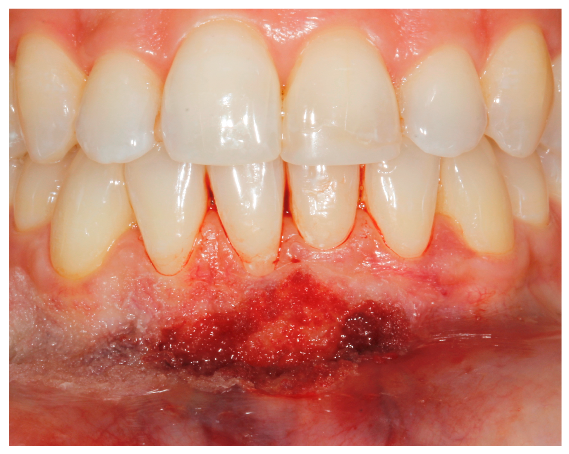
Local anesthesia with mepivacaine and adrenaline 1:100,000 was used. A deep partial thickness incision was made at the level of the external insertion of the frenulum, and subsequently, the portion of superficial and muscular mucous tissue of the frenulum was eliminated. Three detached points were given to periosteal anchorage to apically position the lining mucosa of the lip and allow healing by the second intention of the exposed periosteum [Figure 4].
A layer of Ifabond surgical glue (Peters Surgical, Boulogne-Billancourt, France) was then applied to isolate the tissues from the oral cavity and between the alveolar mucosa and the lip mucosa [Figure 5].
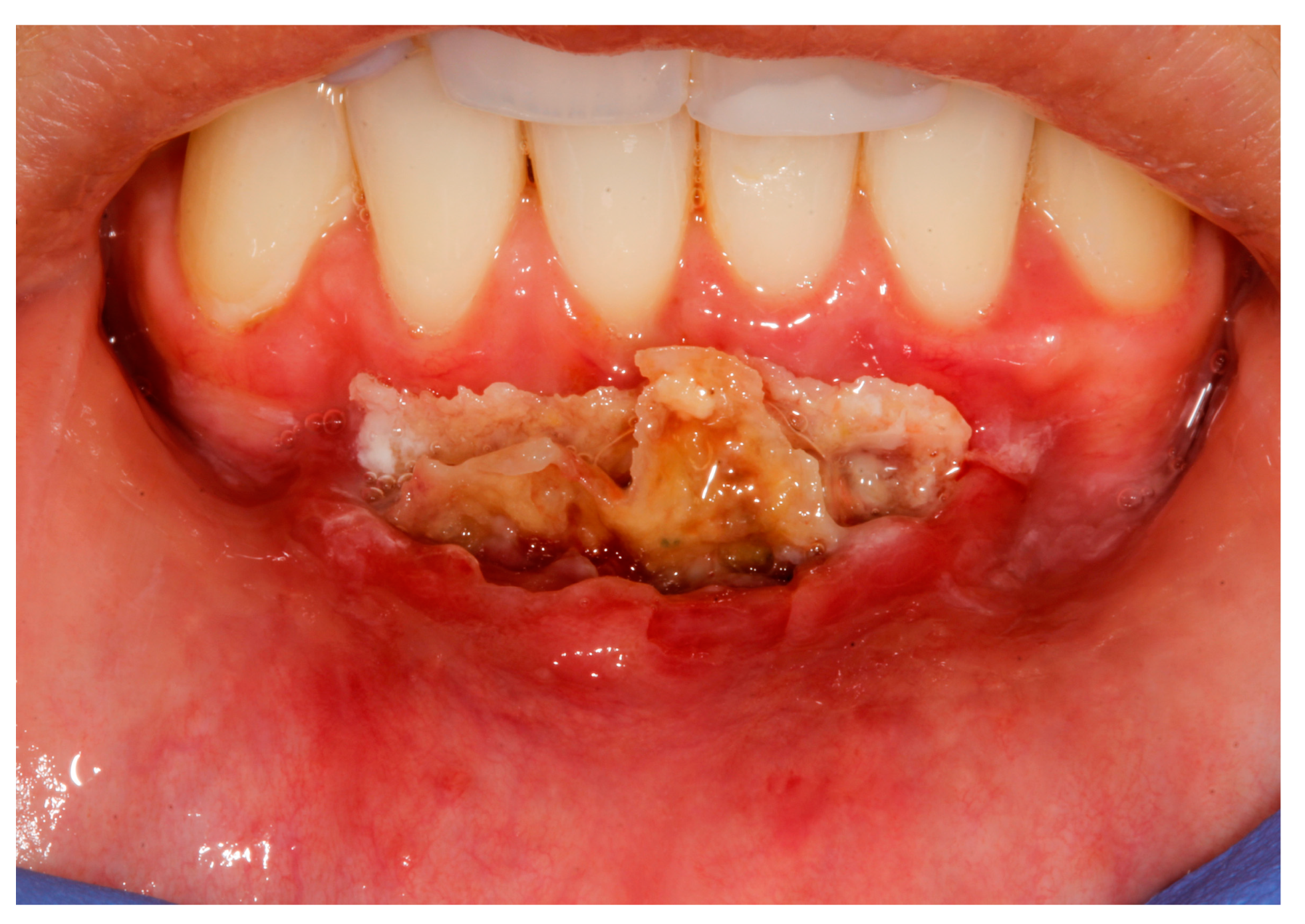
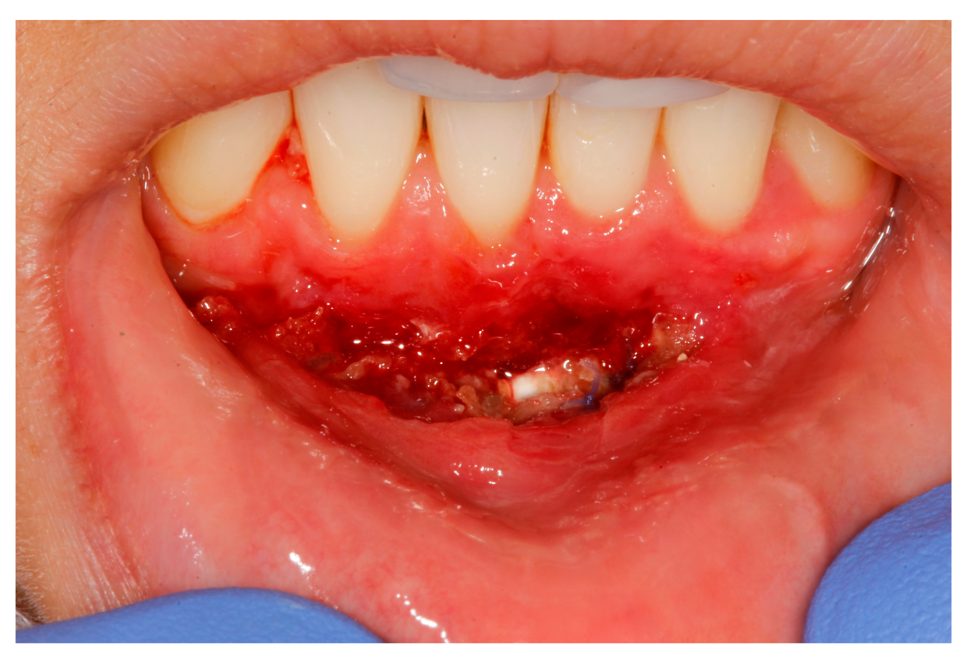
The patient took a dose of analgesics (Ibuprofen cpr 600 mg) at the end of the surgery. No additional painkillers had to be hired. No antibiotic therapy was prescribed. The patient was instructed in hygiene maneuvers and rinsed with 0.12% chlorhexidine-based mouthwash for 15 days. The glue was partially removed at 7 days, and for the remaining, the natural disposal time was expected [Figure 6 and Figure 7].
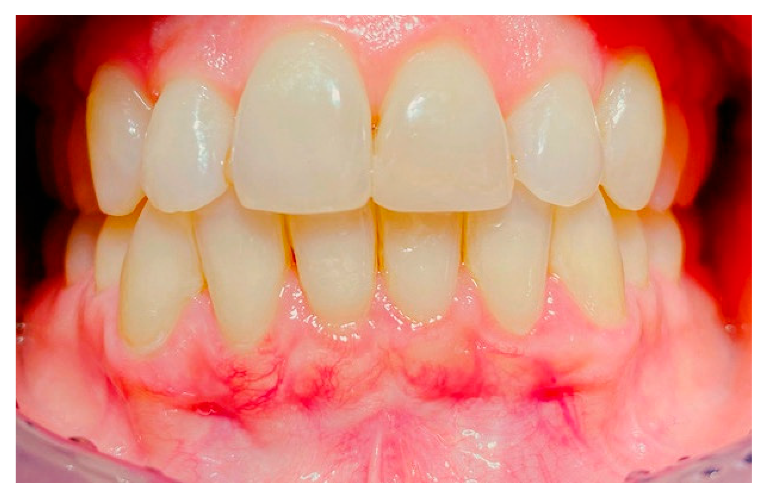
The healing was monitored at 2 and 6 months [Figure 8 and Figure 9] of follow-up.

3. Discussion

There have been numerous attempts by various authors to classify frenula from a clinical-anatomical point of view [5]. One of the most accredited identifies the various types of frenula about the insertion site by dividing them into:
  • mucous attachment frenulum when the frenulum inserts into the mucosa.
  • frenulum with gingival attachment when it is inserted into adherent gingiva.
  • the papillary-attached frenulum when inserted into the papilla.
  • frenulum with penetrating papillary attachment when the frenulum fibers pass through the interincisive papilla and insert into the adherent palatine or lingual gingiva.
Previous studies classified the frenulum into two main morphotypes—thick frenulum and thin frenulum—and about the insertion site: high (alveolar mucosa), middle (adherent gingiva), and low (marginal gingiva) [19,20]. More than 80% of patients who have diastema at nine years of age cease to have it at sixteen, without any therapy [21]. However, sometimes the frenectomy is indicated to reduce the possibility of a “pull syndrome” due to the presence of a pathological frenulum configuration [5,6].
Classical frenulectomy (V-shaped) is a technique proposed by Edwards, which consists of a V-shaped incision of the edges of the frenulum, the removal of its coronal insertion, and its apical repositioning [22]. The intervention is completed by the horizontal and vertical incisions of the periosteal fibers and the affixing of sutures. It is clinically the simplest, but it determines the formation of scar tissue that can cause periodontal problems [22,23,24]. The Z-plasty incision is more complicated and aggressive, but it leads to reduced scar formation. It involves a Z-incision to make two triangular flaps of equal size that are transposed and sutured [23,24]. This technique does not remove the frenulum but is intended to soften the traction of the frenulum on the interdental soft tissue. The Z-plasty technique was considered ideal for large, thick, and hypertrophic frenula, with low insertion associated with diastema and short vestibule. Nevertheless, it allows both the removal of fibrous tissue and the increase in the height of the vestibule [24,25]. During a vestibular sulcus extension, the mucous membrane is raised and placed apically to the gingival margin and sutured to the periosteum while the wound area is uncovered. However, it has a high recurrence rate [8,26]. The frenectomy associated with a free gingiva peduncle positioned laterally is a technique indicated after orthodontic treatment of the diastema to minimize recurrence and excessive scar tissue formation [24,25,26].
Electrosurgery is recommended in patients with low compliance and bleeding problems, as traditional surgery would cause problems in obtaining adequate hemostasis [27]. However, laser frenulectomy has been recently proposed. Diode lasers, YAG lasers, carbon dioxide lasers, and erbium lasers were implemented. Usually, with laser frenulectomy, patients experience less pain, less bleeding during the surgical procedure, no need for suturing, and fewer surgical and functional complications and need for analgesics [4,28].
However, the need for proper sealing of the surgical wound is mandatory to achieve complete healing of the site. The suture is the most used technique to ensure a repositioning of the flap, even if the needle penetration during suturing also causes tissue trauma [9]. Silk is the most universally used suture material in dentistry and many other surgical disciplines [9,29]. However, it is non-absorbable; thus, there is a need for a second appointment to remove the sutures. Moreover, although absorbable sutures are available, most have unpredictable resorption rates in the oral cavity; they may weaken and dissolve early or remain in the incision area for too long [30]. These have prompted the need for an alternative method of wound closure.
Previous studies reported the use of plastic adhesives for tissue suturing in surgical procedures [31,32]. Among them, cyanoacrylates were applied to two opposing, wet, living tissue surfaces, and they showed the ability to fix them perfectly. Their general chemical formula is CH2©C(CN)-COOR, where R can be substituted for any alkyl group, ranging from methyl to decyl. Usually, cyanoacrylate glue generates heat for faster curing, and this may damage soft tissue and hamper blood supply [31]. Therefore, manufacturers have incorporated long chains of a methyl group, due to which the polymerization process is elongated and the rate of heat generation prolonged. Because intraoral mucosa is naturally moist due to saliva, the use of cyanoacrylate glue may find itself as an involuntary participant in the bonding process [32]. Polymerization of the cyanoacrylates occurs within 10–15 s, and they are not absorbable, so they are sloughed from the surface of the skin and mucosa 7–10 days after adhesive application [33,34,35]. Previously ethyl and methyl cyanoacrylate were used for wound closure; however, they were discarded, given their potential toxicities. Newer generation cyanoacrylate includes n-butyl-2-cyanoacrylate, octyl-2-cyanoacrylate, and n-hexyl-cyanoacrylate. They have unique properties, including faster tissue bonding capacity and curing. In addition, their excellent tensile strength, fast polymerization, biocompatibility, immediate hemostasis, ease of application, and bacteriostatic properties make them a good choice for fixing surgical wounds [33,34,35].
Recently, the literature reported the widespread use of cyanoacrylate adhesives during general surgery procedures, including gynecology, gastroenterology, neurosurgery, orthopedics, plastic surgery, dermatology, urology, and vascular and cardiac surgery. Common indications for use included esophageal fistula closure, myocardial surgery, bilateral mammoplasty, skin wound closure, bone and cartilage grafting, corneal surgeries, varicose vein occlusion, and embolization of arteriovenous malformations [36,37]. In particular, the innovative n-hexyl-cyanoacrylate Ifabond glue has been used in laparoscopy, and this new technique is aimed at reducing the operating time, the number of surgical complications, and the required sutures [36,37,38,39,40].
In the maxillofacial field, some cyanoacrylates have been proposed for wound closure on extraction sockets, sinus perforation, mucogingival surgery, and biopsy [17]. However, despite the potential advantages of the use of innovative butyl and hexyl-cyanoacrylate surgical glues in oral surgery, fewer studies have been conducted on this topic. A study showed a reduced inflammatory response after the use of cyanoacrylates during the first postoperative days [30]. These results are in accordance with other studies reporting positive clinical outcomes in combination with surgical glues [29,30,31,32]. Some studies highlighted the hypothesis that faster healing with cyanoacrylate could be promoted by the evidence that the adhesive may act as a scab, maintaining a moist surface over which epithelial migration occurs more readily, as well as preventing secondary infection [29,30,31,32]. Alhourani et al. reported that tissue adhesive significantly reduced the time needed for wound healing when a free gingival graft is positioned compared with traditional sutures [33]. Moreover, the absence of seepage under the covering formed by cyanoacrylate may be responsible for reduced postoperative pain [14,41].
Therefore, in the present study, the use of n-hexyl-cyanoacrylate surgical glue has been described in oral surgery. The case report documented this glue’s sealing and hemostatic ability during frenectomy, with positive results after 2 months of healing. Therefore, this n-hexyl-cyanoacrylate surgical glue could be proposed as a viable alternative to sutures during surgical procedures involving wide flaps, the impossibility of first-time healing, and patients with low compliance. Moreover, it could also be indicated to reduce operative time and postoperative pain, even if these hypotheses should be confirmed by future clinical studies.

Author Contributions

Conceptualization, M.A. and E.B.; methodology, F.E., B.L. and L.B.; validation, M.A. and F.E.; formal analysis, D.P. and N.S.; investigation, M.A., F.E., B.L. and L.B.; resources: B.L. and L.B.; data curation, M.A. and F.E.; writing—original draft preparation, F.E. and B.L.; writing—review and editing, M.A. and F.E.; visualization, D.P., B.L. and N.S.; supervision, E.B.; project administration, B.L., L.B., F.E. and M.A.; funding acquisition, E.B. All authors have read and agreed to the published version of the manuscript.

Funding

This research received no external funding.

Institutional Review Board Statement

This study was conducted in accordance with the Declaration of Helsinki and approved by the Institutional Ethics Committee of AOU Città della Salute e della Scienza di Torino (protocol number 730.503).

Informed Consent Statement

Written informed consent was obtained from all subjects involved in the study.

Data Availability Statement

Not applicable.

Conflicts of Interest

The authors declare no conflict of interest.

References

  1. Gartner, L.P.; Schein, D. The superior labial frenum: A histologic observation. Quintessence Int. 1991, 22, 443–445. [Google Scholar] [PubMed]
  2. Özener, H.; Meseli, S.E.; Sezgin, G.; Kuru, L. Clinical Efficacy of Conventional and Diode Laser-Assisted Frenectomy in Patients with Different Abnormal Frenulum Insertions: A Retrospective Study. Photobiomodulation Photomed. Laser Surg. 2020, 38, 565–570. [Google Scholar] [CrossRef] [PubMed]
  3. Delli, K.; Livas, C.; Sculean, A.; Katsaros, C.; Bornstein, M. Facts and myths regarding the maxillary midline frenum and its treatment: A systematic review of the literature. Quintessence Int. 2013, 44, 177–187. [Google Scholar] [CrossRef] [PubMed]
  4. Dioguardi, M.; Ballini, A.; Quarta, C.; Caroprese, M.; Maci, M.; Spirito, F.; Caloro, G.A.; Alovisi, M.; Basile, E.; Muzio, L.L. Labial Frenectomy using Laser: A Scoping Review. Int. J. Dent. 2023, 2023, 7321735. [Google Scholar] [CrossRef]
  5. Biradar, S.M.; Patil, A.Y.; Kotnoor, S.S.; Bacha, S.; Bijjaragi, S.C.; Kattimani, P.T. Assessment of Diverse Frenal Morphology in Primary, Mixed, and Permanent Dentition: A Prevalence Study. J. Contemp. Dent. Pract. 2020, 21, 562–567. [Google Scholar] [CrossRef]
  6. Mintz, S.; Siegel, M.; Seider, P. An overview of oral frena and their association with multiple syndromic and non- syndromic conditions. Oral Surg. Oral Med. Oral Pathol. Oral Radiol. Endodontol. 2005, 99, 321–324. [Google Scholar] [CrossRef]
  7. Harris, R.J. Creeping Attachment Associated With the Connective Tissue With Partial-Thickness Double Pedicle Graft. J. Periodontol. 1997, 68, 890–899. [Google Scholar] [CrossRef]
  8. Devishree; Gujjari, S.; Gujjari, S.K.; Shubhashini, P.V. Frenectomy: A review with the reports of surgical techniques. J. Clin. Diagn. Res. 2012, 6, 1587–1592. [Google Scholar]
  9. Silverstein, L.H.; Kurtzman, G.M.; Shatz, P.C. Suturing for Optimal Soft-Tissue Management. J. Oral Implantol. 2009, 35, 82–90. [Google Scholar] [CrossRef]
  10. Borie, E.; Rosas, E.; Kuramochi, G.; Etcheberry, S.; Olate, S.; Weber, B. Oral Applications of Cyanoacrylate Adhesives: A Literature Review. BioMed Res. Int. 2019, 2019, 8217602. [Google Scholar] [CrossRef]
  11. Zheng, K.; Gu, Q.; Zhou, D.; Zhou, M.; Zhang, L. Recent progress in surgical adhesives for biomedical applications. Smart Mater. Med. 2021, 3, 41–65. [Google Scholar] [CrossRef]
  12. Shupak, R.P.; Blackmore, S.; Kim, R.Y. Skin hypersensitivity following application of tissue adhesive (2-octyl cyanoacrylate). Bayl. Univ. Med. Cent. Proc. 2021, 34, 736–738. [Google Scholar] [CrossRef]
  13. Yusuf, I.H.; Patel, C.K. A sticky sight: Cyanoacrylate ‘superglue’ injuries of the eye. BMJ Case Rep. 2010, 2010, bcr1120092435. [Google Scholar] [CrossRef] [PubMed] [Green Version]
  14. Oladega, A.A.; James, O.; Adeyemo, W.L. Cyanoacrylate tissue adhesive or silk suture for closure of surgical wound following removal of an impacted mandibular third molar: A randomized controlled study. J. Cranio-Maxillofac. Surg. 2018, 47, 93–98. [Google Scholar] [CrossRef]
  15. Maia, G.V.; Sousa, C.W.; de Lima, J.C.R.; Vecchio, J.L.; Silva, P.G.d.B.; Carvalho, A.C.G.d.S. Cyanoacrylate glue in socket repair: A comparative study. Br. J. Oral Maxillofac. Surg. 2021, 60, 145–151. [Google Scholar] [CrossRef] [PubMed]
  16. Vastani, A.; Maria, A. Healing of Intraoral Wounds Closed Using Silk Sutures and Isoamyl 2-Cyanoacrylate Glue: A Comparative Clinical and Histologic Study. J. Oral Maxillofac. Surg. 2013, 71, 241–248. [Google Scholar] [CrossRef]
  17. Gümüş, P.; Buduneli, E. Graft stabilization with cyanoacrylate decreases shrinkage of free gingival grafts. Aust. Dent. J. 2014, 59, 57–64. [Google Scholar] [CrossRef] [PubMed]
  18. Ghoreishian, M.; Gheisari, R.; Fayazi, M. Tissue adhesive and suturing for closure of the surgical wound after removal of impacted mandibular third molars: A comparative study. Oral Surg. Oral Med. Oral Pathol. Oral Radiol. Endodontol. 2009, 108, e14–e16. [Google Scholar] [CrossRef]
  19. Kramer, P.F.; de Amorim, L.M.; Alves, N.d.M.; Ruschel, H.C.; Bervian, J.; Feldens, C.A. Maxillary Labial Frenum in Preschool Children: Variations, Anomalies and Associated Factors. J. Clin. Pediatr. Dent. 2022, 46, 51–57. [Google Scholar] [CrossRef]
  20. Tadros, S.; Ben-Dov, T.; Catháin, É.Ó.; Anglin, C.; April, M.M. Association between superior labial frenum and maxillary midline diastema—A systematic review. Int. J. Pediatr. Otorhinolaryngol. 2022, 156, 111063. [Google Scholar] [CrossRef]
  21. Wheeler, B.; Carrico, C.K.; Shroff, B.; Brickhouse, T.; Laskin, D.M. Management of the Maxillary Diastema by Various Dental Specialties. J. Oral Maxillofac. Surg. 2018, 76, 709–715. [Google Scholar] [CrossRef] [PubMed]
  22. Ranjbaran, M.A.; Aslani, F.; Jafari-Naeimi, A.; Rakhshan, V. Associations among the occurrence or types of maxillary canine impaction, labial frenum attachment types, lateral incisor anomalies, and midline diastema in patients with and without impaction: A case-control study. Int. Orthod. 2023, 21, 100743. [Google Scholar] [CrossRef] [PubMed]
  23. Ahn, J.H.; Newton, T.; Campbell, C. Labial frenectomy: Current clinical practice of orthodontists in the United Kingdom. Angle Orthod. 2022, 92, 780–786. [Google Scholar] [CrossRef] [PubMed]
  24. Sinha, J.; Kumar, V.; Tripathi, A.K.; Saimbi, C.S. Untangle lip through Z-plasty. BMJ Case Rep. 2014, 2014, bcr2014206258. [Google Scholar] [CrossRef]
  25. Chaubey, K.; Thakur, R.; Arora, V.; Narula, I. Perio-esthetic surgery: Using LPF with frenectomy for prevention of scar. J. Indian Soc. Periodontol. 2011, 15, 265–269. [Google Scholar] [CrossRef] [PubMed]
  26. Miller, P.D. The Frenectomy Combined with a Laterally Positioned Pedicle Graft: Functional and Esthetic Considerations. J. Periodontol. 1985, 56, 102–106. [Google Scholar] [CrossRef]
  27. Hema, D.; Prasanna, J.S. Radiosurgery in periodontics: Have we forgotten it? J. Indian Soc. Periodontol. 2021, 25, 6–10. [Google Scholar] [CrossRef]
  28. Gontijo, I.; Navarro, R.S.; Haypek, P.; Ciamponi, A.L.; Haddad, A.E. The applications of diode and Er:YAG lasers in labial frenectomy in infant patients. J. Dent. Child. 2005, 72, 10–15. [Google Scholar]
  29. Kulkarni, S.; Dodwad, V.; Chava, V. Healing of periodontal flaps when closed with silk sutures and N-butyl cyanoacrylate: A clinical and histological study. Indian J. Dent. Res. 2007, 18, 72. [Google Scholar]
  30. Giray, C.B.; Sungur, A.; Atasever, A.; Araz, K. Comparison of silk sutures and n-butyl-2-cyanoacrylate on the healing of skin wounds. A pilot study. Aust. Dent. J. 1995, 40, 43–45. [Google Scholar] [CrossRef]
  31. Raj, M.; Raj, G.; Sheng, T.K.; Jsp, L. Use of cyanoacrylate tissue adhesives for wound closure in the head and neck region: A systematic review. J. Plast. Reconstr. Aesthetic Surg. 2021, 75, 183–198. [Google Scholar] [CrossRef]
  32. Nitsch, A.; Pabyk, A.; Honig, J.F.; Verheggen, R.; Merten, H.-A. Cellular, Histomorphologic, and Clinical Characteristics of a New Octyl-2-Cyanoacrylate Skin Adhesive. Aesthetic Plast. Surg. 2005, 29, 53–58. [Google Scholar] [CrossRef]
  33. Alhourani, M.A.; Kasem, T.; Hamadah, O. Comparative study between using a tissue adhesive (N-BCA & OCA) and surgical sutures in free gingival graft surgery: A randomized controlled clinical trial. Dent. Med. Probl. 2022, 59, 241–248. [Google Scholar] [CrossRef]
  34. Montanaro, L.; Arciola, C.; Cenni, E.; Ciapetti, G.; Savioli, F.; Filippini, F.; Barsanti, L. Cytotoxicity, blood compatibility and antimicrobial activity of two cyanoacrylate glues for surgical use. Biomaterials 2000, 22, 59–66. [Google Scholar] [CrossRef]
  35. Verma, D.; Ahluwalia, T. Cyanoacrylate tissue adhesives in oral and maxillofacial surgery. J. Indian Dent. Assoc. 2002, 73, 171–174. [Google Scholar]
  36. Bellón, J.M.; Fernández-Gutiérrez, M.; Rodríguez, M.; Pérez-López, P.; Pérez-Köhler, B.; Kühnhardt, A.; Pascual, G.; Román, J.S. Behavior of a new long-chain cyanoacrylate tissue adhesive used for mesh fixation in hernia repair. J. Surg. Res. 2016, 208, 68–83. [Google Scholar] [CrossRef]
  37. Bellón, J.M.; Fernández-Gutiérrez, M.; Rodríguez, M.; Sotomayor, S.; Pérez-Köhler, B.; Kuhnhardt, A.; Pascual, G.; Román, J.S. Bioassay of cyanoacrylate tissue adhesives used for intraperitoneal mesh fixation. J. Biomed. Mater. Res. Part B Appl. Biomater. 2015, 105, 312–319. [Google Scholar] [CrossRef]
  38. Pascual, G.; Rodríguez, M.; Pérez-Köhler, B.; Mesa-Ciller, C.; Fernández-Gutiérrez, M.; Román, J.S.; Bellón, J.M. Host tissue response by the expression of collagen to cyanoacrylate adhesives used in implant fixation for abdominal hernia repair. J. Mater. Sci. Mater. Med. 2017, 28, 58. [Google Scholar] [CrossRef] [PubMed]
  39. Pascual, G.; Rodríguez, M.; Mesa-Ciller, C.; Pérez-Köhler, B.; Fernández-Gutiérrez, M.; Román, J.S.; Bellón, J.M. Sutures versus new cyanoacrylates in prosthetic abdominal wall repair: A preclinical long-term study. J. Surg. Res. 2017, 220, 30–39. [Google Scholar] [CrossRef]
  40. Estrade, J.-P.; Gurriet, B.; Franquebalme, J.-P.; Chinchole, J.-M.; Glowaczower, E.; Ferry, C.; Crochet, P.; Agostini, A. Laparoscopic sacrocolpopexy with a vaginal prosthetic adhesive. Gynecol. Obstet. Fertil. 2015, 43, 419–423. [Google Scholar] [CrossRef]
  41. Giudice, A.F.S.-L.; Ortiz, A.M.; Sánchez-Labrador, L.; Brinkmann, J.C.-B.; Cobo-Vázquez, C.M.; Meniz-García, C. Current status of split-mouth controlled clinical trials comparing cyanoacrylate vs. conventional suture after lower third molar surgeries: A systematic literature review. Acta Odontol. Scand. 2022, 81, 349–357. [Google Scholar] [CrossRef]
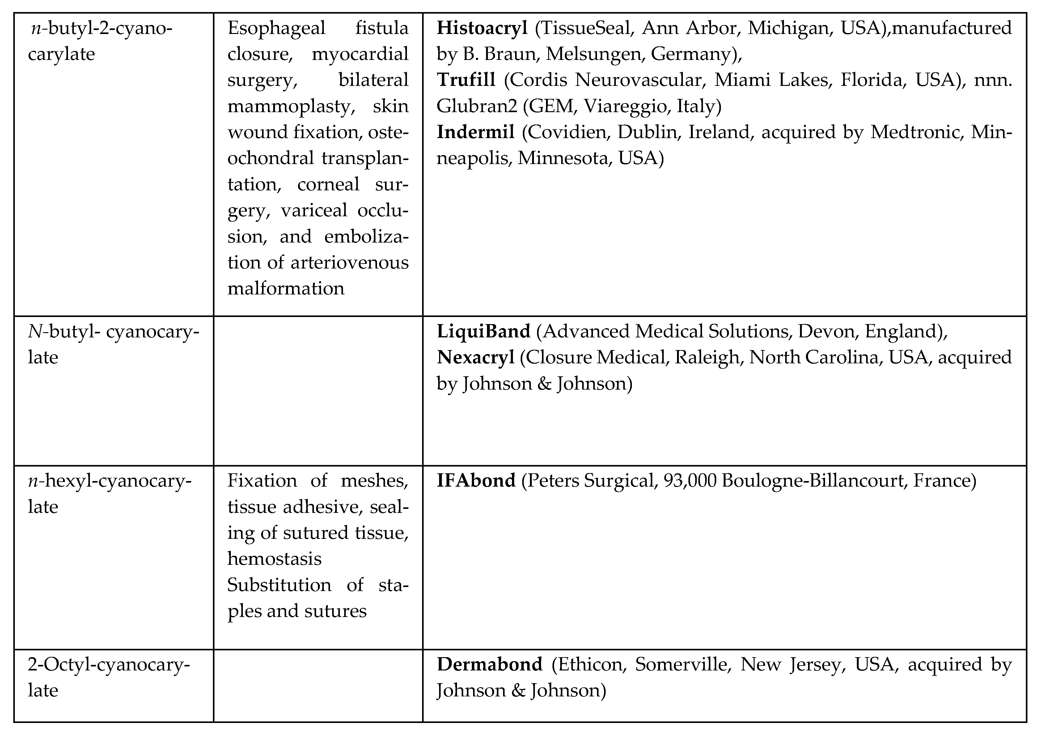
Figure 1. Various types of available cyanoacrylate glues (modified from Kay et al. [11]).
Applsci 13 08979 g001
Figure 2. Preoperative vestibular gingival frenulum showing a “pull syndrome” action on the right central incisor.
Applsci 13 08979 g002
Figure 3. Lateral view of the mandibular frenulum.
Applsci 13 08979 g003
Figure 4. Postoperative vestibular gingival frenulum after the surgical procedure.
Applsci 13 08979 g004
Figure 5. After the surgical procedure, a layer of surgical glue was placed to fix the wound and achieve hemostasis.
Applsci 13 08979 g005
Figure 6. Clinical image of the glue on the surgical site after 1 week.
Applsci 13 08979 g006
Figure 7. Clinical image of the surgical site after the glue has been partially removed.
Applsci 13 08979 g007
Figure 8. Clinical image 2 months after surgery showing the correct healing of the tissues and the repositioning of the buccal lip frenulum.
Applsci 13 08979 g008
Figure 9. Follow-up 6 months after surgery showing the complete healing of the soft tissues.
Applsci 13 08979 g009
Disclaimer/Publisher’s Note: The statements, opinions and data contained in all publications are solely those of the individual author(s) and contributor(s) and not of MDPI and/or the editor(s). MDPI and/or the editor(s) disclaim responsibility for any injury to people or property resulting from any ideas, methods, instructions or products referred to in the content.