Article

Preliminary Results of Measurements of Frontal Plane Knee Alignment Using a Standardized, Sonographic-Assisted Digital Photograph—A Reliable and Accurate Alternative to a Full-Leg Radiograph?

1
Department of Pediatric Surgery and Orthopedics, Clinic St. Hedwig, Barmherzige Brueder Regensburg, KUNO Pediatric University Medical Center and FIFA Medical Center of Excellence, University Medical Center Regensburg, 93053 Regensburg, Germany
2
Department of Trauma Surgery and FIFA Medical Center of Excellence, University Medical Center Regensburg, 93053 Regensburg, Germany
3
Department of Orthopedic Surgery, Koenig-Ludwig-Haus, University of Wuerzburg, 97074 Wuerzburg, Germany
*
Author to whom correspondence should be addressed.
Appl. Sci. 2023, 13(16), 9074; https://doi.org/10.3390/app13169074
Submission received: 15 July 2023 / Revised: 6 August 2023 / Accepted: 7 August 2023 / Published: 9 August 2023
(This article belongs to the Section Applied Biosciences and Bioengineering)

Abstract

:
(1) Clinical alternatives to the full-leg radiograph (FLR) for determining frontal plane lower limb alignment have shown mixed results so far concerning reliability and accuracy. In addition, intercondylar respectively intermalleolar distance (ICD/IMD) measurement is limited in the interpretation of its results. The purpose of the study is to present a standardized, sonographic-assisted photographic measurement (SPM) of the leg axis in the frontal plane using a digital photograph and compare it with the gold standard of the anterior–posterior FLR. (2) Adults who had recently received an FLR were included in the study. After determining the center of the hip joint and knee joint gap in a standing position via ultrasound, a standardized digital photograph was taken. Subsequently, the hip–knee–ankle angle (HKA) was measured using an image editing program for SPM and FLRs. Mean deviation (MD), Pearson’s correlation coefficient and the clinical method’s interclass correlation coefficient (ICC) were calculated. (3) Of 18 subjects (8 male and 10 female), respectively, 34 lower extremities have been included in the study. Their mean BMI was 25.5 kg/m2. The correlation between the two measurement methods for subjects with BMI < 30 kg/m2 was reliably high (r = 0.93, p < 0.001). The HKAs derived from SPM and FLRs showed a mean deviation (MD) of 2.4° (range 0.1–6.0°) for subjects with BMI < 30 kg/m2. The average ICC among all examiners conducting the SPM was 0.99 (CI 0.95–0.99, p < 0.001). The MD for subjects ≥ 30 kg/m2 increased significantly (MD = 5.5°; n = 10, p < 0.001). (4) SPM of the leg axis in the frontal plane allows sufficiently accurate results in patients with a BMI lower than 30 kg/m2. The methodology is limited in cases of increased BMI and needs well-trained examiners. Nevertheless, as a screening method in the field of children and adolescents, this method can be recommended in clinical daily routine.

1. Introduction

Knee malalignment is considered to be one of the main causes of the development of cartilage lesions, ligament instability and in the end, knee arthrosis [1,2]. Lower limb misalignment—next to other factors like female sex, obesity, trauma and sports activities—is already proven to be associated with osteoarthritis of the lower limb [3,4]. Sharma et al. have shown that lower limb malalignment influences the risk of subsequent primary osteoarthritis disease progression and, furthermore, the decline in the functional status of the knee joint. These effects could already be detected after 18 months of observation [1]. Tanamas et al. even showed with their literature review that malalignment of the knee joint, in either the valgus or varus direction, is an independent risk factor for the progression of radiographic knee osteoarthritis [2]. Due to the continuous increase in life expectancy, the rise in the mean age of society and the resulting expected increase in osteoarthritis patients [5,6], a proper and safe detection and examination of the lower extremity’s alignment is very important in daily clinical routine and even for study purposes. In order to understand whether and to what extent factors such as mechanical loading through sports, for example, have an influence on the development of the leg axis and its malalignment, it is important for study purposes to have a measurement technique that allows a quick statement about the leg axis in the frontal plane that is as accurate as possible. Needless to say, a target group of such radiographic-sparing examinations comprises children and adolescents. Until nowadays, the gold standard for determining the frontal axis in lower extremities in clinical practice has been the full-leg radiograph (FLR), as described by Lamark and, for example, performed by Colyn et al. [7,8]. So far, the anterior–posterior FLR seems to be irreplaceable and is mostly needed as a diagnostic tool for assessing lower limb malalignment, the planning of surgical treatment in adults and children (e.g., total knee arthroplasty, tibial osteotomy, tibial or femoral hemiepiphysiodesis), assessment of leg length discrepancies and even postsurgical follow-up examinations [9].
In contrast to the daily routine in clinical orthopedic practice for individual patients, radiographs and MRI are not suitable as a screening method for, for example, prevalence studies with a lack of a large study population size. Few studies existing on the clinical determination of the leg axis show acceptable results when, for example, measuring the intercondylar respectively intermalleolar distance (ICD/IMD) or by using digital photographs [10,11,12,13,14,15,16]. One advantage of the ICD/IMD method is obviously that the measurement is easy to carry out, and only a few instruments are needed. One of the disadvantages is that the ICD/IMD measurement is age dependent in early age (ICD increased in 2- to 4-year-olds) but stays constant in pre-adolescents and adolescents [17]. Additionally, the method loses significance and is limited due to the soft tissue component at the knee joint. Furthermore, statements about lateral differences (right/left or dominant/non-dominant leg) are not possible with this measurement method. Since methods using radiographs or MRI imaging are not feasible in large prevalence studies, methods have already been developed that do not use ionizing radiation and make use of standardized photographic images [14,15]. Schmitt et al. tried to find out how knee joint alignment can be reliably measured from a standardized photograph and, more importantly for our study, what influence changes in the standing position have on the angles measured [14]. Sheehy et al. assessed the intra-rater, inter-rater and test–retest reliability and concurrent validity of lower-extremity alignment, also estimated from a standardized photograph, and showed satisfying results [15]. By using a photographic image to estimate the leg axis, in contrast to the ICD/IMD measurement, statements about right/left differences are possible, and by orienting to anatomical landmarks to assess the alignment angles, e.g., the hip–knee–ankle angle, a sufficient reduction of the soft tissue component can be achieved, which leads to better accuracy, especially for subjects with a higher BMI. We took these shown approaches as an example and tried to develop a new, modified method of lower limb alignment measurement using standardized digital photographs.
The idea of developing the presented method was given by a study which is currently being performed in junior football (registered in the German Clinical Trials Registry DRKS; Clinical trial registration number DRKS00020446) [18]. The purpose of this study is to evaluate the presented method of frontal plane lower limb alignment measurement without using ionizing radiation. The authors want to describe the method as well as compare the results with the gold standard of the FLR in order to determine the accuracy of this new clinical measurement of the leg axis.

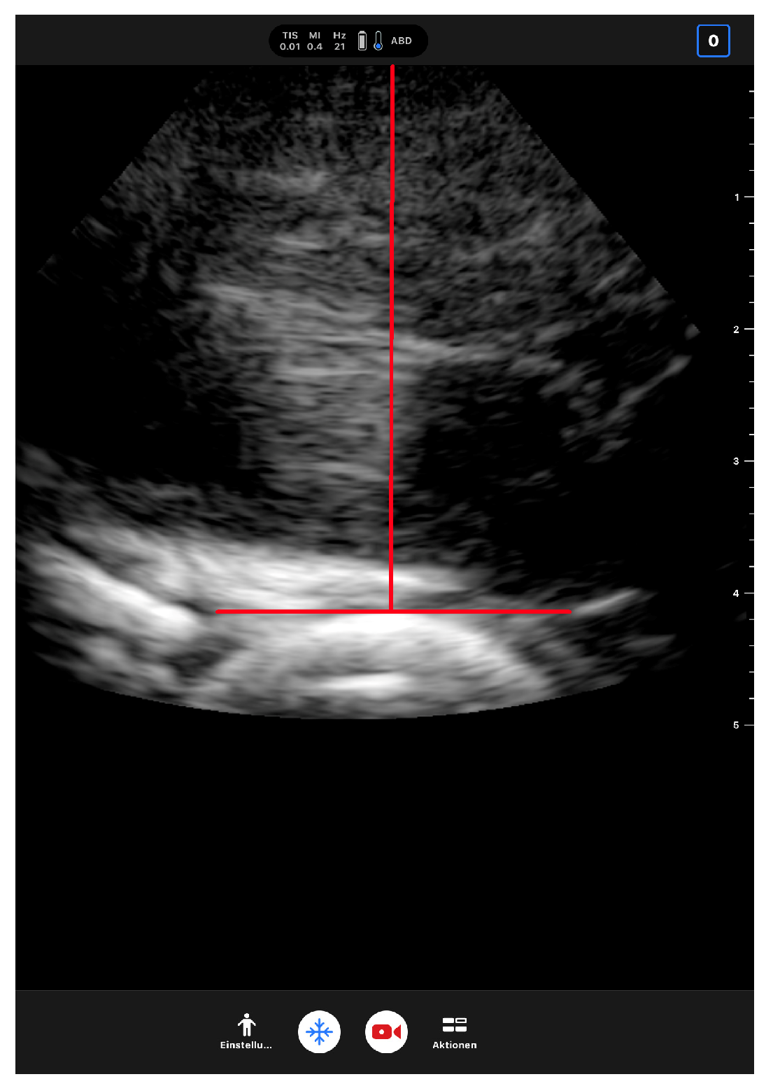
2. Materials and Methods

The study was conducted according to the guidelines of the Declaration of Helsinki and approved by the Ethics Committee of the University of Regensburg, Germany (date of approval: 20 October 2021; protocol code 21-2612-101). Prior to recruitment, the study was registered in the German Clinical Trials Registry DRKS (clinical trial registration number DRKS00027937). Participants were included if they were able to stand unassisted in an upright position with straightened leg(s), which is required for proper measurement, if they were able to understand and follow the examiner’s instructions and if an acute bony, ligamentous, meniscal or cartilage injury could be excluded by clinical and radiographic anamnesis. Each participant has given written informed consent to participate in the study. Subjects were divided into two groups according to their BMI (group I: <30 kg/m2; group II: ≥30 kg/m2). The maximum allowed time span between SPM and FLR was 4 months. In addition, those with an acute injury and a surgical intervention on the leg were excluded. A further exclusion criterion was an implanted hip joint replacement, as this makes sonographic detection of the femoral head impossible. In 13 of the total of 18 participants, both leg axis measurement methods were performed on the same day.
The SPM method was performed under precisely standardized conditions. The participant, undressed but for his or her underwear, was positioned on level ground, standing at a distance of 3 m from the camera. The camera (Apple® iPad Air 3rd generation, 8-megapixel front camera, Apple Inc, Cupertino, CA, USA) stood on a tripod (Tripod Stativ 106 3D by Hama® GmbH & Co. KG, Monheim, Germany), which was positioned at the participant’s knee height in order to minimize the parallactic error. The participant was then asked to remain in a relaxed position with their weight equally distributed on both feet, which were positioned hip-width standing. The legs were then aligned to achieve the exact orthograde positioning of the patellae by the examiner, analogous to the FLR [19]. Next, three different joint points were defined to determine the mechanical femorotibial axis: (1) Hip joint: first, the femoral head was sonographically detected in the anterior–posterior direction with a portable ultrasound device (Butterfly iQ®, Butterfly Network, Inc., Burlington, MA, USA) in a standing position (see Figure 1). The skin was marked with an adhesive electrode at the point that showed the highest point of convexity of the femoral head on ultrasound. (2) Knee joint: the next step was palpatory detection of the knee joint gap both medially and laterally and the application of an elastic band at this level. If palpation was difficult due to the soft tissue mantle, detection was also assisted sonographically. A line was then drawn on the digital image from the medial to the lateral intersection of the rubber band with the background where the rubber band rests in the knee joint gap, and the midpoint of this line was determined as the knee joint center. (3) Ankle joint: the center of the line connecting the bony prominences of the medial and lateral malleoli was determined, which quite accurately intersects the center of the talus (see Figure 2).
One weight-bearing, full-leg anterior–posterior digital radiograph was taken in the identic standardized position, analogously to the positioning described above for the SPM, and recommended by Paley et al. [19]. The HKA of the photograph and the corresponding full-leg radiograph (transferred anonymized as .jpeg file) were measured with an angle measuring application (Angle Meter 360, ©AK App Develop). The FLR’s HKA was defined by the angle of the straight lines between the center of the femoral head/center of the femoral intercondylar notch and the center of the intercondylar notch/center of the talus. All examiners were instructed in the usage of the ultrasound device, sonographic detection of the femoral head and knee joint gap and the operation mode and usage of the camera and digital measurement software and had performed the measurement multiple times prior to the start of the study. Inter-rater reliability was tested by measuring ten leg axes from three different examiners using the photographic method, including measurement of the angles using the software. Prior to the study, a sample size calculation was made with a confidence level of 95%, a margin of error of ±5% and a standard deviation of 0.5, which resulted in a sample size of 35, which approximately fit the overall number of measurements included in the survey.
Analysis was performed using SPSS Statistics 26 (SPSS Inc., Chicago, IL, USA). For each method, the mean deviation (MD), standard deviation, minimum and maximum deviation of the HKA were calculated. To assess inter-rater reliability between all examiners, the intraclass correlation coefficient (ICC) was used, using a 2-way random model with absolute agreement definition [20,21]. The 95% confidence interval was also applied. Correlation between the HKA of the radiograph and the tested photographic method was determined using Pearson’s correlation coefficient. A correlation coefficient of 0.5 to 0.75 was evaluated as good, values > 0.75 were regarded as excellent [22]. Whenever suitable, two-tailed t-tests for comparing mean values were conducted, with a significance level of p < 0.05 as significant.

3. Results

For this study, 34 lower extremities of 18 (8 male and 10 female) participants could be included, and the mean age was 50.1 years (see Table 1). Two lower extremities had to be excluded due to hip joint replacement having been undergone.

3.1. Inter-Rater Reliability

For determining the inter-rater reliability, ten leg axis alignments of five subjects were evaluated by three examiners. The average ICC among all examiners was 0.99 (CI (95): 0.95–0.99, p < 0.001). The MD rate between the examiners was 0.5° with a minimum–maximum span of 0.0 to 2.0°.

3.2. Comparison between SPM and FLR of Subjects with BMI < 30 kg/m2

The HKA of 24 lower extremities of subjects with BMI < 30 kg/m2 were measured using SPM and FLRs. The FLRs’ HKA had an absolute range of 166.4° (varus) to 195.1° (valgus); the SPM’s HKA showed a range from 168.6° (varus) to 195.0° (valgus). Results show that the HKA of both methods highly correlate within this group (Pearson’s r = 0.93; p < 0.001, see Figure 3). The mean deviation (MD) of the HKA amounted to 2.4° (range 0.1–6.0°). The HKA’s mean value of SPM (182.0°) was 1.6° higher (more towards valgus) than the one of the FLRs (180.4°; see Table 2).

3.3. Comparison between SPM and FLRs of Subjects with BMI ≥ 30 kg/m2

In the second group, of n = 5 subjects, respectively n = 10 lower limbs could be measured with SPM and FLRs, of subjects with a mean BMI of 32.4 kg/m2 (range 30–34 kg/m2). The FLRs had an absolute range of 177.0° (varus) to 183.8° (valgus); the SPM’s HKA ranged from 182.5° to 190.0° (valgus). Pearson’s r between both methods within this group was lower than the one of the subpopulation with BMI < 30 kg/m2 (r = 0.84 vs. r = 0.93, p < 0.001), but still shows a high correlation. The HKA’s MD amounted 5.5° (±1.5°), with a minimum–maximum span of 2.6–8.1°. SPM resulted in a higher valgus angle in every case, with an MD of 5.5° (±1.5°), compared to the FLRs’ HKA (see Table 2).

4. Discussion

Determining lower limb alignment is essential to detect malalignment of the lower extremities, as it is considered to be a critical factor for developing degeneration of the knee joint [1,2]. As ionizing radiation is needed in standardized methods right now, there is a need for a non-radiographic measurement. Admittedly, it has to be said that several studies have already shown that the dose of radiographic radiation from even “large-area” images such as FLRs could be reduced through technical developments and better filters, without any loss of relevant diagnostic information. A standardized whole leg radiograph is estimated to have an effective dose of approximately 10 mSv [23,24]. For comparison, the German Federal Office for Radiation Protection BfS estimates that a German citizen is exposed to an average of an effective dose of 2.1 mSv of natural radiation exposure [25]. Against this background, the dose of such a radiological examination as an FLR seems quite relevant, especially in the field of children and adolescents. One of the aims of the development of the presented photographic method was the establishment of a new diagnostic method in the field of children, not only for study purposes but also in clinical practice to reduce radiation exposure. Secondly, there is a need for quick and easy measurement in a clinical setting that could easily be used as a screening method in a broad, extraclinical setting.
To avoid radiation, several research teams have already been introduced to find alternatives to the FLR, and have already tested them for their validity in comparison to the FLR [10,11,12,13,14,15,16]. Sheehy et al. have shown that digital photographs can be used to determine lower limb alignment properly. They defined a PA (photographic alignment) angle, measured from a pelvis-to-floor photograph, which is the angle between the estimated center of the hip, knee and ankle joints. The estimation of the joint centers follows specific landmarks. Sheehy et al. also showed that the PA angle may be used in place of the HKA of the FLR if a bias of 4.5° is added [15]. Schmitt et al. described another way of determining lower limb alignment by precising the anatomical and mechanical axis using a digital photograph. They also focused on factors that influence the measured results most, like leg rotation (interclass correlation coefficient (ICC): 0.658), changes in radiograph–object distance or standing position with feet apart or together. Schmitt et al. concluded that measurement using their method is highly reliable and suitable for routine clinical use, inter alia for cross-sectional analysis of groups wherein legs are under particular stress [14]. What distinguishes the presented method from the studies just mentioned is, among other things, the approach of using sonographic detection of the femoral head and the measurement of the HKA in both SPM and FLRs for better comparison.
The present study shows that measurement of lower limb alignment in the frontal plane using a digital photograph can provide reliable and accurate results. SPM adds 1.6° more valgus, in average, to the FLRs’ HKA in the subpopulation with a BMI lower than 30 kg/m2. The SPM’s mean deviation rises significantly if BMI exceeds 30 kg/m2 (p < 0.001), with negative effects on precision rates in comparison to the radiographic method. The effect of BMI on the non-radiographic measurement of lower limb alignment has already been stated by other studies. Legrand et al. tried to determine frontal plane knee alignment using a three-dimensional reconstruction model based on movement analysis and also compared their results to full-leg radiographs. Although validity could be confirmed for subjects with a BMI lower than 25 kg/m2, the measurement error increased for those with a BMI greater than 25 kg/m2 [26]. These results clearly show that clinical measurement methods are sensitive to methodological errors if the BMI rises above a certain level. Reasons for this can be, on the one hand, the more difficult detection and marking of the femoral head, and on the other hand, the assessment of the rotation of the lower extremity, as determination of the orthograde alignment of the patella is difficult. The question now arises as to what extent the restriction of the BMI of subjects to <30 kg/m2 represents a relevant limitation of the population that is eligible for the presented measurement technique. A broad epidemiological study of children and adolescents whose age-specific BMI was measured showed that in childhood and adolescence, the 97th BMI percentile is 21.1 kg/m2 for 8-year-olds and 28.8 kg/m2 for 18-year-olds. This, in turn, means that limiting the BMI of subjects to below 30 kg/m2 only affects far less than 3% of the total population and should, therefore, not be considered a relevant limitation [27]. Conversely, this means that the new technique can be applied to at least 97% of children and adolescents with accurate results. In the adult population, on the other hand, approximately one in five subjects (20.5%) would not be suitable for measurement due to a BMI > 30 kg/m2, as the average BMI increases continuously with age and is significantly higher in adults than in children and adolescents [28,29,30]. McCann et al. have already highlighted the effect of tibial rotation on varus deformity measurement, showing that especially internal rotation and varus angulation of the tibia are associated with a higher deviation of the measured frontal plane alignment compared to neutral controls [31]. Schmitt et al. purposely measured lower limb alignment in different positions concerning leg rotation using a digital photography technique and showed that leg rotation has a big influence on the measured results [14]. This highlights the importance of establishing a precise, standardized measuring setup. The correct positioning of the lower limb and orthograde alignment of the patella, regardless of the type of measurement of the frontal leg axis, is especially important [9,19].
The reliability of digital full-leg radiography—even if it is considered to be the gold standard so far [32,33]—is currently not fully clear in clinical results. Although Boewer et al. show a high inter-rater and intra-rater reliability [34], other studies, e.g., the one conducted by Schmidt et al., state mediocre intra-rater and quite poor inter-rater reliability [35]. Ilahi et al. similarly suggest, after examining the inter- and intra-observer variability of radiographic measurements of knee alignment, that this variability must be considered in the interpretation of measurements [36]. Against this background, the inter-rater reliability of SPM (ICC 0.99; CI (95) 0.95–0.99, p < 0.001) seems to be quite comparable to that of the FLR. Schmitt et al. estimate that measurement errors of radiographic full-leg radiographs range from 1.1 to 1.6°, which represents a non-negligible factor that must be taken into account when making therapeutic decisions [14,31,37,38,39].
The HKA’s mean value from the photograph measurement was 1.6° higher (more towards valgus) than the FLR-derived mean value. The degree of deviation does not allow this method to replace a full-leg radiograph for the more accurate diagnostics of frontal plane lower limb malalignment needed when planning a surgical treatment, but it can certainly pose as a reliable diagnostic alternative for known lower limb deformities during follow-up examinations, for screening methods or for further study purposes. Factors that can be blamed for the SPM’s MD of 2.4° are measurement errors due to soft tissue and errors in the (re)alignment of the lower limb before the measurement (SPM and FLR). The method’s ICC shows that SPM provides high reliability.
Of course, this study is also subject to certain limitations. Even though, in the majority of the measurements, the radiograph was taken on the same day as the SPM (13 out of 18 subjects), some radiographic measurements were performed with a delay of up to 4 months, which could have affected the precision rates due to small changes in the leg axis. An important point to consider is that although the alignment of the subjects for both methods was standardized, minor deviations due to repositioning for the second method cannot be excluded. The influence of soft tissues on the accuracy of the measurement is also an important factor in the interpretation of the results. Due to their mostly smaller soft tissue mantle compared to adults, the authors assume an even higher accuracy in normal-weight children and adolescents. In the future, this method could not be used as a substitute for the FLR, but much more as an initial screening method, and if the findings are conspicuous, it could then be completed by an FLR.

5. Conclusions

The presented photographic method could provide reliable results for subjects with a BMI lower than 30 kg/m2 in determining leg axis, compared to a radiographic control. Even though the accuracy of the method is not suitable to replace radiographic diagnostics, especially in therapy decisions, it might provide sufficiently accurate values to deliver statements about knee alignment in clinical practice. The advantages of this method are its ability to be performed in an extraclinical setting, the ability of clinicians to make side-separated statements about the leg axes, in contrast to ICD/IMD measurement, and its rapid feasibility without using ionizing radiation. Quite apart from the radiation protection reasons, there are further advantages in that it saves not only personnel and thus costs, but also time compared to the FLR. Full-leg radiography requires a radiology department with the appropriate equipment (e.g., radiography tube, long film cassette), a trained radiological technical assistant, and compliance with the necessary radiation protection requirements, which is associated with financial and time expenditures. This poses problems not only for orthopedic colleagues in private practice, but also for researchers. It, thus, also makes the SPM method attractive to orthopedic colleagues in private practice once it has been thoroughly validated, including with regard to cut-off values for physiological respectively pathological leg axes. Future studies must clarify how accurately the measurements can be performed on children and adolescents, as it can be assumed that SPM might have even better accuracy for them than for adult subjects. This preliminary study can serve as a basis for a larger comparative study with children and adolescents, with the help of which validation can take place and cut-off points can be determined. Only then will it be possible to establish this new and simple method in clinical practice.

Author Contributions

Conceptualization, C.M. (Clemens Memmel) and C.M. (Christoph Meier); Methodology, C.M. (Clemens Memmel); Software, L.H.; Validation, J.W., L.A. and W.K.; Formal Analysis, M.K.; Investigation, C.M. (Christoph Meier); Resources, V.A.; Data Curation, C.M. (Christoph Meier); Writing—Original Draft Preparation, C.M. (Clemens Memmel); Writing—Review and Editing, C.M. (Clemens Memmel) and J.W.; Visualization, D.S.; Supervision, W.K.; Project Administration, C.M. (Clemens Memmel); Funding Acquisition, C.M. (Clemens Memmel). All authors have read and agreed to the published version of the manuscript.

Funding

This research received no external funding.

Institutional Review Board Statement

The study was conducted according to the guidelines of the Declaration of Helsinki, and approved by the Ethics Committee of the University of Regensburg, Germany (date of approval: 20 October 2021; protocol code 21-2612-101).

Informed Consent Statement

The authors confirm that any participant has given informed consent to participate in the research. The subject pictured in Figure 2 has given written consent to the publication of the photographic material.

Data Availability Statement

The data that support the findings of this study are available on request from the corresponding author. The data are not publicly available due to restrictions, e.g., their containing information that could compromise the privacy of research participants.

Acknowledgments

The authors would like to express their sincere gratitude to all doctoral students and subjects, without whom it would not have been possible to conduct the study.

Conflicts of Interest

The authors report no conflict of interest.

References

  1. Sharma, L.; Song, J.; Felson, D.T.; Cahue, S.; Shamiyeh, E.; Dunlop, D.D. The role of knee alignment in disease progression and functional decline in knee osteoarthritis. JAMA 2001, 286, 188–195. [Google Scholar] [CrossRef] [Green Version]
  2. Tanamas, S.; Hanna, F.S.; Cicuttini, F.M.; Wluka, A.E.; Berry, P.; Urquhart, D.M. Does knee malalignment increase the risk of development and progression of knee osteoarthritis? A systematic review. Arthritis Rheum 2009, 61, 459–467. [Google Scholar] [CrossRef]
  3. Kadam, U.T.; Croft, P.R. Clinical comorbidity in osteoarthritis: Associations with physical function in older patients in family practice. J. Rheumatol. 2007, 34, 1899–1904. [Google Scholar]
  4. Kadam, U.T.; Croft, P.R. Clinical multimorbidity and physical function in older adults: A record and health status linkage study in general practice. Fam. Pract. 2007, 24, 412–419. [Google Scholar] [CrossRef]
  5. Hunter, D.J.; Bierma-Zeinstra, S. Osteoarthritis. Lancet 2019, 393, 1745–1759. [Google Scholar] [CrossRef] [PubMed]
  6. Wallace, I.J.; Worthington, S.; Felson, D.T.; Jurmain, R.D.; Wren, K.T.; Maijanen, H.; Woods, R.J.; Lieberman, D.E. Knee osteoarthritis has doubled in prevalence since the mid-20th century. Proc. Natl. Acad. Sci. USA 2017, 114, 9332–9336. [Google Scholar] [CrossRef]
  7. Lamark, K. The whole leg radiograph and radiation exposure in paediatrics. In Proceedings of the European Congress of Radiology 2013, Vienna, Austria, 7–11 March 2013. [Google Scholar]
  8. Colyn, W.; Agricola, R.; Arnout, N.; Verhaar, J.A.N.; Bellemans, J. How does lower leg alignment differ between soccer players, other athletes, and non-athletic controls? Knee Surg. Sports Traumatol. Arthrosc. 2016, 24, 3619–3626. [Google Scholar] [CrossRef]
  9. Becker, C.; Ross, C. Ganzbeinaufnahmen—Wieso, weshalb, warum? [Whole-leg radiographs—Why?]. Radiopraxis 2010, 3, 11–21. [Google Scholar] [CrossRef]
  10. Abreu, A.V.D.; Barbosa, J.R.P.; Paiva, F.J.D. Alinhamento dos joelhos no plano frontal dos 12 aos 17 anos [Alignment of the knees in the frontal plane from 12 to 17 years]. Rev. Bras. Ortop. 1996, 31, 83–88. [Google Scholar]
  11. Asadi, K.; Mirbolook, A.; Heidarzadeh, A.; Mardani Kivi, M.; Emami Meybodi, M.K.; Rouhi Rad, M. Association of Soccer and Genu Varum in Adolescents. Trauma Mon. 2015, 20, e17184. [Google Scholar] [CrossRef] [PubMed] [Green Version]
  12. Hinman, R.S.; May, R.L.; Crossley, K.M. Is there an alternative to the full-leg radiograph for determining knee joint alignment in osteoarthritis? Arthritis Rheumatol. 2006, 55, 306–313. [Google Scholar] [CrossRef] [PubMed]
  13. Navali, A.M.; Bahari, L.A.S.; Nazari, B. A comparative assessment of alternatives to the full-leg radiograph for determining knee joint alignment. Sports Med. Arthrosc. Rehabil. Ther. Technol. 2012, 4, 40. [Google Scholar] [CrossRef] [PubMed] [Green Version]
  14. Schmitt, H.; Kappel, H.; Moser, M.T.; Cardenas-Montemayor, E.; Engelleiter, K.; Kuni, B.; Clarius, M. Determining knee joint alignment using digital photographs. Knee Surg. Sports Traumatol. Arthrosc. 2008, 16, 776–780. [Google Scholar] [CrossRef]
  15. Sheehy, L.; Cooke, T.D.V.; McLean, L.; Culham, E. Standardized standing pelvis-to-floor photographs for the assessment of lower-extremity alignment. Osteoarthr. Cartil. 2015, 23, 379–382. [Google Scholar] [CrossRef] [PubMed] [Green Version]
  16. Witvrouw, E.; Danneels, L.; Thijs, Y.; Cambier, D.; Bellemans, J. Does soccer participation lead to genu varum? Knee Surg. Sports Traumatol. Arthrosc. 2009, 17, 422–427. [Google Scholar] [CrossRef]
  17. Omololu, B.; Tella, A.; Ogunlade, S.O.; Adeyemo, A.A.; Adebisi, A.; Alonge, T.O.; Salawu, S.A.; Akinpelu, A.O. Normal values of knee angle, intercondylar and intermalleolar distances in Nigerian children. West Afr. J. Med. 2003, 22, 301–304. [Google Scholar] [CrossRef] [Green Version]
  18. Memmel, C.; Denzlein, A.; Szymski, D.; Huber, L.; Achenbach, L.; Gerling, S.; Alt, V.; Krutsch, W.; Koch, M. Playing Football as a Risk Factor for Lower Leg Malalignment?—Comparing Lower Leg Axis of Male Adolescent Football Players and Referees. Appl. Sci. 2023, 13, 7928. [Google Scholar] [CrossRef]
  19. Paley, D. Radiographic Assessment of Lower Limb Deformities. In Principles of Deformity Correction; Paley, D., Ed.; Springer: Berlin/Heidelberg, Germany, 2002; pp. 31–60. [Google Scholar]
  20. McGraw, K.O.; Wong, S.P. Forming inferences about some intraclass correlation coefficients. Psychol. Methods 1996, 1, 30–46. [Google Scholar] [CrossRef]
  21. Shrout, P.E.; Fleiss, J.L. Intraclass correlations: Uses in assessing rater reliability. Psychol. Bull. 1979, 86, 420–428. [Google Scholar] [CrossRef]
  22. Portney, L.G.; Watkins, M.P. Foundations of Clinical Research: Applications to Practice, 2nd ed.; Prentice Hall: Upper Saddle River, NJ, USA, 2000. [Google Scholar]
  23. Jeon, M.R.; Park, H.J.; Lee, S.Y.; Kang, K.A.; Kim, E.Y.; Hong, H.P.; Youn, I. Radiation dose reduction in plain radiography of the full-length lower extremity and full spine. Br. J. Radiol. 2017, 90, 20170483. [Google Scholar] [CrossRef]
  24. Kloth, J.K.; Neumann, R.; von Stillfried, E.; Stiller, W.; Kauczor, H.-U.; Ewerbeck, V.; Weber, M.-A. Quality-controlled dose reduction of full-leg radiography in patients with knee malalignment. Skeletal Radiol. 2015, 44, 423–429. [Google Scholar] [CrossRef]
  25. Bundesamt für Strahlenschutz. Natural Radiation in Germany. 2022. Available online: https://www.bfs.de/DE/themen/ion/umwelt/natuerliche-strahlung/natuerliche-strahlung_node.html (accessed on 18 May 2023).
  26. Legrand, T.; Richard, V.; Bonnefoy-Mazure, A.; Armand, S.; Miozzari, H.H.; Turcot, K. The impact of body-mass index on the frontal knee alignment estimation using three-dimensional reconstruction based on movement analysis. Knee 2020, 27, 89–94. [Google Scholar] [CrossRef]
  27. Kromeyer-Hauschild, K.; Wabitsch, M.; Kunze, D.; Geller, F.; Geiß, H.C.; Hesse, V.; von Hippel, A.; Jaeger, U.; Johnsen, D.; Korte, W.; et al. Percentiles for the body mass index for childhood and adolescence using different German samples. Monatsschrift Kinderheilkd. 2001, 149, 807–818. [Google Scholar] [CrossRef] [Green Version]
  28. Bergmann, K.E.; Mensink, G.B.M. Body measurements and overweight. Gesundheitswesen 1999, 61, 115–120. [Google Scholar]
  29. Hauner, H. Obesity. Med. Klin. 2009, 104, 851–866. [Google Scholar] [CrossRef] [PubMed]
  30. Max-Rubner-Institute—Federal Research Institute of Nutrition and Food; Result Report Part 1—National Consumption Study II. 2008. Available online: https://www.bmel.de/SharedDocs/Downloads/DE/_Ernaehrung/NVS_Ergebnisbericht.html (accessed on 6 August 2023).
  31. McCann, H.; Stanitski, D.F.; Barfield, W.R.; Leupold, J.A.; Nietert, P.J. The effect of tibial rotation on varus deformity measurement. J. Pediatr. Orthop. 2006, 26, 380–384. [Google Scholar] [CrossRef]
  32. Cooke, T.D.V.; Sled, E.A.; Scudamore, R.A. Frontal plane knee alignment: A call for standardized measurement. J. Rheumatol. 2007, 34, 1796–1801. [Google Scholar]
  33. Sabharwal, S.; Zhao, C. The Hip-Knee-Ankle Angle in Children: Reference Values Based on a Full-Length Standing Radiograph. J. Bone Jt. Surg. 2009, 91, 2461–2468. [Google Scholar] [CrossRef]
  34. Boewer, M.; Arndt, H.; Ostermann, P.A.W.; Petersein, J.; Mutze, S. Length and angle measurements of the lower extremity in digital composite overview images. Eur. Radiol. 2005, 15, 158–164. [Google Scholar] [CrossRef] [PubMed]
  35. Schmidt, G.L.; Altman, G.T.; Dougherty, J.T.; DeMeo, P.J. Reproducibility and reliability of the anatomic axis of the lower extremity. J. Knee Surg. 2004, 17, 141–143. [Google Scholar] [CrossRef]
  36. Ilahi, O.A.; Kadakia, N.R.; Huo, M.H. Inter- and intraobserver variability of radiographic measurements of knee alignment. Am. J. Knee Surg. 2001, 14, 238–242. [Google Scholar]
  37. Goker, B.; Block, J.A. Improved precision in quantifying knee alignment angle. Clin. Orthop. Relat. Res. 2007, 458, 145–149. [Google Scholar] [CrossRef] [PubMed]
  38. Moreland, J.R.; Bassett, L.W.; Hanker, G.J. Radiographic analysis of the axial alignment of the lower extremity. J. Bone Jt. Surg. 1987, 69, 745–749. [Google Scholar] [CrossRef]
  39. Specogna, A.V.; Birmingham, T.B.; DaSilva, J.J.; Milner, J.S.; Kerr, J.; Hunt, M.A.; Jones, I.C.; Jenkyn, T.R.; Fowler, P.J.; Giffin, J.R. Reliability of lower limb frontal plane alignment measurements using plain radiographs and digitized images. J. Knee Surg. 2004, 17, 203–210. [Google Scholar] [CrossRef] [PubMed] [Green Version]
Figure 1. Screenshot off the iPad® when detecting the femoral head. The red lines mark the anterior–posterior positioning of the ultrasound device. The skin was marked with an adhesive electrode at the point that showed the highest point of convexity of the femoral head.
Applsci 13 09074 g001
Figure 2. (Left). Standardized sonographic-assisted digital photograph of lower limb alignment in frontal plane, hip–knee–ankle angles measured for each leg using digital software. (Right). Corresponding anterior–posterior full-leg radiography.
Applsci 13 09074 g002
Figure 3. Scatterplot depicting the relationship between the sonographic-assisted photographic measurement (SPM) and full-leg radiographs (FLRs) of hip–knee–ankle angles (HKAs) for subjects with BMI < 30 kg/m2 (n = 24).
Applsci 13 09074 g003
Table 1. General data of the study population. cm: centimeters; SD: standard deviation; kg: kilogram; BMI: body mass index; m: meter.
GroupOverallIII
Inclusion criterion BMI < 30 kg/m2BMI ≥ 30 kg/m2
n (participants)18135
n (lower extremities)342410
Height in cm
(mean/SD/range)
173/10/158–190169/7/158–183183/9/165–190
Weight in kg
(mean/SD/range)
79.5/24.6/43–12066.4/14.4/43–85110.8/13.4/85–120
BMI in kg/m2
(mean/SD/range)
25.5/5.8/15–3422.6/4.2/15–2932.4/1.6/30–34
Table 2. Comparison of SPM and FLR measurements. n: number of legs; FLR: full-leg radiograph; SPM: sonographic-assisted photographic measurement; HKA: hip–knee–ankle angle; SD: standard deviation.
GroupIIIOverall
BMI < 30 kg/m2BMI ≥ 30 kg/m2
n241034
methodFLRSPMFLRSPMFLRSPM
HKA range (°)166.4–195.1168.6–195.2177.0–183.8182.5–190.0166.4–195.1168.6–195.2
HKA (mean ± SD; °)180.4 ± 5.9182.0 ± 6.4180.3 ± 2.2185.8 ± 2.7180.4 ± 5.1183.1 ± 5.8
mean deviation and range (MD/range; °)2.4/0.1–6.05.5/2.6–8.13.3/0.1–8.1
Disclaimer/Publisher’s Note: The statements, opinions and data contained in all publications are solely those of the individual author(s) and contributor(s) and not of MDPI and/or the editor(s). MDPI and/or the editor(s) disclaim responsibility for any injury to people or property resulting from any ideas, methods, instructions or products referred to in the content.

