Case Report

Photobiomodulation and Orthodontic Treatment with Clear Aligners: A Case Report of Severe Crowding and Agenesis

1
Department of Life, Health & Environmental Sciences, Postgraduate School of Orthodontics, University of L’Aquila, 67100 L’Aquila, Italy
2
Department of Clinical Sciences and Stomatologies, Polytechnic University of Marche, 60121 Ancona, Italy
*
Author to whom correspondence should be addressed.
Appl. Sci. 2023, 13(16), 9198; https://doi.org/10.3390/app13169198
Submission received: 24 June 2023 / Revised: 5 August 2023 / Accepted: 9 August 2023 / Published: 12 August 2023

Abstract

:
This paper aims to demonstrate the effectiveness of photobiomodulation in accelerating orthodontic treatment with clear aligners in correcting an orthodontic malocclusion characterized by severe crowding and agenesis in an adult subject. An adult male patient with Class I malocclusion, a mild Class III skeletal base and severe crowding was treated with 88 Invisalign® (Align Technology, Santa Clara, CA, USA) pairs of aligners replaced every 5 days in combination with an Orthopulse™ (Biolux Research, Vancouver, BC, Canada) photobiomodulation device for home use by the patient. The total orthodontic treatment time was 440 days. Orthopulse™ is a photobiomodulation device that generates continuous beams of near-infrared light (NIR—near-infrared) with a wavelength of 850 nm and a power of 42 mW/cm2 to produce an average energy density on the surface of the silicone impression of 9.3 J/cm2. The combined use of clear aligners and the Orthopulse™ device allowed the patient to replace the aligners according to an experimental 5-day protocol to speed up the orthodontic therapy and, thus, resolve the malocclusion in less time than the manufacturer’s standard.

1. Introduction

In recent years, there has been an increase in adults undergoing orthodontic treatment; therefore, this therapy is no longer the simple prerogative of subjects in the development phase [1,2].
However, there are numerous problems that still hold adults back from approaching orthodontic treatment. First and foremost is the desire to be able to wear an effective, comfortable, and discreet appliance that is a valid alternative to fixed multibracket therapy [3]. Clear aligners have proven to be a valid answer to this aesthetic demand of patients, and the literature available to date has shown that it is possible to achieve results that are now fully comparable to those obtained with fixed orthodontic treatment, without forgetting, however, that both therapies have specific and different strengths and limitations [4].
The second factor holding adult patients back from orthodontic treatment is that it takes a long time to complete. According to a study on individual preferences with regard to orthodontic treatment carried out in 2020 on a sample of 100 patients, 72% stated that orthodontic treatment was generally too long, and 50% stated that they could accept a treatment that was at most 6 months long [5,6].
Therefore, to shorten the duration of orthodontic treatment without compromising clinical results and, at the same time, maintain high patient compliance, it would be useful to have an orthodontic acceleration system [7].
In the range of possibilities available today to speed up orthodontic treatment, clinicians can take advantage of both surgical and nonsurgical techniques.
Among surgical techniques, some of the most popular are the corticotomy techniques. They are rather efficient and predictable, leading to a reduction in treatment time of up to 6–8 months, but at the same time, they are relatively invasive as they require mucoperiosteal flaps, cortical incisions, and sutures, with post-surgical discomfort that can result in pain, swelling, hemorrhages, infections, gingival recessions, etc. [8].
However, nonsurgical acceleration techniques are less invasive, among which we can include biomechanical, biological, and physical approaches as different subcategories. The latter techniques, in particular, are among the least invasive, as they often use additional devices capable of transmitting energy to the alveolar structures through ultrasound or electromagnetic radiation of infrared length [9,10,11,12,13].
Ultrasound techniques have been applied to orthodontic therapy with the idea of increasing the extent of bone remodeling and reducing the incidence of root resorption but have been shown to exhibit pulpal heat damage as a side effect [14].
The second physical technique mentioned is based on the emission of electromagnetic waves of infrared length (600–1000 nm) produced by an LED or laser source [10,11,12,13], called photobiomodulation (PBM) or low-level light therapy (LLLT). This therapy can stimulate the proliferation of osteoblasts, osteoclasts, and fibroblasts of the dentoalveolar complex by activating molecular and chemical mechanisms: the photons emitted by these devices are absorbed by the mitochondria of the cells mentioned and stimulate the enzyme cytochrome C oxidase, which is part of the cell’s respiratory chain [14]. This leads to an increase in the synthesis of three important molecules: endogenous ATP, the cell’s main source of energy; superoxide anions O2−/− and other reactive oxygen species (ROS), which can activate numerous intracellular signaling pathways; and nitric oxide (NO), a vasodilator molecule that allows for greater blood flow to the affected areas and, consequently, an increased supply of oxygen and nucleic acids. All this is reflected in an increased rate of bone remodeling, resulting in accelerated tooth movement [15].
Furthermore, by inhibiting the depolarization of nerve endings and reducing serum prostaglandin concentrations, PBM can also control the perception of painful stimuli associated with orthodontic treatment [16,17].
Memè et al. recently published a paper in which they demonstrated, on a large sample of patients, the effectiveness in terms of acceleration of tooth movement resulting from the combined use of clear aligners and photobiomodulation in resolving cases of anterior tooth crowding, confirming the results emerging from previous studies [18].
Therefore, the combined use of treatment with clear aligners and a photobiomodulation device for home use provides numerous advantages at the same time: it makes it more acceptable to undergo orthodontic treatment, even for adult patients with little time available to devote to dental sessions, as monthly check-ups and adjustments are not strictly necessary as with fixed multibracket therapy, and thanks to the total digitalization of the treatment, it is also possible in extreme cases to monitor the progress of the therapy remotely; the patient’s aesthetic needs can be accommodated through the use of a more discreet and less-invasive alignment system; the total duration of orthodontic treatment can be reduced thanks to the use of a tooth movement acceleration technique performed using a domestic device that is easy to manage and has no side effects.
It is from these assumptions that the presented case report arises. It aims to demonstrate that the combined use of Invisalign® (Align Technology, Santa Clara, CA, USA) clear aligners and photobiomodulation with the Orthopulse™ (Biolux Research, Vancouver, BC, Canada) device was able to resolve a case of major malocclusion characterized by severe tooth crowding with agenesia of an upper-left lateral incisor in a very short time, with aligner replacement more frequent than the 7 days recommended by the manufacturer, even in an adult subject, and with the possibility of ending therapy with implant prosthetic rehabilitation.

2. Case Report

A 61-year-old male patient of Caucasian origin came to the private practice for an orthodontic examination to align his teeth and prosthetically rehabilitate the lower-left first molar (3.6).
He is a healthy patient and smoker with a negative history of remote and upcoming diseases.
The patient presented with Class I malocclusion on a mild Class III skeletal base, complicated by severe crowding in the lower arch, missing the upper-left lateral incisor (2.2), absence of lower-left first molar (3.6), midline discrepancy, anterior crossbite, and impacted wisdom teeth (1.8, 3.8, 4.8).
He wanted to align his teeth, which he perceived to be crowded, and noticed an asymmetrical smile. He also made it clear from the outset that he would not undergo any traditional orthodontic therapy with brackets and arches or any surgery other than to place implant fixtures. Authorization was sought and obtained to process personal and sensitive data in accordance with the regulations on force, and explicit authorization was given for their dissemination for purely scientific purposes.
The patient was examined, and extra- and intraoral photographs were taken (Figure 1).
Afterward, an orthopanoramic radiograph and a teleradiography of the skull in latero-lateral projection were performed (Figure 2).
A scan was also performed with the iTero® Flex element™ intraoral scanner (Align Technology, Santa Clara, CA, USA) to perform dental measurements with the OrthoCAD® program (Cadent Inc., Align Technology, Santa Clara, CA, USA), as shown in Figure 3.
The clinical examination of the face showed a flat, hyperdivergent profile with a retrognatic maxilla and a protruded mandible. The face frontally revealed a harmonious and symmetrical oval with a predominance of the lower third over the other parts. The lips were well represented through the vermilion border.
Intraoral examination showed the absence of the left upper incisor (2.2) and the left first molar (3.6). The periodontal tissue was healthy, and there were no radiological or clinical signs of active disease. There were no functional lateral deviations of the mandible in the opening, and there was no detected pain and clicking in the TMJ areas. The dental evaluation showed a molar and canine Class I on the right, while on the left, a canine Class III and an undetectable molar Class were evident, with crossbite in position 3.3–4.3. The upper midline deviated 5.5 mm to the patient’s left. The mandibular midline was centered on the midline of the face.
The intraoral dental measurements shown in the table below were taken at the first visit and the end of the treatment. The Little index, defined as the sum of the measurements of the linear distances between the anatomical contact points of the lower elements between canine and canine, was used to assess the degree of inferior crowding [18] (Table 1).
Following the diagnosis, the patient was offered orthodontic rehabilitation with Invisalign® Aligners (in combination with a home photobiomodulation program using an Orthopulse® device).
The orthodontic treatment involved wearing the clear aligners full time and, differently from the manufacturer’s recommendations, replacing them every 5 days instead of every 7. Both conventional and optimized attachments were used with, respectively, the former to passively increase the contact surface between tooth and aligner and consequently increase retention, and the latter to achieve better control of the point-of-force application, its direction, and modulus in a tooth-specific manner. Furthermore, the application of the PBM device was recommended for 5 min per day per arch for the duration of therapy.
A total of 88 aligners per arch were used, which, with a progression timing of 5 days, resulted in a total of 440 days of orthodontic therapy. If the same number of aligners had been run with a conventional timing of 7 days, this would have resulted in a total treatment duration of 616 days.
Control photographs (Figure 4) and scans were taken during the orthodontic treatment to assess the progress of the patient’s treatment and at the end of the treatment to ascertain that the clinical goals had been achieved, also in view of the subsequent implant prosthetic rehabilitation phase.
The objectives of the treatment included the alignment and leveling of the arches, the centering of the midline, with the opening of the space in site 2.2 and the maintenance of the space in site 3.6.
Alignment was accomplished by mild dentoalveolar expansion and a reduction in interproximal enamel. The centering of the upper median was made possible by shifting the upper frontal elements to the right of the patient without losing the canine Class I. This was possible with an increase in the anterior diameter of the arch and with a simultaneous reduction in the interproximal enamel.
The patient reached canine and molar class I, overbite and overjet were corrected, the midlines were centered, and a space opening in position 2.2 of a size consistent with the missing element in the arch was obtained. The initial and final cephalometric values are shown in Table 2.
The patient was then finalized with implant prosthetic therapy at the level of elements 2.2 and 3.6.
The implant in the 2.2 position is a Osstem® (Osstem Implant Co., Ltd., Seoul, Republic of Korea) with a diameter of 3.0 mm and a length of 11.5 mm (TSIII SA), which did not require any bone or soft tissue graft. In the 3.6 position, an Osstem® implant (Osstem Implant Co., Ltd., Seoul, Republic of Korea) with a diameter of 4.0 mm and a length of 10.0 mm (ETIII SA) was inserted.
At the 2.2 position, as the space was opened in a short time, it was not necessary to operate a hard tissue implement either with a graft or with a split crest; the space was sufficient to insert a 3 mm implant, subsequently prosthetized with an Osstem® titanium abutment glued onto a stratified monolithic zirconia crown. The same procedure was used for the implant in position 3.6 [19].
At the end of the prosthetic procedure, two Vivera® retainers were made (Align Technology, Santa Clara, CA, USA), which he wore during the afternoon and night for the first six months and then only at night.
Photographs (Figure 5), scans, an orthopantomograph, and a teleradiograph in later-lateral projection (Figure 6) were also taken following the final implant prosthetic rehabilitation.

3. Discussion

For decades, orthodontic therapy was the prerogative of developing subjects, intercepting skeletal and dental malocclusions, modulating jaw growth by correcting discrepancies in the three spatial dimensions, and ensuring the achievement of static and dynamic occlusal balance. In recent years, the desire for well-aligned and aesthetically pleasing teeth has also prompted many adults to undertake orthodontic treatment; in fact, it seems that the negative self-perception of one’s dental appearance affects self-esteem more than the real severity of the malocclusion, of which the patient is often unaware [20].
However, this choice is often influenced by various factors that are evaluated as negative by the patient, including the duration of the treatment, the fear of perceiving pain during the therapy, discomfort related to the use of the appliance, and a rejection of the unaesthetic aspect of the appliance [21].
The case report described in this article demonstrates how a treatment performed using a combined approach between a clear aligner system and a PBM device to accelerate tooth movement allows one, even in an adult individual, to combine comfort, treatment speed, reduction in the associated pain perception, and reduce the aesthetic impact of the device.
This made it possible to resolve the malocclusion characterized by severe crowding and significant crossbite involving elements 3.3–4.3.
The lack of control of the progression of the orthodontic treatment and the incorrect programming of the spaces or of the dental movements would, in the first line, have precluded the possibility of adequately reopening the space of the lateral incisor and, therefore, prevented the implant prosthetic finalization of the case. The incorrect management of the movements would have secondarily reopened the space in that site, however, losing the references relating to the coincidence of the midline and the canine and molar class. The clinical perfection of this case was possible thanks to the careful planning of all phases and to the constant use by the patient of the Orthopulse® device, which made it possible to speed up treatment times and always maintain high compliance.
A crossbite is one of the most complex malocclusions found in young patients and, if not properly treated, can lead to the development of even more severe pictures in the adult patient, as in the present case [22].
It was also possible to reopen the space required for implant prosthetic rehabilitation, effectively achieving the clinical goals set.
The patient was exceptionally cooperative throughout the duration of treatment, following clinical instructions at every stage, showing up for appointments, and maintaining a decent level of oral hygiene despite being a smoker.
The scientific literature available to date reports in vitro [23] and in vivo studies [24,25,26,27] on reducing the duration of orthodontic treatment with photobiomodulation, both in combination with traditional fixed therapy and the use of clear aligners.
Shaugnessy et al., in a 2016 study, used the Orthopulse® device on a series of 19 patients treated with fixed therapy, divided into a study group of 11 patients who combined the device with a daily PBM treatment and a control group of 8 patients. The results of the study demonstrate a greater speed of tooth movement in the study group than in the control group, with an alignment rate of 1.27 mm/week and 0.44 mm/week, respectively; the duration of treatment was also correspondingly reduced by up to 55% in the study group compared with the control group [25].
Memè et al. recently performed a larger study based on a sample of 170 patients treated with Invisalign® clear aligners. Of these, 90 patients were in the study group also undergoing PBM therapy with the Orthopulse® device, while the remaining 80 patients were placed in the control group. Again, a significant increase in weekly tooth displacement was observed in the study group, with an average of 0.33 mm/week compared to 0.21 mm/week in the control group, resulting in a reduction in treatment time of 57.7 weeks [18].
In a very recent review, El-Angbawi et al. stated that the evidence from randomized controlled trials concerning the effectiveness of nonsurgical interventions to accelerated orthodontic treatment is of low to very low certainty [28]. From the limited evidence available, they did not find a benefit from the use of light vibrational forces or photobiomodulation for a reduction in orthodontic treatment duration. However, there could be a potential benefit from photobiomodulation to reduce the length of the early stage of orthodontic treatment only and increase the speed of orthodontic tooth movement; it is important to realize that the results from discrete phases do not necessarily have a similar impact on the full orthodontic treatment duration. Further well-designed studies with longer follow-up are needed because, in any case, the use of photobiomodulation is still limited and the results are limited.
In addition, the case reported in this article is to be considered perfectly in line with the results available in the relevant literature and presents itself as a further example of the effectiveness of photobiomodulation in accelerating orthodontic tooth movement.
Reducing the treatment time also made it possible, in this case, to maintain good patient compliance throughout the duration of the therapy, and, in this way, it was possible to achieve all the set clinical goals.
Of all the acceleration systems available in the orthodontic field today, photobiomodulation is certainly the least invasive and the most easily exploited, together with clear aligners for therapy managed at home by the patient autonomously, following the definition of the simple modalities of use. In addition, the possibility of wirelessly connecting the OrthoPulse® device to the respective digital application allows the physician to monitor therapy progress remotely, opening up to a modern and increasingly contemporary concept of telemedicine also associated with the dental field (teledentistry) [27,29].
In addition, a shortened aligner replacement timing thanks to PBM and, consequently, a reduced use of each aligner over time can reduce the risk of deformation and aging of the aligners themselves due to being in the mouth and contact with food, drink, or substances, e.g., from smoking habits, as in the present case.

4. Conclusions

The reduction in treatment time achieved through PBM, in combination with the discretion of the invisible aligners, makes it possible, as demonstrated in this successful case report, to accommodate the main needs of adult patients and to be able to complete even the most complex treatment plans with excellent clinical results once compliance has been strengthened [30,31]. RCTs are needed to confirm that PBM devices are able to speed up orthodontic treatments, especially if long and complex, and improve the expression of movements induced by aligners.

Author Contributions

Conceptualization, L.M. and G.C.; methodology, E.F.; software, F.S.; validation, L.M. and G.C.; formal analysis, L.M.; investigation, F.B.; resources, E.F.; data curation, A.M.; writing—original draft preparation, A.M.; writing—review and editing, L.M.; visualization, G.C.; supervision, F.B.; project administration, F.S. All authors have read and agreed to the published version of the manuscript.

Funding

This research received no external funding.

Institutional Review Board Statement

Not applicable.

Informed Consent Statement

Written informed consent was obtained from the patient to use his data for scientific publication.

Data Availability Statement

The data presented in this study are available on request from the corresponding author.

Conflicts of Interest

The authors declare no conflict of interest.

References

  1. Saccomanno, S.; Saran, S.; Laganà, D.; Mastrapasqua, R.F.; Grippaudo, C. Motivation, perception, and behavior of the adult orthodontic patient: A survey analysis. BioMed Res. Int. 2022, 2022, 2754051. [Google Scholar] [CrossRef]
  2. Abbasi, M.S.; Lal, A.; Das, G.; Salman, F.; Akram, A.; Ahmed, A.R.; Maqsood, A.; Ahmed, N. Impact of social media on aesthetic dentistry: General practitioners’ perspectives. Healthcare 2022, 10, 2055. [Google Scholar] [CrossRef] [PubMed]
  3. Tamer, I.; Öztas, E.; Marsan, G. Orthodontic treatment with clear aligners and the scientific reality behind their marketing: A literature review. Turk. J. Orthod. 2019, 32, 241–246. [Google Scholar] [CrossRef]
  4. Ke, Y.; Zhu, Y.; Zhu, M. A comparison of treatment effectiveness between clear aligner and fixed appliance therapies. BMC Oral Health 2019, 19, 24. [Google Scholar] [CrossRef]
  5. Linjawi, A.I.; Abushal, A.M. Young adults’ preferences and willingness to pay for invasive and non-invasive accelerated orthodontic treatment: A comparative study. Inquiry 2020, 57. [Google Scholar] [CrossRef]
  6. Torsello, F.; D’Amico, G.; Staderini, E.; Marigo, L.; Cordaro, M.; Castagnola, R. Factors influencing appliance wearing time during orthodontic treatments: A literature review. Appl. Sci. 2022, 12, 7807. [Google Scholar] [CrossRef]
  7. Ganesh, M.L.; Saravana Pandian, K. Acceleration of tooth movement during orthodontic treatment—A frontier in orthodontics. J. Pharm. Sci. Res. 2017, 9, 741–744. [Google Scholar]
  8. Apalimova, A.; Roselló, À.; Jané-Salas, E.; Arranz-Obispo, C.; Marí-Roig, A.; López-López, J. Corticotomy in orthodontic treatment: Systematic review. Heliyon 2020, 6, e04013. [Google Scholar] [CrossRef] [PubMed]
  9. Nimeri, G.; Kau, C.H.; Abou-Kheir, N.S.; Corona, R. Acceleration of tooth movement during orthodontic treatment—A frontier in orthodontics. Prog. Orthod. 2013, 14, 42. [Google Scholar] [CrossRef] [Green Version]
  10. Cronshaw, M.; Parker, S.; Anagnostaki, E.; Mylona, V.; Lynch, E.; Grootveld, M. Photobiomodulation Dose Parameters in Dentistry: A Systematic Review and Meta-Analysis. Dent. J. 2020, 8, 114. [Google Scholar] [CrossRef]
  11. Borzabadi-Farahani, A. A Scoping Review of the Efficacy of Diode Lasers Used for Minimally Invasive Exposure of Impacted Teeth or Teeth with Delayed Eruption. Photonics 2022, 9, 265. [Google Scholar] [CrossRef]
  12. Borzabadi-Farahani, A.; Cronshaw, M. Lasers in Orthodontics. In Lasers in Dentistry—Current Concepts. Textbooks in Contemporary Dentistry; Coluzzi, D., Parker, S., Eds.; Springer: Cham, Switzerland, 2017. [Google Scholar] [CrossRef]
  13. Coluzzi, D.J. Lasers in dentistry. Compend. Contin. Educ. Dent. 2005, 26, 429–435. [Google Scholar] [CrossRef]
  14. Lazăr, L.; Manu, D.R.; Dako, T.; Mârțu, M.-A.; Suciu, M.; Ormenișan, A.; Păcurar, M.; Lazăr, A.-P. Effects of Laser Application on Alveolar Bone Mesenchymal Stem Cells and Osteoblasts: An In Vitro Study. Diagnostics 2022, 12, 2358. [Google Scholar] [CrossRef] [PubMed]
  15. Almpani, K.; Kantarci, A. Nonsurgical methods for the acceleration of the orthodontic tooth movement. Front. Oral Biol. 2015, 18, 80–91. [Google Scholar] [CrossRef] [PubMed]
  16. Hamblin, M.R.; Demidova, T.N. Mechanisms of low level light therapy. Prog. Biomed. Opt. Imaging-Proc. SPIE 2006, 6140, 614001. [Google Scholar] [CrossRef]
  17. Sfondrini, M.F.; Vitale, M.; Pinheiro, A.L.B.; Gandini, P.; Sorrentino, L.; Iarussi, U.M.; Scribante, A. Photobiomodulation and pain reduction in patients requiring orthodontic band application: Randomized clinical trial. BioMed Res. Int. 2020, 2020, 7460938. [Google Scholar] [CrossRef] [PubMed]
  18. Meme’, L.; Gallusi, G.; Coli, G.; Strappa, E.; Bambini, F.; Sampalmieri, F. Photobiomodulation to reduce orthodontic treatment time in adults: A historical prospective study. Appl. Sci. 2022, 12, 11532. [Google Scholar] [CrossRef]
  19. Bambini, F.; Orilisi, G.; Quaranta, A.; Memè, L. Biological Oriented Immediate Loading: A New Mathematical Implant Vertical Insertion Protocol, Five-Year Follow-Up Study. Materials 2021, 14, 387. [Google Scholar] [CrossRef] [PubMed]
  20. Little, R.M. The irregularity index: A quantitative score of mandibular anterior alignment. Am. J. Orthod. 1975, 68, 554–563. [Google Scholar] [CrossRef]
  21. Phillips, C.; Beal, K.N.E. Self-concept and the perception of facial appearance in children and adolescents seeking orthodontic treatment. Angle Orthod. 2009, 79, 12–16. [Google Scholar] [CrossRef] [Green Version]
  22. Oliveira, P.G.S.A.; Tavares, R.R.; de Freitas, J.C. Assessment of motivation, expectations and satisfaction of adult patients submitted to orthodontic treatment. Dent. Press J. Orthod. 2013, 18, 81–87. [Google Scholar] [CrossRef] [Green Version]
  23. Sioustis, I.-A.; Axinte, M.; Prelipceanu, M.; Martu, A.; Kappenberg-Nitescu, D.-C.; Teslaru, S.; Luchian, I.; Solomon, S.M.; Cimpoesu, N.; Martu, S. Finite Element Analysis of Mandibular Anterior Teeth with Healthy, but Reduced Periodontium. Appl. Sci. 2021, 11, 3824. [Google Scholar] [CrossRef]
  24. Ferro, R.; Besostri, A.; Olivieri, A.; Quinzi, V.; Scibetta, D. Prevalence of cross-bite in a sample of Italian preschoolers. Eur. J. Paediatr. Dent. 2016, 17, 307–309. [Google Scholar]
  25. Shaughnessy, T.; Kantarci, A.; Kau, C.H.; Skrenes, D.; Skrenes, S.; Ma, D. Intraoral photobiomodulation-induced orthodontic tooth alignment: A preliminary study. BMC Oral Health 2016, 16, 3. [Google Scholar] [CrossRef] [Green Version]
  26. Quinzi, V.; Paskay, L.C.; Manenti, R.J.; Marzo, G.; Saccomanno, S. Telemedicine for a multidisciplinary assessment of orofacial pain in a patient affected by eagle’s syndrome: A clinical case report. Open Dent. J. 2021, 15, 102–110. [Google Scholar] [CrossRef]
  27. Memè, L.; Notarstefano, V.; Sampalmieri, F.; Orilisi, G.; Quinzi, V. Atr-ftir analysis of orthodontic invisalign® aligners subjected to various in vitro aging treatments. Materials 2021, 14, 818. [Google Scholar] [CrossRef] [PubMed]
  28. El-Angbawi, A.; McIntyre, G.; Fleming, P.S.; Bearn, D. Non-surgical adjunctive interventions for accelerating tooth movement in patients undergoing orthodontic treatment. Cochrane Database Syst. Rev. 2023, 6, CD010887. [Google Scholar] [CrossRef] [PubMed]
  29. Primozic, J.; Federici Canova, F.; Rizzo, F.A.; Marzo, G.; Quinzi, V. Diagnostic ability of the primary second molar crown-to-root length ratio and the corresponding underlying premolar position in estimating future expander anchoring teeth exfoliation. Orthod. Craniofacial Res. 2021, 24, 561–567. [Google Scholar] [CrossRef]
  30. Saccomanno, S.; Mummolo, S.; Giancaspro, S.; Manenti, R.J.; Mastrapasqua, R.F.; Marzo, G.; Quinzi, V. Catering work profession and medico-oral health: A study on 603 subjects. Healthcare 2021, 9, 582. [Google Scholar] [CrossRef]
  31. Quinzi, V.; Scibetta, E.T.; Marchetti, E.; Mummolo, S.; Bruno Giannì, A.; Romano, M.; Marzo, G. Analyze my face. J. Biol. Regul. Homeost. Agents 2018, 32, 149–158. [Google Scholar]
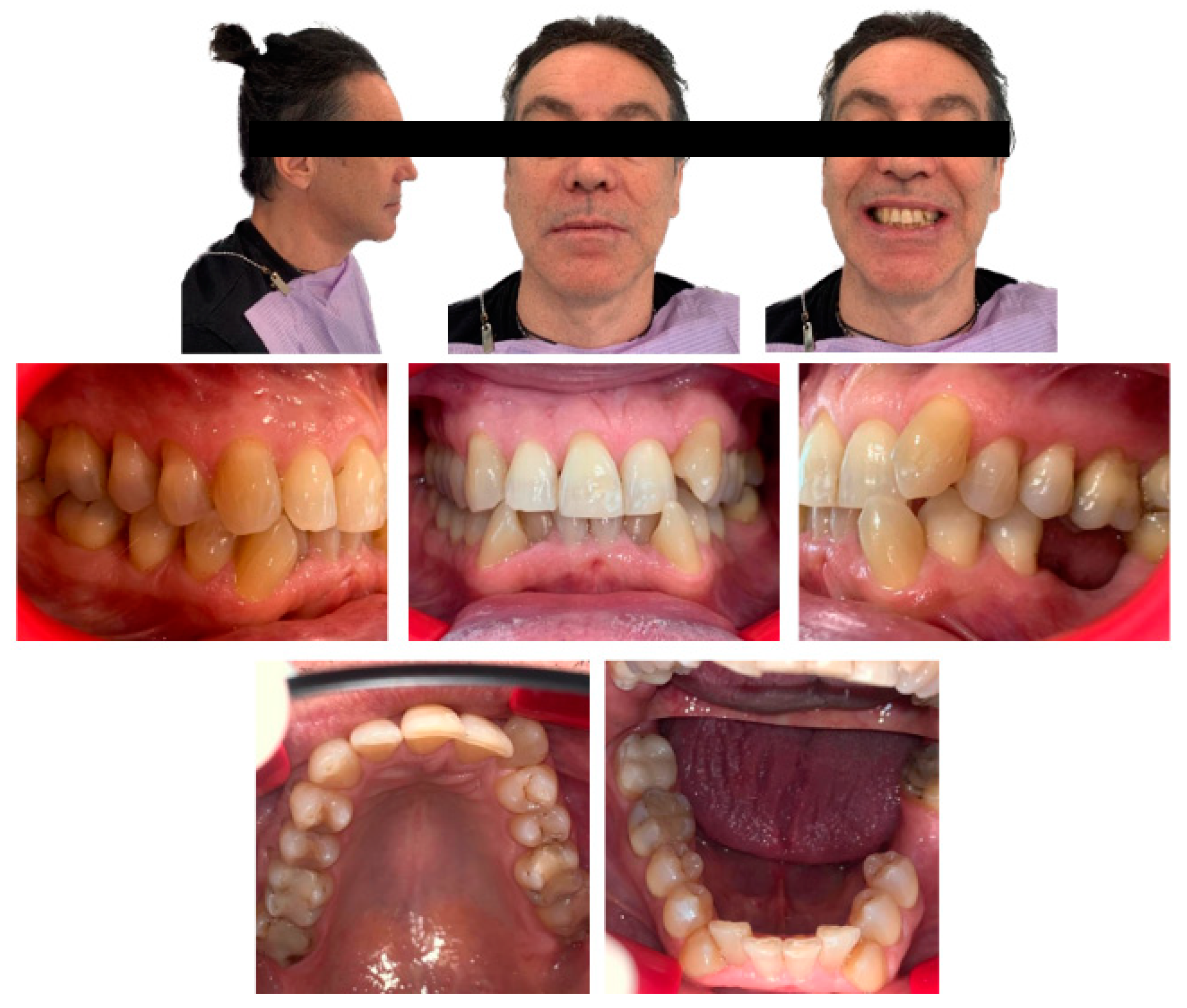
Figure 1. Extraoral and intraoral photographs taken at the first visit.
Applsci 13 09198 g001
Figure 2. Radiological instrumental examinations of the patient performed before starting orthodontic treatment: (a) orthopantomography, which highlights the lack of element 2.2 and element 3.6.—note the absence of space for element 2.2; (b) teleradiography of the skull in latero-lateral position on which cephalometric tracing was performed.
Applsci 13 09198 g002
Figure 3. Intraoral scan of the patient performed before orthodontic treatment.
Applsci 13 09198 g003
Figure 4. Intermediate extraoral and intraoral photographs taken during the orthodontic treatment.
Applsci 13 09198 g004
Figure 5. Extraoral and intraoral photographs taken following implant prosthetic rehabilitation at site 2.2 and 3.6.
Applsci 13 09198 g005
Figure 6. Radiological instrumental examinations of the patient performed at the end of the orthodontic treatment: (a) orthopantomography, which highlights the complete rehabilitation of the arches; (b) teleradiograph of the skull in latero-lateral position on which cephalometric tracing was performed.
Applsci 13 09198 g006
Table 1. Comparison of intraoral dental measurements taken at the start of therapy (T0) and at the end of therapy (T1).
MeasurementsT0T1
Bolton index0.181.87
LII16.6 mm0 mm
Intercanine width sup.33 mm35.2 mm
Interpremolar width sup.33.1 mm37.5 mm
Intermolar width sup.43.3 mm47 mm
Intercanine width inf.23.6 mm23.3 mm
Interpremolar width inf.26.5 mm29.8 mm
Intermolar width inf.--
Arch perimeter sup.66.5 mm70.8 mm
Arch perimeter inf.56.6 mm61 mm
Overjet0.7 mm3.6 mm
Overbite2.1 mm2.7 mm
LII = the Index of Irregularity by Little.
Table 2. Comparison of cephalometric values measured at the beginning and end of orthodontic therapy.
MeasurementsT0T1
SNA76.0°79.1°
SNB76.8°77.1°
ANB (Eastman Correction)1.7°3.0°
WITS INDEX−5.0−3.4
Maxillary incisor inclination112.3°116°
Mandibular incisor inclination86.5°91.6°
Interincisal Angle117.2°124°
SNA: angle between Sella-Nasion-Subspinale, SNB: angle between Sella-Nasion-Supramentale, ANB: difference between SNA and SNB, WITS Index: distance between the perpendicular of A and B on the occlusal functional plane.
Disclaimer/Publisher’s Note: The statements, opinions and data contained in all publications are solely those of the individual author(s) and contributor(s) and not of MDPI and/or the editor(s). MDPI and/or the editor(s) disclaim responsibility for any injury to people or property resulting from any ideas, methods, instructions or products referred to in the content.