Abstract
Since the da Vinci surgical system was approved by the Food and Drug Administration (FDA) in 2000, the development and deployment of various robot-assisted minimally invasive surgery (MIS) systems have been largely expedited and boomed. With the rapid advancement of robotic techniques in recent decades, robot-assisted systems have been widely used in various surgeries including orthopedics. These robot-related techniques are transforming the conventional ways to conduct surgical procedures. Robot-assisted orthopedic surgeries have become more and more popular due to their potential benefits of increased accuracy and precision in surgical outcomes, enhanced reproducibility, reduced technical variability, decreased pain, and faster recovery time. In this paper, robotic systems and navigation techniques in typical orthopedic surgeries are reviewed, especially for arthroplasty. From the perspective of robotics and engineering, the systems and techniques are divided into two main categories, i.e., robotic systems (RSs), and computer-aided navigation systems (CANSs). The former is further divided into autonomous RS, hands-on RS, and teleoperated RS. For the latter, three key elements in CANS are introduced, including 3D modeling, registration, and navigation. Lastly, the potential advantages and disadvantages of the RS and CANS are summarized and discussed. Future perspectives on robotics in orthopedics, as well as the challenges, are presented.
1. Introduction
In 1954, Devol invented the first digitally operated and programmable robot (later known as the Unimate) in the world, which is viewed as the foundation of the modern robotics industry [1]. Together with Engelberger, they founded the first robotics company in the world named Unimation. The company developed the first Unimate robot based on Devol’s patent and sold it to General Motors in 1960 to be used to lift and stack hot pieces of metal [2]. Since then, robots have continually been improved, and have spread their applications to the surgical field. In 1985, the first robotic surgical system, Puma 560, was used for neurosurgical biopsies guided by computed tomography (CT) images [2,3]. In the early 1990s, Minerva was introduced as the next-generation neurosurgical robot [4]. In 1988, ROBODOC (Integrated Surgical Systems, DE, USA) was introduced in orthopedics [4]. In the same year, PROBOT performed a clinical trial at Imperial College London with the earliest robotic procedure in urology. In 1993, a robotic arm called AESOP (Automated Endoscopic System for Optimal Positioning) (Computer Motion, Inc., Santa Barbara, CA, USA) was developed to assist in holding and positioning a laparoscopic camera. In 1998, both the ZEUS robotic surgical system (Computer Motion, Inc., Santa Barbara, CA, USA) and the da Vinci surgical system (Intuitive Surgical, Sunnyvale, CA, USA) were introduced into the market for use in teleoperated surgery [4], with the latter receiving the Food and Drug Administration (FDA) approval in 2000 for use in general laparoscopic surgery, which is considered legendary in the field of surgical robotics. Thereafter, medical and surgical robotics started to boom in various fields.
Minimally invasive surgery (MIS) allows the surgeon to conduct surgical procedures through much smaller incisions than traditional open surgery, thus having a faster recovery rate and shorter rehabilitation time as well as less pain for the patient [5]. Robot-assisted MIS involves a robot to improve the quality and precision of surgical procedures. Since the da Vinci surgical system was approved by the Food and Drug Administration (FDA) in 2000, the development and deployment of various robot-assisted MIS systems have been largely expedited [6,7].
With the rapid advancement of robotic techniques in recent decades, robotic systems have been widely used in various medical fields, such as neurological, laparoscopy, radiosurgery, prosthetics, rehabilitation, orthopedics, ophthalmology, and more [5,8,9,10]: for example, the da Vinci surgical system (Intuitive Surgical, Sunnyvale, CA, USA) and RAVEN II (University of Washington, and University of California, Santa Cruz, CA, USA) for use in teleoperated laparoscopic surgery [8,11,12], the CyberKnife System (Accuray Inc., Sunnyvale, CA, USA) for use in radiosurgery [13], and the JHU Steady-Hand Robot for use in retinal microsurgery [14,15,16]. These robotic systems and techniques are transforming the conventional ways to perform surgical procedures in a large variety of fields.
Through decades of technique evolution and clinical evaluations in orthopedic surgeries, plenty of studies have proven that robotic systems and navigation techniques can be beneficial in improving and enhancing surgical outcomes, such as increasing the accuracy and precision of bone cutting and component alignment, reducing operative time, and enhancing patients’ satisfaction [17,18,19].
Numerous review papers on reviewing robots or navigation systems in orthopedics can be found in the literature. Most of them are focused on a meta-analysis or reviewing clinical outcomes and user studies [2,19,20,21,22], a specific field like MIS [5], or a specific feature like haptic feedback [23]. Instead of those, this paper presents a historical review of the robotic systems and navigation techniques that exist and have ever existed in the field of orthopedics, especially on those systems still commercially available at present. And the primary focus is on the historical evolution of the systems as well as the engineering features and techniques from the perspective of engineering. Correspondingly, two main categories are covered, i.e., robotic systems (RS), and computer-aided navigation systems (CANS). The RS is further divided into autonomous RS, hands-on RS, and teleoperated RS, while the CANS is broken down into three key technical elements, including 3D modeling, registration, and navigation.
It is worth noting that in orthopedics, computer-assisted orthopedic surgery (CAOS) divides the surgical systems into three categories, i.e., autonomous (also known as active), semi-autonomous (also known as semi-active), and passive [17,24,25]. Correspondingly, the autonomous systems are equivalent to the autonomous RS used here, and the semi-autonomous systems are equivalent to the hands-on RS used here, while the passive systems indicate the computer-aided navigation systems (CANS) used in this paper. A hierarchical flowchart of these categories and their components is illustrated in Figure 1.
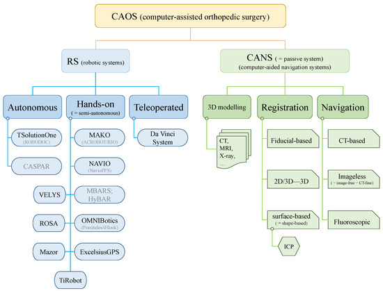
Figure 1.
A hierarchical flowchart for the robotic systems and navigation techniques in orthopedics. Note: system names in gray color means either not in use anymore or upgraded with new names; the equal symbol “=” means “equivalent to”.
Common orthopedic surgeries involving RS and/or CANS may include arthroplasty, arthroscopy, and surgical interventions related to tissues in joints. Note that in orthopedics, joint replacement is equivalent to arthroplasty, and similarly, total hip replacement (THR) is equivalent to total hip arthroplasty (THA), and total knee replacement (TKR) is equivalent to total knee arthroplasty (TKA). A general flowchart of surgical procedures for orthopedic surgeries is illustrated in Figure 2. Both RS and CANS play an important role in the procedures, which is introduced in detail in subsequent sections.
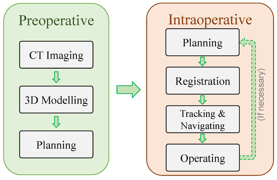
Figure 2.
A general flowchart for orthopedic surgeries.
The remainder of this paper is organized as follows. Section 2 presents the robotic systems (RSs) in orthopedics, including their historical background, applications, main features, and techniques. Section 3 presents CANS-related techniques, including 3D modeling, registration, and navigation. Section 4 provides discussions and future perspectives on RS and CANS in orthopedics, as well as some novel techniques. Section 5 presents a brief conclusion and future challenges.
2. Robotic Systems (RSs)
In this section, the robotic systems (RSs) in orthopedics are historically reviewed. For each of the RS, the background of development, the features, applications, techniques, advantages, and disadvantages are introduced.
Robotic systems (RSs) are divided into three subcategories, i.e., autonomous, hands-on, and teleoperated [23], as illustrated in Figure 1. The autonomous RS indicates that the robot can conduct the surgery completely on its own, while the surgeon can only interrupt it by using an emergency stop [26]. The hands-on RS is equivalent to semi-autonomous or semi-active robots in the literature of orthopedic surgeries. It indicates that the surgeon and robot cooperatively move the surgical instrument installed on the robot end effector (EE), which means it requires physical human–robot interaction (pHRI) [23]. The teleoperated RS indicates a standard leader–follower teleoperation system in which two robots are required. The leader robot is physically operated by the surgeon, while a follower robot on the remote site (e.g., on the patient side) is controlled by the leader robot via the internet or Ethernet.
2.1. Autonomous RS
2.1.1. ROBODOC → TSolution One
The ROBODOC surgical system (Curexo Technology Corporation, Fremont, CA, USA) is a fully autonomous robotic system initially designed for total hip arthroplasty (THA) in the 1980s [9,27], and introduced to be used on patients in 1992 [26,28]. It is the first robot that was clinically used in orthopedic surgery [2]. ROBODOC had its first system installed in Germany after being approved for sale by the European Union in 1994 [2,4]. ROBOTDOC was approved by the FDA (ROBODOC FDA 2008 file (accessed on 28 June 2023): https://www.accessdata.fda.gov/scripts/cdrh/cfdocs/cfpmn/pmn.cfm?ID=K072629) in 2008 for use in THA [19].
The whole system includes ORTHODOC (a 3D preoperative computer modeling and planning workstation) and ROBODOC surgical assistant (a 5-axis SACARA-type surgical robot) [25]. The system conducted its work, e.g., bone milling and preparing for stem implantation, based on preoperative computed tomography (CT) imaging. The workstation generates a 3D virtual model and produces a customized surgical plan [19]. The system employed fiducials implanted about 5 mm deep in the bone for bone motion detection and tracking; thus, the tracking accuracy is high and unaffected by debris and fluids [9]. Since ROBODOC is a fully autonomous system, once it starts to work, the surgeon only has control over the emergency stop [19].
The company Curexo Technology Corporation changed its name to Think Surgical, Inc. in 2014 [2,29]. Based on ROBODOC technology, its next-generation system named TSolution One (THINK Surgical, Inc., formerly Curexo Technology Corporation, Fremont, CA, USA) (THINK Surgical website (accessed on 28 June 2023): https://thinksurgical.com/) was developed. Its applications have been expanded to total knee arthroplasty (TKA) [4,19]. The TSolution One was approved by the FDA (TSolution One FDA 2019 file (accessed on 28 June 2023): https://www.accessdata.fda.gov/scripts/cdrh/cfdocs/cfpmn/pmn.cfm?ID=K191369) in 2019 for use in TKA. Currently, the TSolution One system has upgraded to a computer-aided system from the earlier CT-based system [2,4].
2.1.2. CASPAR
CASPAR (computer-assisted surgical planning and robotics) (OrthoMaquet/URS, Rastatt, Germany) was another early autonomous 6-degree-of-freedom (DOF) robotic system for THA and TKA [2,9]. It was introduced by OrthoMaquet in 1997, and acquired by Getinge (Gothenburg, Sweden) in 2000, and further acquired and discontinued by Universal Robot Systems (URS, Rastatt, Germany) in 2001 [8]. One example photo of the system is shown in Figure 3 [30].
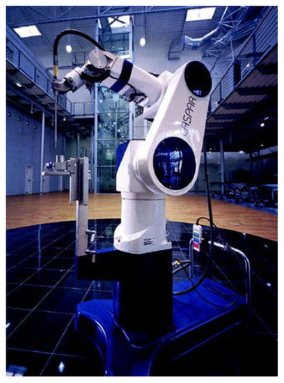
Figure 3.
CASPAR. (Reproduced with permission from J. Bellemans, Robotics in TKA, 2013, Springer Nature).
Similar to ROBODOC, the CASPAR also has an interactive computer system used for preoperative planning based on CT images [9]. The CASPAR also has similar functions, including automatically milling a bone and guiding the implant position in THA [19]. Therefore, it directly competed with the ROBODOC. Although some research studies showed increased accuracy of bone preparation and implant positioning in both TKA and THA with CASPAR [2,31,32], many other studies showed significantly less improvement in Harris Hip Scores, significantly longer procedure times, more blood loss, and higher rates of complications and revision surgeries compared to ROBODOC [2,9,33]. Therefore, CASPAR is no longer in use due to these drawbacks [2].
2.1.3. CyberKnife
CyberKnife (CyberKnife website (accessed on 28 June 2023): https://cyberknife.com/cyberknife-technology/) (Accuray Inc., Sunnyvale, CA, USA) is an image-guided robotic system that is specially designed for radiosurgery and radiotherapy [13,34]. The CyberKnife system can deliver stereotactic radiosurgery and radiation therapy anywhere in the body, including the spine and bone, although it is not a robotic system specially designed for orthopedics. The system autonomously conducts the non-invasive treatment procedures under the surgeons’ supervision. Initially conceived in 1992 [35] and fully described in 1997 [36], CyberKnife received FDA approval in 1999 (CyberKnife FDA 1999 file (accessed on 28 June 2023): https://www.accessdata.fda.gov/scripts/cdrh/cfdocs/cfpmn/pmn.cfm?ID=K984563) for use in intracranial treatment and in 2001 (CyberKnife FDA 2001 file (accessed on 28 June 2023): https://www.accessdata.fda.gov/scripts/cdrh/cfdocs/cfpmn/pmn.cfm?ID=K011024) for use in extracranial treatment [13]. As of 2020, CyberKnife has delivered treatments to over 400,000 patients worldwide [13]. A technical overview of the CyberKnife system can be found in [13].
2.2. Hands-On RS
Compared with autonomous RS, hands-on RS is much more preferred by surgeons due to the feature of surgeon-in-the-loop and full control by the surgeon. The following ACROBOT, RIO, and MAKO have the same core system but different names at different periods [37].
2.2.1. ACROBOT
The ACROBOT system (Acrobot Co., Ltd., London, UK) is a semi-autonomous system designed for robot-assisted MIS for unicompartmental knee arthroplasty (UKA) [3,26]. It is the first robot-assisted system used in UKA [37]. One example photo of the ACROBOT system is shown in Figure 4 [38].

Figure 4.
ACROBOT system. (Reproduced with permission from F. Rodriguez y Baena and B. Davies, Robotic Surgery: From Autonomous Systems to Intelligent Tools, Robotica, 28(2), 163–170, 2010, Cambridge University Press).
The ACROBOT was named as an acronym for active-constraint robot and was initially designed for knee surgeries [39]. Therefore, it employs active-constraint control, which can constrain the robot’s movement within a predefined zone [2,37], and thus the surgeon can safely cut the bone with high precision [2]. It provides haptic feedback to the surgeon and is considered a prototype of modern haptic systems [37]. ACROBOT is a 6DOF robot that allows only predefined trajectories [40]. It uses CT scans for preoperative planning. During the surgery, a small robot called ACROBOT was mounted on a gross positioning device and operated on by the surgeon [2]. A non-invasive anatomical registration will be conducted intraoperatively, based on which the drill can be tracked. If the surgical tool is detected to be away from the predefined cutting zone, the system will actively prevent it [37].
Acrobot Co., Ltd. was acquired by Stanmore Implants Worldwide in 2010, and based on the ACROBOT system, the Stanmore Sculptor Robotic Guidance Arm (RGA) System (Stanmore Implants, Elstree, UK) was released [37] and approved by the FDA (RGA FDA 2013 file (accessed on 28 June 2023): https://www.accessdata.fda.gov/scripts/cdrh/cfdocs/cfpmn/pmn.cfm?ID=K121765) in 2013 for use in UKA. MAKO Surgical Corp. obtained some confidential patents in 2013 as part of a patent infringement settlement [2,21]. Stryker Corporation acquired MAKO Surgical Corp. in 2013 and acquired Stanmore Implants Worldwide in 2016.
2.2.2. RIO (←ACROBOT)
The robotic arm interactive orthopedic system (RIO, previously called the tactile guidance system) (MAKO Surgical Corp., Fort Lauderdale, FL, USA) is the commercialized version of the ACROBOT system [8,23]; thus, it inherited many features from ACROBOT [26]. It can be used for UKA [26], and TKA [23]. It received FDA approval for use in TKA (RIO-MCK FDA 2009 file (accessed on 28 June 2023): https://www.accessdata.fda.gov/scripts/cdrh/cfdocs/cfpmn/pmn.cfm?ID=K090763) in 2009 and for use in THA (RIO-THA FDA 2010 file (accessed on 28 June 2023): https://www.accessdata.fda.gov/scripts/cdrh/cfdocs/cfpmn/pmn.cfm?ID=K093425) in 2010 [9].
The RIO system features haptic and auditory feedback, force-controlled tip, and surgeon-in-the-loop. The preoperative CT images are used to construct a 3D model of the patient’s knee. Then the 3D model is used by the surgeon to create a preoperative plan. The preoperative plan and the 3D model are used to finalize an intraoperative plan at the beginning of the operation, which includes defining an exact cutting zone for the robot. During the operation, the surgeon physically operates the RIO robot to perform bone resection while referencing the 3D bone model on a monitor. The robot will provide haptic and auditory feedback, while constraining the force-controlled tip of the tool (e.g., a rotating burr) to work only within the predefined cutting zone. The robot will automatically stop if the burr is outside of the predefined zone, or the computer has detected more bone being resected than necessary. Therefore, the RIO robot helps to monitor the operation, and provides necessary real-time data for accurate bone cutting and accurate components placement, thus potentially improving the outcomes of the UKA [26]. Therefore, the RIO system heavily relies on the preoperative plan and the surgeon’s skill. It is reported that the RIO system has a short learning curve, which is beneficial for surgeons with less experience in operating this system [26,41,42].
2.2.3. MAKO (←RIO)
MAKO system (Stryker Corporation, Kalamazoo, MI, USA) (MAKO SmartRobotics website (accessed on 28 June 2023): https://www.stryker.com/us/en/joint-replacement/systems/Mako_SmartRobotics_Overview.html) can be viewed as a new generation of the RIO (MAKO Surgical Corp., Fort Lauderdale, FL, USA) system, which means it is also a semi-autonomous system [10,19]. Founded in 2004, MAKO Surgical Corp., together with its most notable products RIO (robotic arm interactive orthopedic system) and MAKOplasty (for partial knee and total hip arthroplasty), was acquired by the Stryker Corporation in 2013. MAKOplasty received FDA approval in 2014 for use in THA (MAKOplasty FDA 2014 file (accessed on 28 June 2023): https://www.accessdata.fda.gov/scripts/cdrh/cfdocs/cfpmn/pmn.cfm?ID=K141989).
Being further developed and rebranded from the RIO, the MAKO system is an image-based system with haptic and auditory feedback. Preoperative CT images are acquired and used to conduct preoperative planning, which is further confirmed and adjusted intraoperatively based on the patient’s true bone anatomy before executing the surgery [2]. A special feature of the MAKO system is a haptic technology named AccuStop that provides the auditory beep alert, tactile vibration feedback, and visual feedback with color changes. This haptic technology can assist surgeons in making incisions, saving soft tissues and healthy bones [9]. One drawback of the MAKO system is the high purchasing and surgical costs [9].
Since 2021, the MAKO robots have been enhanced by integrating intraoperative sensor technology. MAKO is integrated with a computer navigation system, and it uses preoperative CT scans to generate a 3D model, then the robot arm is guided by the 3D model. MAKO is a human–robot collaboration system making use of user input and robotic guidance. Besides the haptic, visual, and audio feedback, it also provides virtual fixture (VF) protection and emergency auto-shutdown as a safety strategy. MAKO performed its first THA case in 2010 and received FDA (MAKO-THA FDA 2017 file (accessed on 28 June 2023): https://www.accessdata.fda.gov/scripts/cdrh/cfdocs/cfpmn/pmn.cfm?ID=K170593) approval in 2017. MAKO has also been used for UKA and TKA [19].
The MAKO system is gaining more popularity in clinical practice for UKA, THA, and TKA. In addition to its long history of nearly two decades, there is a large body of research literature about the MAKO system [2]. Many studies have revealed the positive outcomes of using the MAKO system compared to manual techniques. For more clinical case study results on MAKO, please refer to [2].
2.2.4. NavioPFS/NAVIO
NavioPFS (precision freehand sculpting) (Blue Belt Technologies, Plymouth, MN, USA) system is a computer-aided orthopedic navigation and surgical burring system, and is also a semi-autonomous, hand-held, and image-free system. It was approved by the FDA (NavioPFS FDA 2012 file (accessed on 28 June 2023): https://www.accessdata.fda.gov/scripts/cdrh/cfdocs/cfpmn/pmn.cfm?ID=K121936) in 2012 for unicondylar knee arthroplasty (uKA) [2].
The NavioPFS is a hand piece that has a cutting tool (a motorized burr) installed at the robot end effector, and the burr can extend and retract. A related safety strategy is that by modulating the retraction (exposure control) and speed of the burr (speed control) [27], inadvertent bone removal can be avoided, although there is no haptic feedback in this system. Another big advantage of this system is its imageless feature, meaning that preoperative CT imaging is not needed, and all the registration, planning, and navigation will be performed during the surgery. Note that the imageless feature is equivalent to the image-free feature in this paper. NavioPFS employs an optical-based navigation system via a passive infrared (IR) tracking camera and trackers. Intraoperative data from the trackers are collected and displayed in a graphical format, and together with anatomic landmarks and surface painting techniques, a 3D model of the patient’s femur and tibia can be created; thus, a surgical plan can be made by the surgeon intraoperatively [2,9].
The NavioPFS system has been proven to have reduced implant position errors [43], satisfied mechanical axis alignment accuracy, decreased bone cutting time, and improved Oxford Knee Scores [2]. The learning curve for operating the NavioPFS has been shown to be fairly rapid [44,45].
Blue Belt Technologies Inc., a Carnegie Mellon University spin-off company that was founded in 2003, was acquired by Smith & Nephew plc (London, UK) in 2016 [10]. In 2019, Smith & Nephew acquired the orthopedic unit of Brainlab, after which Smith & Nephew’s NAVIO surgical system upgraded its software to Navio 7.0. The software upgrade resulted in a significant reduction in the required data point collection and in the workflow stages, faster surface modeling, and improved usability [9]. The current Smith & Nephew’s NAVIO surgical system (NAVIO Surgical System website (accessed on 28 June 2023): https://www.smithnephewlivesurgery.com/navio-surgical-system) expanded the applications to TKA and UKA, and together with its PFJ software, it can assist the surgeon in making implant plans and help them to prepare the bone for implantation [24].
2.2.5. BRIGIT
The BRIGIT (bone resection instrument guidance by intelligent telemanipulator) system [46] was developed by MedTech SA (Montpellier, France). The system was implemented with a compliant control strategy aiming for more compliant and safer human–robot interaction [47]. It was integrated with a computer navigation system, and can be used for TKA to accurately position bone-cutting guides based on the preoperative plan [27,46]. According to the initial design, the system can be operated either in cooperation mode with physical human–robot interaction or in teleoperation mode with haptic feedback [46]. The system was acquired by Zimmer Biomet (Warsaw, IN, USA) in 2006, but was not commercialized [46].
2.2.6. MBARS/HyBAR
The MBARS (mini bone-attached robotic system) robot, as shown in Figure 5a, is a semi-autonomous robot developed at Carnegie Mellon University for TKA [48]. This type of system employs a special feature of small and bone-mounted robots, which are considered to be more efficient and cost effective than large robotic systems [26,48].
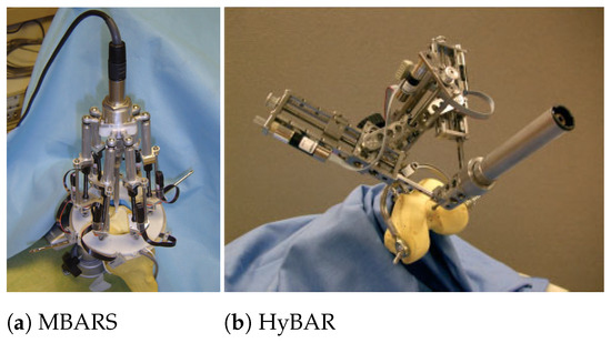
Figure 5.
MBARS and HyBAR. (Reproduced with permission from A. Wolf et al., MBARS: Mini Bone-Attached Robotic System for Joint Arthroplasty, International Journal of Medical Robotics and Computer Assisted Surgery, 1(2): 101–121, 2005, John Wiley and Sons; and with permission from S. Song et al., HyBAR: Hybrid Bone-Attached Robot for Joint Arthroplasty, International Journal of Medical Robotics and Computer Assisted Surgery, 5: 223–231, 2009, John Wiley and Sons).
The MBARS involves attached high-speed orthopedic tools; thus, the major design issues are considered to be structural rigidity and clamping mechanism [49]. To solve this, a HyBAR (hybrid bone-attached robot), as shown in Figure 5b was designed for patellofemoral joint arthroplasty [49]. The HyBAR is an autonomous system with enhanced structural rigidity by using hinged prismatic joints in its novel kinematic configuration, while a new modular clamping system was introduced to enhance the robotic procedure [2,49].
2.2.7. Praxiteles → iBlock
Praxiteles (Praxim Ltd., Grenoble, France), presented in 2005, is also a semi-autonomous system and is in the category of MBARS [50]. The Praxiteles, as shown in Figure 6, was designed to guide the saw blade or to use a passive bone-milling process, and the risk of soft tissue damage can be expected to be reduced by less-invasive exposures [27].

Figure 6.
Praxiteles. (Reproduced with permission from C. Plaskos et al., Praxiteles: A Miniature Bone-Mounted Robot for Minimal Access Total Knee Arthroplasty, International Journal of Medical Robotics and Computer Assisted Surgery, 1(4): 67–79, 2005, John Wiley and Sons).
The OMNINAV iBlock robotic cutting guide (formerly known as Praxiteles, OMNIlife Science (now owned by Corin Group), East Taunton, MA, USA) is a motorized, bone-mounted cutting guide for all femoral resections [51], and was approved by the FDA (iBlock FDA 2010 file (accessed on 28 June 2023): https://www.accessdata.fda.gov/scripts/cdrh/cfdocs/cfpmn/pmn.cfm?ID=K090953) in 2010 for use in TKA applications [2,27]. Similar to the NAVIO system, the iBlock is also imageless. Its OmniBiotics computer station can generate a 3D digital model of the patient’s knee by using patented bone-morphing technology and intraoperative anatomic data [27]. Based on the 3D model, the surgeon can make the implant plan intraoperatively and see the planned bone cuts before they are executed.
Although there are limited clinical data available for this system, some studies have shown that the iBlock system can obtain more efficient, accurate, and repeatable bone resections, as well as having a shorter bone preparation time [2,52]. The limitations of the iBlock system include having no haptic feedback, being limited to TKA applications, having a closed platform, and having limited kinematic assessment after implantation [2].
2.2.8. OMNIBotics (←iBlock)
The OMNIBotics knee system (OMNIBotics website (accessed on 28 June 2023): https://www.coringroup.com/healthcare-professionals/solutions/omnibotics/) (OMNIlife Science, Inc., East Taunton, MA, USA (acquired by Corin Group, Raynham, MA, USA, in 2019)), a new version of iBlock, is an image-free miniature bone-mounted robotic system for bone cutting and ligament balancing in TKA [4]. It initially received FDA (OMNIBotics knee system FDA 2017 file (accessed on 28 June 2023): https://www.accessdata.fda.gov/scripts/cdrh/cfdocs/cfpmn/pmn.cfm?ID=K163338) approval in 2017. The OMNIBotics system includes a bone-mounted robotic cutting guide (iBlock) for guiding bone resections and a robotic ligament tensioning tool (active spacer). The unique feature of integrating an active spacer allows for reproducible tensioning of the soft tissues accurately before and after the bone cuts, and for adjusting the interface fit between the implant and the bone intraoperatively.
The image-free feature does not require preoperative images. Instead, the 3D model of the patient’s anatomy is reconstructed via the bone-morphing technique intraoperatively, while the accuracy can be within 1 mm in all mapped areas [4]. The bone-mounted robot does not need camera-based tracking during robotic positioning and resection guidance. Once the robot is mounted and calibrated, the surgeon can perform the bone resection in sequence by operating on the robot-attached oscillating saw [4].
2.2.9. PiGalileo
PiGalileo (Plus Orthopedics AG, Switzerland, now owned by Smith & Nephew, UK), as shown in Figure 7, is a miniaturized and bone-mounted robotic cutting jig guided by a computer-aided navigation system for TKA and THA [27,53]. The robot is clamped to the bone before surgery, and a cutting guide is accurately positioned with the help of the navigation system [27]. Once the position is confirmed, the surgeon can perform the operation.
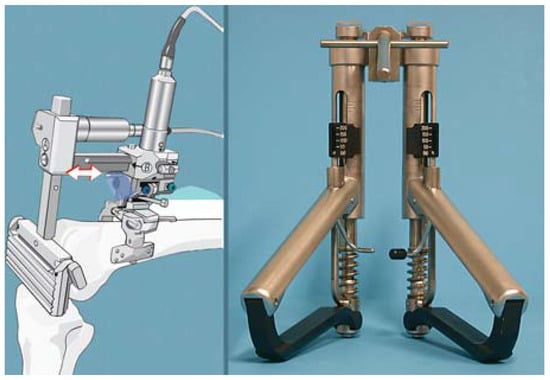
Figure 7.
PiGalileo System and ligament balancer. The ligament balancer with force and distance scale is used to measure the ligament tension and gap size. (Reproduced with permission from P. Ritschl et al., Modern Navigated Ligament Balancing in Total Knee Arthroplasty with the PiGalileo System, 2007, Springer Nature).
Its two computer-aided navigation systems for TKA (PiGalileo-TKR FDA 2006 file (accessed on 28 June 2023): https://www.accessdata.fda.gov/scripts/cdrh/cfdocs/cfpmn/pmn.cfm?ID=K061362) and THA (PiGalileo-THR FDA 2007 file (accessed on 28 June 2023): https://www.accessdata.fda.gov/scripts/cdrh/cfdocs/cfpmn/pmn.cfm?ID=K070731) were approved by the FDA in 2006 and in 2007, respectively [54]. The PiGalileo’s imageless navigation systems can assist the surgeon in bone cutting and implant positioning during joint replacement by collecting intraoperative data and tracking surgical tools and bone positions via stereotaxic technology and infrared (IR) markers.
The PiGalileo has been demonstrated to have good surgical outcomes, including high-accuracy implant positioning, improved bone-cutting precision, and shorter surgical times [27].
2.2.10. ROSA
ROSA (robotic surgical assistant) (Zimmer Biomet Robotics, formerly MedTech SA, Montpellier, France) is a robotic system for TKA and THA [17]. The FDA approved its ROSA hip system (ROSA Hip System FDA 2021 file (accessed on 28 June 2023): https://www.accessdata.fda.gov/scripts/cdrh/cfdocs/cfpmn/pmn.cfm?ID=K210998) in 2021, and the ROSA knee system (ROSA Knee System FDA 2022 file (accessed on 28 June 2023): https://www.accessdata.fda.gov/scripts/cdrh/cfdocs/cfpmn/pmn.cfm?ID=K213708) in 2022.
According to the FDA documents, the ROSA hip system (ROSA Hip System website (accessed on 28 June 2023): https://www.zimmerbiomet.com/en/products-and-solutions/zb-edge/robotics/rosa-hip-system.html) (as shown in Figure 8a) is a CT-free, fluoroscopic-guided system that can be used to assist surgeons in accurately positioning and implanting hip components, while the robotic arm is used to assist in the guidance of the surgical tools. Fluoroscopic images, intraoperatively acquired by a C-arm, will be used to determine the surgical tools’ orientation in relation to the patient’s anatomy and as a guide for bone component orientation. The system provides pre-, intra-, and post-operative measurements relative to the patient’s anatomy. The robotic arm is kept stationary to keep the instruments in a fixed orientation during bone component implanting.

Figure 8.
ROSA systems. (Reproduced with permission from © Zimmer Biomet, Warsaw, IN, USA).
The ROSA knee system (ROSA Knee System website (accessed on 28 June 2023): https://www.zimmerbiomet.com/en/products-and-solutions/specialties/knee/rosa-knee-system.html) (as shown in Figure 8b) can assist surgeons with bone resections and assessing the state of soft tissues to facilitate implant positioning intraoperatively. This system can be either image based or imageless. For the image-based option, a preoperative 3D virtual bone model needs to be generated preoperatively, which can be used by the surgeon to make a preoperative surgical plan. For the imageless option, the landmarks data on the patient’s bony anatomy are collected intraoperatively and used to create an intraoperative surgical plan. The accuracy of the resections, knee state evaluation, and soft tissue assessment are the same between the two options since both of them are always based on intraoperative landmarks. The robotic arm can assist in precisely positioning the component relative to the implantation plan.
ROSA Spine was developed for minimally invasive spine procedures around 2015 [55,56] and approved by the FDA (ROSA Spine FDA 2016 file (accessed on 28 June 2023): https://www.accessdata.fda.gov/scripts/cdrh/cfdocs/cfpmn/pmn.cfm?ID=K151511) in 2016. The system includes a patient-side cart bearing for a 6DOF robotic arm and a workstation, and an optical camera serving navigation purposes. In addition to pedicle screw placement, it is also promising to be used for spinal fusion, percutaneous endoscopic lumbar discectomy, intracorporeal implant positioning, and radiofrequency ablation [55,56,57]. By coupling with intraoperative flat-panel CT guidance, the system can perform accurate pedicle screw placement [55]. Registration can be performed automatically by using a fiducial box (held by the robotic arm) and a percutaneous reference pin. The robot can monitor and follow the patient’s body movements in real time by tracking the movement of the vertebrae; and thus, real-time robotized navigation guidance can be provided by the system. Preoperative and postoperative 3D CT scans can be acquired by an O’arm device and transferred to the workstation. Then, by co-registration, the difference between the initial 3D planning and the actual screw positions can be measured [55]. Accurate screw placement can be achieved due to the robot’s capability for real-time movement tracking on the vertebrae [55]. Later, the ROSA Spine System was integrated into the ROSA ONE System (ROSA ONE Spine website (accessed on 28 June 2023): https://www.zimmerbiomet.eu/en/products-and-solutions/zb-edge/robotics/rosa-spine.html) (as shown in Figure 8c) and approved by the FDA in 2019 [57].
2.2.11. VELYS
The VELYS robotic-assisted solution (VRAS) (VELYS Robotic-Assisted Solution website (accessed on 28 June 2023): https://www.jnjmedtech.com/en-US/patient/velys/robotic-assisted-solution) (DePuy Synthes, now owned by Johnson & Johnson, Warsaw, IN, USA), as shown in Figure 9 [58], is a new system designed from proprietary technology developed by Orthotaxy. It performed its first TKA case in Auckland, New Zealand in 2020 [59], and was approved by the FDA (VELYS FDA 2021 file (accessed on 28 June 2023): https://www.accessdata.fda.gov/scripts/cdrh/cfdocs/cfpmn/pmn.cfm?ID=K202769) in 2021.
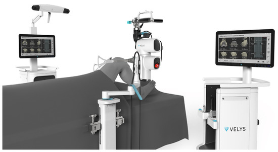
Figure 9.
VELYS robotic-assisted solution (Reproduced from G.W. Doan et al., Image-Free Robotic-Assisted Total Knee Arthroplasty Improves Implant Alignment Accuracy: A Cadaveric Study, The Journal of Arthroplasty, 37(4): 795–801, 2022, Elsevier).
VELYS employs a novel patient-specific TKA technique that can intraoperatively collect accurate data on both the bony anatomy and the soft tissue envelope of the knee [59]. This technique allows the surgeon to make an intraoperative plan, while preserving the soft tissues during the TKA surgery. The robot-assisted saw can assist in conducting the implantation plan precisely, accurately, and efficiently [59]. Early outcome results and limited data indicate favorable outcome scores and high patient satisfaction [59].
2.2.12. Mazor
Spine Assist (Mazor Robotics, Caesarea, Israel) is the first robotic system used in spine surgery and was approved by the FDA (Mazor SpineAssist FDA 2004 file (accessed on 28 June 2023): https://www.accessdata.fda.gov/scripts/cdrh/cfdocs/cfpmn/pmn.cfm?ID=K033413) in 2004 [57]. The Spine Assist system evolved into the Renaissance system, which was approved by the FDA (Mazor Renaissance FDA 2011 file (accessed on 28 June 2023): https://www.accessdata.fda.gov/scripts/cdrh/cfdocs/cfpmn/pmn.cfm?ID=K110911) in 2011, and then Mazor X, which was approved by the FDA (Mazor X FDA 2017 file (accessed on 28 June 2023): https://www.accessdata.fda.gov/scripts/cdrh/cfdocs/cfpmn/pmn.cfm?ID=K163221) in 2017, and then Mazor X Stealth (Medtronic, Dublin, Ireland) which was approved by the FDA (Mazor X Stealth FDA 2018 file (accessed on 28 June 2023): https://www.accessdata.fda.gov/scripts/cdrh/cfdocs/cfpmn/pmn.cfm?ID=K182077) in 2018. The example products of Mazor systems are shown in Figure 10. Note that Mazor Robotics (Mazor Spine Robotics website (accessed on 28 June 2023): https://www.medtronic.com/ca-en/healthcare-professionals/therapies-procedures/spinal-orthopaedic/spine-robotics.html) was acquired by Medtronic in 2018. A detailed comparison between these four versions of the Mazor systems can be found in [57]. Nowadays, the robotic system is just called Mazor. A systematic review on robotics in spine surgery can be found in [60,61].
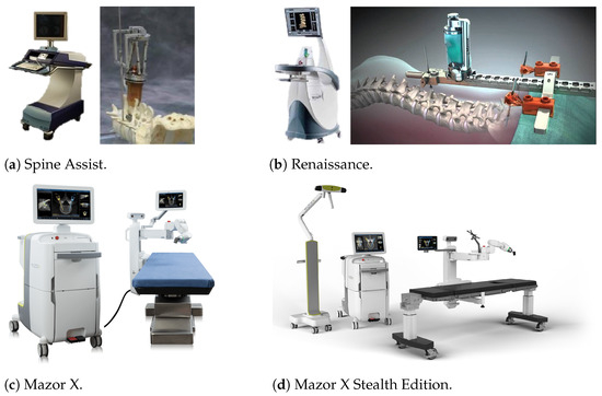
Figure 10.
Mazor systems for spine surgery. (Reproduced with permission from © Medtronic, Dublin, Ireland).
The Spine Assist robotic system can be used for pedicle screw placement in spine surgery, but the accuracy of the screw placement is relatively low [55,57]. The updated version, the Renaissance system, preserves a similar operational workflow but with significant software changes [57]. Finally, the latest version of the Mazor system, Mazor X Stealth, has an accuracy of around 99–100% for the screw placement. Both Spine Assist and Renaissance require preoperative CT scans, based on which preoperative planning (e.g., optimal implant size and trajectory) is conducted. Nowadays, the Mazor system can be used in conjunction with the imaging system O-arm for 3D images, besides the use of a CT scan (Scan and Plan workflow). Before the spine surgery, the robot is mounted to the patient’s spine. During the surgery, intraoperative fluoroscopic images of the anatomy are acquired and matched with the preoperative CT scans in real time, and this procedure is called the CT-to-Fluoro workflow. Alternatively, the Scan and Plan workflow can be used. In both workflows, the robotic arm provides guidance according to the preplanned trajectory [57].
As mentioned earlier, the upgraded versions of Mazor (Mazor X Stealth website (accessed on 28 June 2023): https://www.medtronic.com/ca-en/healthcare-professionals/products/spinal-orthopaedic/spine-robotics/mazor-x-stealth-edition.html) do not require preoperative CT scans anymore, while a feature of instrument tracking is also added [57,62]. Although preoperative CT is not mandatory, Mazor can accept preoperative or intraoperative CT for screw planning. Before the spine surgery, the robot is attached to a table and then mounted to the patient’s spine. During the surgery, 3D images are acquired, and then the intraoperative anatomy is matched with the CT scan via fluoroscopic images for intraoperative guiding purposes. The robot arm performs procedures according to the preplanned trajectory while a 3D camera offers real-time instrument tracking [57]. The latest version of Mazor is integrated with Medtronic’s Stealth navigation system, which can further improve the navigation accuracy.
2.2.13. ExcelsiusGPS
ExcelsiusGPS (ExcelsiusGPS website (accessed on 28 June 2023): https://www.globusmedical.com/musculoskeletal-solutions/excelsiustechnology/excelsiusgps/) (Globus Medical, Audubon, PA, USA), as shown in Figure 11, is a robotic system with real-time image guidance for spine surgery and was approved by the FDA (ExcelsiusGPS FDA 2017 file (accessed on 28 June 2023): https://www.accessdata.fda.gov/scripts/cdrh/cfdocs/cfpmn/pmn.cfm?ID=K171651) in 2017 [57]. The robotic arm is mounted on the floor rather than the patient’s bone. Preoperative CT scans are not mandatory but optional for screw trajectory planning. Optionally, intraoperative CT or radiographs can also be used for the planning. The ExcelsiusGPS system employs a shock-absorbing dynamic reference base and a separate surveillance marker and associated surveillance software to improve the system navigation integrity [57,63]. Also, the ExcelsiusGPS system employs an extremely rigid arm for its robotic guidance system, which can achieve an accuracy of less than 1 mm of tool deflection under a lateral disturbance force of 200 N. Additionally, the system alerts the surgeon if any tool deflection is detected by the surveillance software during the surgery [57,63].
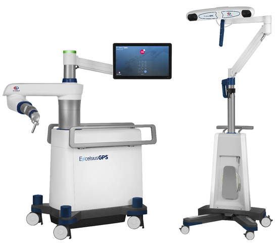
Figure 11.
Globus ExcelsiusGPS for spine surgery. (Reproduced with permission from © Globus Medical, Inc., Audubon, PA, USA).
2.2.14. TiRobot
TiRobot (TiRobot website (accessed on 28 June 2023): https://en.tinavi.com/index.php?c=article&a=type&tid=1) (TINAVI Medical Technologies, Beijing, China) is a robotic system developed in China for use in spine surgery and received China FDA approval in 2016 [64]. The TiRobot platform consists of three components, including a workstation, an optical tracking camera, and one 6-DOF floor-mounted robotic arm as shown in Figure 12 [64]. The fluoroscopic image-based registration employs a cross-referencing approach by using a dynamic reference base on the patient’s body and another dynamic reference base on the robotic arm [60]. The TiRobot system can achieve real-time 3D navigation by tracking the relative positions of the patient and the robotic arm [60]. The workstation houses the interface for screw planning and visual feedback [64]. The robotic arm can help surgeons accurately position the surgical tools and implants. TiRobot can also be used for other surgeries, like intramedullary nail fixation for intertrochanteric fractures [65].

Figure 12.
TINAVI TiRobot for spine surgery. (Reproduced from H. Lan et al., Intramedullary Nail Fixation Assisted by Orthopaedic Robot Navigation for Intertrochanteric Fractures in Elderly Patients, Orthopaedic Surgery, 11: 255–262, 2019, John Wiley and Sons).
2.3. Teleoperated RS
Da Vinci Surgical System
The da Vinci surgical system (Da Vinci website (accessed on 28 June 2023): https://www.intuitive.com/en-us/products-and-services/da-vinci) (Intuitive Surgical, Sunnyvale, CA, USA), as shown in Figure 13, is the most successful teleoperated robotic system for MIS (minimally invasive surgery) in the market. Initially, the system was designed for microvascular surgery [66]. It was used to perform a robot-assisted heart bypass procedure in 1998 in Germany and a robot-assisted radical prostatectomy procedure in 2000 in Paris, France [4]. It received FDA (Da Vinci FDA 2000 file (accessed on 28 June 2023): https://www.accessdata.fda.gov/scripts/cdrh/cfdocs/cfpmn/pmn.cfm?ID=K990144) approval in July 2000 for laparoscopic surgery, and since then, the system has expanded its applications into various surgical fields and procedures [66]. Lots of surgeries in a wide variety of fields have been successfully performed by this system, such as prostatectomies, cardiac valve repair, and gynecologic surgical procedures [9]. The da Vinci surgical system further received FDA approval in 2001 for use in prostate surgery, in 2002 for mitral valve repair surgery, in 2005 for gynecological surgery [4].

Figure 13.
The da Vinci surgical system. (© 2023 Intuitive Surgical Operations, Inc.).
The da Vinci system is a teleoperated system, and it consists of two patient-side manipulators (PSMs), one endoscopic camera manipulator (ECM), and two master tool manipulators (MTMs). The surgeon will remotely control the PSMs by physically operating on the MTMs, while the remote scene on the PSM site will be presented to the surgeon via a console by using the ECM. The da Vinci system can translate the surgeon’s hands manipulation movements from MTMs to PSMs via the console in real time, such as bending, rotating, grasping, palpating, and cutting, while providing haptic feedback to the surgeon for an immersive experience.
In orthopedics, the applications of the da Vinci surgical system can be largely limited due to the fact that the system is designed to be more suitable for manipulating soft tissues (e.g., suturing, ablation, needle insertion) than rigid bones (e.g., cutting, burring). Some surgeries regarding soft tissues or nerves in orthopedics have been performed using the da Vinci system. For example, the da Vinci system was successfully used for ulnar nerve decompression at the elbow [6] and supraclavicular brachial plexus dissection and nerve root grafting at the shoulder [67]. Some cases of anterior lumbar interbody fusion (ALIF) in spine surgery were also reported to be successful by using the da Vinci surgical system [68,69].
2.4. RS Remarks
The RS systems in the timeline are illustrated in Figure 14. Currently, the time-honored robotic systems, including TSolution One (formerly ROBODOC; THINK Surgical, Fremont, CA, USA), MAKO (formerly ACROBOT/RIO; Stryker Corporation, Kalamazoo, MI, USA), NAVIO (Smith & Nephew plc, London, UK), and Mazor (Mazor Robotics, Caesarea, Israel), are still available and continue to improve and thrive in the market. Compared to their relatively large robot body, on the other hand, MBAR represents an emerging trend of mini bone-attached robots with potentially more efficiency and lower cost. ROSA and VELYS are newly developed products in the market. The da Vinci surgical system represents a more versatile robot that has a large potential to be used in a large variety of surgeries for the teleoperated systems used in MIS. In general, by reviewing the robotic systems developed for orthopedics in the past decades, we can see that their functions are mainly focused on bone cutting, positioning, and alignment; additionally, precision has been increasing, whereas the variability has been decreasing [2].
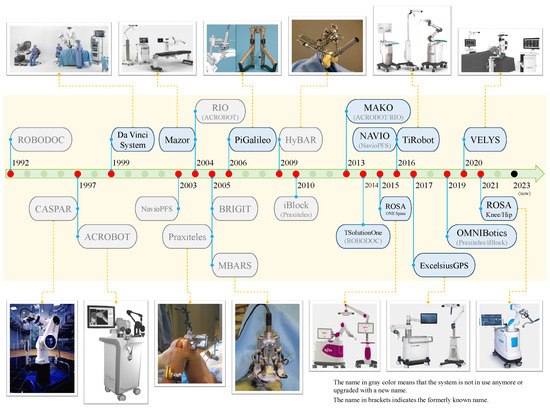
Figure 14.
Robotic systems (RSs) in the timeline.
For autonomous RS, there is still caution about using it in orthopedic surgery due to the ethical issues and safety concerns surrounding autonomous operation [26]. Typical issues and concerns include potential muscle/nerve damage and technical complications. For example, when a procedure stop occurs during bone cutting, re-registration is required, while, sometimes, frequent registration failures may cause the surgery to abort [25].
Compared to autonomous RS, hands-on RS may be more acceptable by surgeons due to the feature of human-in-the-loop [25]. When operating a hands-on RS, the surgeons have full control of the robot and can stop the surgical operations (e.g., bone cutting) at any time they want. This can ensure the maximal safety of the patient.
For teleoperated RS, their applications in orthopedics are limited. The main reason is that orthopedic surgeries are more related to manipulation with rigid bone cutting and implant alignments, which are not suitable for teleoperated RS. However, for those orthopedic surgeries regarding soft tissues, nerves, and vascular, the teleoperated RS could still be applicable and useful.
3. Computer-Aided Navigation Systems (CANSs)
Computer-aided navigation systems (CANSs) can be taken as a parallel category to RS. The CANS focuses on navigation with the help of computers. It can be either integrated with an RS or independent from an RS. When integrated together, all the coordinates of the CANS (e.g., CT image frame, and external camera frame) are registered into the coordinates of the RS, then for navigation. When independent from an RS, i.e., no robots appear in surgery, all the coordinates are registered into the digital patient’s model/image or the camera frame, then for navigation.
In the category of computer-aided navigation systems (CANSs), three basic elements are included, i.e., 3D modeling, registration, and navigation. Strictly, the CANS are computer-assisted systems rather than robot-assisted systems. However, a robot-assisted system (here equivalent to RS) usually includes a CANS system implicitly or explicitly. Implicitly means that an RS itself can be viewed as a special navigation system since any point in the robot workspace can be tracked based on robot kinematics [17]. Explicitly means that a CANS can be integrated with a robotic system to enhance the system’s ability (e.g., tracking and visualization). Therefore, a CANS can be used either independently or integrated with an RS, which means a CANS can be an essential part of assisting surgeons in surgery, no matter whether a robot is involved or not. This also means that the CANS has wider and more general applications than RS in orthopedics and beyond.
In CAOS tripartite categories, the term of passive systems is equivalent to the CANS here [17,25]. The CANS does not perform any actions on patients, thus having no relevant safety concerns. Instead, the CANS only collects intraoperative data and provides visualized information and guidance to the surgeons, thus helping them to better achieve their surgical objectives accurately and precisely.
A large amount of case studies have shown that the CANS can offer more accurate surgical outcomes, such as placement of the components in UKA, alignment of the femoral and tibial components, tibial slope, and the mechanical axis [17,25].
3.1. Three Basic Elements
A complete set of CANS techniques in orthopedics includes three basic elements, i.e., 3D modeling, registration, and navigation [10].
3.1.1. 3D Modeling
Three-dimensional (3D) modeling is about reconstructing a three-dimensional digital model of the patient’s bone, and then the model can be further used to make preoperative planning by the surgeon. Typically, the 3D model is reconstructed from preoperative images (e.g., CT, X-ray, MRI). For example, most of the previously introduced RSs conduct the 3D modeling based on preoperative CT scans. Some other systems (e.g., NAVIO) generate the 3D model by using bone-morphing techniques and intraoperative tracking data, then visualizing the 3D model in a graphical format.
3.1.2. Registration
Registration is a core and compulsory procedure for any CANS system or RS system being used in orthopedic surgeries. The quality of registration fundamentally and directly determines the accuracy and precision of the surgical outcomes [17]. It is worth mentioning that both RS and CANS are heavily reliant on registration, with RS being more so than an independent CANS due to the fact that the surgical work is performed by the robotic-attached tool in RS rather than the surgeon alone [17].
Before the surgery in the operating room, registrations between the patient’s true bone, the corresponding 3D model, the robotic system (if applicable), and the surgical tool need to be conducted first. Some methods may be needed to ensure accurate registration. There are three typical registration methods, including fiducial-based paired-point matching, surface-based, and 2D/3D–3D registration [25]. ROBODOC initially used fiducial-based registration and changed to surface-based registration in 1999; studies have proved that surface-based registration is as accurate as fiducial-based registration [25].
The fiducial-based paired-point matching method is a modified version of the paired-point matching method [25]. To obtain high-accuracy and reproducible registration, fiducials need to be placed into the target bone before the preoperative CT scanning such that these fiducials appear in the CT images. These fiducials are used for registration by using the paired-point matching method. During the surgery, these fiducials are used as the reference points for the patient’s bone, and related to the preoperative plan. However, fiducial-based registration requires an additional minor operation of placing the fiducials into the bone before the preoperative CT scanning, which brings fiducial site pain or inflammation to the patients [25].
Surface-based (also known as shape-based) registration does not need fiducials. It employs the widely used iterative closest point (ICP) algorithm and the least-squares method to match the points on the 3D model surface with those on the patient’s bone surface [70,71]. To avoid local minima, the paired-point method is used first to perform baseline registration, then a certain number of points is used to perform a refinement of the surface-based registration [25]. An advantage of surface-based registration is that it can be conducted and updated intraoperatively and in real time, which makes it more robust than other registration methods.
The 2D-3D or 3D-3D registration method makes use of intraoperative fluoroscopic images [25]. Although this method has been shown to be accurate for use in robot-assisted THA in a lab setting, it has not yet been widely accepted by clinical robotic surgeries [25].
3.1.3. Navigation
Navigation is the kernel element and function of a navigation system. Given an accurate 3D model and accurate registration, the surgical tools and the patient’s bone landmarks can be precisely tracked, either by a set of tracking devices or by the robot coordinate system. A sensor-based navigation system usually uses optical sensors or magnetic sensors to track the 3D positions of the target bones, the surgical tools, and any other objects (e.g., implants) that need to be tracked [25]. An optical IR-based system can track objects with high accuracy by using infrared (IR) light-tracking cameras and infrared light-reflecting markers [25]. However, the optical sensor-based tracking system can be easily affected by light–sight blocking. The magnetic sensor does not have this problem, but it may be affected by metallic objects within its workspace [25]. A novel approach for active optical navigation was proposed recently, where the optical tracking system (OTS) is installed on a robot that can actively adjust the pose of the OTS [72].
Based on the working principles, CANS includes three typical types of navigation systems being used in orthopedic surgeries, i.e., CT-based, imageless, and fluoroscopic [25]. In 1994, two years after the ROBODOC was first used on humans, the first CT-based navigation system was developed and used in THA in Pittsburgh [17,73]. CT-based navigation prevails among most of the previously introduced robotic systems. Although CT-based navigation has the highest accuracy, a big disadvantage is that acquiring preoperative CT images and making preoperative plans based on them are time consuming, which may bring more cost and radiation exposure to patients [25].
Imageless navigation, also known as “surgeon-defined anatomy” technology, employs some other techniques, such as infrared (IR)-based tracking, stereotaxic technology, and bone-morphing technology. Note that, here, imageless or image-free means that no preoperative CT images are required but may still need camera-based intraoperative tracking for navigation. Together with the intraoperatively collected patient’s anatomic data, an abstract of the patient’s anatomy can be generated [17]. This kind of imageless technique is adopted by some robotic systems, such as the NAVIO system and Praxiteles. The accuracy of imageless navigation depends on the techniques adopted.
Fluoroscopic navigation has a similar principle to imageless navigation but uses fluoroscopic images. Fluoroscopic navigation is good for use in trauma and spine surgeries but is limited in other orthopedic surgeries due to its cumbersome registration procedures [25].
3.2. Typical Systems
3.2.1. Stryker Navigation system
Stryker Navigation system II Cart (Stryker NAV3i website (accessed on 28 June 2023): https://www.stryker.com/us/en/navigation/products/nav3i.html) (Stryker, Kalamazoo, MI, USA) is an optical-based navigation system with an optical localizer accuracy of about 0.07 mm [71]. The navigation system can reconstruct the 3D bone model by using the original CT data. Dynamic reference trackers are placed on the patient’s bone for later registration with considerations of avoiding nerve injuries and interrupting navigation pointer operations [71]. A study on validating the registration accuracy of a Stryker navigation system (Stryker II cart Navigation system) for elbow arthroscopic debridement showed that the registration accuracy can be within 1 mm [71].
3.2.2. OrthoPilot
OrthoPilot (OrthoPilot website (accessed on 28 June 2023): https://www.bbraun.com/en/products-and-solutions/therapies/orthopaedic-surgery/orthopilot.html) (Aesculap/BBraun, Germany), as shown in Figure 15, is a pure navigation system that conducted its first surgery in 1997 [18]. It is an imageless navigation system, and it works based on intraoperative data acquired by an optical tracking system [18]. The tracking system consists of an infrared (IR) tracking camera and IR Spectra localizer (Northern Digital Inc., Waterloo, ON, Canada). To date, the OrthoPilot has been used in TKA, revision TKA, and THA. A detailed introduction to using OrthoPilot over two decades can be found in [18].
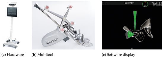
Figure 15.
OrthoPilot navigation system. (Reproduced with permission from © Aesculap AG, Tuttlingen, Germany).
3.2.3. Brainlab Knee3
Brainlab Knee3 (Brainlab Knee3 website (accessed on 28 June 2023): https://knee3.brainlab.com/#/main-menu) (Brainlab AG, Munich, Germany), as shown in Figure 16, is a new imageless navigation system for use in TKA [59]. It is featured with real-time assessment of the knee as a complete kinematic structure and predicting final joint stability at each surgical step. It can display X-rays and templated plans during navigation. It can also predict and visualize the virtual interaction between 3D kinematics, joint stability, and implant alignment [74].
Figure 16.
Brainlab Knee3. (Reproduced with permission from © Brainlab AG, Munich, Germany).
3.3. CANS Remarks
The three basic elements of CANS, i.e., 3D modeling, registration, and navigation, are critical conditions for each other. Three-dimensional modeling is the necessary prerequisite for registration, while registration is the necessary prerequisite for navigation. The accuracy of 3D modeling directly affects the accuracy of registration, and the latter will further determine the accuracy of the navigation.
The CANS is an independent technique that can be either used together with the RS or used alone during surgical procedures. The applications of CANS are much wider and more general than RS since a robot may be not demanded or applicable in some surgeries, while navigation can be always helpful. From another perspective, the existing RS mainly focuses on hip/knee arthroplasty, while the CANS has a relatively wider variety of applications, such as for the shoulder [22], elbow [75], and ankle. A systematic review of CT-based intraoperative navigation techniques used in total shoulder arthroplasty (TSA) can be found in [22].
4. Discussions and Future Perspectives
Both CANS and RS have been playing an increasingly critical role in modern orthopedic surgeries. According to a review study [17,76], the ratio of patents to publications related to CANS and RS in knee arthroplasty increased from around 1:10 in 2004 to around 1:3 in 2014. The benefits brought by RS and CANS include but are not limited to augmenting the surgical procedures, fine tuning surgical plans to personalized patient profiles, and proving intraoperative data and real-time visualization to the surgeons for a more accurate and precise surgical outcome [17].
With decades of evolution, RS and CANS themselves have gone through critical improvement and upgrading. Without a doubt, RS and CANS will continue to thrive and play an indispensable role in orthopedics.
A critical unique feature of RS in orthopedics is that they must be capable of dealing with high forces and stiffness due to the rigid nature of their target object of bones, while the da Vinci surgical system is more suitable for soft-tissue-related procedures. The main advantages of RS applied in orthopedics include increased accuracy and precision of implant positioning, enhanced reproducibility, improved implant stability, and less resulting pain. On the other hand, the main disadvantages of RS include potential safety concerns, high economic costs, and potentially longer operative times.
CANS will continue to develop along two parallel paths. One is to integrate with robotic systems, another is to be used alone without involving RS. For the latter, CANS is capable of being used in more versatile surgeries, where robots are not needed or not yet available. In that case, with the help of CANS, surgeons can perform conventional surgeries with potentially better and more accurate surgical outcomes.
4.1. RS and CANS for Various Orthopedic Surgeries
From Table 1, it can be found that most of the robotic systems are applied for hip/knee surgeries, some are for spine surgeries, and no specific robotic system is exclusively for elbow/shoulder surgeries. For example, ROSA has robot-assisted systems for the hip, knee, and spine, respectively, but only has a computer-aided system for assisting in planning and navigation for the shoulder. RSs have been abundantly developed and frequently used in hip/knee surgeries but not in elbow/shoulder/foot/ankle surgeries, while the latter seems more favorable for CANSs [77]. The possible reason could be that there are much more cases and higher demand for knee/hip surgery than elbow/shoulder surgery. The knee is the largest hinge joint in the body, while the hip is a large ball-and-socket joint. Both the knee and hip joints take a lot of wear, tear, and stress from daily activities (e.g., walking, running, and jumping) while supporting the body weight in horizontal and vertical directions, and they are more vulnerable to injury and osteoarthritis than the elbow/shoulder or any other joints.

Table 1.
Robotic systems (RSs) in orthopedics.
Technically, all joint replacement/arthroplasty surgeries are open surgeries since a significant incision needs to be made in order to expose the bone for bone cutting or implant positioning. It is worth noting that in joint replacement/arthroplasty, minimally invasive approaches/procedures have different definitions, such as shorter incision length, nondissection of quadriceps tendon, noneversion of the patella, or nondislocation of the tibiofemoral joint [78]. Therefore, strictly speaking, all joint replacement/arthroplasty are open surgeries rather than MIS, the latter of which only requires several trocars to be made in order to insert the surgical instruments for performing the surgery. In this sense, arthroscopic surgeries belong to the category of MIS. For arthroscopic surgeries, several portals are made to insert an arthroscope and surgical tools, and the surgical tools are used to perform the surgery with the help of the arthroscope. Arthroscopic surgeries can be used for a large variety of indications. For example, elbow arthroscopy can be used for the management of stiffness related to degenerative arthritis, loose bodies, lateral epicondylitis, synovitis, osteochondritis dissecans, symptomatic plica, infection, contracture, instability, and fracture management [79].
Arthroscopic surgery (e.g., arthroscopic debridement) is an active field that is being transformed by techniques of RS and CANS, although there is yet no specific robotic system specially designed for them. One possible reason is that there is high demand for the accuracy and precision of surgeries for joint replacement/arthroplasty but not for arthroscopic surgeries. Also, for those arthroscopic surgeries related to soft tissue manipulation rather than rigid bone cutting, laparoscopic-type robotic systems like the da Vinci surgical system can be employed [6,67]. A robotic system can be helpful in arthroscopic surgeries, such as holding with the arthroscope as a robotic assistance. On the other hand, a navigation system can also bring benefits, such as tracking and visualizing the real-time location of the tool tip. Accompanying the wide usage of arthroscopic surgeries with the benefits of MIS, RS and CANS are becoming deeply involved in arthroscopic surgeries [80].
4.2. Novel Robotic Designs
In addition to traditional rigid robots, novel types of robots, instruments, and approaches have been developed for use in orthopedic surgeries [81]. A curved drilling approach was developed by integrating curved drilling tools with a continuum dexterous manipulator (CDM) for use in core decompression of the femoral head osteonecrosis [82]. The curved drilling technique and bendable medical screws have been examined on cadaveric specimens for minimally invasive interventions in orthopedic surgery [83]. A redundant robotic system consisting of a rigid-link robot and a CDM was proposed for the treatment of pelvic osteolysis and for the autonomous debridement of osteolytic bone lesions in confined spaces [84,85]. A miniaturized tendon-driven articulated surgical drill was designed for bone drilling, which can be used in minimally invasive spine fusion [86]. Hand-held robotic systems have also been developed for minimally invasive orthopedic surgeries [87,88]. Recently, a concentric-tube steerable drilling robot was developed for spinal fixation procedures and implanting flexible pedicle screws [89,90]. Although these novel designs have not yet been applied in the clinical setting, their benefits in orthopedic surgeries can be expected in the near future.
4.3. Surgical Simulators
In parallel to robotic systems, another promising robotic technique is surgical simulators for training novices, e.g., virtual reality (VR) arthroscopy trainer, VirtaMed ArthroS Hip/Knee/Shoulder/Ankle (VirtaMed AG, Zurich, Switzerland), and insightArthroVR (GMV, Madrid, Spain) [91,92]. By using surgical simulators, the surgical skills of the novice can be improved before they start to conduct surgeries on human patients. This can largely enhance the novice’s confidence and reduce the risk of surgical mistakes in patients caused by lacking practical experience and unfamiliar operations on surgical robotic systems.
4.4. Artificial Intelligence (AI)
As a prominent topic in recent years, artificial intelligence (AI) is becoming extraordinarily popular, especially after the breakthrough made by ChatGPT (OpenAI, San Francisco, CA, USA), although AI itself is not a novel concept [93]. Some AI-based features have already been applied in robot-assisted surgeries, for example, the AI algorithms presented in [93,94,95]. Benefiting from the huge amount of patient data available in literature and hospitals, a series of reliable AI-based techniques can be expected, such as AI-based diagnosis, AI-based pre- and intra-operative planning, AI-based intraoperative navigation, AI-based decision making, and AI-based control of robotic systems [93]. By appropriately incorporating these AI features, the capability of the robot and navigation systems can be further improved and enhanced. On the other hand, this is also an opportunity for developing fully autonomous robotic systems and pushing them a step forward. One can imagine that AI-powered fully automated robotic systems can be developed and accepted by the public in the future.
The future of CANS and RS in orthopedics is promising with the rapidly advancing and evolving new technologies, such as image-guided techniques, virtual reality (VR), augmented reality (AR), mixed reality (MR) [96,97], advanced robotic control strategies, AI, and even novel biodegradable materials [98,99].
5. Conclusions and Future Challenges
As a brief conclusion, it is an unstoppable trend for the RS and CANS to be introduced into a greater variety of surgical scenarios besides hip/knee surgeries, and it is rapidly happening. Robotics and navigation techniques have been playing increasingly important roles in elbow/shoulder, foot/ankle, spine surgeries, arthroscopic surgeries, and far beyond [22,75,77,80,100,101]. With the newly emerging techniques, such as AI, VR, and soft/flexible robotics, robots and navigation systems in orthopedics will become more intelligent, more reliable, and more economical in the future.
Accompanying the increasingly thriving potential of the RS and CANS in orthopedics and beyond, many challenges may emerge. First of all, fully autonomous robotic systems may still face big challenges in being accepted. Safety is always of the utmost concern both for surgeons and patients. To ensure safe surgery, surgeon-in-the-loop is usually a more preferable solution for robot-assisted surgery than fully autonomous robotic systems. In return, this will slow down the development of autonomous systems. In this situation, finding a way to improve the quality and stability of autonomous systems could be challenging. Artificial intelligence (AI) is increasingly popular nowadays. Another challenge could be how developers should incorporate AI features into the existing robotic and navigation systems in order to enhance the capabilities of the system while ensuring system stability and safety. Economic costs can be also a challenge in making robotic systems widely accepted both by patients and hospitals. Without a doubt, to make robot-assisted surgery affordable to patients, more efforts and collaborations need to be made by manufacturers, hospitals, developers, and surgeons. Last but not least, surgeons with traditional surgical skills may face a challenging situation for operating robot-assisted surgeries. Novel training approaches and strategies need to be established in order to help surgeons acquire robot-assisted surgical skills based on their own traditional surgical skills.
Author Contributions
Conceptualization, T.L., A.B. and M.T.; writing—original draft preparation, T.L.; writing—review and editing, T.L., A.B., F.A. and M.T.; supervision, M.T. and A.B.; project administration, M.T.; funding acquisition, M.T. and A.B. All authors have read and agreed to the published version of the manuscript.
Funding
This research is supported in part by the Canada Foundation for Innovation (CFI) under grants LOF 28241 and JELF 35916, in part by the Government of Alberta under grants IAE RCP-12-021 and EDT RCP-17-019-SEG, in part by the Government of Alberta’s grant to Centre for Autonomous Systems in Strengthening Future Communities (RCP-19-001-MIF), in part by the Natural Sciences and Engineering Research Council (NSERC) of Canada under grants RTI-2018-00681, RGPIN-2019-04662, and RGPAS-2019-00106, and in part by the Edmonton Civic Employee Charitable Assistance Fund.
Institutional Review Board Statement
Not applicable.
Informed Consent Statement
Not applicable.
Conflicts of Interest
The authors declare no conflict of interest.
Abbreviations
The following abbreviations are used in this manuscript:
| RS | robotic system |
| CANS | computer-aided navigation system |
| CAOS | computer-assisted orthopedic surgery |
| FDA | Food and Drug Administration |
| CT | computed tomography |
| MIS | minimally invasive surgery |
| VF | virtual fixture |
| VR | virtual reality |
| MR | mixed reality |
| AR | augmented reality |
| AI | artificial intelligence |
| uKA | unicondylar knee arthroplasty |
| UKA | unicompartmental knee arthroplasty |
| UKR | unicompartmental knee replacement (=UKA) |
| TKA | total knee arthroplasty |
| TKR | total knee replacement (=TKA) |
| THA | total hip arthroplasty |
| THR | total hip replacement (=THA) |
| TSA | total shoulder arthroplasty |
| pre-CT | preoperative CT image |
| preop. | preoperative |
| intraop. | intraoperative |
| IR | infrared |
| ICP | iterative closest point |
| DOF | degree of freedom |
| CDM | continuum dexterous manipulator |
| pHRI | physical human–robot interaction |
| MTM | master tool manipulator |
| PSM | patient side manipulator |
| ECM | endoscopic camera manipulator |
| OTS | optical tracking system |
References
- Devol, G.C. Programmed Article Transfer. U.S. Patent 2988237A, 13 June 1961. [Google Scholar]
- Jacofsky, D.J.; Allen, M. Robotics in arthroplasty: A comprehensive review. J. Arthroplast. 2016, 31, 2353–2363. [Google Scholar] [CrossRef]
- Davies, B. A review of robotics in surgery. Proc. Inst. Mech. Eng. Part J. Eng. Med. 2000, 214, 129–140. [Google Scholar] [CrossRef]
- Lonner, J.H. Robotics in Knee and Hip Arthroplasty: Current Concepts, Techniques and Emerging Uses; Springer: Cham, Switzerland, 2019. [Google Scholar]
- Vitiello, V.; Lee, S.L.; Cundy, T.P.; Yang, G.Z. Emerging robotic platforms for minimally invasive surgery. IEEE Rev. Biomed. Eng. 2012, 6, 111–126. [Google Scholar] [CrossRef]
- Garcia, J.C., Jr.; de Souza Montero, E.F. Endoscopic robotic decompression of the ulnar nerve at the elbow. Arthrosc. Tech. 2014, 3, e383–e387. [Google Scholar] [CrossRef]
- D’Ettorre, C.; Mariani, A.; Stilli, A.; Valdastri, P.; Deguet, A.; Kazanzides, P.; Taylor, R.H.; Fischer, G.S.; DiMaio, S.P.; Menciassi, A. Accelerating surgical robotics research: Reviewing 10 years of research with the dVRK. arXiv 2021, arXiv:2104.09869. [Google Scholar]
- Beasley, R.A. Medical robots: Current systems and research directions. J. Robot. 2012, 2012, 401613. [Google Scholar] [CrossRef]
- Ginoya, T.; Maddahi, Y.; Zareinia, K. A historical review of medical robotic platforms. J. Robot. 2021, 2021, 6640031. [Google Scholar] [CrossRef]
- Abdelaal, A.E.; Mathur, P.; Salcudean, S.E. Robotics in vivo: A perspective on human–robot interaction in surgical robotics. Annu. Rev. Control. Robot. Auton. Syst. 2020, 3, 221–242. [Google Scholar] [CrossRef]
- Pugin, F.; Bucher, P.; Morel, P. History of robotic surgery: From AESOP® and ZEUS® to da Vinci®. J. Visc. Surg. 2011, 148, e3–e8. [Google Scholar] [CrossRef] [PubMed]
- Hannaford, B.; Rosen, J.; Friedman, D.W.; King, H.; Roan, P.; Cheng, L.; Glozman, D.; Ma, J.; Kosari, S.N.; White, L. Raven-II: An open platform for surgical robotics research. IEEE Trans. Biomed. Eng. 2012, 60, 954–959. [Google Scholar] [CrossRef]
- Kilby, W.; Naylor, M.; Dooley, J.R.; Maurer, C.R., Jr.; Sayeh, S. A technical overview of the CyberKnife system. In Handbook of Robotic and Image-Guided Surgery; Elsevier: Amsterdam, The Netherlands, 2020; pp. 15–38. [Google Scholar]
- Taylor, R.; Jensen, P.; Whitcomb, L.; Barnes, A.; Kumar, R.; Stoianovici, D.; Gupta, P.; Wang, Z.; Dejuan, E.; Kavoussi, L. A steady-hand robotic system for microsurgical augmentation. Int. J. Robot. Res. 1999, 18, 1201–1210. [Google Scholar] [CrossRef]
- Mitchell, B.; Koo, J.; Iordachita, I.; Kazanzides, P.; Kapoor, A.; Handa, J.; Hager, G.; Taylor, R. Development and application of a new steady-hand manipulator for retinal surgery. In Proceedings of the 2007 IEEE International Conference on Robotics and Automation, Roma, Italy, 10–14 April 2007; pp. 623–629. [Google Scholar]
- Ahronovich, E.Z.; Simaan, N.; Joos, K.M. A review of robotic and OCT-aided systems for vitreoretinal surgery. Adv. Ther. 2021, 38, 2114–2129. [Google Scholar] [CrossRef] [PubMed]
- Picard, F.; Deakin, A.H.; Riches, P.E.; Deep, K.; Baines, J. Computer assisted orthopaedic surgery: Past, present and future. Med. Eng. Phys. 2019, 72, 55–65. [Google Scholar] [CrossRef]
- Saragaglia, D. More Than 20 Years Navigation of Knee Surgery with the Orthopilot Device. In Handbook of Robotic and Image-Guided Surgery; Elsevier: Amsterdam, The Netherlands, 2020; pp. 425–441. [Google Scholar]
- Perets, I.; Mu, B.H.; Mont, M.A.; Rivkin, G.; Kandel, L.; Domb, B.G. Current topics in robotic-assisted total hip arthroplasty: A review. Hip Int. 2020, 30, 118–124. [Google Scholar] [CrossRef] [PubMed]
- Shatrov, J.; Parker, D. Computer and robotic–assisted total knee arthroplasty: A review of outcomes. J. Exp. Orthop. 2020, 7, 1–15. [Google Scholar] [CrossRef]
- St Mart, J.P.; Goh, E.L.; Shah, Z. Robotics in total hip arthroplasty: A review of the evolution, application and evidence base. EFORT Open Rev. 2020, 5, 866. [Google Scholar] [CrossRef]
- Eng, K.; Eyre-Brook, A.; Shields, D.W. A Systematic Review of the Utility of Intraoperative Navigation During Total Shoulder Arthroplasty. Cureus 2022, 14, e33087. [Google Scholar] [CrossRef]
- Enayati, N.; De Momi, E.; Ferrigno, G. Haptics in robot-assisted surgery: Challenges and benefits. IEEE Rev. Biomed. Eng. 2016, 9, 49–65. [Google Scholar] [CrossRef]
- Battenberg, A.K.; Netravali, N.A.; Lonner, J.H. A novel handheld robotic-assisted system for unicompartmental knee arthroplasty: Surgical technique and early survivorship. J. Robot. Surg. 2020, 14, 55–60. [Google Scholar] [CrossRef] [PubMed]
- Sugano, N. Computer-assisted orthopaedic surgery and robotic surgery in total hip arthroplasty. Clin. Orthop. Surg. 2013, 5, 1–9. [Google Scholar] [CrossRef]
- Lang, J.; Mannava, S.; Floyd, A.; Goddard, M.; Smith, B.; Mofidi, A.; Seyler, T.M.; Jinnah, R. Robotic systems in orthopaedic surgery. J. Bone Jt. Surg. Br. Vol. 2011, 93, 1296–1299. [Google Scholar] [CrossRef]
- Koenig, J.H.; Hepinstall, M.S. Available robotic platforms in partial and total knee arthroplasty. Oper. Tech. Orthop. 2015, 25, 85–94. [Google Scholar] [CrossRef]
- Davies, B.; Rodriguez y Baena, F.; Barrett, A.; Gomes, M.; Harris, S.; Jakopec, M.; Cobb, J. Robotic control in knee joint replacement surgery. Proc. Inst. Mech. Eng. Part H J. Eng. Med. 2007, 221, 71–80. [Google Scholar] [CrossRef] [PubMed]
- Jinnah, A.H.; Luo, T.D.; Plate, J.F.; Jinnah, R.H. General Concepts in Robotics in Orthopedics. In Robotics in Knee and Hip Arthroplasty: Current Concepts, Techniques and Emerging Uses; Lonner, J.H., Ed.; Springer International Publishing: Cham, Switzerland, 2019; pp. 27–35. [Google Scholar] [CrossRef]
- Bellemans, J. Robotics in TKA. In Knee Surgery Using Computer Assisted Surgery and Robotics; Catani, F., Zaffagnini, S., Eds.; Springer: Berlin/Heidelberg, Germany, 2013. [Google Scholar] [CrossRef]
- Decking, J.; Gerber, A.; Kränzlein, J.; Meurer, A.; Böhm, B.; Plitz, W. The primary stability between manual and robot assisted implantation of hip prostheses: A biomechanical study on synthetic femurs. Z. Orthop. Ihre Grenzgeb. 2004, 142, 309–313. [Google Scholar] [CrossRef]
- Wu, L.D.; Hahne, H.; Hassenpflug, J. The dimensional accuracy of preparation of femoral cavity in cementless total hip arthroplasty. J. Zhejiang Univ.-SCIENCE A 2004, 5, 1270–1278. [Google Scholar] [CrossRef] [PubMed]
- Siebel, T.; Käfer, W. Clinical outcome following robotic assisted versus conventional total hip arthroplasty: A controlled and prospective study of seventy-one patients. Z. Orthop. Ihre Grenzgeb. 2005, 143, 391–398. [Google Scholar] [CrossRef] [PubMed]
- Kilby, W.; Dooley, J.; Kuduvalli, G.; Sayeh, S.; Maurer, C., Jr. The CyberKnife® robotic radiosurgery system in 2010. Technol. Cancer Res. Treat. 2010, 9, 433–452. [Google Scholar] [CrossRef]
- Guthrie, B.; Adler, J., Jr. Computer-assisted preoperative planning, interactive surgery, and frameless stereotaxy. Clin. Neurosurg. 1992, 38, 112–131. [Google Scholar]
- Adler, J.R., Jr.; Chang, S.D.; Murphy, M.J.; Doty, J.; Geis, P.; Hancock, S.L. The Cyberknife: A frameless robotic system for radiosurgery. Stereotact. Funct. Neurosurg. 1997, 69, 124–128. [Google Scholar] [CrossRef]
- Liu, P.; Lu, F.F.; Liu, G.J.; Mu, X.H.; Sun, Y.Q.; Zhang, Q.D.; Wang, W.G.; Guo, W.S. Robotic-assisted unicompartmental knee arthroplasty: A review. Arthroplasty 2021, 3, 15. [Google Scholar] [CrossRef]
- y Baena, F.R.; Davies, B. Robotic surgery: From autonomous systems to intelligent tools. Robotica 2010, 28, 163–170. [Google Scholar] [CrossRef]
- Davies, B.; Harris, S.; Lin, W.; Hibberd, R.; Middleton, R.; Cobb, J. Active compliance in robotic surgery—The use of force control as a dynamic constraint. Proc. Inst. Mech. Eng. Part H J. Eng. Med. 1997, 211, 285–292. [Google Scholar] [CrossRef] [PubMed]
- Bargar, W.L. Robots in orthopaedic surgery: Past, present, and future. Clin. Orthop. Relat. Res. 2007, 463, 31–36. [Google Scholar] [CrossRef] [PubMed]
- Pearle, A.D.; O’Loughlin, P.F.; Kendoff, D.O. Robot-assisted unicompartmental knee arthroplasty. J. Arthroplast. 2010, 25, 230–237. [Google Scholar] [CrossRef]
- Rees, J.; Price, A.; Beard, D.; Dodd, C.; Murray, D. Minimally invasive Oxford unicompartmental knee arthroplasty: Functional results at 1 year and the effect of surgical inexperience. Knee 2004, 11, 363–367. [Google Scholar] [CrossRef] [PubMed]
- Lonner, J.H.; Smith, J.R.; Picard, F.; Hamlin, B.; Rowe, P.J.; Riches, P.E. High degree of accuracy of a novel image-free handheld robot for unicondylar knee arthroplasty in a cadaveric study. Clin. Orthop. Relat. Res. 2015, 473, 206–212. [Google Scholar] [CrossRef]
- Wallace, D.; Gregori, A.; Picard, F.; Bellemans, J.; Lonner, J.; Marquez, R.; Smith, J.; Simone, A.; Jaramaz, B. The learning curve of a novel handheld robotic system for unicondylar knee arthroplasty. Orthop. Proc. 2014, 96-B (Suppl. S16), 13. [Google Scholar]
- Simons, M.; Riches, P. The learning curve of robotically-assisted unicondylar knee arthroplasty. Bone Jt. J. 2014, 96-B (Suppl. S1), 152. [Google Scholar]
- Maillet, P.; Nahum, B.; Blondel, L.; Poignet, P.; Dombre, E. BRIGIT, a robotized tool guide for orthopedic surgery. In Proceedings of the 2005 IEEE International Conference on Robotics and Automation, Barcelona, Spain, 18–22 April 2005; pp. 211–216. [Google Scholar]
- Kuang, S.; Tang, Y.; Lin, A.; Yu, S.; Sun, L. Intelligent control for human-robot cooperation in orthopedics surgery. In Intelligent Orthopaedics: Artificial Intelligence and Smart Image-Guided Technology for Orthopaedics; Springer: Singapore, 2018; pp. 245–262. [Google Scholar]
- Wolf, A.; Jaramaz, B.; Lisien, B.; DiGioia, A. MBARS: Mini bone-attached robotic system for joint arthroplasty. Int. J. Med. Robot. Comput. Assist. Surg. 2005, 1, 101–121. [Google Scholar] [CrossRef]
- Song, S.; Mor, A.; Jaramaz, B. HyBAR: Hybrid bone-attached robot for joint arthroplasty. Int. J. Med. Robot. Comput. Assist. Surg. 2009, 5, 223–231. [Google Scholar] [CrossRef]
- Plaskos, C.; Cinquin, P.; Lavallée, S.; Hodgson, A. Praxiteles: A miniature bone-mounted robot for minimal access total knee arthroplasty. Int. J. Med. Robot. Comput. Assist. Surg. 2005, 1, 67–79. [Google Scholar] [CrossRef]
- Allen, M.W.; Jacofsky, D.J. Evolution of Robotics in Arthroplasty. In Robotics in Knee and Hip Arthroplasty: Current Concepts, Techniques and Emerging Uses; Lonner, J.H., Ed.; Springer International Publishing: Cham, Switzerland, 2019; pp. 13–25. [Google Scholar] [CrossRef]
- Koulalis, D.; O’Loughlin, P.F.; Plaskos, C.; Kendoff, D.; Cross, M.B.; Pearle, A.D. Sequential versus automated cutting guides in computer-assisted total knee arthroplasty. Knee 2011, 18, 436–442. [Google Scholar] [CrossRef] [PubMed]
- Ritschl, P.; Machacek, F.; Fuiko, R. Modern Navigated Ligament Balancing in Total Knee Arthroplasty with the PiGalileo System. In Navigation and MIS in Orthopedic Surgery; Stiehl, J.B., Konermann, W.H., Haaker, R.G., DiGioia, A.M., Eds.; Springer: Berlin/Heidelberg, Germany, 2007; pp. 135–140. [Google Scholar] [CrossRef]
- Netravali, N.A.; Shen, F.; Park, Y.; Bargar, W.L. A perspective on robotic assistance for knee arthroplasty. Adv. Orthop. 2013, 2013, 970703. [Google Scholar] [CrossRef]
- Lefranc, M.; Peltier, J. Accuracy of thoracolumbar transpedicular and vertebral body percutaneous screw placement: Coupling the Rosa® Spine robot with intraoperative flat-panel CT guidance—A cadaver study. J. Robot. Surg. 2015, 9, 331–338. [Google Scholar] [CrossRef]
- Lefranc, M.; Peltier, J. Evaluation of the ROSA™ Spine robot for minimally invasive surgical procedures. Expert Rev. Med. Devices 2016, 13, 899–906. [Google Scholar] [CrossRef]
- Jiang, B.; Azad, T.D.; Cottrill, E.; Zygourakis, C.C.; Zhu, A.M.; Crawford, N.; Theodore, N. New spinal robotic technologies. Front. Med. 2019, 13, 723–729. [Google Scholar] [CrossRef] [PubMed]
- Doan, G.W.; Courtis, R.P.; Wyss, J.G.; Green, E.W.; Clary, C.W. Image-free robotic-assisted total knee arthroplasty improves implant alignment accuracy: A cadaveric study. J. Arthroplast. 2022, 37, 795–801. [Google Scholar] [CrossRef] [PubMed]
- Clatworthy, M.; Surgeon, O.K.; Bone, A.; Auckland, N.Z. Patient-Specific TKA with the VELYS™ Robotic-Assisted Solution. Surg. Technol. Int. 2022, 40, 315–320. [Google Scholar] [CrossRef] [PubMed]
- Perfetti, D.C.; Kisinde, S.; Rogers-LaVanne, M.P.; Satin, A.M.; Lieberman, I.H. Robotic spine surgery: Past, present, and future. Spine 2022, 47, 909–921. [Google Scholar] [CrossRef]
- Zhang, Q.; Han, X.G.; Fan, M.X.; Zhao, J.W.; Lang, Z.; Jiang, J.L.; He, D.; Liu, B.; Tian, W. Robotic navigation during spine surgery: An update of literature. Expert Rev. Med. Devices 2023, 20, 1–6. [Google Scholar] [CrossRef]
- Gosrisirikul, C.; Don Chang, K.; Raheem, A.A.; Rha, K.H. New era of robotic surgical systems. Asian J. Endosc. Surg. 2018, 11, 291–299. [Google Scholar] [CrossRef]
- Crawford, N.; Johnson, N.; Theodore, N. Ensuring navigation integrity using robotics in spine surgery. J. Robot. Surg. 2020, 14, 177–183. [Google Scholar] [CrossRef] [PubMed]
- Huang, M.; Tetreault, T.A.; Vaishnav, A.; York, P.J.; Staub, B.N. The current state of navigation in robotic spine surgery. Ann. Transl. Med. 2021, 9, 86. [Google Scholar] [CrossRef] [PubMed]
- Lan, H.; Tan, Z.; Li, K.N.; Gao, J.H.; Liu, T.H. Intramedullary nail fixation assisted by orthopaedic robot navigation for intertrochanteric fractures in elderly patients. Orthop. Surg. 2019, 11, 255–262. [Google Scholar] [CrossRef] [PubMed]
- Conditt, M. History of Robots in Orthopedics. In Surgical Robotics: Systems Applications and Visions; Rosen, J., Hannaford, B., Satava, R.M., Eds.; Springer: Boston, MA, USA, 2010; pp. 661–677. [Google Scholar]
- Facca, S.; Hendriks, S.; Mantovani, G.; Selber, J.C.; Liverneaux, P. Robot-assisted surgery of the shoulder girdle and brachial plexus. Semin. Plast. Surg. 2014, 28, 39–44. [Google Scholar] [CrossRef]
- Beutler, W.J.; Peppelman, W.C., Jr.; DiMarco, L.A. The da Vinci robotic surgical assisted anterior lumbar interbody fusion: Technical development and case report. Spine 2013, 38, 356–363. [Google Scholar] [CrossRef]
- Lee, Z.; Lee, J.; Welch, W.; Eun, D. Technique and surgical outcomes of robot-assisted anterior lumbar interbody fusion. J. Robot. Surg. 2013, 7, 177–185. [Google Scholar] [CrossRef]
- Besl, P.J.; McKay, N.D. Method for registration of 3-D shapes. In Proceedings of the Sensor Fusion IV: Control Paradigms and Data Structures, Boston, MA, USA, 12–15 November 1991; SPIE: Bellingham, WA, USA, 1992; Volume 1611, pp. 586–606. [Google Scholar]
- Shigi, A.; Oka, K.; Tanaka, H.; Abe, S.; Miyamura, S.; Takao, M.; Mae, T.; Yoshikawa, H.; Murase, T. Validation of the registration accuracy of navigation-assisted arthroscopic debridement for elbow osteoarthritis. J. Shoulder Elb. Surg. 2019, 28, 2400–2408. [Google Scholar] [CrossRef]
- Qin, Y.; Geng, P.; You, Y.; Ma, M.; Wang, H.; Han, J. Collaborative Preoperative Planning for Operation-Navigation Dual-Robot Orthopedic Surgery System. IEEE Trans. Autom. Sci. Eng. 2023. [Google Scholar] [CrossRef]
- Digioia, A.M.; Simon, D.; Jaramaz, B.; Blackwell, M.; Morgan, F.; O’Toole, R.V.; Colgan, B.; Kischell, E. HipNav: Pre-operative planning and intra-operative navigational guidance for acetabular implant placement in total hip replacement surgery. In Proceedings of the Computer Assisted Orthopaedic Surgery Symposium, Bern, Switzerland, 30 November–1 December 1995; Volume 30. [Google Scholar]
- Pan, Y.; Jiang, B.; Li, Y.; Yu, Y.; Chen, Y. Alignment analysis of Brainlab knee 3 navigation-guided total knee arthroplasty using the adjusted mechanical method. Front. Surg. 2022, 9, 1040025. [Google Scholar] [CrossRef]
- McDonald, C.P.; Johnson, J.A.; Peters, T.M.; King, G.J. Image-based navigation improves the positioning of the humeral component in total elbow arthroplasty. J. Shoulder Elb. Surg. 2010, 19, 533–543. [Google Scholar] [CrossRef]
- Dalton, D.M.; Burke, T.P.; Kelly, E.G.; Curtin, P.D. Quantitative analysis of technological innovation in knee arthroplasty: Using patent and publication metrics to identify developments and trends. J. Arthroplast. 2016, 31, 1366–1372. [Google Scholar] [CrossRef]
- Chen, A.F.; Kazarian, G.S.; Jessop, G.W.; Makhdom, A. Robotic technology in orthopaedic surgery. JBJS 2018, 100, 1984–1992. [Google Scholar] [CrossRef]
- Leopold, S.S. Minimally invasive total knee arthroplasty for osteoarthritis. N. Engl. J. Med. 2009, 360, 1749–1758. [Google Scholar] [CrossRef] [PubMed]
- Athwal, G.S. PART IV, Elbow and Forearm; Chapter 26, Elbow Arthroscopy; Elsevier: Amsterdam, The Netherlands, 2017. [Google Scholar]
- Hahn, B.S.; Park, J.Y. Incorporating new technologies to overcome the limitations of endoscopic spine surgery: Navigation, robotics, and visualization. World Neurosurg. 2021, 145, 712–721. [Google Scholar] [CrossRef] [PubMed]
- Sendrowicz, A.; Scali, M.; Culmone, C.; Breedveld, P. Surgical drilling of curved holes in bone—A patent review. Expert Rev. Med. Devices 2019, 16, 287–298. [Google Scholar] [CrossRef]
- Alambeigi, F.; Wang, Y.; Sefati, S.; Gao, C.; Murphy, R.J.; Iordachita, I.; Taylor, R.H.; Khanuja, H.; Armand, M. A curved-drilling approach in core decompression of the femoral head osteonecrosis using a continuum manipulator. IEEE Robot. Autom. Lett. 2017, 2, 1480–1487. [Google Scholar] [CrossRef]
- Alambeigi, F.; Bakhtiarinejad, M.; Sefati, S.; Hegeman, R.; Iordachita, I.; Khanuja, H.; Armand, M. On the use of a continuum manipulator and a bendable medical screw for minimally invasive interventions in orthopedic surgery. IEEE Trans. Med. Robot. Bionics 2019, 1, 14–21. [Google Scholar] [CrossRef] [PubMed]
- Sefati, S.; Hegeman, R.; Alambeigi, F.; Iordachita, I.; Kazanzides, P.; Khanuja, H.; Taylor, R.H.; Armand, M. A surgical robotic system for treatment of pelvic osteolysis using an FBG-equipped continuum manipulator and flexible instruments. IEEE/ASME Trans. Mechatron. 2020, 26, 369–380. [Google Scholar] [CrossRef]
- Sefati, S.; Hegeman, R.; Iordachita, I.; Taylor, R.H.; Armand, M. A dexterous robotic system for autonomous debridement of osteolytic bone lesions in confined spaces: Human cadaver studies. IEEE Trans. Robot. 2021, 38, 1213–1229. [Google Scholar] [CrossRef]
- Wang, Y.; Yip, H.W.; Zheng, H.; Lin, H.; Taylor, R.H.; Au, K.W.S. Design and experimental validation of a miniaturized robotic tendon-driven articulated surgical drill for enhancing distal dexterity in minimally invasive spine fusion. IEEE/ASME Trans. Mechatron. 2021, 26, 1858–1866. [Google Scholar] [CrossRef]
- Ma, J.H.; Sefati, S.; Taylor, R.H.; Armand, M. An active steering hand-held robotic system for minimally invasive orthopaedic surgery using a continuum manipulator. IEEE Robot. Autom. Lett. 2021, 6, 1622–1629. [Google Scholar] [CrossRef] [PubMed]
- Wang, Y.; Zheng, H.; Taylor, R.H.; Au, K.W.S. A Handheld Steerable Surgical Drill with a Novel Miniaturized Articulated Joint Module for Dexterous Confined-Space Bone Work. IEEE Trans. Biomed. Eng. 2022, 69, 2926–2934. [Google Scholar] [CrossRef] [PubMed]
- Sharma, S.; Sun, Y.; Go, S.; Amadio, J.P.; Khadem, M.; Eskandari, A.H.; Alambeigi, F. Towards Biomechanics-Aware Design of a Steerable Drilling Robot for Spinal Fixation Procedures with Flexible Pedicle Screws. In Proceedings of the 2023 International Symposium on Medical Robotics (ISMR), Atlanta, GA, USA, 19–21 April 2023; pp. 1–6. [Google Scholar]
- Sharma, S.; Mohanraj, T.G.; Amadio, J.P.; Khadem, M.; Alambeigi, F. A Concentric Tube Steerable Drilling Robot for Minimally Invasive Spinal Fixation of Osteoporotic Vertebrae. IEEE Trans. Biomed. Eng. 2023, 1–11. [Google Scholar] [CrossRef]
- Wang, M.; Li, D.; Shang, X.; Wang, J. A review of computer-assisted orthopaedic surgery systems. Int. J. Med. Robot. Comput. Assist. Surg. 2020, 16, 1–28. [Google Scholar] [CrossRef]
- Putzer, D.; Dammerer, D.; Baldauf, M.; Lenze, F.; Liebensteiner, M.C.; Nogler, M. A Prospective Assessment of Knee Arthroscopy Skills Between Medical Students and Residents—Simulator Exercises for Partial Meniscectomy and Analysis of Learning Curves. Surg. Innov. 2022, 29, 398–405. [Google Scholar] [CrossRef]
- Nallamothu, P.T.; Bharadiya, J.P. Artificial intelligence in orthopedics: A concise review. Asian J. Orthop. Res. 2023, 9, 17–27. [Google Scholar]
- El-Galaly, A.; Grazal, C.; Kappel, A.; Nielsen, P.T.; Jensen, S.L.; Forsberg, J.A. Can machine-learning algorithms predict early revision TKA in the Danish knee arthroplasty registry? Clin. Orthop. Relat. Res. 2020, 478, 2088. [Google Scholar] [CrossRef]
- Kurmis, A.P.; Ianunzio, J.R. Artificial intelligence in orthopedic surgery: Evolution, current state and future directions. Arthroplasty 2022, 4, 9. [Google Scholar] [CrossRef]
- Lohre, R.; Warner, J.J.; Athwal, G.S.; Goel, D.P. The evolution of virtual reality in shoulder and elbow surgery. JSES Int. 2020, 4, 215–223. [Google Scholar] [CrossRef]
- Van der Putten, K.; Anderson, M.B.; van Geenen, R.C. Looking through the Lens: The Reality of Telesurgical Support with Interactive Technology Using Microsoft’s HoloLens 2. Case Rep. Orthop. 2022, 2022, 5766340. [Google Scholar]
- Hussain, M.; Khan, S.M.; Al-Khaled, K.; Ayadi, M.; Abbas, N.; Chammam, W. Performance analysis of biodegradable materials for orthopedic applications. Mater. Today Commun. 2022, 31, 103167. [Google Scholar] [CrossRef]
- Hussain, M.; Ullah, S.; Raza, M.R.; Abbas, N.; Ali, A. Recent Developments in Zn-Based Biodegradable Materials for Biomedical Applications. J. Funct. Biomater. 2022, 14, 1. [Google Scholar] [CrossRef] [PubMed]
- Karthik, K.; Colegate-Stone, T.; Dasgupta, P.; Tavakkolizadeh, A.; Sinha, J. Robotic surgery in trauma and orthopaedics: A systematic review. Bone Jt. J. 2015, 97, 292–299. [Google Scholar] [CrossRef] [PubMed]
- Stauffer, T.P.; Kim, B.I.; Grant, C.; Adams, S.B.; Anastasio, A.T. Robotic Technology in Foot and Ankle Surgery: A Comprehensive Review. Sensors 2023, 23, 686. [Google Scholar] [CrossRef] [PubMed]
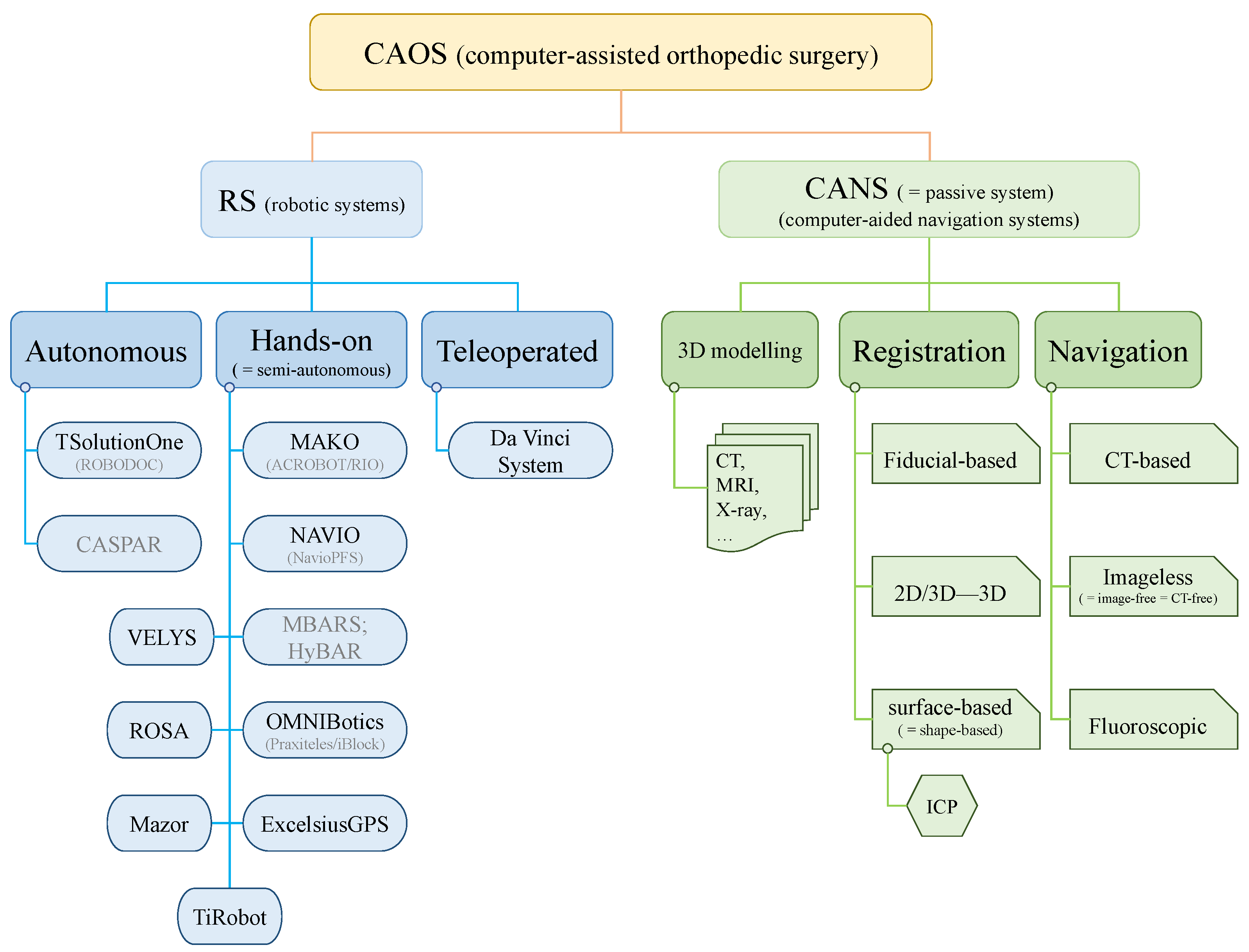
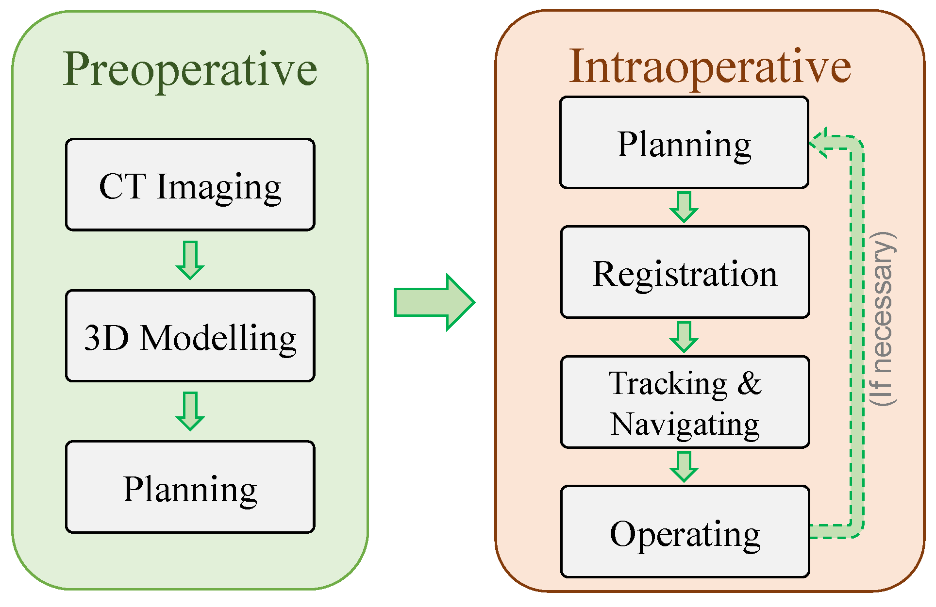

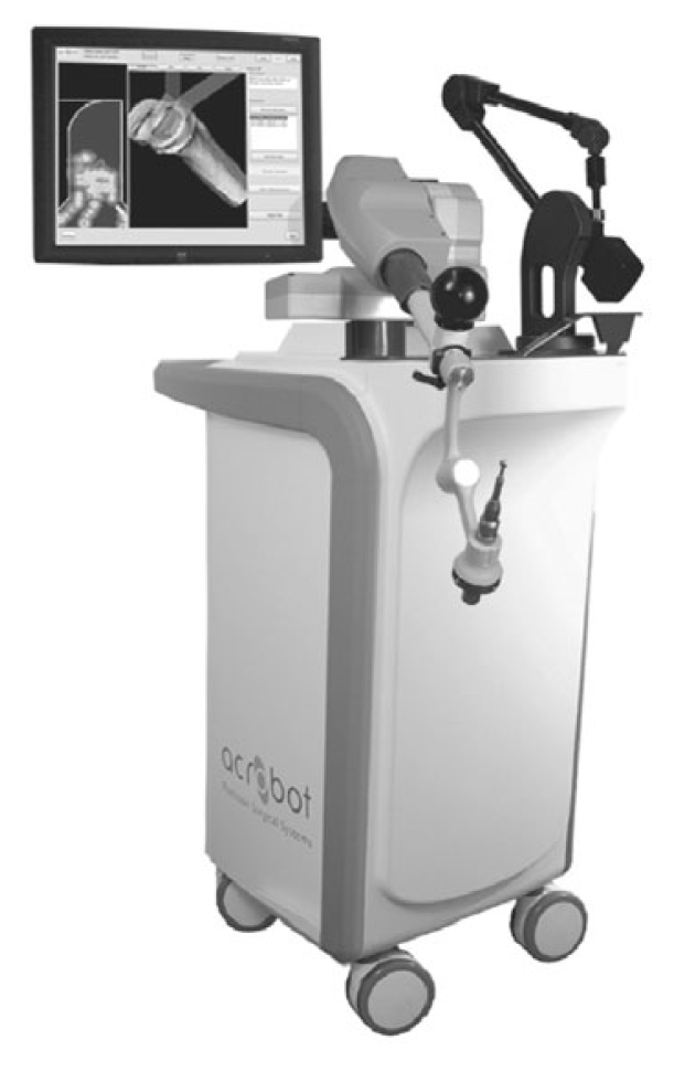
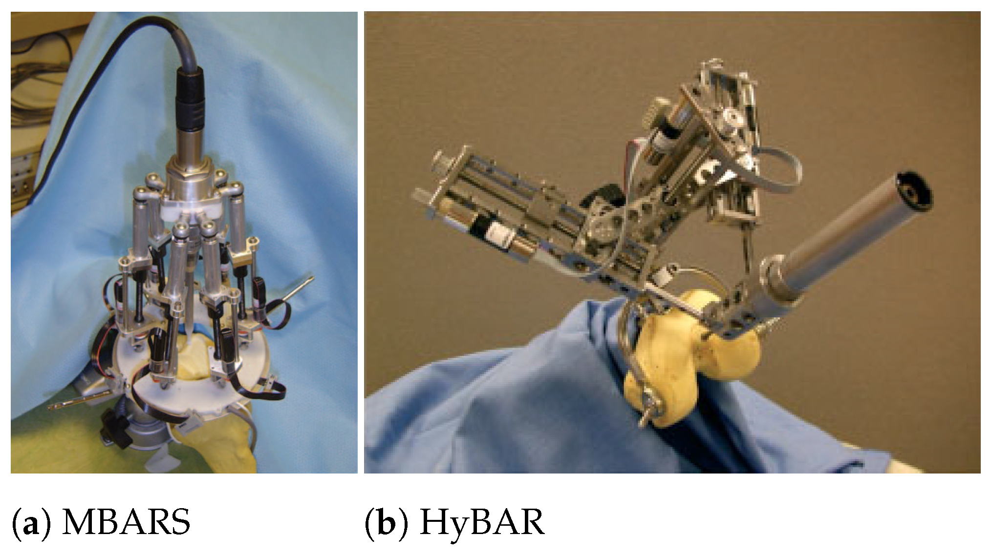
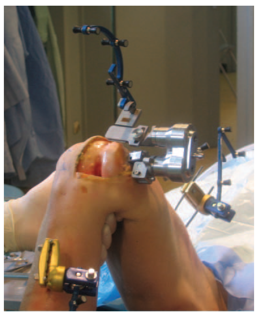
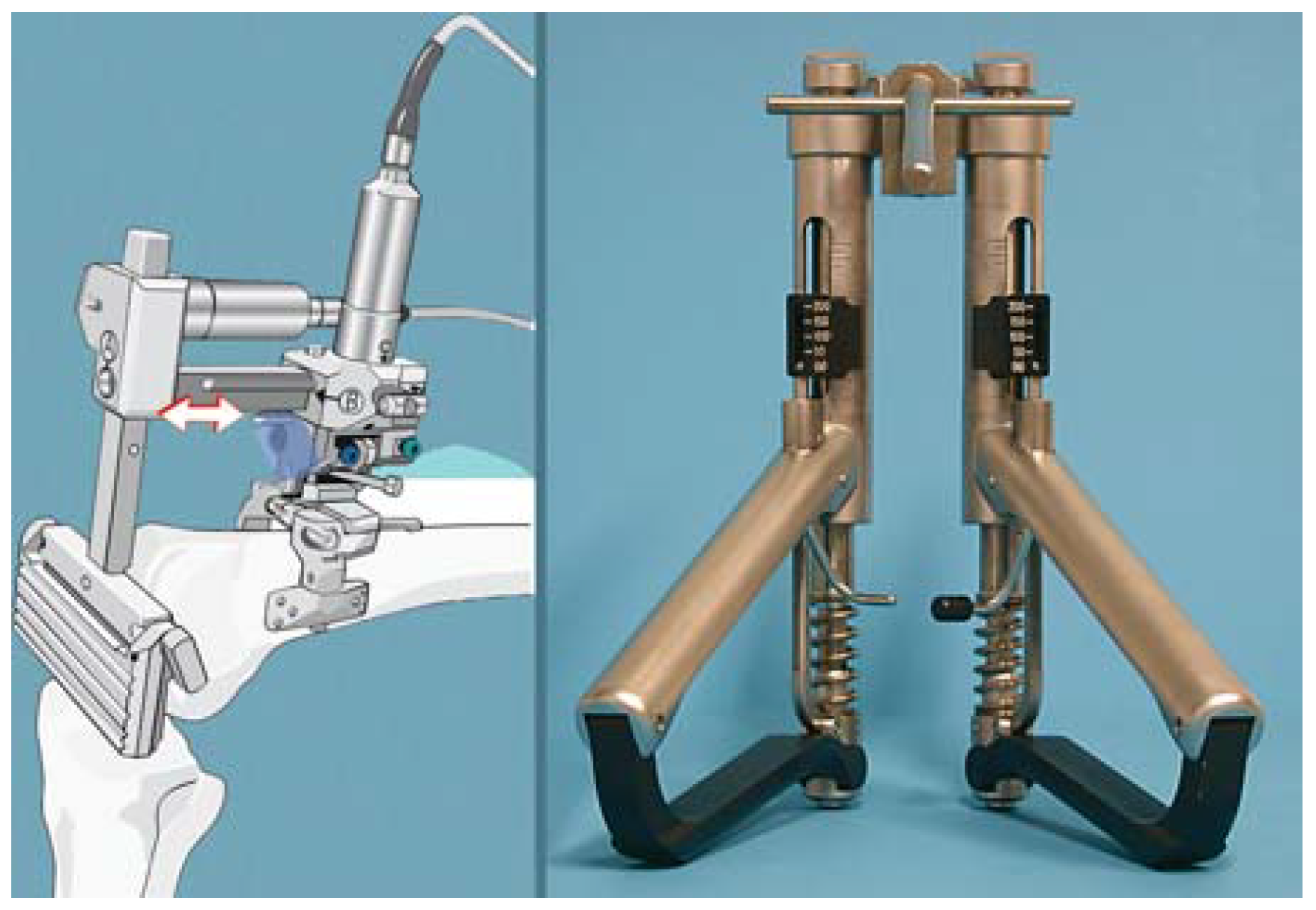

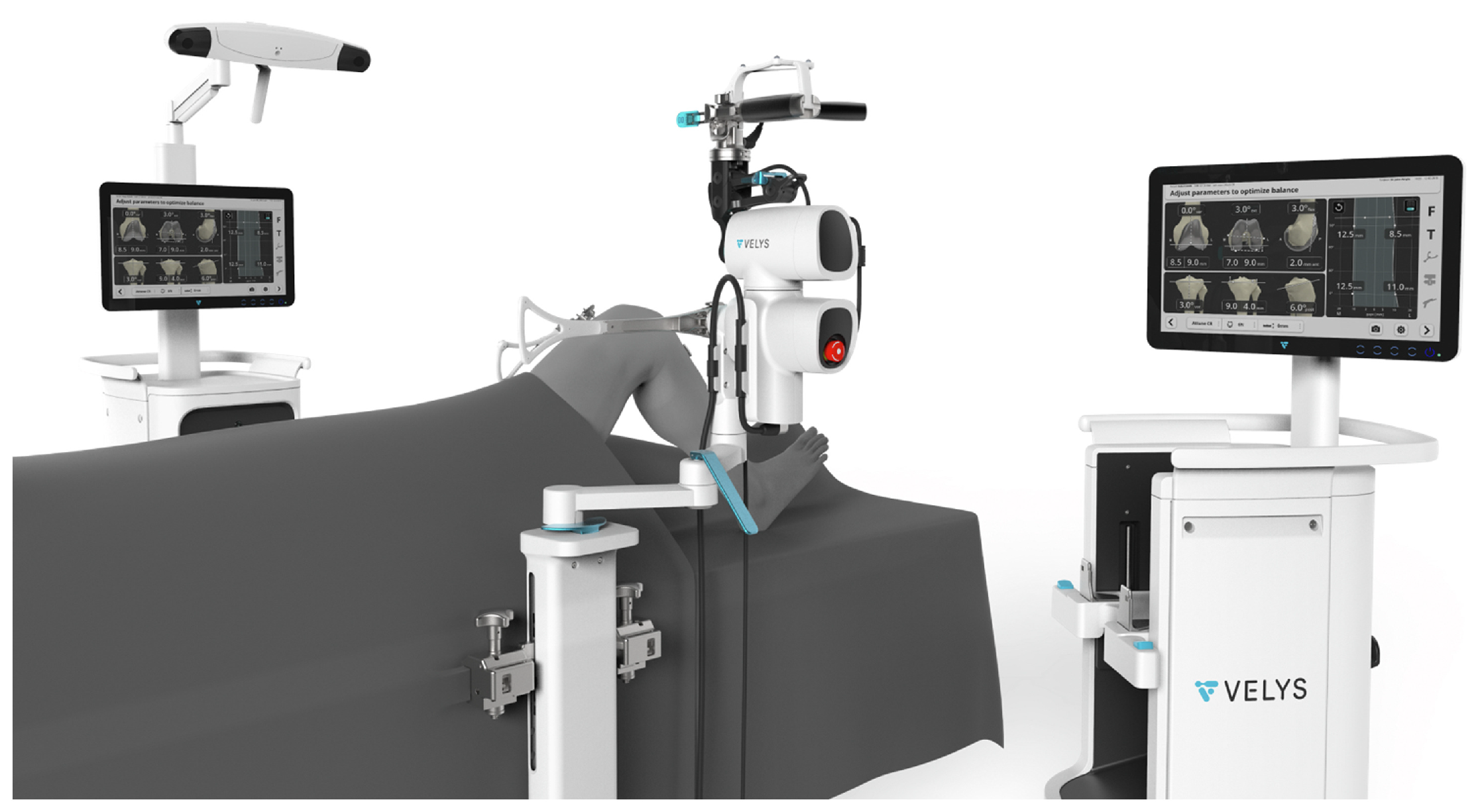


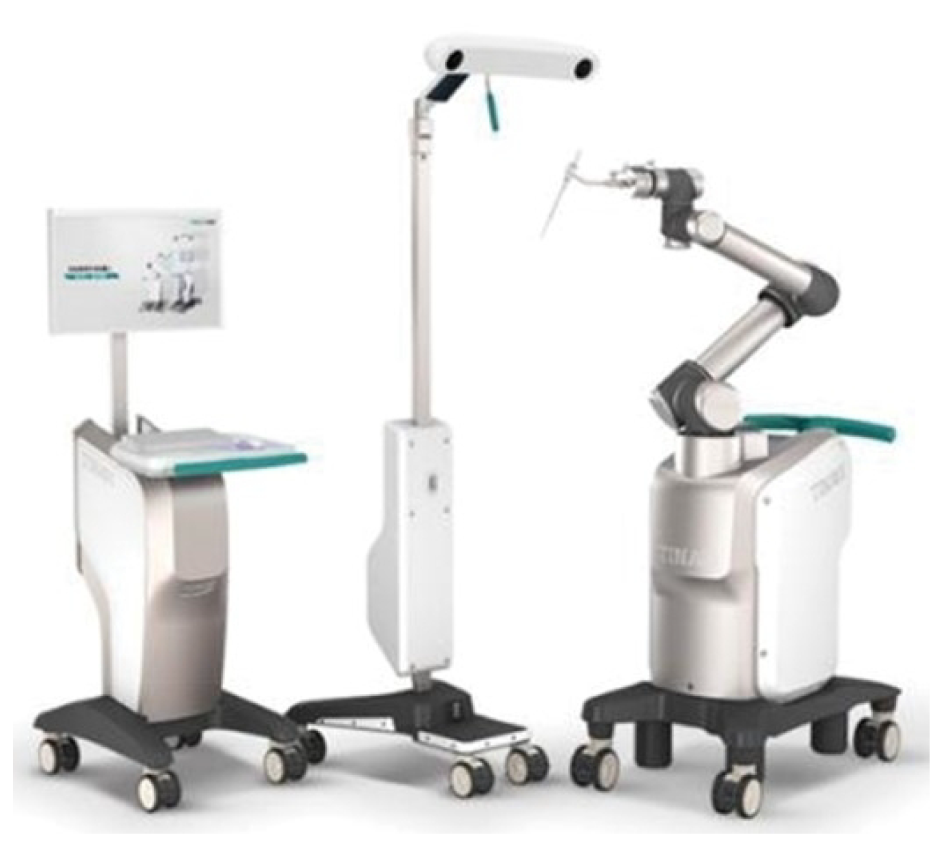
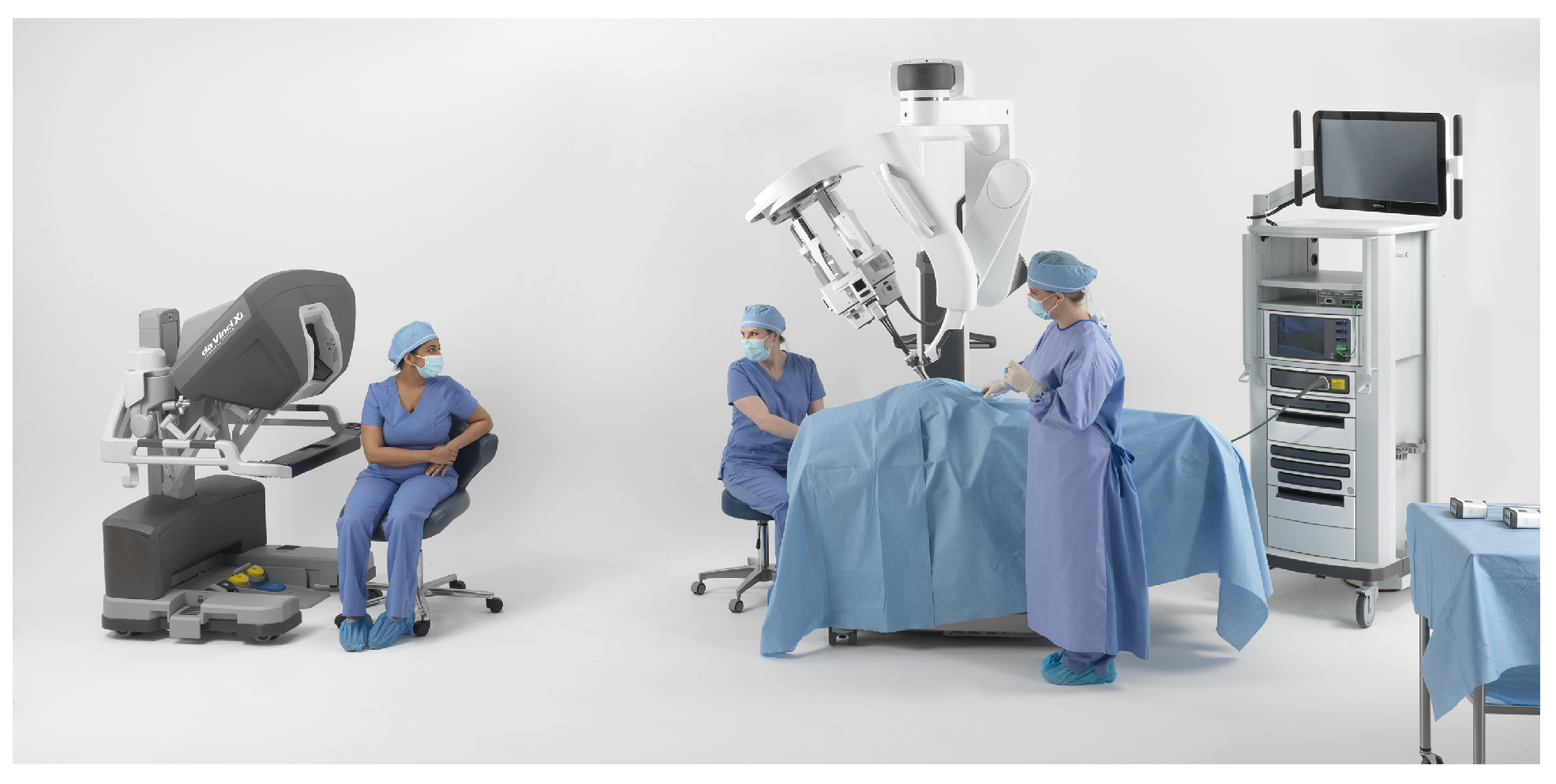
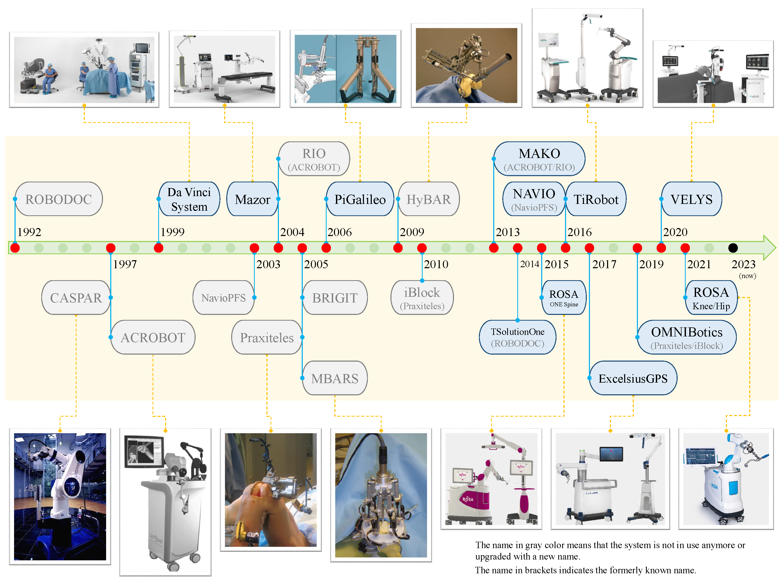
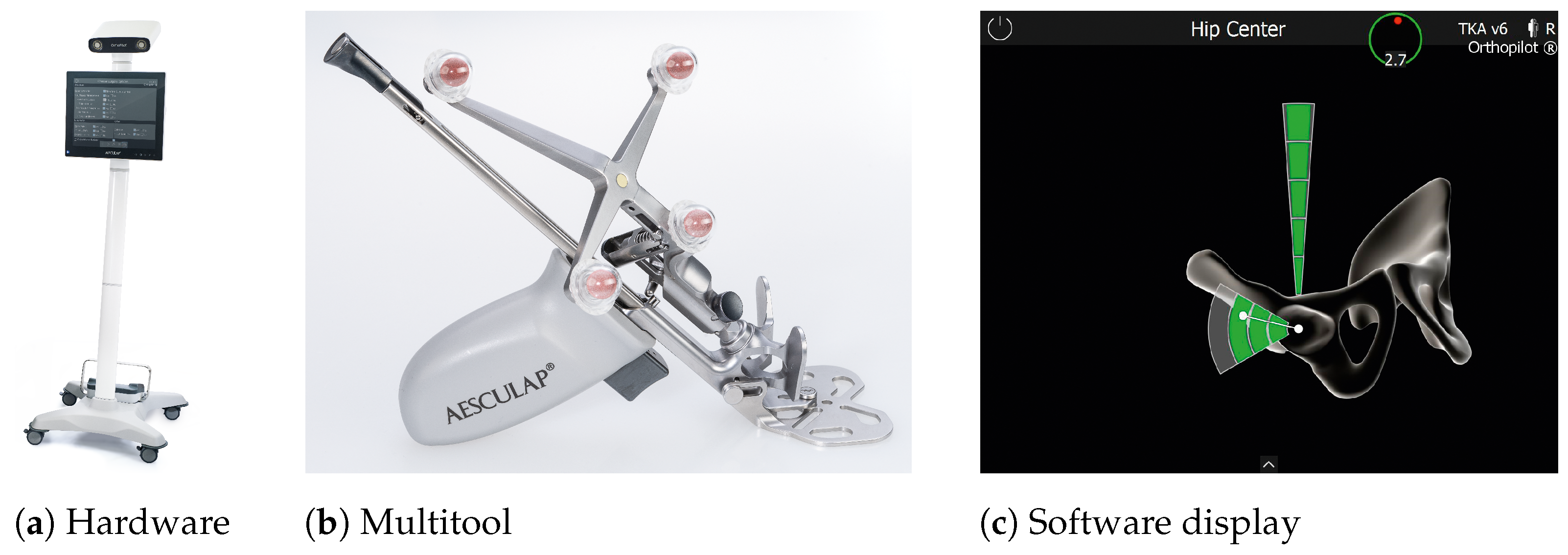
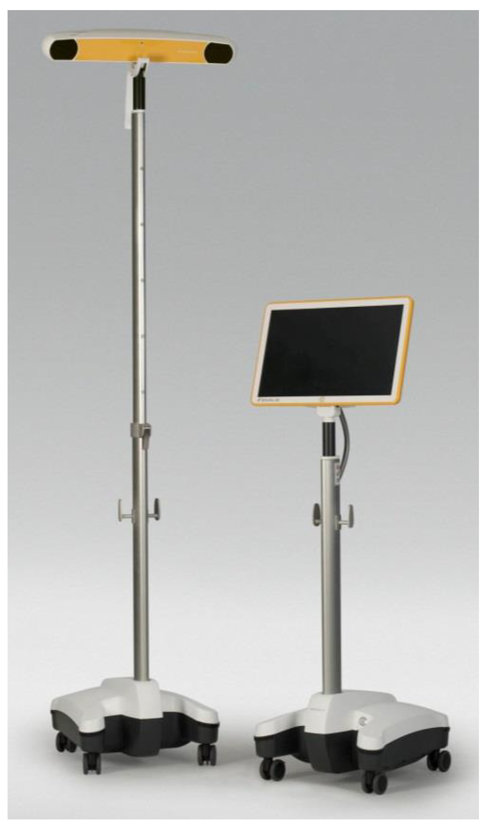
Disclaimer/Publisher’s Note: The statements, opinions and data contained in all publications are solely those of the individual author(s) and contributor(s) and not of MDPI and/or the editor(s). MDPI and/or the editor(s) disclaim responsibility for any injury to people or property resulting from any ideas, methods, instructions or products referred to in the content. |
© 2023 by the authors. Licensee MDPI, Basel, Switzerland. This article is an open access article distributed under the terms and conditions of the Creative Commons Attribution (CC BY) license (https://creativecommons.org/licenses/by/4.0/).