Article

The Relationship between Ankle Joint Kinematics and Impact Forces during Unilateral Jump-Landing Tasks in University-Level Netball Players: A Pilot Study

by
Thembisile T. Jolingana-Seoka
1,
Henriëtte V. Hammill
1,
Yolandi Willemse
2 and
Mark Kramer
1,*
1
Physical Activity, Sport, and Recreation (PhASRec) Research Focus Area, North-West University (NWU), Potchefstroom 2531, South Africa
2
Centre for Health and Human Performance, North-West University (NWU), Potchefstroom 2531, South Africa
*
Author to whom correspondence should be addressed.
Appl. Sci. 2023, 13(17), 9934; https://doi.org/10.3390/app13179934
Submission received: 28 July 2023 / Revised: 29 August 2023 / Accepted: 31 August 2023 / Published: 2 September 2023

Abstract

:

Featured Application

Analyzing ankle landing kinematics, ground reaction forces and stabilisation times of high-level female netball players.

Abstract

Netball is a sport that involves multiplane- and multidirectional landings, which results in a high proportion of injuries, particularly to the ankle joint. The purpose of this study was to analyse the ankle kinematics in multiple planes during multidirectional single-leg landings in high-level netball players. A total of ten (n = 10) netball players voluntarily participated in the study. All netball players performed 25 single-leg jump landing maneuvers per leg (dominant and non-dominant) from a 0.30 m high platform, landing onto a 0.70 m away force plate platform. Their ankle kinematic, landing kinetic and time to stabilisation (TTS) data were collected in sagittal-, frontal- and transverse planes. Netball players showed mean differences in peak landing forces (F (8,91) = 2.68, p = 0.009) but not in TTS (F (8,91) = 2.27, p = 0.260). There was evidence of differences in ankle kinematics across all three planes [Sagittal: (F (9,81) = 3.48, p = 0.001); Frontal: (F (9,81) = 8.01, p < 0.001); Transverse: (F (9,81) = 8.80, p < 0.001)]. Furthermore, small to large negative (r = −0.55) correlations were observed between ankle range of motion (ROM) and peak landing forces. Associated landing forces can be moderated by greater sagittal plane ankle ROM during multidirectional landings to minimise the risk of ankle injuries in netball.

1. Introduction

Netball is a team sport consisting of 7 players per squad and incorporates a large number of accelerations, decelerations, jumps, and directional changes [1]. Landing from a jump involves a complex interplay of forces, joint movements, and muscle actions where the primary focus, especially in netball, is on the ankle joint due to its role in absorbing forces, which tends to predispose it to injury [2,3]. More specifically, elite netball players perform an average of 58 high-intensity and multi-directional jumps (and, therefore, landings) per player per game, where incorrect landing techniques are ascribed as the primary mechanism of injury [4,5,6,7].
Ankle sprains, in particular, are among the most prominent injuries in netball (23% to 37% of netball injuries occur in the ankle joint) [5,7,8,9], and the mechanism for lateral ankle sprains results when the foot moves into sudden excessive foot inversion during landing or sidestep-cutting movements [10,11,12,13]. Whether the multiplanar kinematics of the ankle change as a function of the landing direction and the extent to which this may influence the landing kinetics has not been previously investigated and, therefore, presents a gap in the literature. Moreover, netball players tend to perform single-leg landings in multiple directions during match play and practices [14], where the landing in certain directions is known to invariably influence the kinematics of the hip-, knee- and ankle joints [6,15,16]. However, the preponderance of the literature tends to report on lateral-, horizontal- and vertical landings, which has transferred to the training recommendations but fails to account for diagonality and unilaterality of the landing movement (e.g., landing in a diagonal direction while moving towards the outside of the landing leg [varus stress] versus (vs.) moving towards inside of the landing leg [valgus stress]). Therefore, future research should rationalise the observed variability in landing performances by accounting for diagonality and unilaterality. To date, to the knowledge of the authors, only Kamffer et al. [17] have investigated both the directional- and unilateral components of landing within the sport of netball but did not account for the effect of such movements on lower extremity kinematics.
Furthermore, an evaluation of the ground reaction force (GRF) can be used to appraise both technique [18] and dynamic postural stability during landing by exploring the time-to-stabilisation (TTS) [13,19]. Given the dynamic nature of TTS, a more integrative measure of balance, postural control, mechanical stability, and proprioception can be obtained, especially for the evaluation of netball players [20]. Examining the ankle ROM across different jump landing directions in multiple planes, along with the GRF and TTS, could reveal novel insights into landing mechanics, which could inform future training customs for netball. The latter is especially true on the basis that novel assessments (e.g., multi-planar landing) provide a unique challenge to the neuromuscular system that may be revealed through differences in landing forces, the ability to absorb such forces, and the capacity to rapidly stabilise [21].
The objectives of the current study were to (i) analyse differences in ankle kinematics in all three cardinal planes during single-leg landing across five different landing directions, (ii) assess differences in unilateral peak landing forces across multiple directions, and (iii) evaluate the association between ankle ROM and peak forces as a function of multidirectional unilateral landing. We hypothesised that there would be significant differences in landing kinetics (specifically GRF) and kinematics and that there would be a significant negative correlation between ankle kinematics and kinetics during multidirectional single-leg landings in high-level netball players. The findings of this study could provide useful information related to ankle joint positioning when landing from a jump, which could identify the characteristics that could inform both netball-specific training and practices.

2. Materials and Methods

2.1. Participants

The a priori sample size calculations stipulated that a total of 13 would be required to attain sufficient statistical power (alpha = 0.05; power = 0.80; repeated measures = 5, effect size (f) = 0.25; correlation among repeated measures = 0.70). A total of 15 participants volunteered for testing, with a final sample of 10 participants being retained for analysis (injury: n = 3; incomplete data: n = 2). Ten (n = 10) female netball players (weight: 80.19 ± 13.00 kg; height: 176.96 ± 7.62 cm; BMI: 25.50 ± 3.30 kg·m−2; and %fat: 20.84 ± 3.91%) from the official netball team were recruited to participate in this study voluntarily. The age group of the netball players varied between 19 and 25 years and presented no current (<6 months) musculoskeletal injuries to the lower extremities. On the appointed day, voluntary written informed consent was obtained from participants prior to data collection.

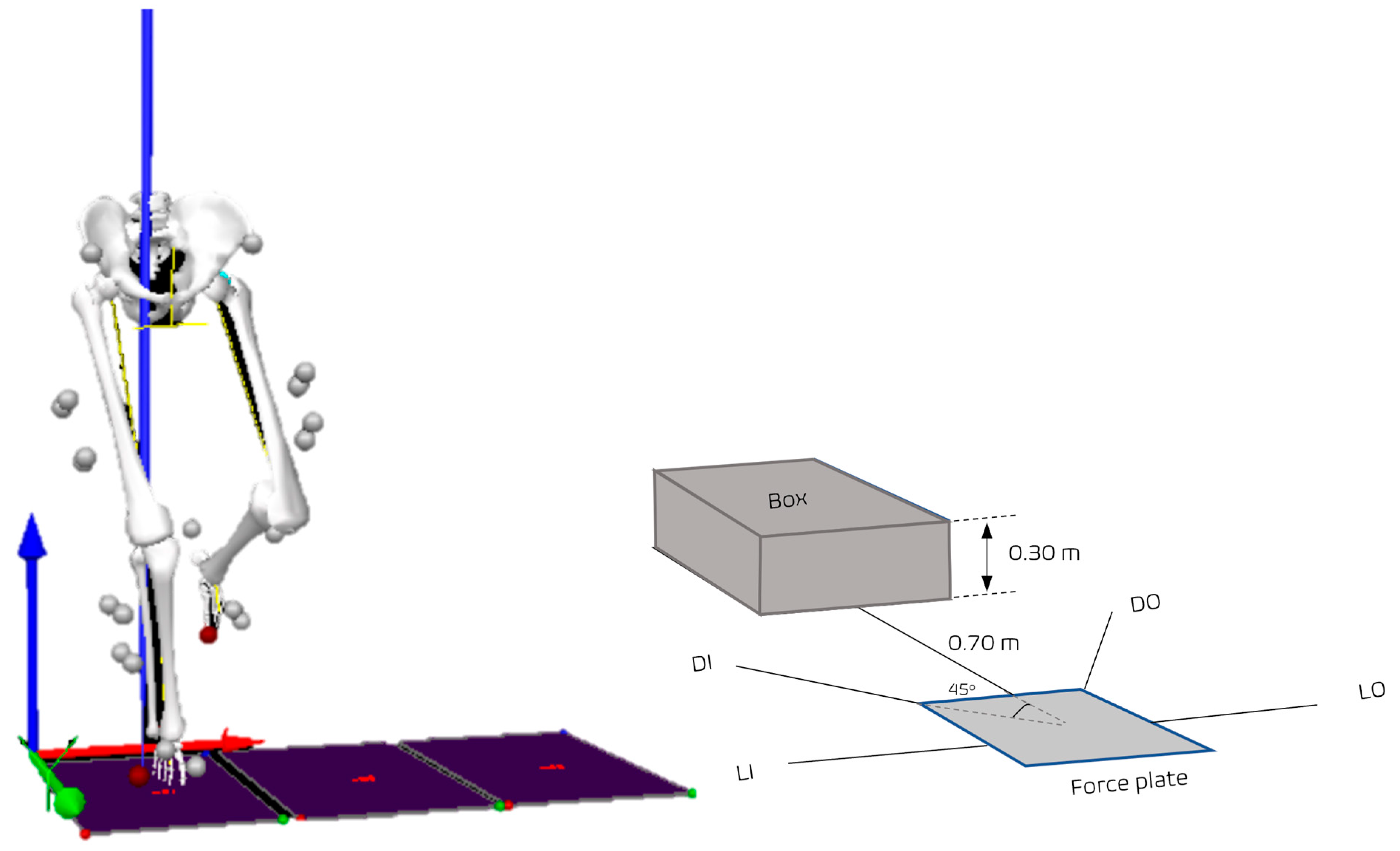
2.2. Experimental Design

This study was a pilot study that employed a cross-sectional study design. Testing and assessment of the netball players were performed in the well-controlled facilities during the 2022 netball season. The analysis of ankle kinematics and GRF at different landing directions between dominant (D) and non-dominant (ND) lower extremities during single-leg multidirectional landings were obtained within the same environmental testing conditions (21–22 °C and 55–60% humidity).

2.3. Ethics

The testing protocol for this research study was conducted in accordance with guidelines established by the Declaration of Helsinki of 1975, revised in 2013. Ethical approval for the research study was obtained from the North-West University Health Research Ethics Committee (NWU-00154-21-S1). Furthermore, the approval to test netball players as participants in the research study was also obtained from the Research Data Gatekeeper Committee (NWU-GK-21-071).

2.4. Testing Procedures

Prior to testing, participants were instructed to avoid strenuous exercise, alcohol, and caffeine consumption for 24 h. Participants were also instructed to arrive at the testing venue approximately 4 h post-prandial and eu-hydrated.

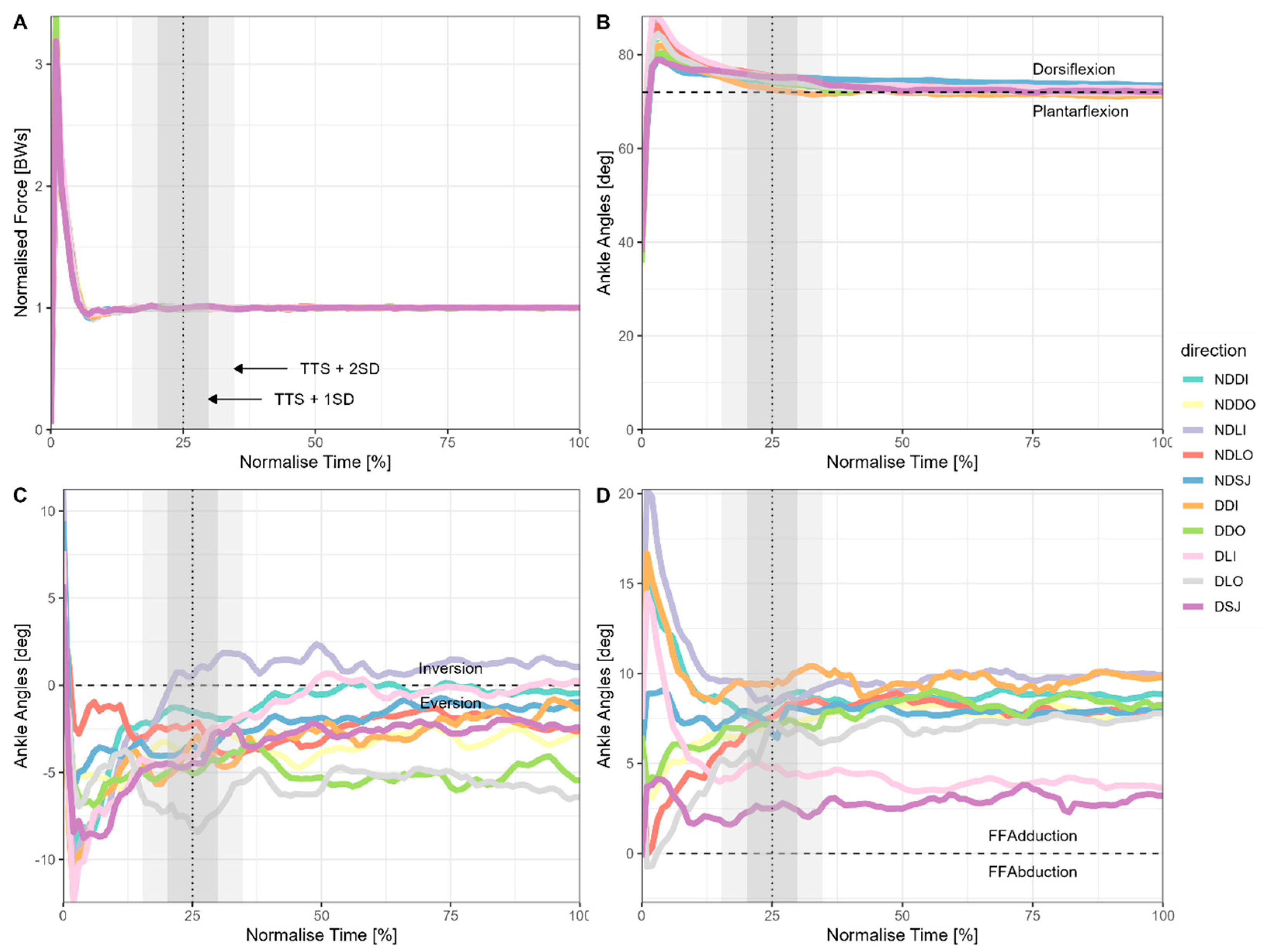
2.4.1. Landing Kinematics

Before data collection, an eight-camera motion capture system (Qualisys, Goteborg, Sweden) was fully calibrated. The CAST marker set was used with 38 reflective markers placed on participants as follows: bilateral markers on the anterior superior iliac spines, posterior superior iliac spines, greater trochanter, medial and lateral femoral epicondyles, medial and lateral malleoli, calcaneus, as well as on the first, second and fifth metatarsal heads. Marker clusters consisting of four markers were used for the thigh and shin segments [22,23,24]. Markers on the femoral condyles, malleoli and greater trochanters were retained only for the static calibration trials. Whereafter, they were removed for the dynamic trials. All motion capture data were sampled at a rate of 200 Hz, whereas kinetic data were captured from an AMTI digital force plate at a rate of 2000 Hz (AMTI, Watertown, MA, USA) [25]. More specifically, ankle ROM was recorded throughout the trial as this was of specific importance during the landing phase (i.e., how much range is present in the ankle during landing and weight acceptance).

2.4.2. Landing Kinetics

Participants were instructed to stand bilaterally on a 0.30-m high platform and to perform a single-leg landing onto an AMTI force plate located 0.70 m [16,17,26] from the platform. Jump-landing trials were completed in the following directions: straight jump (SJ), diagonal inside (DI), diagonal outside (DO), lateral inside (LI), lateral outside (LO), with each direction being orientated at a 45° angle relative to each other [17] (see Figure 1). Participants were asked to stabilise as quickly as possible at ground contact until the end of the trial (8 s) [27]. Each participant performed the single-leg landing task five times on each leg for each direction, with 30 s of recovery between jumps of the same direction and two minutes between directions. Unsuccessful trials (e.g., loss of balance) were repeated and were not retained for analysis. All landing tasks were performed with shoes on to closely mimic match and training scenarios. Limb dominance was determined by asking participants which leg they were likely to use to kick a ball as far as possible or feel comfortable landing on a single leg [18,28,29]. All participants included in the sample were right-leg dominant. The kinetic data were all normalised to body weight such that the forces experienced during landing can be compared across all individuals (i.e., GRF normalised [unitless] = landing force [N] ÷ body weight [N]).
The kinematic data were processed using Visual3D (C-Motion Inc., Gaithersburg, MD, USA), where 3-D kinematics of the ankle joints were calculated and presented using an XYZ Cardan sequence of rotations, where X is plantar/dorsiflexion, Y is inversion/eversion and Z is forefoot adduction-abduction (see Figure 1). The kinematic data were then low-pass filtered using a fourth-order zero-lag Butterworth filter with a cut-off frequency of 6 Hz. All kinetic and kinematic data were ensemble averaged, where the mean of all five trials was retained for analysis.

2.4.3. Time-to-Stabilization

The time-to-stabilisation (TTS) was obtained by evaluating the point at which the resultant force trace remained within an average range of variation of body weight (mean + 5SD) of the final second of the capture period [30].

2.4.4. Body Fat and Body Mass Index Measurements

The body mass and body fat of participants were measured using the InBody system, which utilises hand-to-foot bioelectrical impedance via a tetrapolar 8-point tactile electrode system (InBody770, InBodyUSA, Cerritos, CA, USA). Participants were instructed to wear minimal clothing and to remove shoes and socks when standing on the InBody analyser. Participant information was entered into the system (e.g., age, sex), and participants were instructed to stand as still as possible during the recording process, as per manufacturer instructions. The InBody system has been deemed valid and reliable for the measurement of body fat (BF%) and fat mass with large intraclass correlation coefficients of ≥0.98 and ≥0.98, respectively [31]. Height was measured to the nearest 0.01 m using a stadiometer (Holtain) with the participant in the Frankfort plane. The body mass index (BMI [kg·m−2]) was then calculated for each participant once both height and mass were known.

2.5. Statistical Analyses
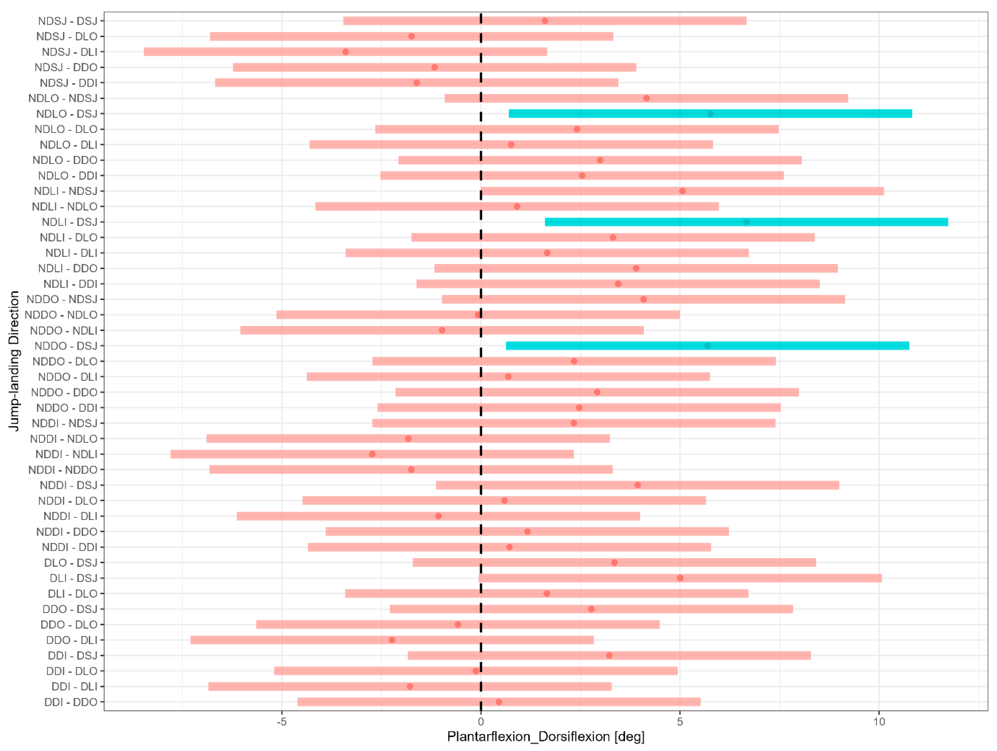
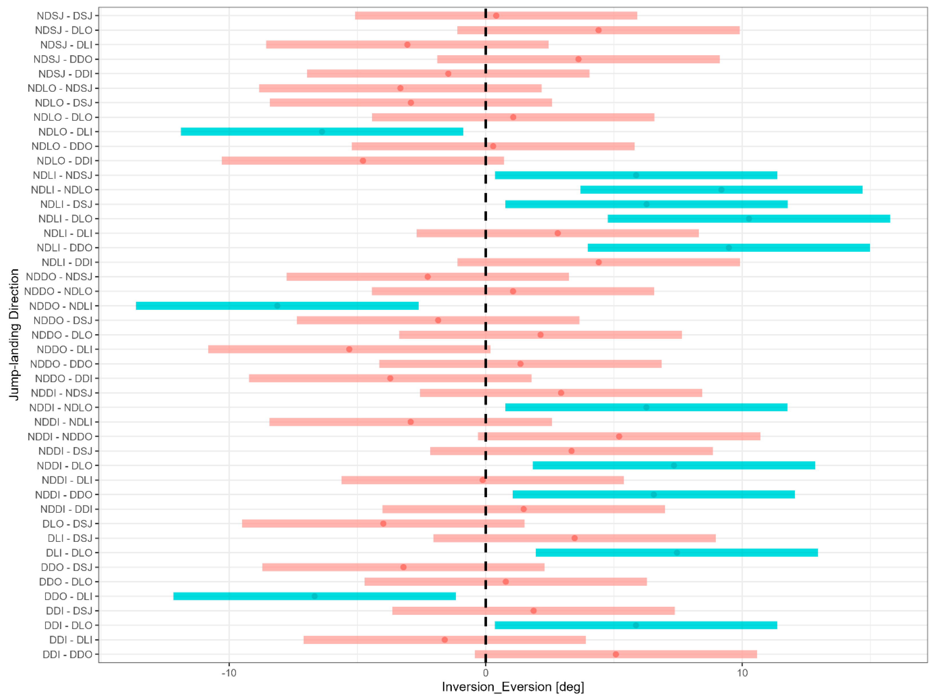
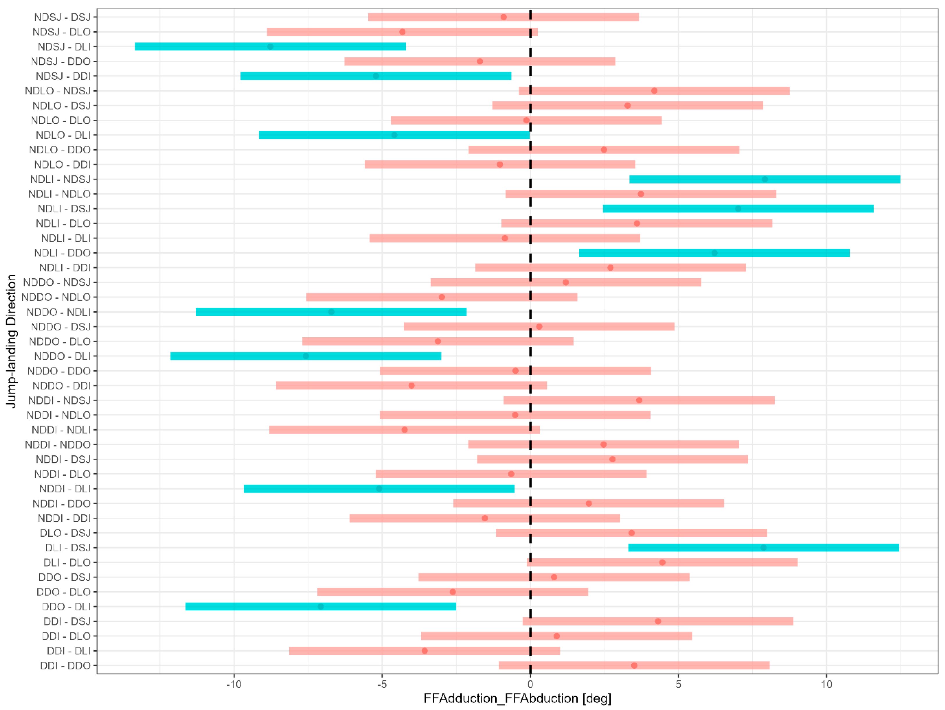
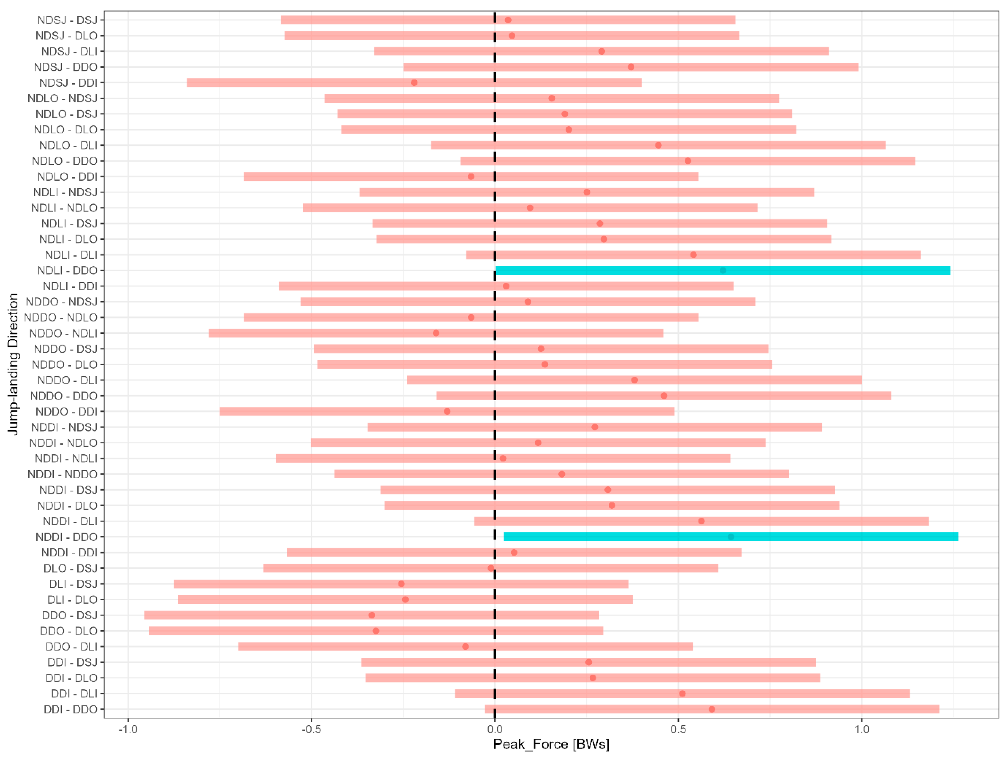
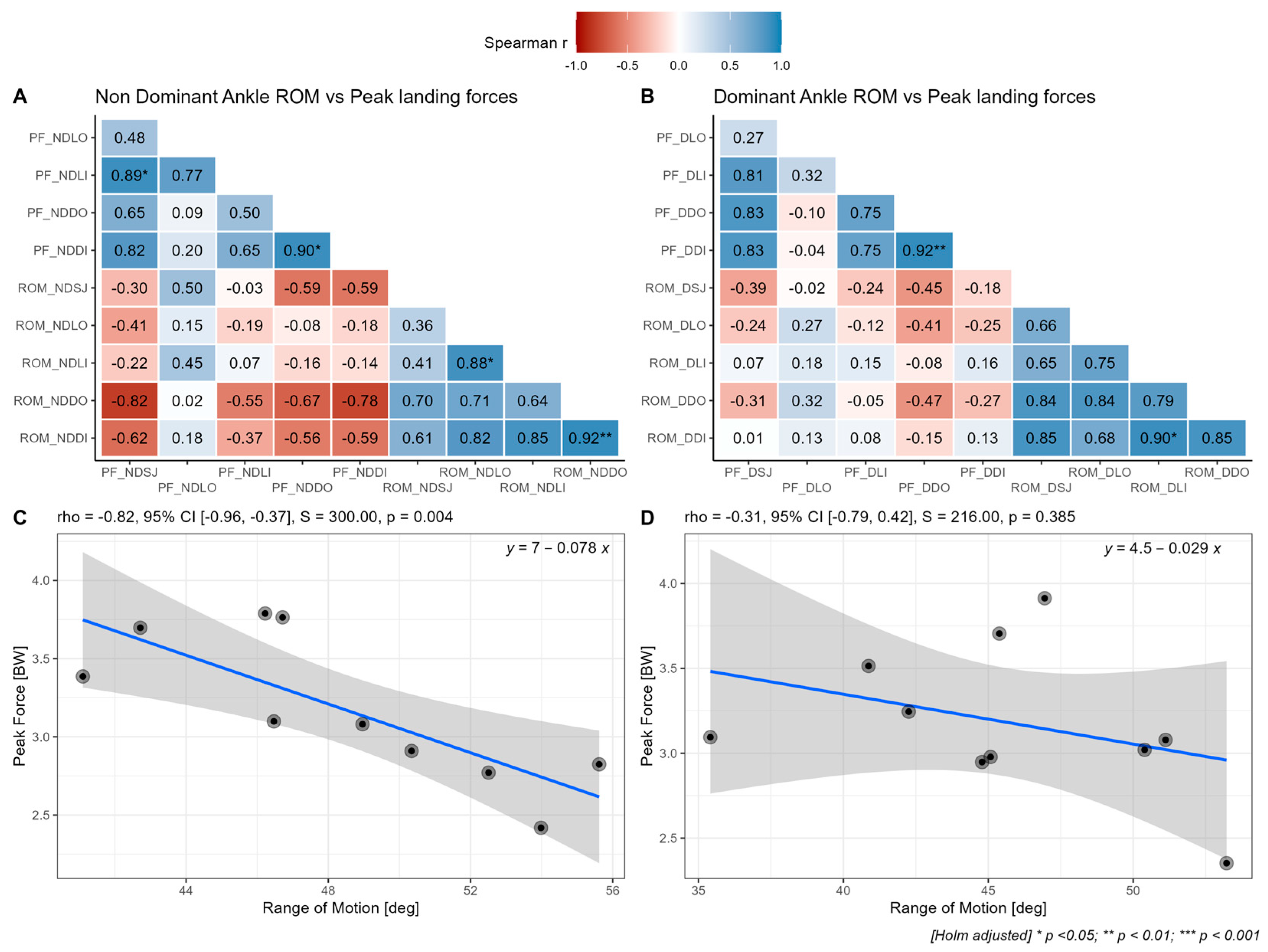
The normality of data was evaluated using the Shapiro-Wilk test, with deviations from normality being accepted at p < 0.05. Within-group differences for each parameter of interest were evaluated using repeated measures ANOVA with a Holm correction for multiple pairwise comparisons. A correlation analysis between mean ankle ROM and peak landing forces was completed using Spearman’s rank correlation analysis with a Holm correction to adjust for multiple comparisons. The coefficients are qualitatively interpreted in absolute terms as follows: negligible: r = |0.00–0.10|; weak: r = |0.11–0.39|; moderate: r = |0.40–0.69|; strong: r = |0.70–0.89|; very strong: r = |0.90–1.00| [32]. Statistical significance was set at p ≤ 0.05. All statistical analyses were completed using the R-programming language [33,34].

3. Results

The multi-directional GRF normalised to body weight (BWs) and the tri-planar ankle range of motion (ROM) of high-level netball players are presented in Figure 1. Participants experienced landing forces of 3.24 ± 0.59 BW (Figure 2A). Participants landed with varying degrees of plantar flexion (Figure 2B), which fluctuated according to landing direction. The degree of inversion/eversion (Figure 2C) and forefoot adduction/abduction (Figure 2D) fluctuated considerably as a function of landing direction.
An overview of the mean, standard deviation (SD), and coefficient of variation (CV%) across all jump-landing directions is provided in Table 1.
Significant within-group differences were present for mean sagittal plane ankle kinematics as a function of the landing direction (F (9,81) = 3.48, p = 0.001). The estimated marginal mean differences for the pairwise comparisons are highlighted in Figure 3. It is evident that although there is direction- and limb-dependent variability in sagittal plane ankle ROM during landing, the differences are typically within ~5° and, therefore, likely not meaningful.
Significant within-group differences for mean inversion/eversion ROM were evident as a function of the landing direction (F (9,81) = 8.01, p < 0.001), where mean differences for each of the pairwise comparisons are presented in Figure 4. The overarching implication is that the mean differences for the lateral inside, lateral outside and diagonal outside jump-landing directions of the dominant (D) and non-dominant (ND) are significantly different from zero (p < 0.05).
Mean forefoot ROM during landing also exhibits significant within-group differences (F (9,81) = 8.80, p < 0.001). The pairwise comparisons of the mean differences for forefoot adduction/abduction as a function of the landing direction are presented in Figure 5. There appears to be considerable variability in the available ankle ROM within the transverse plane that is both direction- and limb-dependent, whereby differences of ~10° are evident. The practical implications of such large differences are presently unclear but should provide impetus for further research.
Peak landing forces also showed significant within-group differences related to landing direction (F (8,91) = 2.68, p = 0.009). Post-hoc analyses (see Figure 6) showed that the disparities could be ascribed to differences in leg dominance, specifically when landing in the diagonal-outside direction. Variability in landing forces is evident whereby the magnitude and direction of the observed differences appear to be dependent on the limb and directionality of landing. However, most observed differences are within ~0.5 BW and likely not meaningful, although future research should verify such inferences.
Although the TTS showed direction-specific within-group differences (F (8,91) = 2.27, p = 0.026), these differences did not hold true following the post-hoc pairwise analyses (see Figure 7). Given the rapidity with which athletes could stabilise regardless of landing limb or direction, our study lacked the statistical power to detect very small differences in TTS.
The correlation analysis between ankle range of motion and peak multidirectional landing forces is highlighted in Figure 7. There is evidence of weak to very strong negative correlations, specifically for the diagonal directions for the non-dominant leg (Figure 8A), whereas small to moderate negative correlations are evident for the dominant leg (Figure 8B). For greater clarity and insights, the relationship between ROM in the diagonal outside (DO) direction and the peak force in the straight (SJ) direction is shown for the non-dominant (Figure 8C) and dominant (Figure 8D) limbs, respectively. A clear negative relationship is apparent, thereby highlighting that greater sagittal plane ROM during landing may favourably moderate (i.e., reduce) the peak landing forces.
It is important to note that, although the sample size was small, the post-hoc analysis revealed that the correlation between repeated measures for ankle ROM was 0.80–0.84, indicating that a sample of 8–9 participants would have been sufficient to achieve the desired statistical power.

4. Discussion

The current study sought to analyze the ankle kinematics in three cardinal planes during multidirectional single-leg landings in high-level netball players. The novel findings of the present study showed that (i) mean differences were present in peak landing forces but not stabilisation times and are likely dependent on leg dominance and landing direction, (ii) significant differences were evident across all planes regarding ankle kinematics as a function of landing direction, and (iii) small-to-large negative correlations were present between ankle ROM and peak landing forces indicating that landing forces can be moderated by changes in sagittal plane ankle kinematics.
Netball players typically perform approximately 58 explosive and multidirectional jump-landings per game, of which 42% are directed forward, 32% are vertical, and 26% are lateral [1,6]. Moreover, approximately 65% of these landings are unilateral, which highlights the importance of ensuring adequate ability to tolerate high unilateral loads across varied directions [6,14]. When landing on a single leg, a considerable amount of impact force needs to be adequately absorbed by the musculoskeletal system to guard against injury and allow for repeated high-intensity efforts [6,12]. The ankle plays an especially dominant role during vertical landings, whereby it contributes 36.5% of the total work during this phase [35]. Our study showed that the kinematics of the ankle varied considerably depending on the landing direction (i.e., lateral, forward, diagonal) as well as the direction in which momentum was orientated (i.e., inside vs. outside). Our findings coincide with and extend those of Fong et al. [36], who showed that ankle dorsiflexion ROM was negatively associated with vertical ground reaction force (r = −0.41, p = 0.14). Importantly, Fong et al. [36] investigated only mono-directional bilateral landing, whereas we showed that such associations are likely dependent on the direction of landing. Intriguingly, Howe et al. [37] concluded that ankle kinematics were not associated with bilateral landing kinetics despite observing small negative associations (r = −0.28, p = 0.08). Our findings suggest that it would be important to consider the relative contributions of each leg during landing, given that limb dominance appears to be a factor (see Figure 8).
Within the context of our study, the ankle was typically everted, and the forefoot adducted across almost all landing directions, where significant direction-specific differences were evident for both frontal (p < 0.001) and transverse (p < 0.001) plane kinematics. The presence of such direction-specific differences is likely an attempt to accommodate for the changes in landing-orientated momentum and to facilitate a better absorption of energy [2,6,22]. These findings seem to hold true across different directions and show that netball coaches and practitioners should incorporate varied movement strategies during strength and conditioning practices which, based on the literature, tend to be skewed towards uniplanar exercises that tend to favour the sagittal plane [1,38]. Whether those with differing tri-planar ankle kinematics exhibited more favourable landing kinetics was not investigated in the present study but would offer a potential area of future research.
Higher landing loads can have two potential outcomes where, on the one hand, the soft-tissue structures adapt over time to become stronger and more resilient, or on the other hand, they may be subject to injury if inadequately stressed to tolerate higher repetitive loads [39]. In the present study, the reported mean average GRF is 3.24 ± 0.59 BW among high-level netball players (Figure 2A), which overlaps with the expected values of 3.0–5.7 BW from other studies [6,12,17]. It is well understood that several factors, such as landing height, jumping distance, foot placement, and landing strategy, tend to moderate landing forces [6]. The present study adds to the available literature by showing that landing directionality and ‘momentum’ should also be considered, in addition to the ability to enhance the sagittal plane ROM during landing, given that the greater the ROM, the lower the expected peak landing forces tend to be.
Landing with one limb requires different muscular recruitment patterns compared to bilateral landing, which likely influences multidirectional dynamic postural stability [6,17,26]. Dynamic postural control has been evaluated with single-leg landing by measuring the TTS required for an individual to stabilise to a stable posture after landing [19,40,41,42]. The TTS should be evaluated across all planes as this may address important aspects required for neuromuscular control when landing and possibly identify the direction that could potentially cause ankle injuries in netball players. The present study reported no significant difference in mean TTS across different landing directions (p > 0.05) and appeared to be independent of limb dominance (Figure 7). The results of the present study are, at least to some extent, contrary to those reported by Wikstrom et al. [26], who showed that the medio-lateral and vertical dynamic postural stability was significantly affected by lateral and diagonal jump landing directions in the healthy population (F (2,50) = 41.41, p < 0.001). The study by Kunugi et al. [22] went further and reported significantly longer TTS in the lateral landing direction (p < 0.001) compared to the medial and forward directions in 49 male collegiate soccer players landing from a 30 cm high platform situated just 5 cm away from force plates. Interestingly, Kunugi et al. [22] showed that the ankle and rear foot kinematics during lateral landing exhibited smaller eversion and pronation positions than forward landing and medial landing (p < 0.010), which seem to employ a similar landing mechanism adopted by the participants of our current study. The recent results of the study by Kamffer et al. [17] confer with the findings of Wikstrom et al. [26] and observed significant directional differences for TTS, specifically between the DI and LI directions (Mdiff = −0.37 s, t (14) = −3.14, pTukey = 0.048) for elite netball players. These findings signify that when landing laterally and diagonally, the centre of mass likely oscillates in the frontal plane (medially and laterally), and the neuromuscular components of the ankle may be challenged more during landing and that this may be directionally dependent [22]. Given the rapidity with which the athletes in our study managed to stabilize independent of landing limb or direction, it is important to note that we likely lacked the statistical power to detect very small differences. Whether the presence of lower limb injuries would substantially amplify the differences in TTS as a function of landing limb and/or direction for netball players would certainly require further research. Despite the preliminary findings of the present study, the TTS data have shown utility as a potential diagnostic tool to evaluate those athletes with the likelihood to have functionally unstable ankles and should be considered for safe return-to-play of netball players [40]. However, the sensitivity and specificity of TTS for such purposes requires further investigation.
Research has shown that inversion ankle sprains tend to dominate the injury spectrum in netball [10,22]. The greater ankle inversion angle coupled with longer TTS in lateral landings compared to forward landings can potentially cause greater loads on muscles and ligaments that resist ankle inversion [16,22]. In the present study, participants presented with ankle eversion and forefoot adduction upon landing, which may imply that the participants of the current study land with a more pronated foot posture, further implying that athletes may potentially be at a lesser risk of sustaining non-contact lateral ankle sprains. However, whether such results transfer to practice and match-play settings would require further research as well as more advanced technologies capable of capturing the biomechanics of in-situ performances. A decrease in ankle ROM (e.g., through the use of a brace) has been shown to require compensatory ROM by the knee and hip joints during various landing tasks, thereby affecting the landing technique but not landing GRF [36,37,43,44,45,46,47]. However, the findings of the present study do show that changes in sagittal plane ankle kinematics during multidirectional landings are negatively associated with peak landing forces to varying yet substantial degrees (see Figure 8C,D). The present results are also supported by those of Sinsurin et al. [16], who showed that the landing direction significantly influences the peak angles of an ankle during landing in male volleyball and basketball players. Although the findings presented here may suggest that greater ankle ROM is imperative for reducing peak landing forces during multidirectional single-leg landings, further research is required to determine whether there is carry-over to the field setting during match-play and whether such strategies mitigate non-contact injury risks. Moreover, the extent to which the relationship between ankle kinematics and landing kinetics can be moderated by various interventions would also require further research. While the present study did not investigate participants with ankle-specific injuries, it is important to note the mechanisms associated with injury are multifactorial [3,11].
Although there are strengths identified in conducting the present study, limitations must also be acknowledged. The present study was conducted specifically on female netball players as the vulnerable population to an ankle injury. However, future studies should consider conducting the same research on male netball players to investigate if they exhibit similar ankle kinematics upon landing. The sample size of the participants was small and should be replicated with a larger number of high-level participants. Also, the results of the current study should not be generalised to other sports and can only apply to high-level netball players as the needs at different levels of play may depend on game demands, and specific adaptations may be required. Therefore, we recommend that the results of the present study should be used as a guideline to coaches and conditioning staff when designing training programs (to consider including multidirectional training in other planes of motion as an ankle is a multiplane joint) and when conducting pre-season testing battery. It is also important to note that, to make specific inferences about landing limb and direction, our study was highly controlled in a laboratory environment. Whether these results would transfer to real-world match-play and practice scenarios would require further research. Given the observational nature of the study, the extent to which the measured variables are amenable to change following specific interventions is not clear. Lastly, our results are based on athletes who were free of injury at the time of testing. Therefore, the extent to which all of the measured parameters change in the presence of injury is presently uncertain and would provide a fruitful area of future research.

5. Conclusions

This study aimed to analyse the three-dimensional ankle kinematics during multidirectional single-leg landing tasks in high-level netball players during a single netball season. We observed that the ankle kinematics were significantly influenced by the jump-landing direction as well as leg dominance, which may necessitate the inclusion of more varied training practices for netball players. Within the findings, there is evidence of small-to-large negative correlations between ankle ROM and peak landing forces during landing and that these are moderated by both the landing direction and leg dominance. More specifically, it would appear that the diagonal and lateral jumping directions posed a unique challenge to the athletes, given the variability observed in the kinetic and kinematic metrics that were measured. Therefore, multiple directions should be accounted for during both strength training and match practices such that between-leg differences are minimised and that appropriate tissue loading and adaptation occur to optimise performance. Whether the relationship between ankle kinematics and landing kinetics can be moderated by various interventions and whether such changes can mitigate the potential risk of injury would require further research.

Author Contributions

Conceptualization, H.V.H. and M.K.; methodology, H.V.H. and T.T.J.-S.; software, M.K.; formal Analysis, M.K.; writing—original draft preparation, T.T.J.-S. and M.K.; writing—review and editing, T.T.J.-S., M.K., H.V.H. and Y.W.; visualization, M.K.; supervision, H.V.H., M.K., and Y.W. All authors have read and agreed to the published version of the manuscript.

Funding

This research received no external funding.

Institutional Review Board Statement

The study was conducted in accordance with the Declaration of Helsinki and approved by the Human Research Ethics Committee of North-West University (NWU-00154-21-S1).

Informed Consent Statement

Informed consent was obtained from all subjects involved in the study. Written informed consent has been obtained from the participants to publish this paper.

Data Availability Statement

The dataset for the study is freely available in the Harvard Dataverse and can be accessed via the following link: https://doi.org/10.7910/DVN/X4PNJQ (accessed on 30 August 2023).

Acknowledgments

The authors would like to thank the netball team for their participation in the study.

Conflicts of Interest

The authors declare no conflict of interest.

References

  1. Chandler, P.T.; Pinder, S.J.; Curran, J.D.; Gabbett, T.J. Physical Demands of Training and Competition in Collegiate Netball Players. J. Strength Cond. Res. 2014, 28, 2732–2737. [Google Scholar] [CrossRef]
  2. Rowley, K.M.; Richards, J.G. Increasing Plantarflexion Angle during Landing Reduces Vertical Ground Reaction Forces, Loading Rates and the Hip’s Contribution to Support Moment within Participants. J. Sports Sci. 2015, 33, 1922–1931. [Google Scholar] [CrossRef] [PubMed]
  3. Downs, C.; Snodgrass, S.J.; Weerasekara, I.; Valkenborghs, S.R.; Callister, R. Injuries in Netball-A Systematic Review. Sports Med. Open 2021, 7, 3. [Google Scholar] [CrossRef]
  4. Ferreira, M.; Spamer, E. Biomechanical, Anthropometrical and Physical Profile of Elite University Netball Players and the Relationship to Musculoskeletal Injuries. S. Afr. J. Res. Sport Phys. Educ. Recreat. 2010, 32, 57–68. [Google Scholar] [CrossRef]
  5. Langeveld, E.; Coetzee, F.F.; Holtzhausen, L.J. Epidemiology of Injuries in Elite South African Netball Players. S. Afr. J. Res. Sport Phys. Educ. Recreat. 2012, 34, 83–93. [Google Scholar]
  6. Mothersole, G.A.; Cronin, J.B.; Harris, N.N. Key Prerequisite Factors Influencing Landing Forces in Netball. Strength Cond. J. 2013, 35, 47–54. [Google Scholar] [CrossRef]
  7. Pillay, T.; Frantz, J.M. Injury Prevalence of Netball Players in South Africa: The Need for in Jury Prevention. S. Afr. J. Physiother. 2012, 68, 7–10. [Google Scholar] [CrossRef]
  8. Ellapen, T.; Schoeman, K.; Zaca, L.; Heerden, H.; Ramiah, P. Prevalence of Musculoskeletal Injuries among Adolescent Recreational Netballers in Kwa-Zulu Natal, South Africa. Br. J. Med. Med. Res. 2015, 9, 1–5. [Google Scholar] [CrossRef]
  9. Sinclair, C.; Coetzee, F.; Schall, R. Epidemiology of Injuries among U18, U19, U21 and Senior Elite Netball Players. S. Afr. J. Sports Med. 2020, 32, 1–6. [Google Scholar] [CrossRef]
  10. Fong, D.T.; Chan, Y.-Y.; Mok, K.-M.; Yung, P.S.; Chan, K.-M. Understanding Acute Ankle Ligamentous Sprain Injury in Sports. BMC Sports Sci. Med. Rehabil. 2009, 1, 14. [Google Scholar] [CrossRef]
  11. Fong, D.T.-P.; Hong, Y.; Chan, L.-K.; Yung, P.S.-H.; Chan, K.-M. A Systematic Review on Ankle Injury and Ankle Sprain in Sports. Sports Med. 2007, 37, 73–94. [Google Scholar] [CrossRef]
  12. Gamble, P. Physical Preparation for Netball—Part 1: Needs Analysis and Injury Epidemiology. Prof. Strength Cond. 2011, 22, 10–15. [Google Scholar]
  13. Hertel, J. Functional Anatomy, Pathomechanics, and Pathophysiology of Lateral Ankle Instability. J. Athl. Train. 2002, 37, 364–375. [Google Scholar]
  14. Hopper, D.; Lo, S.K.; Kirkham, C.; Elliott, B. Landing Patterns in Netball: Analysis of an International Game. Br. J. Sports Med. 1992, 26, 101–106. [Google Scholar] [CrossRef] [PubMed]
  15. Dello Iacono, A.; Ayalon, M.; Wang, W. The Influence of Single-Leg Landing Direction on Lower Limbs Biomechanics. J. Sports Med. Phys. Fit. 2019, 59, 195–203. [Google Scholar] [CrossRef] [PubMed]
  16. Sinsurin, K.; Vachalathiti, R.; Jalayondeja, W.; Limroongreungrat, W. Different Sagittal Angles and Moments of Lower Extremity Joints during Single-Leg Jump Landing among Various Directions in Basketball and Volleyball Athletes. J. Phys. Ther. Sci. 2013, 25, 1109–1113. [Google Scholar] [CrossRef]
  17. Kamffer, L.; Hammill, H.V.; Willemse, Y.; Kramer, M. Multidirectional Landing Kinetics, Stabilisation Times, and Associated Isokinetic Knee Torques of High-Level Female Netball Players. Appl. Sci. 2023, 13, 1261. [Google Scholar] [CrossRef]
  18. Niu, W.; Wang, Y.; He, Y.; Fan, Y.; Zhao, Q. Kinematics, Kinetics, and Electromyogram of Ankle during Drop Landing: A Comparison between Dominant and Non-Dominant Limb. Hum. Mov. Sci. 2011, 30, 614–623. [Google Scholar] [CrossRef]
  19. Liu, K.; Heise, G.D. The Effect of Jump-Landing Directions on Dynamic Stability. J. Appl. Biomech. 2013, 29, 634–638. [Google Scholar] [CrossRef]
  20. Brown, C.; Ross, S.; Mynark, R.; Guskiewicz, K. Assessing Functional Ankle Instability with Joint Position Sense, Time to Stabilization, and Electromyography. J. Sport Rehabil. 2004, 13, 122–134. [Google Scholar] [CrossRef]
  21. Peng, H.-T.; Chang, H.-K.; Chen, H.-W.; Huang, T.-I.; Chen, H. Neuromuscular Changes in Drop Jumps on Different Common Material Surfaces with Incremental Drop Heights. Appl. Sci. 2023, 13, 5123. [Google Scholar] [CrossRef]
  22. Kunugi, S.; Koumura, T.; Myotsuzono, R.; Masunari, A.; Yoshida, N.; Miyakawa, S.; Mukai, N. Directions of Single-Leg Landing Affect Multi-Segment Foot Kinematics and Dynamic Postural Stability in Male Collegiate Soccer Athletes. Gait Posture 2020, 80, 285–291. [Google Scholar] [CrossRef] [PubMed]
  23. Lee, Y.-M.; Lee, H.-J. Effects of Different Jump-Landing Directions on Sagittal Plane Kinematics, Kinetics and Energy Dissipation at Lower Extremity Joints. In Proceedings of the 30 International Conference on Biomechanics in Sports (2012), Melbourne, Australia, 2–6 July 2012. [Google Scholar]
  24. Fong, D.T.-P.; Ha, S.C.-W.; Mok, K.-M.; Chan, C.W.-L.; Chan, K.-M. Kinematics Analysis of Ankle Inversion Ligamentous Sprain Injuries in Sports: Five Cases From Televised Tennis Competitions. Am. J. Sports Med. 2012, 40, 2627–2632. [Google Scholar] [CrossRef] [PubMed]
  25. McNitt-Gray, J.L.; Hester, D.M.E.; Mathiyakom, W.; Munkasy, B.A. Mechanical Demand and Multijoint Control during Landing Depend on Orientation of the Body Segments Relative to the Reaction Force. J. Biomech. 2001, 34, 1471–1482. [Google Scholar] [CrossRef] [PubMed]
  26. Wikstrom, E.A.; Tillman, M.D.; Schenker, S.M.; Borsa, P.A. Jump-Landing Direction Influences Dynamic Postural Stability Scores. J. Sci. Med. Sport 2008, 11, 106–111. [Google Scholar] [CrossRef]
  27. Decker, M.J.; Torry, M.R.; Wyland, D.J.; Sterett, W.I.; Richard Steadman, J. Gender Differences in Lower Extremity Kinematics, Kinetics and Energy Absorption during Landing. Clin. Biomech. 2003, 18, 662–669. [Google Scholar] [CrossRef] [PubMed]
  28. Ali, N.; Robertson, D.G.E.; Rouhi, G. Sagittal Plane Body Kinematics and Kinetics during Single-Leg Landing from Increasing Vertical Heights and Horizontal Distances: Implications for Risk of Non-Contact ACL Injury. Knee 2014, 21, 38–46. [Google Scholar] [CrossRef]
  29. Maulder, P.S. Dominant Limb Asymmetry Associated with Prospective Injury Occurrence. S. Afr. J. Res. Sport Phys. Educ. Recreat. 2013, 35, 121–131. [Google Scholar]
  30. Fransz, D.P.; Huurnink, A.; De Boode, V.A.; Kingma, I.; Van Dieën, J.H. Time to Stabilization in Single Leg Drop Jump Landings: An Examination of Calculation Methods and Assessment of Differences in Sample Rate, Filter Settings and Trial Length on Outcome Values. Gait Posture 2015, 41, 63–69. [Google Scholar] [CrossRef]
  31. McLester, C.N.; Nickerson, B.S.; Kliszczewicz, B.M.; McLester, J.R. Reliability and Agreement of Various InBody Body Composition Analyzers as Compared to Dual-Energy X-Ray Absorptiometry in Healthy Men and Women. J. Clin. Densitom. 2020, 23, 443–450. [Google Scholar] [CrossRef]
  32. Schober, P.; Schwarte, L.A. Correlation Coefficients: Appropriate Use and Interpretation. Anesth. Analg. 2018, 126, 1763–1768. [Google Scholar] [CrossRef]
  33. RStudio Team. RStudio: Integrated Development for R, 4.5.1.; RStudio, PBC: Boston, MA, USA, 2020. [Google Scholar]
  34. Patil, I. Visualizations with Statistical Details: The “ggstatsplot” Approach. J. Open Source Softw. 2021, 6, 3167. [Google Scholar] [CrossRef]
  35. Kotsifaki, A.; Korakakis, V.; Graham-Smith, P.; Sideris, V.; Whiteley, R. Vertical and Horizontal Hop Performance: Contributions of the Hip, Knee, and Ankle. Sports Health 2021, 13, 128–135. [Google Scholar] [CrossRef]
  36. Fong, C.-M.; Blackburn, J.T.; Norcross, M.F.; McGrath, M.; Padua, D.A. Ankle-Dorsiflexion Range of Motion and Landing Biomechanics. J. Athl. Train. 2011, 46, 5–10. [Google Scholar] [CrossRef] [PubMed]
  37. Howe, L.P.; Bampouras, T.M.; North, J.; Waldron, M. Ankle Dorsiflexion Range of Motion Is Associated with Kinematic but Not Kinetic Variables Related to Bilateral Drop-Landing Performance at Various Drop Heights. Hum. Mov. Sci. 2019, 64, 320–328. [Google Scholar] [CrossRef] [PubMed]
  38. Thomas, C.; Comfort, P.; Jones, P.A.; Dos’Santos, T. Strength and Conditioning for Netball: A Needs Analysis and Training Recommendations. Strength Cond. J. 2017, 39, 10–21. [Google Scholar] [CrossRef]
  39. Baar, K. Minimizing Injury and Maximizing Return to Play: Lessons from Engineered Ligaments. Sports Med. 2017, 47, 5–11. [Google Scholar] [CrossRef] [PubMed]
  40. Ross, S.E.; Guskiewicz, K.M.; Gross, M.T.; Yu, B. Balance Measures for Discriminating between Functionally Unstable and Stable Ankles. Med. Sci. Sports Exerc. 2009, 41, 399–407. [Google Scholar] [CrossRef]
  41. Williams, V.J.; Nagai, T.; Sell, T.C.; Abt, J.P.; Rowe, R.S.; McGrail, M.A.; Lephart, S.M. Prediction of Dynamic Postural Stability During Single-Leg Jump Landings by Ankle and Knee Flexibility and Strength. J. Sport Rehabil. 2016, 25, 266–272. [Google Scholar] [CrossRef]
  42. Wright, C.J.; Arnold, B.L.; Ross, S.E. Altered Kinematics and Time to Stabilization During Drop-Jump Landings in Individuals With or Without Functional Ankle Instability. J. Athl. Train. 2016, 51, 5–15. [Google Scholar] [CrossRef]
  43. Devita, P.; Skelly, W.A. Effect of Landing Stiffness on Joint Kinetics and Energetics in the Lower Extremity. Med. Sci. Sports Exerc. 1992, 24, 108–115. [Google Scholar] [CrossRef] [PubMed]
  44. Mason-Mackay, A.R.; Whatman, C.; Reid, D.; Lorimer, A. The Effect of Ankle Bracing on Landing Biomechanics in Female Netballers. Phys. Ther. Sport 2016, 20, 13–18. [Google Scholar] [CrossRef] [PubMed]
  45. Theodorakos, I.; Rueterbories, J.; Lund, M.E.; Andersen, M.S.; De Zee, M.; Kersting, U.G. Ankle Bracing Effects on Knee and Hip Mechanics during Landing on Inclined Surfaces. Int. Biomech. 2016, 3, 22–32. [Google Scholar] [CrossRef]
  46. Malloy, P.; Morgan, A.; Meinerz, C.; Geiser, C.; Kipp, K. The Association of Dorsiflexion Flexibility on Knee Kinematics and Kinetics during a Drop Vertical Jump in Healthy Female Athletes. Knee Surg. Sports Traumatol. Arthrosc. 2015, 23, 3550–3555. [Google Scholar] [CrossRef] [PubMed]
  47. Taylor, J.B.; Wright, E.S.; Waxman, J.P.; Schmitz, R.J.; Groves, J.D.; Shultz, S.J. Ankle Dorsiflexion Affects Hip and Knee Biomechanics During Landing. Sports Health 2022, 14, 328–335. [Google Scholar] [CrossRef] [PubMed]
Figure 1. Generalised experimental setup for a representative athlete. The marker set, biomechanical model used, and jumping setup are shown for an athlete landing on their right leg (dominant). The box height (0.30 m) and location of the box relative to the force plate (0.70 m) are also indicated. The different jump-landing directions are also indicated for this specific landing task (i.e., if landing on the right leg, the jump-landing directions would be labelled as lateral inside [LI], lateral outside [LO], diagonal inside [DI], diagonal outside [DO] and straight jump [SJ—not shown]. The x-y-z axes are shown (mediolateral [red], anteroposterior [green], and vertical [blue]), as well as the resultant GRF force vector (long blue line).
Applsci 13 09934 g001
Figure 2. Multidirectional ground reaction forces and ankle range of motion for all three cardinal planes across different landing directions. Panel (A) shows the mean multidirectional force values of all participants normalised to body weight for each jump-landing direction. The mean sagittal, frontal, and transverse plane ankle kinematics are highlighted in panels (BD), respectively, for each jump-landing direction. The vertical dashed line indicates the mean TTS, whereas the deviation around the mean is indicated by the dark grey bands (+1SD) and light grey bands (+2SD). DLO—dominant lateral outside; DSJ—dominant straight jump; DDI—dominant diagonal inside; DDO—dominant diagonal outside; DLI—dominant lateral inside; NDLO—non-dominant lateral outside; NDSJ—non-dominant straight jump; NDDI—non-dominant diagonal inside; NDDO—non-dominant diagonal outside; NDLI—non-dominant lateral inside.
Applsci 13 09934 g002
Figure 3. Plantar/dorsiflexion ROM in sagittal plane. DLO—dominant lateral outside; DSJ—dominant straight jump; DDI—dominant diagonal inside; DDO—dominant diagonal outside; DLI—dominant lateral inside; NDLO—non-dominant lateral outside; NDSJ—non-dominant straight jump; NDDI—non-dominant diagonal inside; NDDO—non-dominant diagonal outside; NDLI—non-dominant lateral inside; deg—degree; red bars indicate no significant mean difference; blue bars indicate significant mean difference; vertical black line indicates zero difference.
Applsci 13 09934 g003
Figure 4. Inversion/eversion range of motion in the frontal plane. DLO—dominant lateral outside; DSJ—dominant straight jump; DDI—dominant diagonal inside; DDO—dominant diagonal outside; DLI—dominant lateral inside; NDLO—non-dominant lateral outside; NDSJ—non-dominant straight jump; NDDI—non-dominant diagonal inside; NDDO—non-dominant diagonal outside; NDLI—non-dominant lateral inside; deg—degree; red bars indicate no significant mean difference; blue bars indicate significant mean difference; vertical black line indicates zero difference.
Applsci 13 09934 g004
Figure 5. Forefoot adduction/abduction range of motion in the transverse plane. DLO—dominant lateral outside; DSJ—dominant straight jump; DDI—dominant diagonal inside; DDO—dominant diagonal outside; DLI—dominant lateral inside; NDLO—non-dominant lateral outside; NDSJ—non-dominant straight jump; NDDI—non-dominant diagonal inside; NDDO—non-dominant diagonal outside; NDLI—non-dominant lateral inside; deg—degree; red bars indicate no significant mean difference; blue bars indicate significant mean difference; vertical black line indicates zero difference.
Applsci 13 09934 g005
Figure 6. Mean difference in peak ground reaction force. DLO—dominant lateral outside; DSJ—dominant straight jump; DDI—dominant diagonal inside; DDO—dominant diagonal outside; DLI—dominant lateral inside; NDLO—non-dominant lateral outside; NDSJ—non-dominant straight jump; NDDI—non-dominant diagonal inside; NDDO—non-dominant diagonal outside; NDLI—non-dominant lateral inside; deg—degree; red bars indicate no significant mean difference; blue bars indicate significant mean difference; vertical black line indicates zero difference.
Applsci 13 09934 g006
Figure 7. Mean differences in TTS as a function of leg dominance and landing direction. DLO—dominant lateral outside; DSJ—dominant straight jump; DDI—dominant diagonal inside; DDO—dominant diagonal outside; DLI—dominant lateral inside; NDLO—non-dominant lateral outside; NDSJ—non-dominant straight jump; NDDI—non-dominant diagonal inside; NDDO—non-dominant diagonal outside; NDLI—non-dominant lateral inside; deg—degree; red bars indicate no significant mean difference; TTS (time-to-stabilization); vertical black line indicates zero difference.
Applsci 13 09934 g007
Figure 8. Spearman rank correlation for ankle range of motion and peak multidirectional landing forces. Positive correlations are blue-shifted, and negative correlations are red-shifted. Panel (A): correlations between sagittal plane ROM and multidirectional peak forces for the non-dominant (ND) limb. Panel (B): correlations between sagittal plane ROM and multidirectional peak forces for the dominant (D) limb. Panel (C): regression for the relationship between ROM for diagonal-outside direction for the non-dominant limb (ROM NDDO) and peak forces in SJ direction (PF NDSJ) (blue line ± 95% CI (grey shaded area). Panel (D): regression for the relationship between ROM for diagonal-outside direction for the dominant limb (ROM DDO) and peak forces in SJ direction (PF DSJ) (blue line ± 95% CI (grey shaded area). ROM—range of motion; PF—peak force; DLO—dominant lateral outside; DSJ—dominant straight jump; DDI—dominant diagonal inside; DDO—dominant diagonal outside; DLI—dominant lateral inside; NDLO—non-dominant lateral outside; NDSJ—non-dominant straight jump; NDDI—non-dominant diagonal inside; NDDO—non-dominant diagonal outside; NDLI—non-dominant lateral inside.
Applsci 13 09934 g008
Table 1. Descriptive data for ankle kinematics and kinetics during the landing phase of a jump.
MeasureMeanSDCV%
Plantarflexion-Dorsiflexion (°)46.584.8810.48
Inversion-Eversion (°)17.612.9516.78
FFAdduction-FFAbduction (°)10.521.3412.76
Peak Landing Forces (BWs)3.240.4112.58
Time-to-stabilisation (s)1.990.4120.59
Note: FF—forefoot; SD—standard deviation; CV%—coefficient of variation.
Disclaimer/Publisher’s Note: The statements, opinions and data contained in all publications are solely those of the individual author(s) and contributor(s) and not of MDPI and/or the editor(s). MDPI and/or the editor(s) disclaim responsibility for any injury to people or property resulting from any ideas, methods, instructions or products referred to in the content.

