Communication

Multipotent Antiviral Effects of Deacetylated Chitosan Supplemented with Grapefruit Seed Extract against Influenza and Parainfluenza Viruses

1
Department of Biology and Chemistry, Changwon National University, Changwon 51140, Republic of Korea
2
Amicogen Inc., 64 Dongburo 1259, Jinsung, Jinju 52621, Republic of Korea
*
Author to whom correspondence should be addressed.
These authors contributed equally to this work.
Appl. Sci. 2023, 13(17), 9938; https://doi.org/10.3390/app13179938
Submission received: 17 July 2023 / Revised: 28 August 2023 / Accepted: 30 August 2023 / Published: 2 September 2023

Abstract

:
Chitosan, poly[β-(1–4)-linked-2-amino-2-deoxy-d-glucose], is a polysaccharide with broad applications in various biomaterial and biomedical fields, primarily because of its nontoxicity. Because the biodegradable biopolymer chitosan can be catalyzed by the alkaline partial deacetylation of chitin in shrimp and crab shells, its degree of deacetylation is crucial for proper application. However, the antiviral properties of deacetylated chitosan in combination with other compounds remain unclear. We used a plaque assay to examine the antiviral activity of deacetylated chitosan against human influenza A virus (H1N1) and parainfluenza virus type 3 (HPIV3) when supplemented with grapefruit seed extract (GSE). We demonstrated that deacetylated chitosan reduced H1N1- and HPIV3-induced plaque formation in Madin–Darby canine kidney (MDCK) cells. Chitosan also decreased viral RNA expression in H1N1- and HPIV3-infected A549 cells. Furthermore, antiviral activity analysis in human A549 cells revealed that deacetylated chitosan prevented virus-induced p38 MAPK activation and mitochondrial fragmentation. These findings indicate that deacetylated chitosan with GSE may be a promising antiviral biomaterial against influenza and parainfluenza viruses.

1. Introduction

Influenza epidemics and the severe acute respiratory syndrome coronavirus (SARS-CoV-2) pandemic are important issues threatening human health. Influenza is an enveloped, segmented, single-stranded RNA virus categorized into subtypes based on hemagglutinin (HA) and neuraminidase (N): A, B, C, and D. Among the subtypes, influenza A is predominantly prevalent during winter (e.g., H1N1pdm09, the 2009 pandemic H1N1 strain) [1]. The Centers for Disease Control and Prevention in the United States reported 60.8 million cases, 274,304 hospitalizations, and 12,469 deaths caused by H1N1pdm09 between 2009 and 2010 [2]. In contrast, the human parainfluenza virus (HPIV) is an enveloped, nonsegmented, single-stranded RNA virus classified into four serotypes and two subtypes (HPIV1, 2, 3, 4a, and 4b) [3]. As an influenza-like respiratory pathogen, HPIV primarily targets lung epithelial cells in the respiratory tract. Respiratory viral infections are the foremost causes of asthma and pneumonia, causing arduous social and financial burden [4].
The raw materials and manufacturing methods used affect the characteristics of chitosan. For instance, comparative studies on chitosan yields from various raw materials have revealed high concentrations of chitosan in crab and shrimp shells. Chitosan, a modified natural cationic polymer comprising β-(1→4)-linked D-glucosamine residues, is obtained via a partial N-deacetylation of chitin. This poly-aminosaccharide polymer has excellent chemical and biological properties owing to the presence of several amino groups [5,6]. Chitosan can be produced using chemical or enzymatic methods that differ according to the degree of acetylation, acetyl group distribution along the chains, molecular weight (MW), and chitosan viscosity in solution [7,8]. Chitosan is a natural biopolymer obtained through the enzymatic deacetylation of chitin to different degrees [9], and its derivatives inhibit several prominent pathogenic viruses, such as the human immunodeficiency virus (HIV), influenza A, and SARS-CoV-2 [10,11,12]. Interestingly, the degree of deacetylation, MW, and derivatives make chitosan a promising antiviral drug candidate [10,13]. A recent study showed that covalent conjugation of the influenza A antigen to chitosan could be an effective nasal vaccine candidate against the influenza A H1N1 virus [14]. Sulfated chitooligosaccharide showed potent, low-toxicity anti-influenza activity owing to its sulfation modification, targeting of HA protein, and oral treatment efficacy similar to that of oseltamivir [15]. These findings suggest that chitosan has potential for antiviral and medical applications in combating influenza viruses. Grapefruit seed extract (GSE) is a natural antioxidant compound containing tocopherols, citric and ascorbic acids, and flavonoids, with potential effects against viruses and bacteria [16,17,18,19]. A recent study reported that quercetin and naringenin, which are representative GSE flavonoids, exhibit potent anti-inflammatory and antiviral effects through nuclear factor kappa B (NFκB), Toll-like receptor (TLR), and interleukin-6 (IL-6) signaling [20,21].
This study aimed to assess the antiviral efficacy of chitosan and grapefruit seed extract against H1N1 and HPIV3. Since the transformation of chitin to chitosan only occurs at over 50% deacetylation, we procured samples exhibiting more than 85% deacetylation through time-controlled settings for high-level deacetylation. To discern the differences brought about by varying degrees of deacetylation, we utilized chitosan that had been subjected to 60% and 85% deacetylation. While the antiviral efficacy and various physiological activities of chitosan and grapefruit seed extract are known, the antiviral efficacy of chitosan based on its degree of acetylation remains unexplored. This study verified the antiviral effects according to the degree of deacetylation of chitosan against human influenza and parainfluenza viruses in vitro. Deacetylated chitosan with GSE is a promising natural compound with effective antiviral properties, as evidenced in MDCK and A549 cells. Subsequent mechanistic studies corroborated that deacetylated chitosan prevented virus-induced stress signaling and mitochondrial fragmentation.

2. Materials and Methods

2.1. Deacetylated Chitosan and GSE Preparation Methods

Chitosan used in this study was obtained from Amicogen, Inc. (Jinju, Republic of Korea). Chitosan was produced by deacetylating chitin extracted from crab shells. First, the crab shells were deproteinized via a treatment with 5% NaOH at 100 °C for 4 h. Subsequently, the samples were decalcified in 5% HCl at room temperature for 4 h to produce chitin. For this experiment, chitosan was produced through either 60% or 85% deacetylation. Deacetylation was performed using 50% NaOH for 70 min or 105 min to obtain 60% or 85% deacetylated chitosan, respectively (Figure 1A). Next, GSE was obtained as a DF-100 Organic Grapefruit Extract-Liquid from Chemie Research & Manufacturing Co., Inc. (Casselberry, FL, USA) and extracted using glycerin (Figure 1B). After drying the prepared chitosan, 0.5 g was weighed and dissolved in 5% v/v acetic acid to prepare a 100 mL test solution. Briefly, 1 mL of this test solution was mixed with 30 mL of distilled water, and 2–3 drops of a 0.1% toluidine blue solution were added. The mixture was titrated with a 0.0025 N polyvinyl potassium sulfate solution and the degree of deacetylation was calculated using the appropriate formula (Supplementary Figure S1).

2.2. Experimental Sample Composition

Samples in this experiment were used at a 100 μg/mL concentration of deacetylated chitosan, GSE, or a combination of both (Table 1).

2.3. Antibodies and Chemicals

Materials were obtained from the manufacturers as follows: Cell Signaling Technology, Danvers, MA, USA—anti-phospho NFkB (4806), anti-NFkB (4717), p38 MAPK (9212), and HSP90 (4874) antibodies; BD Biosciences, Franklin Lakes, NJ, USA—anti-phospho ERK1/2 (612358), anti-ERK (610030), and anti-phospho p38 MAPK (612280) antibodies; BioLegend, San Diego, CA, USA—anti-β-actin (622102) antibodies; Sigma-Aldrich, Saint Louis, MO, USA—anti-HA (H3663) antibodies for influenza A and TPCK-treated trypsin (4370285) and Hoechst dye; Santa Cruz Biotechnology, Dallas, TX, USA—anti-parainfluenza virus type 3 HA (SC58177) antibodies; BioActs, Incheon, Republic of Korea—anti-MitoTracker Green dye (RMS1101); and Promega, Madison, WI, USA—low melting agarose (V2111).

2.4. Cell Culture and Viability Assay

Dog MDCK and human A549 lung cancer cells obtained from American Type Culture Collection (Manassas, VA, USA) were cultured in a growth medium of Dulbecco’s modified eagle medium with 10% fetal bovine serum (FBS) and 1% penicillin/streptomycin. Images of cells were obtained using a CELENA® S Digital Imaging System fluorescence microscope (Logos Biosystems, Anyang, Republic of Korea). Cell viability was measured using an 3-(4,5-dimethylthiazol-2-yl)-2,5-diphenyl-2H-tetrazolium bromide (MTT) assay kit (CellTiter 96®; Promega, Madison, WI, USA) following the manufacturer’s instructions. Briefly, cells were seeded in 96-well plates and incubated under the indicated experimental conditions. MTT reagent was added to each well, and the plates were incubated at 37 °C for 4 h. After incubation, stop solution was added to each well and absorbance was measured at 570 nm using a Multiskan SkyHigh microplate spectrophotometer (Thermo Fisher Scientific, Waltham, MA, USA).

2.5. Plaque Assay

The human influenza A virus H1N1 (KBPV-VR-33) and parainfluenza virus type 3 (KBPV-VR-46) strains were obtained from the Korean Bank for Pathogenic Viruses. The plaque assay was performed as previously described [22]. Briefly, MDCK cells were seeded at 5 × 10⁵ cells/well in six-well plates 12 h before infection with serially diluted H1N1 or HPIV3 from 10 2 to 10 6 in media containing 2 µg/mL TPCK-treated trypsin (Sigma-Aldrich) for 1 h. The cells were washed with phosphate-buffered saline and incubated in 0.75% low-melting-point UltraPureTM agarose (Promega) and 0.1% FBS overlay medium for 4 d. Next, the cells were fixed with 4% paraformaldehyde at room temperature for 1 h and stained with 0.1% crystal violet (Amresco, Solon, OH, USA) in 20% ethanol for 30 min. Plaques were visualized and counted using a white-light transilluminator to confirm the antiviral effect of the deacetylated chitosan.

2.6. Western Blot Analysis

Western blotting was performed as previously described [23]. Whole-cell lysate preparation comprised the collection and lysis of cells in radioimmunoprecipitation assay (RIPA) lysis buffer, cleared by centrifugation, boiled in LDS sample buffer (Thermo Fisher Scientific), separated by SDS-PAGE, and transferred onto PVDF membranes (GE Healthcare, Chicago, IL, USA). Following antibody incubation, target proteins were detected using chemiluminescent ECL solution on an iBrightTM 1500 ChemiDoc system (Thermo Fisher Scientific). The density of the blots was analyzed using the ImageJ software 1.53t (National Institutes of Health, Bethesda, MD, USA).

2.7. qRT-PCR

Total RNA was extracted using TRIzol (Thermo Fisher Scientific) and reverse-transcribed using a ProtoScript cDNA synthesis kit (New England Biolabs, Ipswich, MA, USA) following the manufacturer’s instructions. Gene expression was measured on a QuantStudio™ 6 qPCR machine (Thermo Fisher Scientific; Gyeongnam Bio and Anti-aging Core Facility Center) using Luna® Universal qPCR Master Mix (New England Biolabs) following the manufacturer’s instructions. The primer sets used are as follows: 5′-ACC GAG GTC GAA ACG ACG T-3′ and 5′-CCA GTC TCT GCG CGA TCT C-3′ for H1N1 M1; 5′-AAG CAG ATA CTG GGC CAT AAG G-3′ and 5′-GAG AAT GTA GGC TGC ACA CTG ATC-3′ for H1N1 NP; 5′-GTG GTT AAG ACG AGA GAG ATG-3′ and 5′-GTC TGA AAG CCT CTA ATC GAG T-3′ for HPIV3 N; 5′-CCA AGA GAT AAA TCA ACT AAT-3′ and 5′-TCA ATA TTT CTA TCT TTT GC-3′ for HPIV3 P; and 5′-GTC TCC TCT GAC TTC AAC AGC G-3′ and 5′-ACC ACC CTG TTG CTG TAG CCA A-3′ for GAPDH.

2.8. Statistical Analysis

The results are expressed as the mean ± standard error of the mean (SEM) from at least three independent experiments. The statistical significance of differences was determined using variance analysis or a two-tailed Student’s t-test. Statistical significance was set at p < 0.05.

3. Results

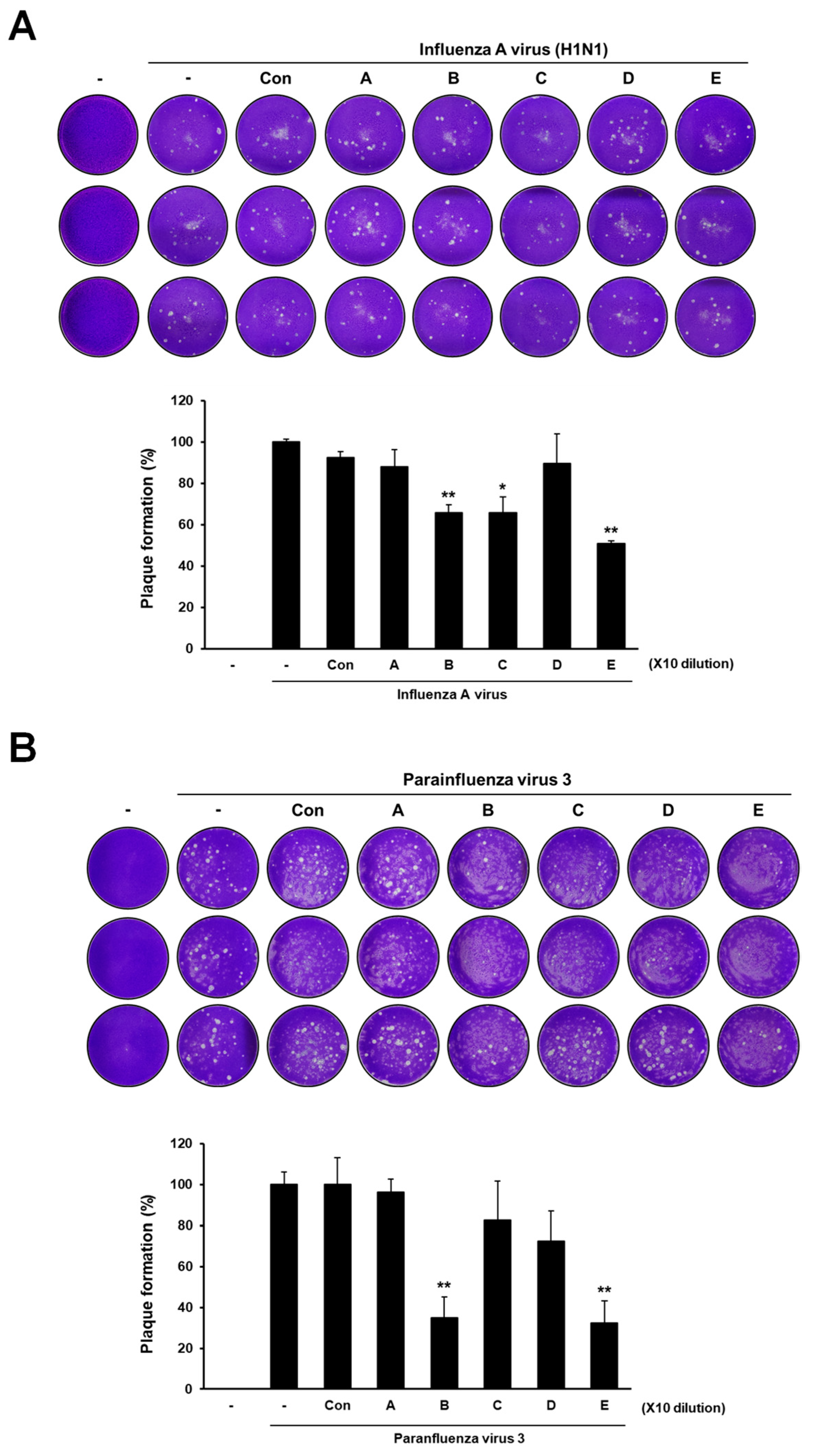
3.1. Antiviral Activities of Deacetylated Chitosan with GSE against Human Influenza A Virus and Parainfluenza Virus Type 3

MDCK and A549 cell viability was determined following treatment with deacetylated chitosan and GSE, revealing that the five deacetylated chitosan and GSE combinations had little effect on cytotoxicity (Figure 2). Therefore, MDCK cells infected with H1N1 were treated with five combinations of deacetylated chitosan and GSE, and plaque volumes were measured to evaluate the antiviral efficacy of this treatment. The plaque assay showed that chitosan with an 85% deacetylation degree reduced plaque formation from H1N1 more effectively than with 60% deacetylation degree (Figure 3A). HPIV3-induced plaque formation was induced under similar conditions. Similarly, chitosan with an 85% deacetylation degree reduced plaque formation caused by HPIV3 more than with a 60% deacetylation degree (Figure 3B). Interestingly, although GSE alone induced less H1N1-induced plaque formation, it did not affect HPIV3. These findings substantiate that chitosan with an 80% deacetylation or higher and GSE induce antiviral activity against H1N1 and HPIV3.

3.2. Antiviral Activity of Deacetylated Chitosan in Human A549 Cells

The composition of deacetylated chitosan was further explored by determining its antiviral potential in A549 human lung cancer cells. Consistent with the plaque formation in MDCK cells, western blot analyses indicated that HA protein levels were reduced in both virus-infected A549 cells with 85% deacetylated chitosan, but not 60% (Figure 4A,B and Figure S2). In comparison, GSE alone reduced the HA protein level of H1N1. Next, H1N1 and HPIV3 viral RNAs in A549 cells treated with deacetylated chitosan were examined via qRT-PCR. The gene expression of H1N1 viral matrix M1 and nucleoprotein NP was reduced in H1N1-infected A549 cells with 85% deacetylated chitosan, but not 60%, suggesting that chitosan inhibits viral infection deacetylation-dependently (Figure 4C and Figure S3). In HPIV3-infected A549 cells, deacetylated chitosan also attenuated the expression of nucleoprotein (N) and phosphoprotein (P) (Figure 4D and Figure S3). These data suggest the antiviral potential of deacetylated chitosan in human cells.

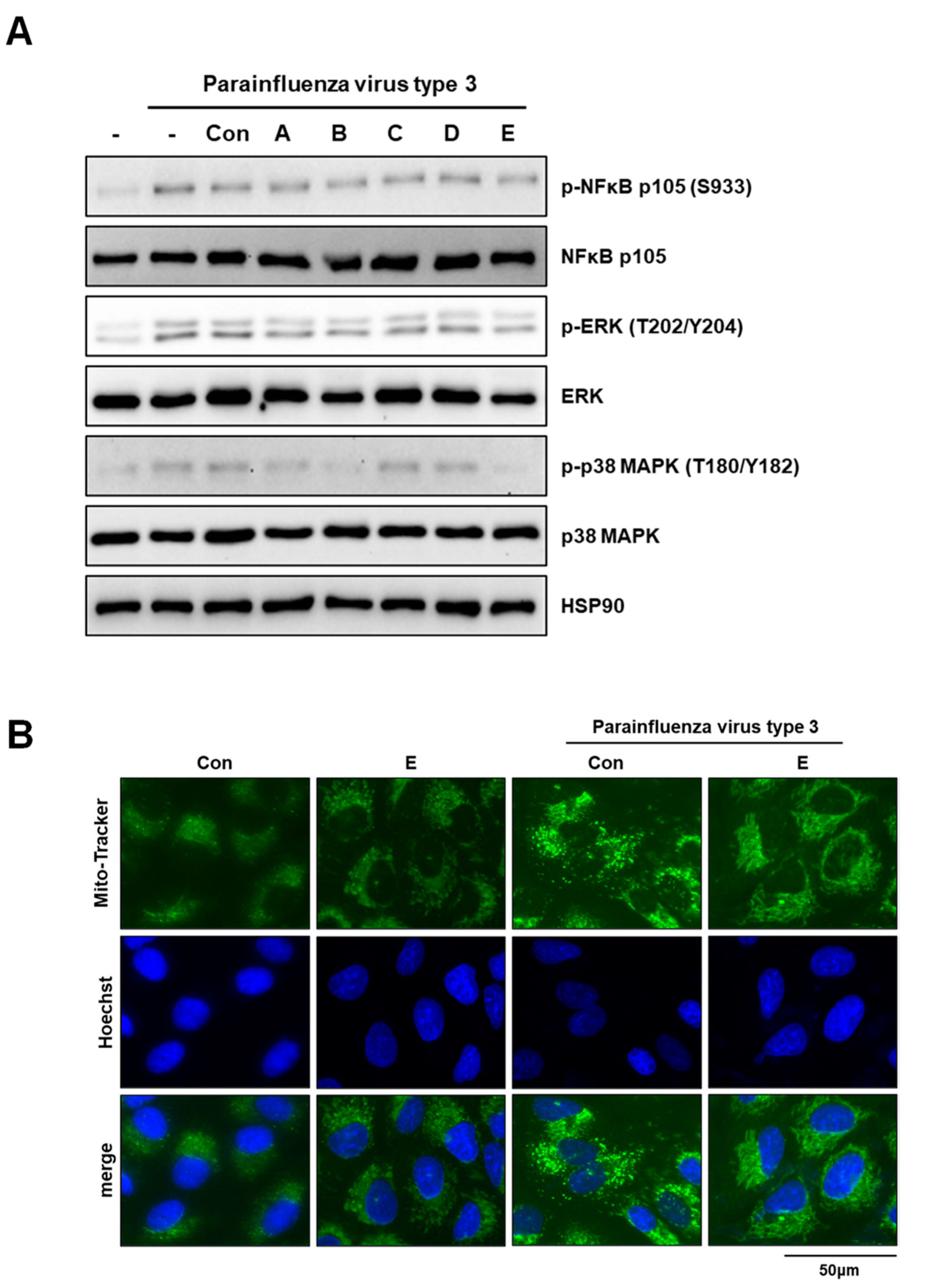
3.3. Deacetylated Chitosan Prevents Virus-Induced P38 MAPK Activation and Mitochondrial Fragmentation in A549 Cells

The antiviral effects of deacetylated chitosan may stem from cellular stress signaling and mitochondrial integrity. Therefore, several stress-signaling pathways potentially affected by the composition of deacetylated chitosan were screened to test this hypothesis. Western blot analyses revealed that the deacetylated chitosan composition inhibited phosphorylation of p38 MAPK but not that of NFκB p105 and ERK (Figure 5A and Figure S4). Next, we determined whether the composition of deacetylated chitosan affected virus-mediated mitochondrial integrity. Interestingly, mitochondrial fragmentation was significantly inhibited (Figure 5B and Figure S5), indicating that deacetylated chitosan obstructed HPIV3-induced p38 MAPK signaling and mitochondrial fragmentation in A549 cells.

4. Discussion

Chitosan is a linear polymer and polysaccharide derivative of deacetylated chitin. It is associated with numerous biological activities and is influenced by the degree of N-deacetylation, positive charge, polymerization degree, and other chemical modifications [24]. Notably, chitosan derivatives exhibit antiviral effects against various viral infections, including HIV, human papillomavirus, influenza A, and human coronavirus [12,25,26,27]. Recent reports have indicated that chitosan and its derivatives can bind to the pocket formed by the spike protein of SARS-CoV-2 [28,29]. Moreover, another study reported that sulfated chitooligosaccharides may block viral adsorption and membrane fusion by targeting the influenza A HA protein [15]. Similarly, our findings demonstrate that deacetylated chitosan combined with GSE reduced H1N1 HA protein levels in A549 cells. Chitosan, with a degree of deacetylation of 85%, was found to be more effective in reducing both H1N1- and HPIV3-induced plaque formation, while GSE alone reduced H1N1-induced plaque formation but not HPIV3-induced plaque formation. However, further studies are required to elucidate the underlying biological mechanisms.
Treatment with high concentrations of deacetylated chitosan had significant antiviral effects on H1N1 and HPIV, whereas GSE did not affect HPIV. GSE alone effectively blocked the H1N1 virus, and high-dose deacetylated chitosan inhibited HPIV infection. However, combining GSE with high-dose deacetylated chitosan proved to be the most effective against H1N1. Studies on the treatment of avian influenza with grapefruit extract also found that GSE was effective against enveloped viruses, such as avian influenza virus and Newcastle disease virus, but not against nonenveloped infectious bursal disease virus (IBDV). Grapefruit extract alone has no bactericidal effect. Instead, it likely destroys the cytoplasmic membrane, which induces a bactericidal effect [16]. A previous study reported that GSE in corn starch-chitosan bio-nanocomposite films could offer an innovative, eco-friendly, active packaging solution for the confectionery industry, prolonging shelf life and maintaining food safety and quality [30]. However, studies on the antiviral activity and food safety of the combination of chitosan and GSE are lacking. Further in vivo studies exploring this combination are required.
Chitosan is characterized by its degree of deacetylation or MW; thus, its solubility, hydrophilicity, crystallinity, and cell response predict its potential for various biological applications [31]. A recent in vitro study concluded that low-MW chitooligosaccharides other than chitosan also inhibit SARS-CoV-2 infection [22]. Deacetylation of chitosan is an important chemical modification that affects its performance and use in biological applications [31]. In addition, chitosan can restore immunity which is initially lowered by obesity [32,33,34]. Obesity intensifies insulin resistance generated by an immune system imbalance between M1 and M2 macrophages [35]. This response augments the susceptibility to H1N1 influenza virus infection in patients with obesity owing to their low immunity [36]. The weight-reducing effect of chitosan can help prevent obesity and maintain immunity, effectively averting influenza viral infections. The antiviral activity of chitosan, which has been tested and proven in practice, has been studied against several human viruses such as herpes simplex virus type 1, human cytomegalovirus, and Rift Valley fever virus [37]. In this study, we examined the antiviral activities of deacetylated chitosan and GSE against representative human respiratory viruses, specifically H1N1 and HPIV3.
HPIV infection causes mitochondrial fragmentation in the lung cells, resulting in cell death and severe lung damage [16,38]. These inflammation-related factors are expressed in response to mitochondrial destruction, which exacerbates the severity of lung damage [39]. The present study demonstrated that supplementing deacetylated chitosan with GSE may be important for enhancing the natural antiviral properties of chitosan. Chitosan, with an 85% deacetylation degree, has demonstrated antiviral potential in human A549 lung cancer cells, reducing viral protein levels and gene expression in both H1N1- and HPIV3-infected cells in a deacetylation-dependent manner. In addition, chitosan with a high degree of deacetylation inhibited the HPIV3-induced p38 MAPK pathway and mitochondrial fragmentation in A549 lung cancer cells. Previously, chitosan oligosaccharide was found to influence the regulation of several cellular signaling pathways, including NFκB and MAPK [40,41]. Interestingly, a study showed that the expression levels of TNF-alpha and IFN-beta increased significantly, suggesting that the antiviral activity of amino-modified chitosan operates by stimulating the immune response [42]. Deacetylated chitosan may exert its antiviral effects by inhibiting p38 MAPK phosphorylation and preserving mitochondrial integrity in virus-infected A549 cells. These studies indicate that chitosan can inhibit inflammation by modulating cellular stress signaling, corroborating our findings. However, further studies are needed to verify whether chitosan exhibits antiviral or anti-inflammatory effects in vivo based on the degree of deacetylation or combination with GSE.
Using MDCK and A549 cells, we demonstrated the antiviral effects of deacetylated chitosan with GSE against H1N1 and HPIV3 in vitro. Chitosan is a nontoxic biopolymer with a broad range of applications, including antimicrobial products. After treatment with deacetylated chitosan and GSE, the viability of MDCK and A549 cells showed no significant cytotoxicity for the five tested combinations. This study highlights the importance of the degree of chitosan deacetylation and its antiviral effects in vitro. Moreover, we confirmed the therapeutic potential of deacetylated chitosan against influenza epidemics. Interestingly, an in vivo study on chitosan implied that efficient mucosal vaccine delivery could be achieved using chitosan and its derivative, trimethyl chitosan nanoparticles, which are readily absorbed by microfold cells in nasal-associated lymphoid tissues and dendritic cells [43].
Recently, interesting findings on chitosan involving various nanoparticles have been reported. Zinc-stabilized chitosan-chondroitin sulfate nanocomposites have shown potential as drug delivery systems to enhance antiviral efficacy in the treatment of HIV-1 infection by inhibiting viral reservoirs [44]. Silver nanoparticle-chitosan composites have also been developed, displaying size-dependent antiviral activity against the H1N1 influenza A virus, with smaller nanoparticles yielding stronger antiviral effects [45]. Acyclovir-loaded chitosan nanospheres generated via a nanoemulsion template showed enhanced antiviral activity against herpes simplex virus compared to free acyclovir, making them useful for the topical treatment of herpes virus infections [46]. Acyclovir-loaded chitosan nanospheres showed improved antiviral efficiency against herpes simplex virus compared to free acyclovir, emphasizing their potential as topical therapies against herpes virus infections [46]. Various chitosan sources and preparation methods allow the production of a wide range of chitosan polymers with distinct physicochemical properties, including degree of deacetylation, MW, crystallinity, residual ash, and protein content [37]. Consequently, research on nanoparticle applications and composite materials is helpful.

5. Conclusions

Chitosan is a nontoxic polysaccharide that is widely used in biomaterials and biomedical applications. The degree of deacetylation is crucial for its application as a biodegradable biopolymer derived from the chitin in shrimp and crab shells. This study demonstrates that deacetylated chitosan, especially when supplemented with GSE, holds promise as an effective antiviral biomaterial against influenza and parainfluenza viruses. Through plaque assay, deacetylated chitosan was found to effectively decrease H1N1- and HPIV3-induced plaque formation in MDCK cells. Interestingly, our observations demonstrate that the antiviral efficacy of chitosan is largely dependent on whether the degree of deacetylation exceeds 80%. Furthermore, deacetylated chitosan effectively prevented virus-induced p38 MAPK activation in human A549 cells. A combination of deacetylated chitosan and GSE significantly suppressed HPIV3-induced mitochondrial fragmentation, thereby indicating their crucial role in blocking virus-induced stress signaling and maintaining mitochondrial integrity. Since the antiviral efficacy of chitosan significantly depends on its degree of deacetylation, further investigation into this chemical mechanism is required. Further studies are needed to verify the mechanism by which deacetylated chitosan and GSE exhibit antiviral activity in vivo. In addition, it is important to optimize the biological tissue penetration and antiviral effects through additional chemical modifications of deacetylated chitosan or its combination with nanoparticles.

Supplementary Materials

The following supporting information can be downloaded at: https://www.mdpi.com/article/10.3390/app13179938/s1, Figure S1: Results of measuring the deacetylation degree of chitosan; Figure S2: Hemagglutinin protein levels of H1N1- and HPIV3-infected A549 cells; Figure S3: Raw data from qPCR; Figure S4: Intracellular stress signaling of HPIV3 infected A549 cells; Figure S5: Mitochondria fragmentation of HPIV3 infected A549 cells.

Author Contributions

Conceptualization, D.S.S., J.S.L. and Y.J.; methodology, D.S.S., J.S.L. and Y.J.; validation, D.S.S., J.S.L. and Y.J.; formal analysis, D.S.S.; investigation, D.S.S., J.S.L., Y.C.S. and Y.J.; writing—original draft preparation, D.S.S., J.S.L. and Y. J.; and writing—review and editing, D.S.S., J.S.L. and Y.J. All authors have read and agreed to the published version of the manuscript.

Funding

This work was supported by Amicogen, Inc., Korea Basic Science Institute (National Research Facilities and Equipment Center) grant funded by the Ministry of Education (2023R1A6C101B022), and the Korea Institute of Marine Science and Technology Promotion (and) funded by the Ministry of Oceans and Fisheries (20220027). This research is funded by the Financial Program for Self-Directed Research Capacity in 2022.

Institutional Review Board Statement

Not applicable.

Informed Consent Statement

Not applicable.

Data Availability Statement

Not applicable.

Conflicts of Interest

The authors declare no conflict of interest.

References

  1. Medina, R.A.; García-Sastre, A. Influenza A viruses: New research developments. Nat. Rev. Microbiol. 2011, 9, 590–603. [Google Scholar] [CrossRef] [PubMed]
  2. Chan, P.K.S.; Lee, N.; Joynt, G.M.; Choi, K.W.; Cheung, J.L.K.; Yeung, A.C.M.; Lam, P.; Wong, R.; Leung, B.-W.; So, H.-Y.; et al. Clinical and virological course of infection with haemagglutinin D222G mutant strain of 2009 pandemic influenza A (H1N1) virus. J. Clin. Virol. 2011, 50, 320–324. [Google Scholar] [CrossRef] [PubMed]
  3. Zhang, L.; Bukreyev, A.; Thompson Catherine, I.; Watson, B.; Peeples Mark, E.; Collins Peter, L.; Pickles Raymond, J. Infection of Ciliated Cells by Human Parainfluenza Virus Type 3 in an In Vitro Model of Human Airway Epithelium. J. Virol. 2005, 79, 1113–1124. [Google Scholar] [CrossRef] [PubMed]
  4. Battles, M.B.; McLellan, J.S. Respiratory syncytial virus entry and how to block it. Nat. Rev. Microbiol. 2019, 17, 233–245. [Google Scholar] [CrossRef]
  5. Santos, V.P.; Marques, N.S.S.; Maia, P.; Lima, M.A.B.; Franco, L.O.; Campos-Takaki, G.M. Seafood Waste as Attractive Source of Chitin and Chitosan Production and Their Applications. Int. J. Mol. Sci. 2020, 21, 4290. [Google Scholar] [CrossRef] [PubMed]
  6. Hajji, S.; Younes, I.; Ghorbel-Bellaaj, O.; Hajji, R.; Rinaudo, M.; Nasri, M.; Jellouli, K. Structural differences between chitin and chitosan extracted from three different marine sources. Int. J. Biol. Macromol. 2014, 65, 298–306. [Google Scholar] [CrossRef] [PubMed]
  7. Berger, J.; Reist, M.; Chenite, A.; Felt-Baeyens, O.; Mayer, J.M.; Gurny, R. Pseudo-thermosetting chitosan hydrogels for biomedical application. Int. J. Pharm. 2005, 288, 197–206. [Google Scholar] [CrossRef]
  8. Younes, I.; Rinaudo, M. Chitin and chitosan preparation from marine sources. Structure, properties and applications. Mar. Drugs 2015, 13, 1133–1174. [Google Scholar] [CrossRef]
  9. Kaczmarek, M.B.; Struszczyk-Swita, K.; Li, X.; Szczęsna-Antczak, M.; Daroch, M. Enzymatic Modifications of Chitin, Chitosan, and Chitooligosaccharides. Front. Bioeng. Biotechnol. 2019, 7, 243. [Google Scholar] [CrossRef]
  10. Tan, R.S.L.; Hassandarvish, P.; Chee, C.F.; Chan, L.W.; Wong, T.W. Chitosan and its derivatives as polymeric anti-viral therapeutics and potential anti-SARS-CoV-2 nanomedicine. Carbohydr. Polym. 2022, 290, 119500. [Google Scholar] [CrossRef]
  11. Artan, M.; Karadeniz, F.; Karagozlu, M.Z.; Kim, M.-M.; Kim, S.-K. Anti-HIV-1 activity of low molecular weight sulfated chitooligosaccharides. Carbohydr. Res. 2010, 345, 656–662. [Google Scholar] [CrossRef] [PubMed]
  12. Zheng, M.; Qu, D.; Wang, H.; Sun, Z.; Liu, X.; Chen, J.; Li, C.; Li, X.; Chen, Z. Intranasal Administration of Chitosan Against Influenza A (H7N9) Virus Infection in a Mouse Model. Sci. Rep. 2016, 6, 28729. [Google Scholar] [CrossRef]
  13. Dimassi, S.; Tabary, N.; Chai, F.; Blanchemain, N.; Martel, B. Sulfonated and sulfated chitosan derivatives for biomedical applications: A review. Carbohydr. Polym. 2018, 202, 382–396. [Google Scholar] [CrossRef] [PubMed]
  14. Liu, Q.; Zheng, X.; Zhang, C.; Shao, X.; Zhang, X.; Zhang, Q.; Jiang, X. Conjugating influenza a (H1N1) antigen to n-trimethylaminoethylmethacrylate chitosan nanoparticles improves the immunogenicity of the antigen after nasal administration. J. Med. Virol. 2015, 87, 1807–1815. [Google Scholar] [CrossRef] [PubMed]
  15. Wang, S.; Luo, Y.; Huang, L.; Wang, S.; Hao, C.; Sun, L.; Zhang, Y.; Wang, W.; Li, C. The inhibition effects and mechanisms of sulfated chitooligosaccharides on influenza A virus in vitro and in vivo. Carbohydr. Polym. 2022, 286, 119316. [Google Scholar] [CrossRef]
  16. Komura, M.; Suzuki, M.; Sangsriratanakul, N.; Ito, M.; Takahashi, S.; Alam, M.S.; Ono, M.; Daio, C.; Shoham, D.; Takehara, K. Inhibitory effect of grapefruit seed extract (GSE) on avian pathogens. J. Vet. Med. Sci. 2019, 81, 466–472. [Google Scholar] [CrossRef] [PubMed]
  17. Go, C.C.; Pandav, K.; Sanchez-Gonzalez, M.A.; Ferrer, G. Potential Role of Xylitol Plus Grapefruit Seed Extract Nasal Spray Solution in COVID-19: Case Series. Cureus 2020, 12, e11315. [Google Scholar] [CrossRef]
  18. Han, H.W.; Kwak, J.H.; Jang, T.S.; Knowles, J.C.; Kim, H.W.; Lee, H.H.; Lee, J.H. Grapefruit Seed Extract as a Natural Derived Antibacterial Substance against Multidrug-Resistant Bacteria. Antibiotics 2021, 10, 85. [Google Scholar] [CrossRef]
  19. Roy, S.; Zhang, W.; Biswas, D.; Ramakrishnan, R.; Rhim, J.W. Grapefruit Seed Extract-Added Functional Films and Coating for Active Packaging Applications: A Review. Molecules 2023, 28, 730. [Google Scholar] [CrossRef]
  20. Di Petrillo, A.; Orrù, G.; Fais, A.; Fantini, M.C. Quercetin and its derivates as antiviral potentials: A comprehensive review. Phytother. Res. PTR 2022, 36, 266–278. [Google Scholar] [CrossRef]
  21. Tutunchi, H.; Naeini, F.; Ostadrahimi, A.; Hosseinzadeh-Attar, M.J. Naringenin, a flavanone with antiviral and anti-inflammatory effects: A promising treatment strategy against COVID-19. Phytother. Res. PTR 2020, 34, 3137–3147. [Google Scholar] [CrossRef] [PubMed]
  22. Jang, D.; Lee, D.; Shin, Y.C.; Lee, J.S.; Jung, J.; Ryoo, S. Low molecular weight chitooligosaccharide inhibits infection of SARS-CoV-2 in vitro. J. Appl. Microbiol. 2022, 133, 1089–1098. [Google Scholar] [CrossRef]
  23. Joo, S.; Baek, S.; Kang, J.; Seo, D.S.; Kwon, T.K.; Jang, Y. α-ketoglutarate suppresses immediate early gene expression in cancer cells. Biochem. Biophys. Res. Commun. 2022, 637, 144–152. [Google Scholar] [CrossRef]
  24. Chirkov, S.N. The antiviral activity of chitosan (review). Prikl. Biokhim. Mikrobiol. 2002, 38, 5–13. [Google Scholar] [PubMed]
  25. Nishimura, S.I.; Kai, H.; Shinada, K.; Yoshida, T.; Tokura, S.; Kurita, K.; Nakashima, H.; Yamamoto, N.; Uryu, T. Regioselective syntheses of sulfated polysaccharides: Specific anti-HIV-1 activity of novel chitin sulfates. Carbohydr. Res. 1998, 306, 427–433. [Google Scholar] [CrossRef] [PubMed]
  26. Gao, Y.; Liu, W.; Wang, W.; Zhang, X.; Zhao, X. The inhibitory effects and mechanisms of 3,6-O-sulfated chitosan against human papillomavirus infection. Carbohydr. Polym. 2018, 198, 329–338. [Google Scholar] [CrossRef]
  27. Ciejka, J.; Wolski, K.; Nowakowska, M.; Pyrc, K.; Szczubiałka, K. Biopolymeric nano/microspheres for selective and reversible adsorption of coronaviruses. Mater. Sci. Eng. C Mater. Biol. Appl. 2017, 76, 735–742. [Google Scholar] [CrossRef]
  28. Milewska, A.; Ciejka, J.; Kaminski, K.; Karewicz, A.; Bielska, D.; Zeglen, S.; Karolak, W.; Nowakowska, M.; Potempa, J.; Bosch, B.J.; et al. Novel polymeric inhibitors of HCoV-NL63. Antivir. Res. 2013, 97, 112–121. [Google Scholar] [CrossRef] [PubMed]
  29. Kalathiya, U.; Padariya, M.; Mayordomo, M.; Lisowska, M.; Nicholson, J.; Singh, A.; Baginski, M.; Fahraeus, R.; Carragher, N.; Ball, K.; et al. Highly Conserved Homotrimer Cavity Formed by the SARS-CoV-2 Spike Glycoprotein: A Novel Binding Site. J. Clin. Med. 2020, 9, 1473. [Google Scholar] [CrossRef]
  30. Jha, P. Effect of grapefruit seed extract ratios on functional properties of corn starch-chitosan bionanocomposite films for active packaging. Int. J. Biol. Macromol. 2020, 163, 1546–1556. [Google Scholar] [CrossRef]
  31. El Knidri, H.; Belaabed, R.; Addaou, A.; Laajeb, A.; Lahsini, A. Extraction, chemical modification and characterization of chitin and chitosan. Int. J. Biol. Macromol. 2018, 120, 1181–1189. [Google Scholar] [CrossRef] [PubMed]
  32. Do, A.R.; Cho, S.J.; Cho, Y.Y.; Kwon, E.Y.; Choi, J.Y.; Lee, J.H.; Han, Y.; Kim, Y.S.; Piao, Z.; Shin, Y.C.; et al. Antiobesity Effects of Short-Chain Chitosan in Diet-Induced Obese Mice. J. Med. Food 2018, 21, 927–934. [Google Scholar] [CrossRef] [PubMed]
  33. Gopan, G.; Jose, J.; Khot, K.B.; Bandiwadekar, A. The use of cellulose, chitosan and hyaluronic acid in transdermal therapeutic management of obesity: A review. Int. J. Biol. Macromol. 2023, 244, 125374. [Google Scholar] [CrossRef] [PubMed]
  34. Nedunchezhiyan, U.; Varughese, I.; Sun, A.R.; Wu, X.; Crawford, R.; Prasadam, I. Obesity, Inflammation, and Immune System in Osteoarthritis. Front. Immunol. 2022, 13, 907750. [Google Scholar] [CrossRef]
  35. Patel, P.S.; Buras, E.D.; Balasubramanyam, A. The role of the immune system in obesity and insulin resistance. J. Obes. 2013, 2013, 616193. [Google Scholar] [CrossRef]
  36. Bhattacharya, I.; Ghayor, C.; Pérez Dominguez, A.; Weber, F.E. From Influenza Virus to Novel Corona Virus (SARS-CoV-2)-The Contribution of Obesity. Front. Endocrinol. 2020, 11, 556962. [Google Scholar] [CrossRef]
  37. Jaber, N.; Al-Remawi, M.; Al-Akayleh, F.; Al-Muhtaseb, N.; Al-Adham, I.S.I.; Collier, P.J. A review of the antiviral activity of Chitosan, including patented applications and its potential use against COVID-19. J. Appl. Microbiol. 2022, 132, 41–58. [Google Scholar] [CrossRef]
  38. Ding, B.; Zhang, L.; Li, Z.; Zhong, Y.; Tang, Q.; Qin, Y.; Chen, M. The Matrix Protein of Human Parainfluenza Virus Type 3 Induces Mitophagy that Suppresses Interferon Responses. Cell Host Microbe 2017, 21, 538–547.e534. [Google Scholar] [CrossRef]
  39. Irazoki, A.; Gordaliza-Alaguero, I.; Frank, E.; Giakoumakis, N.N.; Seco, J.; Palacín, M.; Gumà, A.; Sylow, L.; Sebastián, D.; Zorzano, A. Disruption of mitochondrial dynamics triggers muscle inflammation through interorganellar contacts and mitochondrial DNA mislocation. Nat. Commun. 2023, 14, 108. [Google Scholar] [CrossRef]
  40. Kim, J.H.; Kim, Y.S.; Hwang, J.W.; Han, Y.K.; Lee, J.S.; Kim, S.K.; Jeon, Y.J.; Moon, S.H.; Jeon, B.T.; Bahk, Y.Y.; et al. Sulfated chitosan oligosaccharides suppress LPS-induced NO production via JNK and NF-κB inactivation. Molecules 2014, 19, 18232–18247. [Google Scholar] [CrossRef]
  41. Liu, H.T.; Li, W.M.; Li, X.Y.; Xu, Q.S.; Liu, Q.S.; Bai, X.F.; Yu, C.; Du, Y.G. Chitosan oligosaccharides inhibit the expression of interleukin-6 in lipopolysaccharide-induced human umbilical vein endothelial cells through p38 and ERK1/2 protein kinases. Basic. Clin. Pharmacol. Toxicol. 2010, 106, 362–371. [Google Scholar] [CrossRef] [PubMed]
  42. He, X.; Xing, R.; Liu, S.; Qin, Y.; Li, K.; Yu, H.; Li, P. The improved antiviral activities of amino-modified chitosan derivatives on Newcastle virus. Drug Chem. Toxicol. 2021, 44, 335–340. [Google Scholar] [CrossRef] [PubMed]
  43. Mosafer, J.; Sabbaghi, A.H.; Badiee, A.; Dehghan, S.; Tafaghodi, M. Preparation, characterization and in vivo evaluation of alginate-coated chitosan and trimethylchitosan nanoparticles loaded with PR8 influenza virus for nasal immunization. Asian J. Pharm. Sci. 2019, 14, 216–221. [Google Scholar] [CrossRef] [PubMed]
  44. Wu, D.; Ensinas, A.; Verrier, B.; Primard, C.; Cuvillier, A.; Champier, G.; Paul, S.; Delair, T. Zinc-Stabilized Chitosan-Chondroitin Sulfate Nanocomplexes for HIV-1 Infection Inhibition Application. Mol. Pharm. 2016, 13, 3279–3291. [Google Scholar] [CrossRef]
  45. Mori, Y.; Ono, T.; Miyahira, Y.; Nguyen, V.Q.; Matsui, T.; Ishihara, M. Antiviral activity of silver nanoparticle/chitosan composites against H1N1 influenza A virus. Nanoscale Res. Lett. 2013, 8, 93. [Google Scholar] [CrossRef]
  46. Donalisio, M.; Leone, F.; Civra, A.; Spagnolo, R.; Ozer, O.; Lembo, D.; Cavalli, R. Acyclovir-Loaded Chitosan Nanospheres from Nano-Emulsion Templating for the Topical Treatment of Herpesviruses Infections. Pharmaceutics 2018, 10, 46. [Google Scholar] [CrossRef]
Figure 1. Manufacturing process of deacetylated chitosan and GSE main compounds. (A) Manufacturing process and chemical structure of deacetylated chitosan. (B) GSE and structures of naringin and naringenin.
Applsci 13 09938 g001
Figure 2. Chitosan with GSE exhibits no cytotoxicity. (A,B) Dog MDCK cells (A) or human A549 cells (B) were treated with indicated dilutions of 60% or 85% deacetylated chitosan and GSE for 24 h, followed by MTT cell viability assay. A (60% deacetylated chitosan), B (85% deacetylated chitosan), C (GSE), D (60% deacetylated chitosan + GSE), and E (85% deacetylated chitosan + GSE) are shown in Table 1. The untreated group was compared to the chitosan treatment group. Quantitative data are presented as the mean ± SEM from three biological replicates (n = 3). * p < 0.05, ** p < 0.01.
Applsci 13 09938 g002
Figure 3. Antiviral activity of deacetyl-chitosan in combination with GSE against H1N1 and HPIV3. (A) Plaque assay images of H1N1-infected MDCK cells treated with or without deacetyl-chitosan (upper panel). Quantitative data for plaque formation (%) are presented as the mean ± SEM from three biological replicates (n = 3) (lower panel). (B) Plaque assay images (upper panel) and quantitative data (lower panel) for HPIV3 were performed in the same manner. A (60% deacetylated chitosan), B (85% deacetylated chitosan), C (GSE), D (60% deacetylated chitosan + GSE), and E (85% deacetylated chitosan + GSE) are shown in Table 1. Statistical comparison between groups was performed using Student’s t-test in all figures. Control group was compared to treatment group from three biological replicates (n = 3). * p < 0.05, ** p < 0.01.
Applsci 13 09938 g003
Figure 4. Assessment of antiviral activity of deacetyl-chitosan in human A549 cells. (A,B) HA protein levels of H1N1-infected (A) or HPIV3-infected (B) A549 cells treated with or without deacetyl-chitosan. Whole-cell lysates were analyzed by western blot using antibodies on the right. (C) qRT-PCR of H1N1_M1 and H1N1_NP expression in H1N1-infected A549 cells. (D) qRT-PCR of HPIV3-N and HPIV3-P expression in HPIV3-infected A549 cells. Quantitative data for gene expression are presented as the mean ± SEM from three biological replicates (n = 3). A (60% deacetylated chitosan), B (85% deacetylated chitosan), C (GSE), D (60% deacetylated chitosan + GSE), and E (85% deacetylated chitosan + GSE) are shown in Table 1. Control group was compared to treatment group from three biological replicates (n = 3). * p < 0.05, ** p < 0.01.
Applsci 13 09938 g004
Figure 5. Deacetyl-chitosan prevents virus-induced p38 MAPK activation and mitochondrial fragmentation in A549 cells. (A) Western blot analysis of protein levels of phospho or total NFκB p105, ERK and p38 MAPK using whole-cell lysates from HPIV3-infected A549 cells treated with or without deacetyl-chitosan. HSP90 was used as a loading control. (B) HPIV3-infected A549 cells treated with or without 85% deacetyl-chitosan with GSE, followed by treatment with Mito-Tracker Green dye for 1 h. Mitochondria were observed via fluorescence microscopy.
Applsci 13 09938 g005
Table 1. Combinations of deacetylated chitosan with GSE.
GroupSample Composition
A60% deacetylated chitosan
B85% deacetylated chitosan
CGSE
D60% deacetylated chitosan + GSE
E85% deacetylated chitosan + GSE
Disclaimer/Publisher’s Note: The statements, opinions and data contained in all publications are solely those of the individual author(s) and contributor(s) and not of MDPI and/or the editor(s). MDPI and/or the editor(s) disclaim responsibility for any injury to people or property resulting from any ideas, methods, instructions or products referred to in the content.

