Article

Non-Enzymatic Formation of N-acetylated Amino Acid Conjugates in Urine

by
Jano Jacobs
1,
Cornelia Gertina Catharina Elizabeth van Sittert
2,
Lodewyk Japie Mienie
1,
Marli Dercksen
1,
Monique Opperman
1 and
Barend Christiaan Vorster
1,*
1
Centre for Human Metabolomics, North-West University, Potchefstroom 2520, South Africa
2
Laboratory for Applied Molecular Modelling, Research Focus Area for Chemical Resource Beneficiation, North-West University, Potchefstroom 2520, South Africa
*
Author to whom correspondence should be addressed.
Appl. Sci. 2023, 13(18), 10002; https://doi.org/10.3390/app131810002
Submission received: 26 September 2022 / Revised: 21 August 2023 / Accepted: 29 August 2023 / Published: 5 September 2023

Abstract

:
Unknown N-acylated amino acid (N-AAA) conjugates have been detected in maple syrup urine disease (MSUD) and other inborn errors of metabolism (IEMs). This study aimed to elucidate the mechanism behind the formation of urinary N-AAA conjugates. Liquid–liquid extraction was employed to determine the enantiomeric composition of N-AAA conjugates, followed by liberation of conjugated amino acids through acid hydrolysis. Gas chromatography–mass spectrometry (GC–MS) was used to separate amino acid enantiomers. In vitro experiments were conducted to test the non-enzymatic formation of N-AAA conjugates from 2-keto acids and ammonia, with molecular modelling used to assess possible reaction mechanisms. Adequate amounts of N-AAA conjugates were obtained via organic acid extraction without concurrent extraction of native amino acids, and hydrolysis was complete without significant racemisation. GC–MS analysis successfully distinguished amino acid enantiomers, with some limitations observed for L-isoleucine and D-alloisoleucine. Furthermore, investigation of racemic N-AAA conjugates from an MSUD case confirmed its non-enzymatic origin. These findings highlight the value of employing chiral strategy and molecular modelling to investigate the origin of unknown constituents in biological samples. Additionally, these conjugates warrant further investigation as potential factors contributing to MSUD and other IEMs.

1. Introduction

Inborn errors of metabolism (IEMs) occur due to numerous hereditary genetic variants that affect proper protein functioning; IEMs impair cell metabolism by disrupting growth, energy production, and/or causing metabolite accumulation or depletion [1]. Diagnosis of IEMs involves extensive metabolic profiling to identify disease-specific constituents, primarily by detecting accumulated metabolites. These metabolites are often attributed to induced secondary pathways and contribute to the characteristic phenotypic presentation of the disorders [2].
During routine diagnostics, particularly in untargeted or semi-targeted applications, the presence of unknown constituents in significant concentrations is a common occurrence [3]. These unknown constituents can have various origins, including unwanted reactions during analyses, medication or dietary intake, microbial contamination or degradation, or compounds not included in the target compound library. These unknown contaminants raise doubts about the association of metabolites with a specific disorder or condition and lead to questions of whether all relevant information has been extracted from the data [4,5]. Sometimes, even when a constituent is correctly identified, its origin and functionality may still require clarification. This is illustrated in the case of certain N-acylated amino acid (N-AAA) conjugates found in isolated cases of maple syrup urine disease (MSUD) [6,7].
MSUD (OMIM: 248600) is an autosomal recessive disorder characterised by impaired metabolism of branched-chain amino acids (BCAAs) due to various mutations in the branched-chain α-keto acid dehydrogenase complex (BCKDH) [8]. The condition exhibits a range of phenotypes, from mild thiamine-responsive forms to severe classical MSUD. Metabolic profiling of MSUD typically reveals elevated levels of BCAAs [9,10], particularly alloisoleucine, along with their corresponding 2-keto acids (substrates of BCKDH), such as 2-keto-3-methylvaleric acid, 2-ketoisovaleric acid, and 2-ketoisocaproic acid, as well as their 2-hydroxy acid analogues [10,11]. Additionally, elevated levels of ammonia, N-acetyl BCAA (N-Ac BCAA), N-lactyl BCAA conjugates, and BCAA conjugation of 2-hydroxyisovaleric acid have also been observed in some MSUD cases [6,7,12,13].
Dry [7] conducted research on the induced metabolism of MSUD and identified several metabolites (Figure 1). These include trace amounts of (1) decarboxylated amines (2) 2-hydroxy BCAA conjugates (3) N-lactyl conjugates, (4) N-acetyl conjugates, and (5) hydantoin-like metabolites. Additionally, Dry [7] identified three variant cases of MSUD that presented with N-AAA conjugates derived from the BCAAs valine (Val), leucine (Leu) and isoleucine (Ile), including N-isobutyryl-, N-isovaleryl, and N-2-methylbutyryl conjugates. These findings have been subsequently confirmed [6].
Of these metabolites, N-AAA conjugates are carboxamides formed through the conjugation of an acyl group to an amino acid via a peptide bond [14]. N-AAA conjugates play a role as intermediates in both primary and alternative metabolism, participating in urea cycle regulation [15], endo- and exogenous N-acetylated protein degradation [16], and the myelination of neurons by providing the precursor for myelin lipid biosynthesis, acetate, upon hydrolysis [17]. Furthermore, during Gly-mediated biotransformation, glycine N-acyltransferase (GLYAT) (Enzyme Commission number [EC]: 2.3.1.13) catalyses the conjugation of Gly to various acyl-coenzyme A (CoA) substrates. These biotransformation products are frequently observed in inherited metabolic diseases. It is believed that they play a crucial role in salvaging acetyl-CoA and enhancing the solubility of carboxylic acid intermediates for excretion in urine [18].
In aminoacidopathies, transferases catalyse the acetylation of amino acids to form N-acetylated amino acid (N-AcAA) conjugates [11,19,20,21]. In contrast, in organic acidemias and fatty acid oxidation disorders GLYAT conjugates various acyl-CoA molecules, predominantly with GLY and other amino acids [7,22,23,24,25,26,27,28,29]. Aminoacidopathies thus exhibit elevated levels of N-AcAA conjugates corresponding to the increased amino acids, while organic acidemias exhibit elevated levels of N-acyl-Gly conjugates corresponding to the elevated acyl-CoA molecules. However, exceptions to this pattern include N-phenylacetyl-glutamine (-Gln) in phenylketonuria, N-isovaleryl-glutamate (-Glu), and -alanine (-Ala) in isovaleric acidemia, as they are products of GLYAT-mediated biotransformation [12,22,30]. Similarly, the presence of N-acylated BCAA conjugates in the above-mentioned variant MSUD cases deviates from the expected pattern [6,7].
Previous studies by Dry [7] and Deysel [6] have investigated and proposed hypotheses to elucidate the formation of these N-AAA conjugates in the mentioned cases. Although the biotransformation of N-acylated-CoAs with BCAAs by the GLYAT enzyme seemed a plausible explanation, the presence of N-AAA conjugates in MSUD cases was unexpected as N-acyl-glycine conjugates were not observed [7,22]. Moreover, as can be seen in Figure 2, the enzyme defect in MSUD prevented or depleted the formation of apparent GLYAT substrates (N-isobutyryl-, N-isovaleryl-, and N-2-methylbutyryl-CoA) [6,7]. Deysel [6] proposed a more likely hypothesis, suggesting the non-enzymatic formation of N-AAA conjugates from 2-keto acids and ammonia. This hypothesis built upon the mechanism proposed by Yanagawa et al. [31], which demonstrated that pyruvate and glyoxylic acid react with ammonium sulphate under specific conditions, resulting in a conjugate that yields amino acids upon hydrochloric acid (HCl) hydrolysis [31].
However, the formation mechanism of this unique metabolomic profile and the clinical-biochemical effects of the hydantoin and N-AAA conjugates were deemed unclear. Understanding the origin of these constituents could provide insights into an individual's disorder/condition, shed light on unknown metabolic pathways, and troubleshoot potential issues with diagnostic methods.
Studying variant metabolomic presentations is essential for: (1) further comparison of clinical-biochemical characteristics, (2) discovery of new diagnostic markers, (3) assessing responsiveness to treatment, (4) improving therapeutic approaches, and (5) expanding our knowledge of metabolic mechanisms, which may have implications beyond the specific disease context. Motivated by the confirmation of the alternative mechanism of N-AAA formation proposed by Dry [7] and Deysel [6], this study aims to investigate and validate this mechanism.
In this study, our aim was to investigate the formation of N-AAA conjugates and determine whether this process occurs enzymatically or non-enzymatically. To achieve this, we pursued several objectives. Firstly, we sought to determine the enantiomeric composition of N-AAA conjugates using chiral analysis. This involved liquid–liquid extraction, acid hydrolysis to release conjugated amino acids, and chiral derivatisation followed by GC–MS analysis. Secondly, we attempted to replicate the non-enzymatic formation of N-AAA conjugates in vitro. Lastly, we used molecular modelling to assess the feasibility of this non-enzymatic formation by evaluating the required activation energy (Ea) under physiological and laboratory conditions, comparing it with typical enzymatic Ea values. These objectives allowed us to gain valuable insights into the metabolic pathways involved and shed light on the nature of N-AAA conjugate formation, thus contributing to our understanding of vital biochemical processes.

2. Materials and Methods

2.1. Reagent Preparation and Chemicals

Thermo Fisher Scientific’s Creatinine Enzymatic kit (ref# 981845) was used for creatinine determination. The 'Phenomenex EZ:faast for free (physiological) amino acid analysis by GC–MS’ kit (EZ:faast) was purchased from Phenomenex (Torrance, CA, USA). Amino acid standards (DL-Ala, L-Ala, L-aIle, D-aIle, L-Leu, D-Leu, L-Val, D-Val, DL-norvaline [Nva], L-Ile and DL-Ile), the internal standard (3-phenylbutyric acid), derivatisation reagents (N,O-bis(trimethylsilyl)trifluoroacetamide [BSTFA], trimethylchlorosilane [TMCS], MTBSTFA with 1% t-BDMCS and R-2-butanol), ammonium sulphate, and N-AcAA conjugate standards (N-Ac-L-Ala, N-Ac-D-Ala, N-Ac-L-Val, N-Ac-D-Val, N-Ac-L-Leu, N-Ac-D-Leu, N-Ac-L-Ile and N-Ac-D-aIle) were all purchased from Sigma-Aldrich (St. Louis, MI, USA).
HCl (37%), sulphuric acid, sodium hydroxide (NaOH), pyridine, ethyl acetate, diethyl ether, and anhydrous sodium sulphate were purchased from Merck Millipore (Darmstadt, Germany). Instrument-grade helium (baseline 5.0) was purchased from African Oxygen Limited (Johannesburg, South Africa) as a carrier gas in GC operations. Ca. 6 N Moisture-free enantiopure r-2-butanolic HCl solution was prepared by slowly bubbling HCl gas through r-2-butanol on ice until saturated. The gas was generated by slowly adding concentrated HCl(aq) (37%) to concentrated sulphuric acid. This stock solution was diluted with pure r-2-butanol ca. 3 N.

2.2. Sample Availability

N-AAA conjugates (N-isobutyryl-Val, N-isobutyryl-Leu, N-isobutyryl-Ile, N-isovaleryl-Val, N-isovaleryl-Leu, N-isovaleryl-Ile, N-2-methylbutyryl-Val, N-2-methylbutyryl-Leu and N-2-methylbutyryl-Ile), previously identified and quantified in the urine of a South African MSUD case, were investigated. In addition to this MSUD case, ten urine samples were acquired and used as controls. The study was carried out in accordance with Helsinki Declaration guidelines, and these samples were stored as biological materials collected for diagnostic purposes. Before this investigation, all samples were de-identified, decoded, and anonymised.

2.3. Instrumentation

Creatinine was measured enzymatically according to the instructions included in the Thermo Fisher Scientific's Creatinine Enzymatic kit using an Indiko Clinical Chemistry Analyzer (Thermo Fisher Scientific, Waltham, MA, USA).
A Hewlett Packard 6890 GC system coupled to an Agilent 5973N Mass Selective Detector (MSD) was used for amino acid analysis. The GC was fitted with a Phenomenex CG0-7169 Zebron EZ-AAA amino acid GC column, and instrument settings were in accordance with the kit instructions. Organic acid analysis was performed on an Agilent 7890A GC, 5975C VL MSD fitted with an electron ionisation (EI) ion source using a J&W 122-0132UI GC column. Instrument settings were in accordance with Blau et al. [32]. Diastereomeric amino acid derivative analysis was conducted on an Agilent 7890A GC system with 5977A MSD. The GC system was fitted with a J&W CP8842 CP-Sil 19CB column, and the injector was set to 280 °C. The oven temperature programme was set to remain isothermal at 70 °C for 3 min and was then ramped at 5 °C/min and 2 °C/min in 10-min intervals to reach 140 °C. It was further increased by 30 °C/min to 280 °C and kept isothermal for another 2.5 min. A post-run followed this at 300 °C for 1 min. The MSD was set to scan from 70 to 600 m/z with a threshold of 50 and a scan speed of 1.562 Da/s. In all cases, ionisation was performed with an electron impact ionisation ion source, and helium was used as a carrier gas.
AMDIS version 2.71 from NIST was used to deconvolute, identify, and quantify all data acquired from GC–MS analysis in this study. An in-house library aided in the identification of compounds. This library was updated with spectra obtained from a previous study [7], data acquired from analysis of pure standards, and the NIST Mass Spectral Search Program for the NIST/EPA/NIH mass spectral library version 2.0f. The Agilent Technologies MSD Enhanced ChemStation F.01.00.1903 was used to identify and quantify GC–MS data and overlay chromatograms used in visual comparisons.

2.4. Methods

2.4.1. Organic Acid Analysis

N-AAA conjugates were confirmed using organic acid analysis based on the method described by Blau et al. [32] with minor modifications. An internal standard (3-phenyl butyric acid) was used, and an additional diethyl ether extraction step was included. After confirmation of N-AAA conjugates, organic acid extraction was repeated, followed by hydrolysis using 2.0 N HCl at 110 °C for 1 h. The dried hydrolysate was chirally derivatised in two steps. First, it underwent incubation with R-2-butanolic HCl (150 μL, 3 mol/L) at 110 °C for 2 h. Then, following evaporation to dryness, it was incubated for a second time with MTBSTFA (100 μL, with 1% t-BDMCS) and pyridine (50 μL) at 75 °C for 30 minutes. Chiral derivatised samples were analysed using GC–MS. This approach introduced a chiral auxiliary and aided the separation of diastereomers while improving stability and reducing evaporation temperatures.
The validity of this procedure was confirmed via preceding experiments. The selectivity of the organic acid extraction procedure was assessed by analysing reconstituted organic acid extracts spiked with BCAA standards to confirm the absence of amino acids. Control samples were subjected to organic acid analysis to verify the absence of N-AAA conjugates. These samples were then hydrolysed, reconstituted, and subjected to amino acid analysis to ensure no liberation of amino acids from other unknown N-AAA conjugates. Chiral derivatisation and analysis were subsequently performed on the hydrolysed reconstitutes to ensure no overlapping peaks with amino acid enantiomers.

2.4.2. N-AAA Conjugates Hydrolysis

An iterative approach was used to test different acids and bases for hydrolysis efficiency. The best incubation results were obtained using 2.0 mol/L HCl at 110 °C for 1 h. The hydrolysis process and hydrolysis-induced racemisation were assessed using enantiopure standards and spiked control samples. Hydrolysis efficiency was evaluated by measuring the disappearance of N-AAA conjugates and the appearance of amino acids through organic and amino acid analysis, respectively. Since specific N-AAA conjugate standards (N-isobutyryl-, N-isovaleryl- and N-2-methylbutyryl conjugates of Leu, Val, and Ile) were unavailable, structural analogues (N-AcAA conjugate standards) were used. The near absence of racemic pairs after chiral derivatisation and GC–MS analysis confirmed the insignificance of hydrolysis-induced racemisation.

2.4.3. Non-Enzymatic In Vitro Synthesis of N-AAA Conjugates

The feasibility of non-enzymatic formation of N-AAA conjugates was investigated by incubating various 2-keto acids (refer to Section 3.2) at pH < 2 for 1 h at 37 °C. Reaction conditions were selected to mimic the proposed formation of N-AAA conjugates from 2-keto acids and ammonia (as per the hypothesis described by Deysel [6]). Each reaction used a single 2-keto acid substrate (serving as both the formyl and carboxy donor) to produce a single product, allowing for the examination of specific reactions. The predicted end products and their potential combinations (as shown in Table 1) were determined based on the proposed net reaction given in Figure 3.
The number of possible compounds resulting from product combinations can be calculated as n2, where n is the number of 2-keto acids in the reaction. In this study, with nine selected 2-keto acids, there were 81 potential end products. Additionally, because of incomplete derivatisation, each N-AAA conjugate end product could produce two peaks with different mass spectra during GC–MS analysis with BSTFA. However, mass spectra were only available for 27 out of the 162 possible compounds. As a result, the non-enzymatic reactions were restricted to using a single 2-keto acid substrate per reaction to address this limitation.

2.4.4. Molecular Modelling

Molecular modelling was used to assess the possibility of in vivo formation of N-AAA conjugates. The maximum activation energy (Ea) required for the reaction was calculated and compared with typical Ea values of various enzymes. A reaction mechanism proposed by Yanagawa et al. [31] for the non-enzymatic formation of N-Ac-Ala from pyruvic acid and ammonia served as a starting point.
Intermediates and products involved in the mechanism were visually represented using the Visualiser module in BIOVIA Materials Studio software (v16.1.0.31 Dassault Systèmes, Vélizy-Villacoublay, France). Geometric optimisation of these structures was performed using the density functional theory (DFT) module (DMol3) in Materials Studio. The calculation model employed was the generalised gradient approximation (GGA) functional PW91, using Ortmann, Bechstedt, and Schmidt (OBS) dispersion correction and a double-numerical basis set with polarisation functions (DNP) basis file 4.4.
The molecular modelling parameters were set as follows: An unrestricted electron spin with the initial spin value set to formal spin. The convergence tolerance was set to medium, and the maximum number of iterations was 1 000. The maximum allowable step size for any change in the Cartesian coordinates was 0.3 Å, and integration accuracy was set to medium. Direct inversion in interactive subspace (DIIS) was used to speed up the self-consistent field (SCF) convergence, which was set to medium. The DIIS size was set to 6, and the maximum number of SCF iterations to 1000. The maximum angular momentum function was set to octupole, with a density mixing charge of 0.2 and a spin of 0.5.
Thermal smearing with a value of 0.005 Ha was selected. All electrons were included in the calculations. The orbital cut-off of the atomic basis set was set to medium quality, and the exact value depended on the elements present in the specific structure. The conductor-like screening model (COSMO) continuum solvation model (CSM) was selected to simulate the solvent environment for the model. Water, with the dielectric constant set to 78.54, was chosen as the solvent in this study. Vibrational frequency analysis was performed as part of the DMol3 calculations to confirm that structures at minimum energy were obtained. Coarse-grained parallelisation was used for the numerical displacement frequency calculations. After the convergence of the DMol3 calculations, the vibration analysis tool was used to confirm that no imaginary vibrations were present.
Following geometric optimisation, single-point energy calculations were performed to determine the relative electronic energy (Ee) of the structures at 0 K. The total relative Gibbs free energy (ΔGrel) at 37 °C was calculated by adding the free energy (ΔG) corrections from vibration analyses at 37 °C to the Ee of the geometrically optimised structures. These ΔGrel values were used to construct a preliminary reaction energy profile. Potential energy surface (PES) scans were carried out to explore transition states (TS) between reagents, intermediates, and products. Bonds were broken and formed during the scans, and the “calculate bonds” tool in Materials Studio was employed to locate the points of bond formation or cleavage. The distance between atoms involved in bond cleavage or formation was measured and constrained. The same calculation model used for geometry optimisation was applied to fine-quality PES scans. TS structures were obtained using the built-in TS-search function of Materials Studio, where the optimised structures of the two reagents or intermediates were placed together in one frame of the Visualiser. The set of two reagents was then geometrically optimised, followed by TS search, using the same setup previously used for the geometric optimisation, but with the task set to “TS search.”
The obtained TS structures underwent refinement through TS optimisation calculations to determine the Ee. These TS optimisations utilised the same calculation setup as geometry optimisation, but with the task set to “TS optimisation.” Vibrational frequency calculations were performed to confirm the presence of a real TS structure. One imaginary vibration was identified through vibration analysis. The final energy profile for the proposed reaction mechanism was constructed using the total ΔGrel at 37 °C obtained from both geometric and TS optimisations.

3. Results

3.1. Determining the Enantiomeric Composition of N-AAA Conjugates Observed in the Case Study

The efficacy of our method for separating and hydrolysing N-AAA conjugates is illustrated in Figure 4. Figure 4a shows the organic acid analysis result of N-Ac-L-Ala before hydrolysis, which indicates an adequate quantity of this conjugate for the procedure. Figure 4c confirms the absence of detectable amounts of Ala contamination in the same sample through amino acid analysis before hydrolysis. Thus, the organic acid extraction approach successfully provided sufficient quantities of N-AAA conjugates without extracting native amino acids.
Moreover, Figure 4b displays the organic acid analysis result of N-Ac-L-Ala after hydrolysis, indicating complete hydrolysis of N-Ac-L-Ala as only a trace amount of the N-Ac-L-Ala mono-TMS derivative was detected. Additionally, amino acid analyses of Ala were performed after hydrolysis, as shown in Figure 4d, confirming the presence of sufficient amounts of Ala and the procedure's minimal decomposition of the desired end product.
The method was also effective with all available N-AAA conjugates (results not shown). Figure 4e demonstrates the enantiomeric separation of L- and D-Ala after N-Ac-L-Ala hydrolysis, indicating an acceptably low level of D-Ala. However, the origin of the detected D-Ala remains unclear, potentially stemming from a slightly enantio-impure N-Ac-L-Ala standard, racemisation induced during hydrolysis and derivatisation, the opposing L-Ala diastereomer resulting from an enantio-impure R-2-butanolic HCl derivatisation reagent, or a combination of these factors. Nevertheless, the intensity of the second peak is low enough to determine whether the original sample had a racemic or enantiomeric excess of the original N-AAA conjugate. Thus, hydrolysis was successfully completed without significant hydrolysis-induced racemisation.
The GC–MS separation of enantiomeric N-AAA conjugates is shown in Figure 5, including DL-Ala, -Nva, and -BCAAs. While baseline separation of DL-Leu was not achieved, the opposite enantiomer remained distinguishable when there was an enantiomeric excess of either enantiomer (e.g., if L-Leu was significantly elevated compared to D-Leu, D-Leu could still be differentiated from L-Leu).
Based on these results, it is evident that amino acid enantiomers can be distinguished through GC–MS analysis, although limitations were observed with l-isoleucine and D-alloisoleucine. After method standardisation, this chiral strategy was applied to investigate a specific case of MSUD that contained racemic N-AAA conjugates (Figure 6). More detailed chromatograms, which confirm significant amounts of various 2-keto acids, N-AAA conjugates, and trace amounts of hydantoin conjugates in the case study after organic acid extraction, are available in the Supplementary Material (Figures S1–S7).

3.2. In Vitro Synthesis at Near-Physiological Conditions

As summarised in Table 1, in vitro synthesis experiments at near-physiological conditions resulted in the successful generation of the predicted N-AAA conjugate end products, except for N-(p-hydroxyphenylacetyl)-(p-hydroxy-Phe) from p-hydroxyphenylpyruvate and ammonia. A mass spectrum was unavailable for this compound, and one could not be derived from the surprisingly cluttered chromatogram.

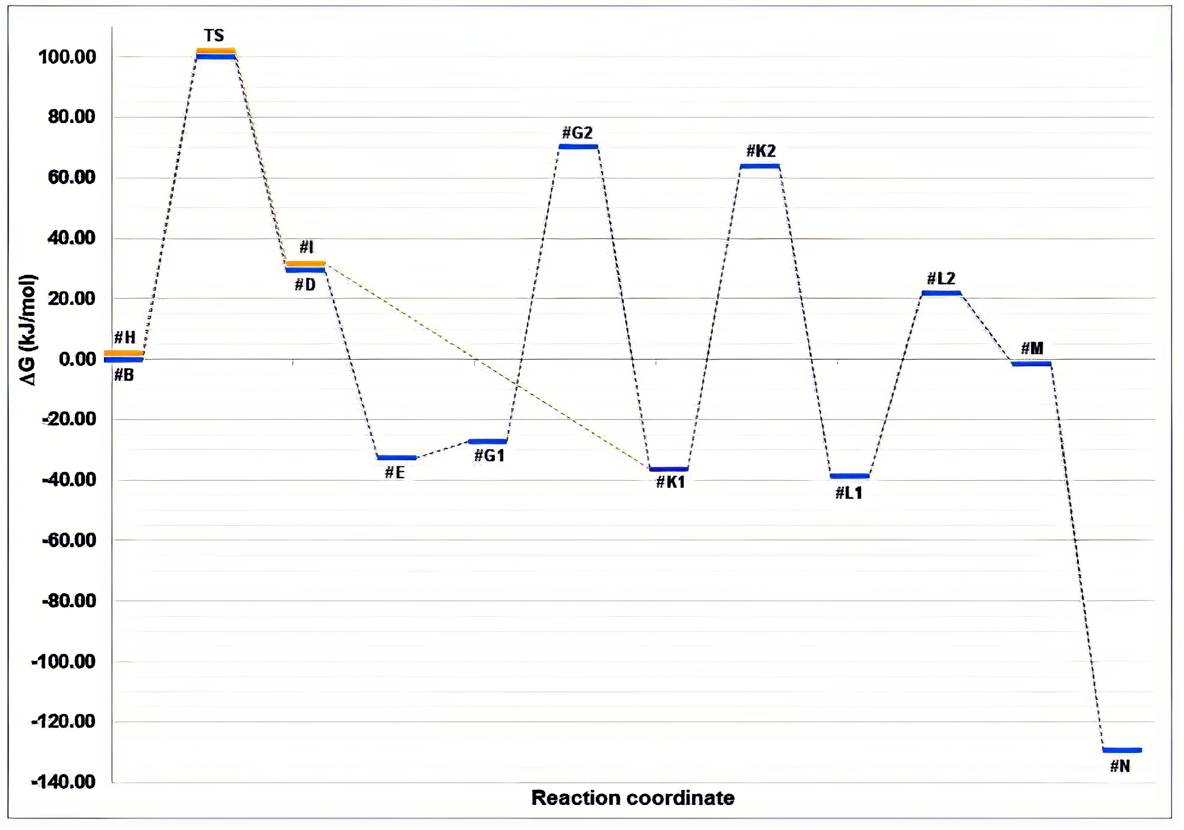
3.3. Molecular Modelling to Investigate the Non-Enzymatic Formation of N-AAA Conjugates

Molecular modelling was used to investigate a newly proposed reaction mechanism for the non-enzymatic formation of N-AAA conjugates (Figure 7). Due to cost constraints, only the reaction mechanism for the simplest structural analogue, N-Ac-Ala formed from pyruvate, was calculated. R1 and R2 in Figure 6 and Figure 7 thus represent only a methyl group, but the principles could also be applied to other 2-keto acids. The hydronium ion was selected to represent Brønsted acid. A TS was found for the protonation of pyruvate.
The results in Figure 8 indicated a slightly higher Ea compared to experimental Ea values reported in the literature [33,34,35,36,37,38,39,40,41,42]. However, considering the energy requirements of enzymatic reactions, the Ea obtained for the non-enzymatic formation of N-Ac-Ala fell within an acceptable range for an in vivo non-enzymatic reaction. It is worth noting that N-AAA conjugates may also form in vitro under higher temperature conditions during sample preparation. A higher Ea slows down the reaction rate but does not prevent the reaction from occurring [43]. The trace amounts of N-AAA conjugates observed in the non-enzymatic in vitro experiment align with the slower reaction rate. Yanagawa et al. [31] also reported slow hydrolysis during N-oxalyl-Gly formation, which could have contributed to the low yields observed.

4. Discussion

The metabolic significance of N-AAA conjugates is evident due to their involvement in various biological processes such as protein function [16], urea cycle regulation [15], myelin synthesis [17], biotransformation, medicine [44], and their association with certain IEMs. Enzymatic synthesis of N-AAA conjugates occurs through acyltransferases and their catabolism involves aminoacylases [16], predominantly yielding L-amino acid enantiomers [45]. Additionally, N-AAA conjugates have been observed in three variant MSUD cases [6,7], and the non-enzymatic synthesis of N-AAA conjugates has been proposed [31].
Enzymatic reactions exhibit high selectivity and are subject to stringent control mechanisms for maintaining homeostasis and targeted product synthesis [37]. In contrast, non-enzymatic reactions are unregulated, which can have supplementary or toxic implications for metabolism [37]. Therefore, it is important to differentiate between enzymatic and non-enzymatic reactions due to their varying impacts.
The chiral analytical approach has been successful in distinguishing between enantiomers by exploiting the stereo-nonspecific nature of non-enzymatic reactions and the formation of racemic mixtures from prochiral substrates [46]. In our case study, the chiral analytical strategy effectively determined that the observed N-AAA conjugates consisted of both L- and D-enantiomers, indicating a non-enzymatic origin. This approach relied on specific steps: (1) urinary extraction of N-AAA conjugates without co-extraction of native BCAAs to avoid pseudo-enantiomeric excess, (2) hydrolysis of N-AAA conjugates without significant racemisation, and (3) conversion of extracted BCAAs to volatile diastereomers for separation via GC–MS without inducing racemisation during derivatisation. Through this qualitative approach, the enantiomeric composition of N-AAA conjugates could be determined, revealing their enzymatic or non-enzymatic origin. Whether this happens in vivo or in vitro merits further discussion following the results from molecular modelling.
To synthesise N-AAA conjugates non-enzymatically, we incubated various 2-keto acids at pH < 2 and 37 °C. Although these conditions are unlikely to occur in vivo, they align with in vitro reaction conditions during sample preparation for organic acid analysis. However, the in vivo non-enzymatic formation of N-AAA conjugates cannot yet be ruled out, because Yanagawa et al. [31] synthesised N-Ac-Ala non-enzymatically at a temperature of 27 °C and a more acceptable pH of 4. While both our study and the work by Yanagawa et al. [31] achieved only low yields of N-AAA conjugates, the presence of these conjugates in an enzyme-free environment supports the occurrence of non-enzymatic reactions. Furthermore, the proposed reaction mechanism was confirmed, as evidenced by the predicted products being produced. It is important to note that this method is only valid when appropriate starting substrates are chosen, as non-enzymatic synthesis of N-AAA conjugates from acyl-chlorides and amino acids would not have provided meaningful results given that acyl-chlorides are not metabolites.
In this study, molecular modelling successfully validated the proposed reaction mechanism and determined the energy requirements (Ea). Although the calculated Ea was relatively high compared to standard in vivo enzymatic reactions, it corresponded to the low yields observed during chemical synthesis. These results support the hypothesis of a slow in vivo reaction that may be facilitated by an unidentified catalyst or more appropriate reaction conditions, or they may indicate that the formation of N-AAA conjugates requires an acceptably low amount of energy during routine sample handling. The results neither confirm nor disprove the occurrence of non-enzymatic reactions in vivo or in vitro, but they support the hypothesis of non-enzymatic formation of N-AAA conjugates in urine.
In conclusion, the findings of this study hold importance in understanding the formation of N-AAA conjugates and their relevance to metabolic processes. By employing a combination of chiral analysis, chemical synthesis, and molecular modelling, we have provided evidence supporting the non-enzymatic formation of N-AAA conjugates from 2-keto acids and ammonia. The distinction between enzymatic and non-enzymatic reactions is crucial for comprehending the underlying mechanisms governing important metabolic pathways, as well as their implications for normal physiology and pathological conditions. This study thus supports previous findings and concludes that N-AAA conjugates can form non-enzymatically from 2-keto acids and ammonia. The insights gained here thereby pave the way for further investigations into the role of N-AAA conjugates in various biological contexts and contribute to our understanding of metabolic pathways and their regulation.

Supplementary Materials

The following supporting information can be downloaded at: https://www.mdpi.com/article/10.3390/app131810002/s1. More detailed figures (Figures S1–S7) of the chromatogram resulting from the organic acid analysis of the case study sample.

Author Contributions

Conceptualisation, L.J.M. and B.C.V.; Data curation, J.J.; Formal analysis, J.J.; Investigation, J.J., C.G.C.E.v.S. and B.C.V.; Methodology, J.J., C.G.C.E.v.S., L.J.M. and B.C.V.; Software, J.J. and C.G.C.E.v.S.; Supervision, C.G.C.E.v.S., M.D. and B.C.V.Validation, J.J., C.G.C.E.v.S. and M.D.; Visualisation, J.J. and M.O.; Writing—original draft, M.O.; Writing—review & editing, J.J., C.G.C.E.v.S., M.D., M.O. and B.C.V. All authors have read and agreed to the published version of the manuscript.

Funding

This research received no external funding.

Institutional Review Board Statement

The study was carried out in accordance with the Helsinki Declaration guidelines, and the samples were stored as biological materials collected for diagnostic purposes. Before this investigation, all samples were de-identified, decoded, and anonymised, thereby, per national guidelines [47], removing the need for ethics review or newly obtained informed consent.

Data Availability Statement

The data presented in this study are available on request from the corresponding author.

Acknowledgments

This manuscript is based on the main author’s MSc Dissertation [48].

Conflicts of Interest

The authors declare no conflict of interest.

References

  1. Ferreira, C.R.; Van Karnebeek, C.D.M. Inborn errors of metabolism. Handb. Clin. Neurol. 2019, 162, 449–481. [Google Scholar]
  2. Mordaunt, D.; Cox, D.; Fuller, M. Metabolomics to improve the diagnostic efficiency of inborn errors of metabolism. Int. J. Mol. Sci. 2020, 21, 1195. [Google Scholar]
  3. Coene, K.L.M.; Kluijtmans, L.A.J.; Van der Heeft, E.; Engelke, U.F.H.; De Boer, S.; Hoegen, B.; Kwast, H.J.T.; Van de Vorst, M.; Huigen, M.C.D.G.; Keularts, I.M.L.W.; et al. Next-generation metabolic screening: Targeted and untargeted metabolomics for the diagnosis of inborn errors of metabolism in individual patients. J. Inherit. Metab. Dis. 2018, 41, 337–353. [Google Scholar]
  4. Krumsiek, J.; Suhre, K.; Evans, A.M.; Mitchell, M.W.; Mohney, R.P.; Milburn, M.V.; Wägele, B.; Römisch-Margl, W.; Illig, T.; Adamski, J.; et al. mining the unknown: A systems approach to metabolite identification combining genetic and metabolic information. PLoS Genet. 2012, 8, e1003005. [Google Scholar] [CrossRef]
  5. Peisl, B.Y.L.; Schymanski, E.L.; Wilmes, P. Dark matter in host-microbiome metabolomics: Tackling the unknowns—A review. Anal. Chim. Acta 2018, 1037, 13–27. [Google Scholar]
  6. Deysel, M.S.M. Biochemiese Karakterisering van ʼn Suid-Afrikaanse MSUD Variant. Master’s Thesis, Potchefstroomse Universiteit vir Christelike Hoër Onderwys, Potchefstroom, South Africa, 2001. [Google Scholar]
  7. Dry, J. ʼn Studie van Geïnduseerde Metaboliese Weë Weens ʼn Aangebore Defek van Die Vertakte Ketting-α-Ketosuur Dehidrogenase-Kompleks. Bachelor’s (Honours) Thesis, Potchefstroomse Universiteit vir Christelike Hoër Onderwys, Pothchefstroom, South Africa, 1997. [Google Scholar]
  8. Menkes, J.H.; Hurst, P.L.; Craig, J.M. A new syndrome: Progressive familial infantile cerebral dysfunction associated with an unusual urinary substance. Pediatrics 1954, 14, 462–467. [Google Scholar] [CrossRef]
  9. Snyderman, S.E.; Norton, P.M.; Roitman, E.; Holt, L.E., Jr. Maple syrup urine disease, with particular reference to dietotherapy. Pediatrics 1964, 34, 454–472. [Google Scholar] [CrossRef]
  10. Dancis, J.; Levitz, M.; Miller, S.; Westall, R.G. Maple syrup urine disease. Br. Med. J. 1959, 1, 91–93. [Google Scholar] [CrossRef]
  11. Meister, A.; Abendschein, P.A. Chromatography of α-Keto Acid 2,4-Dinitrophenylhydrazones and Their Hydrogenation Products. Anal. Chem. 1956, 28, 171–173. [Google Scholar] [CrossRef]
  12. Guder, C.M. Die Gebruik van Filtreerpapier vir Die Versending van Urine vir Die Opsporing van Aangebore Defekte van Die Metabolisme van Organiese Sure. Master’s Thesis, Potchefstroomse Universiteit vir Christelike Hoër Onderwys, Potchefstroom, South Africa, 1988. [Google Scholar]
  13. Hagenfeldt, L.; Naglo, A.S. New conjugated urinary metabolites in intermediate type maple syrup urine disease. Clin. Chim. Acta 1987, 169, 77–83. [Google Scholar] [CrossRef]
  14. Schäfer, G.; Bode, J.W. The synthesis of sterically hindered amides. Chimia 2014, 68, 252–255. [Google Scholar] [CrossRef] [PubMed]
  15. Caldovic, L.; Tuchman, M. N-acetylglutamate and its changing role through evolution. Biochem. J. 2003, 372, 279–290. [Google Scholar] [CrossRef] [PubMed]
  16. Perrier, J.; Durand, A.; Giardina Puigserver, T.A. Catabolism of intracellular N-terminal acetylated proteins: Involvement of acylpeptide hydrolase and acylase. Biochimie 2005, 87, 673–685. [Google Scholar] [CrossRef] [PubMed]
  17. Wijayasinghe, Y.S.; Pavlovskym, A.G.; Violam, R.E. Aspartoacylase catalytic deficiency as the cause of Canavan disease: A structural perspective. Biochemistry 2014, 53, 4970–4978. [Google Scholar] [CrossRef] [PubMed]
  18. Liska, D.; Lyon, M.; Jones, D.S. Detoxification and biotransformational imbalances. Explore 2006, 2, 122–140. [Google Scholar] [PubMed]
  19. Gerlo, E.; Van Coster, R.; Lissens, W.; Winckelmans, G.; De Meirleir, L.; Wevers, R. Gas chromatographic-mass spectrometric analysis of N-acetylated amino acids: The first case of aminoacylase I deficiency. Anal. Chim. Acta 2006, 571, 191–199. [Google Scholar] [CrossRef]
  20. Jellum, E.; Horn, L.; Thoresen, O.; Kvittingen, E.A.; Stokke, O. Urinary excretion of N-acetyl amino acids in patients with some inborn errors of amino acid metabolism. Scand. J. Clin. Lab. Suppl. 1986, 184, 21–26. [Google Scholar]
  21. Sass, J.O.; Mohr, V.; Olbrich, H.; Engelke, U.; Horvath, J.; Fliegauf, M.; Loges, N.T.; Schweitzer-Krantz, S.; Moebus, R.; Weiler, P.; et al. Mutations in ACY1, the gene encoding aminoacylase 1, cause a novel inborn error of metabolism. Am. J. Hum. Genet. 2006, 78, 401–409. [Google Scholar]
  22. Van der Westhuizen, F.H.; Pretorius, P.J.; Erasmus, E. The utilization of alanine, glutamic acid, and serine as amino acid substrates for glycine N-acyltransferase. J. Biochem. Mol. Toxicol. 2000, 14, 102–109. [Google Scholar]
  23. Tanaka, K.; Isselbacher, K.J. The Isolation and Identification of N-Isovalerylglycine from Urine of Patients with Isovaleric Acidemia. J. Biol. Chem. 1967, 242, 2966–2972. [Google Scholar]
  24. Rasmussen, K.; Ando, T.; Nyhan, W.L.; Hull, D.; Cottom, D.; Donnell, G.; Wadlington, W.; Kilroy, A.W. Excretion of Propionylglycine in Propionic Acidaemia. Clin. Sci. 1972, 42, 665–671. [Google Scholar] [CrossRef] [PubMed]
  25. Sweetman, L.; Weyler, W.; Nyhan, W.L.; De Céspedes, C.; Loria, A.R.; Estrada, Y. Abnormal metabolites of isoleucine in a patient with propionyl-CoA carboxylase deficiency. Biomed. Mass. Spectrom. 1978, 5, 198–207. [Google Scholar] [PubMed]
  26. Gompertz, D.; Saudubray, J.M.; Charpentier, C.; Bartlett, K.; Goodey, P.A.; Draffan, G.H. A defect in l-isoleucine metabolism associated with alpha-methyl-beta-hydroxybutyric and alpha-methylacetoacetic aciduria: Quantitative in vivo and in vitro studies. Clin. Chim. Acta 1974, 57, 269–281. [Google Scholar] [CrossRef]
  27. Sweetman, L.; Bates, S.P.; Hull, D.; Nyhan, W.L. Propionyl-CoA carboxylase deficiency in a patient with biotin-responsive 3-methylcrotonylglycinuria. Pediatr. Res. 1977, 11, 1144–1147. [Google Scholar] [CrossRef] [PubMed]
  28. Rinaldo, P.; O’Shea, J.J.; Coates, P.M.; Hale, D.E.; Stanley, C.A.; Tanaka, K. Medium-chain acyl-CoA dehydrogenase deficiency. Diagnosis by stable-isotope dilution measurement of urinary n-hexanoylglycine and 3-phenylpropionylglycine. N. Engl. J. Med. 1988, 319, 1308–1313. [Google Scholar]
  29. Goodman, S.I.; McCabe, E.R.B.; Fennessey, P.V.; Mace, J.W. Multiple acyl-coa dehydrogenase deficiency (glutaric aciduria type ii) with transient hypersarcosinemia and sarcosinuria; possible inherited deficiency of an electron transfer flavoprotein. Pediatr. Res. 1980, 14, 12–17. [Google Scholar] [CrossRef]
  30. Loots, D.T.; Erasmus, E.; Mienie, J. Identification of 19 New Metabolites Induced by Abnormal Amino Acid Conjugation in Isovaleric Acidemia. Clin. Chem. 2005, 51, 1510–1512. [Google Scholar]
  31. Yanagawa, H.; Makino, Y.; Sato, K.; Nishizawa, M.; Egami, F. Novel formation of alpha-amino acids and their derivatives from oxo acids and ammonia in an aqueous medium. J. Biochem. 1982, 91, 2087–2090. [Google Scholar] [CrossRef]
  32. Blau, N.; Duran, M.; Gibson, K.M. Laboratory Guide to the Methods in Biochemical Genetics, 1st ed.; Springer: Berlin/Heidelberg, Germany, 2008. [Google Scholar]
  33. Aksoy, Y.; Ogüs, I.H.; Oauzer, N. Purification and some properties of human placental glucose-6-phosphate dehydrogenase. Protein Expr. Purif. 2001, 21, 286–292. [Google Scholar]
  34. Cherepanov, A.V.; De Vries, S. Kinetics and thermodynamics of nick sealing by T4 DNA ligase. Eur. J. Biochem. 2003, 270, 4315–4325. [Google Scholar]
  35. Craig, D.B.; Arriaga, E.A.; Wong, J.C.Y.; Lu, H.; Dovichi, N.J. Studies on Single Alkaline Phosphatase Molecules:  Reaction Rate and Activation Energy of a Reaction Catalyzed by a Single Molecule and the Effect of Thermal Denaturation—The Death of an Enzyme. J. Am. Chem. Soc. 1996, 118, 5245–5253. [Google Scholar] [CrossRef]
  36. Fidaleo, M.; Lavecchia, R. Kinetic Study of Enzymatic Urea Hydrolysis in the pH Range 4–9. Chem. Biochem. Eng. Q. 2003, 17, 311–318. [Google Scholar]
  37. Kelmer-Bracht, A.M.; Santos, C.P.; Ishii-Iwamoto, E.L.; Broetto-Biazon, A.C.; Bracht, A. Kinetic properties of the glucose 6-phosphatase of the liver from arthritic rats. Biochim. Biophys. Acta 2003, 1638, 50–56. [Google Scholar]
  38. Matschinsky, F.M.; Zelent, B.; Doliba, N.; Li, C.; Vanderkooi, J.M.; Naji, A.; Sarabu, R.; Grimsby, J. Glucokinase activators for diabetes therapy: May 2010 status report. Diabetes Care 2011, 34, S236–S243. [Google Scholar] [PubMed]
  39. Scrutton, M.C. Pyruvate Carboxylase: Studies of activator-independent catalysis and of the specificity of activation by acyl derivatives of coenzyme a for the enzyme from rat liver. J. Biol. Chem. 1974, 249, 7057–7067. [Google Scholar] [CrossRef]
  40. Su, J.-T.; Kim, S.-H.; Yan, Y.-B. Dissecting the pretransitional conformational changes in aminoacylase I thermal denaturation. Biophys. J. 2007, 92, 578–587. [Google Scholar] [PubMed]
  41. Van Zyl, L.J.; Schubert, W.-D.; Tuffin, M.I.; Cowan, D.A. Structure and functional characterization of pyruvate decarboxylase from Gluconacetobacter diazotrophicus. BMC Struct. Biol. 2014, 14, 21. [Google Scholar]
  42. Zorich, N.; Jonas, A.; Pownall, H.J. Activation of lecithin cholesterol acyltransferase by human apolipoprotein E in discoidal complexes with lipids. J. Biol. Chem. 1985, 260, 8831–8837. [Google Scholar]
  43. Keller, M.A.; Piedrafita, G.; Ralser, M. The widespread role of non-enzymatic reactions in cellular metabolism. Curr. Opin. Biotechnol. 2015, 34, 153–161. [Google Scholar]
  44. Mazaleuskaya, L.L.; Sangkuhl, K.; Thorn, C.F.; FitzGerald, G.A.; Altman, R.B.; Klein, T.E. PharmGKB summary: Pathways of acetaminophen metabolism at the therapeutic versus toxic doses. Pharmacogenet. Genom. 2015, 25, 416–426. [Google Scholar]
  45. Cava, F.; Lam, H.; De Pedro, M.A.; Waldor, M.K. Emerging knowledge of regulatory roles of D-amino acids in bacteria. Cell. Mol. Life Sci. 2011, 68, 817–831. [Google Scholar] [PubMed]
  46. Mc Murry, J. Organic Chemistry, 6th ed.; Thomsom Brooks/Cole: Belmount, MA, USA, 2004. [Google Scholar]
  47. Department of Health Republic of South Africa. Ethics in Health Research: Principle, Processes and Structures, 2nd ed.; Department of Health Republic of South Africa: Cape Town, South Africa, 2015.
  48. Jacobs, J. Non-Enzymatic Formation of N-Acylated Amino Acid Conjugates in Urine. Master’s Thesis, North-West University, Pothchefstroom, South Africa, 2018. [Google Scholar]
Figure 1. Overview of induced metabolites detected in urine of maple syrup urine disease (MSUD) cases by Dry [7]. The figure includes identification of unknown N-acylated amino acid (N-AAA) conjugates observed in three variant MSUD cases. Adapted from Dry [7].
Applsci 13 10002 g001
Figure 2. Summary of metabolomic obstructions caused by maple syrup urine disease (MSUD) indicating the prevention of typical GLYAT enzyme-substrate formation, acetyl-CoAs. The known enzymatic and non-enzymatic formations of N-AAA conjugates are also indicated. BCAA: branched-chain amino acid; BCKDH: branched-chain α-keto acid dehydrogenase complex.
Applsci 13 10002 g002
Figure 3. Net reaction for forming N-AAA conjugates from 2-keto acids and ammonia. The net reaction was used for the non-enzymatic in vitro synthesis of N-AAA conjugates to predict which N-AAA conjugates would form from different combinations of 2-keto acid substrates. R1 and/or R2 may refer to methyl, isopropyl, 2-butyl, or isobutyl side chains.
Applsci 13 10002 g003
Figure 4. GC–MS results of extracted and hydrolysed N-AAA conjugates. Organic acid analysis of N-Ac-Ala (a) before hydrolysis and (b) after hydrolysis. Compound A is the mono-TMS derivative of N-Ac-Ala, B is the di-TMS derivative of N-Ac-Ala, and C is the internal standard, 3-phenyl butyric acid. The y-axis (abundance) is scaled to 100% C, and the x-axis (retention time in minutes) is cropped to between 13.8 and 18.4 min to show only the section of interest. Amino acid analysis of free Ala (c) before hydrolysis and (d) after hydrolysis. Compound A is Ala, and B is the internal standard, Nva. The y-axis is scaled to 100% B, and the x-axis is cropped to between 3.5 and 4.5 min. In (d), the graph overlays (blue and red lines) the ions used during amino acid single-ion monitoring. (e) Hydrolysis- and/or derivatisation-induced racemisation of L-Ala. Compound A is L-Ala, and B is D-Ala and/or the opposing diastereomeric derivative of L-Ala. The y-axis is scaled to 100% of the di-tBDMS ester of Ala (not shown), and the x-axis is cropped to between 15.7 and 16.7 min.
Applsci 13 10002 g004
Figure 5. Separation of racemic amino acid via GC–MS. The compound list is as follows: (a) L-Ala, (b) D-Ala, (c) L-Val, (d) D-Val, (e) the di-tBDMS ester of Ala, (f) L-Nva, (g) D-Nva, (h) L-Leu, (i) D-Leu, (j) L-aIle, (k) D-aIle and L-Ile, (l) D-Ile and (m–q) the di-tBDMS esters of Val, Nva, Leu, aIle, and Ile, respectively. The y-axis (abundance) is scaled to 100% of (p), and the x-axis (retention time in minutes) is cropped to between 15.5 and 25.3 min to show only the section of interest.
Applsci 13 10002 g005
Figure 6. Branched-chain amino acid enantiomer composition of the case study (pink peaks) and a control overlay (red line). As indicated in the image, blue peaks are extracted ion chromatograms (from the case study) of the amino acid enantiomers.
Applsci 13 10002 g006
Figure 7. Newly proposed reaction mechanism for the non-enzymatic formation of N-AAA conjugates, constructed from molecular modelling observations. The red, light blue, and dark blue groups correlate to the net reaction shown. The reaction steps are numbered in green, while reaction intermediates are labelled with grey letters preceded by a hash symbol. The symbol 'HX' indicates the Brønsted acid, H30+. Where coloured arrows are used (pink and orange), the reaction may proceed via two separate steps.
Applsci 13 10002 g007
Figure 8. Energy profile for the non-enzymatic formation of N-Ac-Ala from pyruvate and ammonia. To simplify the figure, each alpha-numeral represents the most significant reaction intermediate in a balanced pool of reaction intermediates. The orange lines represent the reaction path of the second pyruvate molecule, starting with a transition state (TS) during protonation and ending with the condensation reaction with #G2 to form #K1. These values are intentionally slightly offset to show the blue line underneath.
Applsci 13 10002 g008
Table 1. N-AAA conjugate products from a single substrate reaction.
2-Keto AcidExpected ResultActual ProductYield (μmol/L)
2-Keto-3-methylvaleric acidN-2-Methylbutyryl-IleN-2-Methylbutyry-IleTrace~0.6
2-Ketobutyric acidN-Propionyl-2-aminobutyric acidN-Propionyl-2-aminobutyric acid10.12
2-Ketocaproic acidN-Pentanoyl-nor-LeuN-Pentanoyl-norleucine14.70
2-Ketoisocaproic acidN-Isovaleryl-LeuN-Isovaleryl-Leu1.35
2-Ketoisovaleric acidN-Isobutyryl-ValN-Isobutyryl-Val20.32
2-Ketovaleric acidN-Butyryl-NvaN-Butyryl-NvaTrace~1.0
PhenylpyruvateN-(Phenylacetyl)-PheN-(Phenylacetyl)-Phe5.22
p-HydroxyphenylpyruvateN-(p-hydroxyphenylacetyl)-(p-hydroxy-Phe)No resultNot detected
PyruvateN-Ac-AlaN-Ac-Ala2.46
Disclaimer/Publisher’s Note: The statements, opinions and data contained in all publications are solely those of the individual author(s) and contributor(s) and not of MDPI and/or the editor(s). MDPI and/or the editor(s) disclaim responsibility for any injury to people or property resulting from any ideas, methods, instructions or products referred to in the content.

