Article

Minimalized Erlangen Correction Method by Hümmer (MEK) Compared with Conventional and Minimally Invasive Correction Methods for Pectus Excavatum Single Center Experience

by
Markus Denzinger
1,*,
Patricia Reis Wolfertstetter
2,
Daniel Sossau
2,
Hans Peter Hümmer
2 and
Christian Knorr
2
1
Faculty of Medicine, University Medical Center Regensburg, D-93053 Regensburg, Germany
2
Department of Pediatric Surgery, Klinik St. Hedwig, University Medical Center Regensburg, D-93053 Regensburg, Germany
*
Author to whom correspondence should be addressed.
Appl. Sci. 2023, 13(18), 10009; https://doi.org/10.3390/app131810009
Submission received: 26 July 2023 / Revised: 28 August 2023 / Accepted: 1 September 2023 / Published: 5 September 2023

Abstract

:
Pectus excavatum (funnel chest) is the most common chest wall deformity in childhood and adolescence with a prevalence in the literature ranging from 1 in 3400 to 1 in 1000. In addition to conservative therapy with a suction cup and physiotherapy to improve posture, fitness, and muscle strength, there are several different surgical techniques, many of which have been improved over time. In evaluating the Minimalized Erlangen Correction Method (MEK), the main purpose of this retrospective analysis is to present the results and to compare them with reports on the Minimally Invasive Repair of Pectus Excavatum (MIRPE) technique, especially regarding long-term patient satisfaction, and with other open surgical methods in terms of operative trauma, as well as flexibility in its application, risk of complications, and patient safety.

1. Introduction

Pectus excavatum (funnel chest) is the most common chest wall deformity in childhood and adolescence with a prevalence in the literature ranging from 1 in 3400 to 1 in 1000 [1,2]. Boys are 3 to 5 times more likely to be affected than girls. In most cases, there is an impression on the anterior chest wall that is either present at birth or develops in early childhood. It is caused by a defect in the cartilage of the ribs, involving the sternum. The deformity increases during growth and persists when growth stops [3,4]. Pectus excavatum is often diagnosed in early childhood and usually does not cause symptoms until after puberty or in adulthood. One of the reasons for this is the increased rigidity of the thorax, i.e., the reduced compliance of the chest wall [3]. There is great variability in the extent, depth, and symmetry of the anatomical findings. The deformity may show up as a hole limited to the inferior end of the sternum and the fourth to seventh ribs, but may extend superiorly as far as to the second costal cartilage and peripherally beyond the costochondral junction.
The funnel may be symmetric (in about 40%) or asymmetric, with sternal rotation usually to the right [2,3,5]. There are also forms that are a mixture of pectus excavatum and pectus carinatum, without clearly belonging to either category, as well as even more complex deformities.
Patients with pectus excavatum show a variety of complaints that do not correlate with the objective extent of the deformity. Breathlessness on exertion, weakness, and early fatigue compared to aged-matched peers are common symptoms, as are inappropriate tachycardia and palpitations on exertion, retrosternal tightness, back pain, and significant psychosocial difficulties [5,6,7,8,9,10]. Objective findings that do not consistently correlate with the anatomic severity include restrictive pulmonary function (35–40% of the operated patients), mitral valve prolapse (25% of the patients), or an altered valve anulus and inappropriate tachycardia on exercise. At rest, there is a displacement and rotation of the heart, which can be seen on cross-sectional imaging, as well as reflected in ECG changes, but is usually without clinical significance.
Deep deformities may compress the right atrium or ventricle. Gastroesophageal reflux may occasionally occur, and, rarely, swallowing may be impaired [3,11,12,13]. Serious complications such as sudden cardiac arrest as an initial presentation of pectus excavatum are discussed in the literature [14].
In addition to conservative therapy with a suction cup and physiotherapy to improve posture, fitness, and muscle strength, there are several different surgical techniques, many of which have improved over time. The first surgical correction of pectus excavatum was performed by Tietze in 1899 with partial sternal resection. Ravitch developed this technique into an open correction method with extensive subperichondral cartilage resection and non-metallic stabilization [15]: This was—in various modified forms—the standard therapy for decades, especially in the USA. Following the hypothesis that funnel chest is caused by growth disorders such as costal overgrowth, as postulated by Ochsner and DeBakey in 1939 [16], this led to an extensive costal cartilage resection sparing the perichondrium in addition to sternal osteotomy [15].
This method, like all other historical techniques, involved very long skin incisions (25–35 cm), extensive soft tissue mobilization, and often a significant blood loss. Subsequently, the trend was almost universally toward minimally invasive surgical techniques, not only for cosmetic reasons but also to reduce the trauma of surgery.
Donald Nuss took a decisive step forward in 1987 with his well-known Minimally Invasive Repair of Pectus Excavatum (MIRPE) technique. The basic principle is to bend rather than cut. Through lateral incisions in the chest wall, a strong curved metal bar based on a preoperative template is inserted retrosternally and rotated by 180° to elevate the sternum. No cartilage or rib resection is performed. The bar is usually fixed laterally with plates and wires and remains in place for 3 years.
The alternative approach described in this article is based on an open technique that has been tried and tested over 5 decades and was established—among others—by Sulamaa and Wallgren in Helsinki [17] and Hegemann in Erlangen [18]. It was further developed by Hümmer and named the Minimalized Erlangen Correction Method (‘Minimalisierte Erlanger Korrekturmethode’, MEK). Compared to the previous open techniques, the incisions are smaller and the soft tissue trauma is significantly reduced; the operation times are shorter; and the complication rates are lower. On average, the patients require less post-operative analgesia and they rarely (<1%) require a blood transfusion. The technique allows correction of even grossly asymmetrical or atypical deformities at all ages, including revision surgery after failure of previous Nuss or other procedures. Figure 1 shows the preoperative and postoperative findings after MEK.
In evaluating MEK, the main purpose of this retrospective analysis is to present its results and to compare them with reports on the MIRPE technique, especially regarding long-term patient satisfaction, and with other open methods in terms of operative trauma, as well as flexibility in its application, risk of complications, and patient safety.

2. Materials and Methods

This study was approved by the local ethics committee (22-2961-104). A retrospective analysis was performed on 301 patients who underwent surgery with the Minimalized Erlangen Correction of Hümmer (MEK) at the University Hospital Erlangen between 1 January 2004 and 31 July 2009. The sample consisted of 46 female and 255 male patients. All the outpatient and inpatient records of these patients were analyzed, including preoperative diagnostic tests, surgery, and the outpatient follow-up. Patient satisfaction was assessed by using a questionnaire 2 to 8 years after the surgery.

3. Preoperative Diagnostic Tests

A detailed general and specific medical history of the patients was obtained. A thorough clinical examination was performed with emphasis on the pectus deformity. This included a documentation with photographs, measurements, and drawings of the external findings, the degree of symmetry of the chest wall and the spine, and the development of the chest, abdominal, and back muscles. The Hümmer Pectus Index was used as an objective measure of the chest wall deformity. This index is defined as the quotient of two sagittal diameters of the chest, measured at the level of the caudal end of the sternum (a) and the manubriosternal angle (b) and multiplied by 100 (index = 100a/b, normal range 133 + 18). It characterizes the position of the sternum in relation to the vertebral column, i.e., its leverage on the mediastinum. The index has been reported to be reliable in assessing the severity of a funnel chest before and after surgical repair [19]. Cardiopulmonary function was assessed with pulmonary function tests and with ECG both at rest and during exercise. Cross-sectional imaging with CT or MRI was performed at five levels (sternal notch, deepest point of the pectus deformity, xiphoid process and the two midpoints between these three planes). Echocardiography, now a standard test, was not routinely performed during the study period.

4. Technique of the Minimalized Erlangen Correction Method (MEK)

The caudal end of the sternum including the ends of the 3rd through 7th costal cartilages is exposed through a median longitudinal incision in male patients and a small transverse submammary incision in female patients. In male patients, the length of the incision is two ribs lower than the upper edge of the deformity to the xiphoid process. The xiphoid process is temporarily detached from the body of the sternum, preserving the insertion of the rectus muscles. While monitoring the tension with a spring balance, the forces retracting the sternum are progressively eliminated by parasternal chondrotomy, the removal of retrosternal mediastinal and diaphragmatic attachments including pericardial and pleural adhesions, and retrosternal dissection of the internal thoracic ligaments. These latter procedures significantly reduce the tension. To stabilize the correction, one or two elastic metal bars (made of stainless steel or titanium, 8 mm wide and 2 mm thick, weighing 20 g each) are implanted transsternally. If further correction of a highly antecurved sternum is required by transverse sternal osteotomy, an additional bar can be inserted longitudinally and intramedullary for reinforcement. The divided costal cartilages are reattached to the sternum with minimal loss of length after the resection of small wedges at their costal ends. Finally, a substernal chest drain is placed along with two subfascial Redon drains. The implants are routinely removed after one year through a stab incision at one end of the bar.

5. Perioperative and Postoperative Course

Patient records were used to assess various aspects of the surgery itself, the postoperative inpatient course, and the removal of the implants.

6. Patient Satisfaction

To assess the long-term performance of the correction method, all patients were given a specially designed questionnaire based on validated quality-of-life questionnaires.
The questionnaire consisted of 47 questions covering various aspects of preoperative quality of life, the perioperative and postoperative course, and satisfaction with both the inpatient stay and the postoperative result in terms of form and function.

7. Statistical Analysis: Software and Tests

Calculations were performed with SPSS version 18. Preoperative diagnostic tests, the intraoperative and postoperative course, and the patient satisfaction questionnaires were analyzed. The means and standard deviations of the different parameters were calculated.

8. Results

During the 5 years and 7 months of sample collection, 301 primary corrections of pectus excavatum were performed at the University Hospital Erlangen.
There were more male patients (255, 84.7%) than female patients (46, 15.3%). The mean age was 20.3 years (standard deviation 9.3 years, range 12.8–29.4 years). When divided into 5-year intervals, the most highly represented age group was 16–20 years (40.5%). The mean preoperative body mass index was 20.3 (standard deviation 2.9). The deformity was asymmetric in 143 patients (47.5%) and symmetric in 107 (35.5%); the remaining 51 patients (16.9%) could not be assigned to either category. The mean Hümmer pectus index was 104 in 131 patients (SD 13.8, range 61–142, normal range 133 +/− 18); in the other 170 patients the pectus index was not recorded in the notes. Preoperative ECG was obtained in 174 patients (57.8%), being reported as abnormal in 67 patients (22.3%) and normal in 107 (35.5%). Pulmonary function tests were performed in 224 patients (74.4%) and were found to be abnormal in 82 patients (27.2%) and normal for age in 142 (47.2%). There were 76 cases of pure restrictive lung function, and in 1 case there was obstructive lung function. Five patients had combined lung function impairment or other atypical findings. Data are shown in Table 1.

9. Surgery

The mean length of the submammary incision in female patients was 8.7 cm (SD 1.5 cm, range 6–12 cm). The mean length of the sternal incision in male patients was 9.0 cm (SD 1.6 cm, range 4 cm–15.5 cm). Of the 301 patients, 270 (89.7%) underwent minimal costal cartilage resection of only a few millimeters; 298 (99.0%) underwent a retrosternal dissection. Tensiometry before retrosternal mobilization showed a mean tension of 190 N (SD 36 N); after median chondrotomy it was 100 N (SD 38 N), and after retrosternal dissection it was 36 N (SD 16 N). Metal implants were placed horizontal/transsternal in 286 patients (95%), transsternal and longitudinal intrasternal in 11 patients (3.7%), transsternal and parasternal in 3 patients (1.0%), and episternal in 1 patient (0.3%). The mean operation time was 89.5 min (SD 19.7 min, range 45 min–175 min). For female patients, the average surgery time was 100.3 min (SD 32.4 min, range 59 min–219 min), and for male patients it was 87.9 min (SD 17.7 min, range 45 min–152 min). Data are shown in Table 2.

10. Postoperative Course

Drains: Three patients (0.4%) did not have a substernal drain inserted because no retrosternal dissection was performed; 97 patients (99.3%) had one drain and 1 patient had two. On average, they could be removed on Day 4 (SD 1.2 d, range 1 d–10 d). In 292 patients (97%), two subpectoral Redon drains were placed, and in 9 patients (3%) only one drain was used. They were generally removed on Day 5 (SD 7.8).
Wound healing: The majority of the patients (182 patients, 62.1%) showed normal wound healing; there was redness of the wound in 76 patients (25.9%), swelling in 9 patients (3.1%), dehiscence in 2 patients (0.7%), and secretion from the wound in 8 patients (2.7%).
Intrathoracic complications: A total of 14 patients (4.7%) required an additional postoperative chest drainage, 5 patients (1.7%) because of a pneumothorax and 9 patients (3.0%) because of pleural effusion. Only 1 patient (0.3%) required drainage of a pericardial effusion and 6 patients (2%) had a pleural effusion. Additional surgery was required in 5 patients (1.7%) due to implant displacement and 3 (1%) due to bleeding; 2 patients (0.7%) underwent thoracoscopy because of a suspected pleural empyema.
Length of hospital stay: On average, the patients were mobilized on Day 3 (SD 0.6 d, range 2 d–5 d). The average length of hospital stay was 9 days (SD 1.7, range 5 d–21 d).
Date are shown in Table 3.

11. Metal Implant Removal

On average, the metal implants were removed after 12 months (SD 2.8 months, range 2 months–29 months). In one case, an accessory implant was removed after 2 months because of persistent wound-healing problems. In two other cases, the implant was removed after 3 months; in one case because of persistent pain and in the other because of impaired wound healing with suspected osteomyelitis of the sternum.
The average length of the hospital stay for the explantation of the metal implants was 2 days (SD 1.3 d). Certain patients underwent additional cosmetic procedures at the time of implant removal. In 27 patients (9.0%), a costal arch correction was performed through local stab incisions, in 66 patients (21.9%) scars were resected, and in 23 patients (7.6%) small irregularities in the cartilage or bone were smoothed out.

12. Patient Satisfaction

In total, 178 (59.1%) out of the 301 patients responded to the patient satisfaction questionnaires. It should be noted that these questionnaires were evaluated up to 8 years after surgery. All procedures were clinically indicated and based on objective abnormalities; none were performed for purely cosmetic reasons. In retrospect, the patients often had a different opinion about the reason for their surgery. In total, 17 patients (9.6%) felt very little affected by their pectus deformity preoperatively, 103 (57.9%) felt slightly or moderately affected, and 55 (30.9%) felt severely or very severely affected. Regarding the reason for surgery, 51 patients (28.7%) reported functional limitations, 13 (7.3%) reported psychological disturbance, and 18 (10.1%) reported mainly aesthetic reasons. The remaining patients reported a combination of these reasons.
The length of the hospital stay was considered adequate by 158 patients (88.8%), too long by 12 (6.7%), and too short by 7 (3.9%). Postoperative pain was rated absent to moderate by 120 patients (67.4%) and severe to very severe by 58 patients (32.6%). Postoperative analgesia was considered adequate in 155 patients (87.1%) and inadequate in 21 (11.8%). A peridural catheter was placed as standard and was removed on average on Day 4 (SD 1.2 d, range 2–11 d). No pain after discharge from the hospital was reported by 7 patients (3.9%). Pain during the first month was reported by 130 patients (73%), of whom 23 (12.9%) rated it severe or very severe and 31 patients (17.4%) still had pain more than 4 weeks after discharge.
In total, 17 patients (9.6%) reported no pain limiting their daily activities, 129 (72.5%) during the first 4 months, and 14 (7.9%) patients reported pain for more than 4 months. Regarding their postoperative appearance, 115 patients (64.6%) were satisfied or very satisfied and 57 (32.0%) were dissatisfied. The most common reason for dissatisfaction was the surgical scar. When asked if they would be willing to undergo another surgery, 135 patients (75.8%) answered yes, while 32 (18.0%) ruled out another surgery.

13. Discussion

The prevalence of pectus excavatum reported in the literature ranges from 1 in 1000 to 3400 [1,2]. Males are more frequently affected than females, with the male:female ratio ranging from 3:1 and 5:1 [2]. Our own sample also contained predominantly male patients (with a ratio of 5.5:1). As reported in the literature [3,4], the deformity was congenital in most cases (80.1%). Other authors have found a preponderance of asymmetric deformities, with a ratio of 1.5:1 [2,3]. This was also observed in our collective results (ratio 1.3:1). As described in previous studies [5,6,7,8], the most common symptoms in our patients were dyspnea on exertion, lack of endurance, palpitations, early fatigue, and back pain.
Over time, several theories have been proposed regarding the etiology of pectus excavatum. It has been hypothesized that the overgrowth of costal cartilage causes indrawing of the sternum [2]. Several groups have performed histologic and biochemical studies of costal cartilage in patients with pectus excavatum. The reports are inconsistent, but most describe degenerative changes and impaired chondrogenesis which may weaken the stability of the cartilage during the growth spurt [2,20,21,22]. Comprehensive studies of cartilage from pectus patients have also shown significantly lower levels of zinc and other trace elements that play a role in chondrogenesis [23].
Studies indicate that 5.3% of PE patients are affected by Marfan syndrome and 2.7% by neurofibromatosis type 1 [24,25], and approximately 20 genetic disorders have already been described to be associated with PE. A familial predisposition to chest wall deformities has long been known, with reported rates of 12–50%. Family trees suggest dominant inheritance with incomplete penetrance. Most cases are thought to be multifactorial [1,26], and single genes have been identified that may cause PE [27,28]. Analysis of gene expression in the costal cartilage of affected patients has not definitively elucidated the underlying genetic changes [23].
These disorders of chondrogenesis and their subsequent etiologically-based therapeutic approaches have had strong implications for issues of radical treatment based on the age of the patient. Early open techniques (Ravitch: Lit) therefore focused on radical resection of the anomalous cartilage in the belief that by leaving the perichondrial sheaths intact, new cartilage would develop to stabilize the thorax and to allow for sufficient growth of the chest. Children were often operated on at an early age, and in many cases the resulting impairments caused typical forms of postcorrectional thoracic dystrophy.
As a result, etiologic attempts at treatment were abandoned in favor of corrective methods based on biomechanical principles. These contemporary concepts also imply and explain a noticeable shift toward treating older patients for pectus correction at the time of cessation of their bone growth.
In principle, modern methods of corrective surgery can be performed at any age, and in cases of severe and debilitating compression symptoms, surgery may be justified even in young children. Especially in these cases, surgical invasiveness must be kept to a minimum to avoid the risks mentioned above. Today, however, most authors recommend surgery at the end of the pubertal growth spurt [3] and to reshape rather than eliminate the malformed segments of the anterior chest wall. In addition, at an older age, the patients can decide for themselves whether or not to undergo surgery. The use of surgery too early carries the risk of developing new or recurrent deformities [3,13,29]. The mean age of our patient population at the time of surgery was 21.5 years (range: 4 years–60 years). This wide age range illustrates the applicability of the Hümmer technique at any age.
The Nuss procedure was developed for young adults and the implant should be placed just before the cessation of bone growth. Younger patients are at risk for re-deformation of the chest wall, and approximately 25% of patients develop significant changes in the ribs, such as step deformities in the costal cartilage of greenstick fractures [2]. These complications particularly affect patients who are older than 10 years at the time of surgery. In adults, the Nuss procedure is usually modified, for example, by an additional anterior chest wall incision or extensive endoscopic mobilization to reduce tension. The total length of the incisions in these cases can therefore be significantly longer than in the Hümmer procedure.
In preoperative diagnostic testing, more than 20% of patients in general and more than 40% of patients undergoing surgery have restrictive pulmonary function or abnormal cardiac findings (such as mitral valve prolapse). Postoperatively, these functions almost always return to normal [13]. Functional back complaints usually improve as well. Consistent with the literature, our patient cohort showed no correlation between the severity of the deformity and pulmonary function test and ECG findings [2,30,31,32].
Approximately 70% of our patients felt that their physical fitness had improved after corrective surgery. Some studies suggest that there is no improvement in pulmonary function after the Nuss procedure [33], while others have found opposite results, even postulating better results after the Nuss procedure than after the Ravitch procedure [34]. Studies after the Nuss procedure have not shown any improvement in lung function or physical fitness [35,36]. During the long period (routinely 3 years) that the Nuss implant is in place, thoracic compliance is inevitably compromised. Long-term objective improvement in lung function is expected only in severe cases [37]. Echocardiography shows improvement in the motion of both ventricles [35,37].
The Hümmer procedure has an average operating time of 89.5 min (including a large number of procedures in adults), which is slightly longer than in the MIRPE procedure, which takes an average of 68 min in children and adolescents [38]. It is difficult to compare the figures from samples with such different patient ages. Larger adults require longer operating times than children, regardless of the technique used.
The average incision length for the Hümmer method is 8.9 cm. In children, a 5 cm incision is usually sufficient. Intraoperative tensiometry shows that the important step in overcoming retraction forces is the retrosternal dissection. This can be performed endoscopically; but in our experience, this is time consuming. Direct access to the deformity—either through a longitudinal incision over the sternum or through a submammary transverse incision—allows for correction of both symmetrical and asymmetrical deformities.
The Nuss MIRPE procedure typically involves bilateral longitudinal incisions with an average of 6.8 cm in length and a much longer and stronger metal implant (the “pectus bar”), that is inserted retrosternally and stabilized with reinforcing plates—a total of approximately 200 g of metal. Chondrotomy and dissection and direct access to the deformity are avoided in the original procedure but are required in the modified adult procedure. The original method is suitable for the malleable tissues of the chest wall in children and for symmetrical deformities. Modifications are required for adults, with an already rigid chest wall, and for asymmetric deformities. This usually involves additional incisions [39]. Lateral stabilization is almost always required to prevent displacement of the pectus bar, making removal of the implant a major undertaking [40]. In our study, 64.6% of the patients were not satisfied, mainly because of the scar in the middle of the chest. According to other authors [41], 95.6% of the patients were either satisfied or very satisfied after the Nuss procedure and 87.0% of patients stated that they would undergo the procedure again, which is higher than in our cohort.
In our cohort of 301 patients treated with the Hümmer method, the most common complications were minor wound-healing problems, pneumothorax (5 requiring therapy), pleural effusion (6 aspirated, 9 drained), transient paresthesia, and hypertrophic or widened scars. Implant dislocation occurred in 5 patients (1.7%).
The Nuss MIRPE procedure is also frequently associated with complications such as pneumothorax, infection, and metal migration. There are some reported cases of cardiac perforation and other organ injury [39,40,42], although the overall incidence of life-threatening complications of minimally invasive pectus surgery is estimated to be less than 0.1% [43]. Many modifications have been described focusing on reducing the risk of complications, improving the aesthetics, pain control [44], or adapting to new circumstances, such as use in adults whose chest is not as flexible [45]. The study by Molik et al. in 2001 found a displacement of the metalwork in 11% of cases and a re-operation in 29% of cases, with other causes including infection, hemothorax, pain, and the development of secondary pectus carinatum. Long-term complications occurred in 17% of the patients, such as pain requiring early implant removal, pectus carinatum, or significant chest wall asymmetry [40]. Major complications were reported in 11.1% and minor complications in 8.1% of patients [46]. In a study by Araújo et al., the Nuss procedure was associated with an increased incidence of hemothorax, pneumothorax, and the need for reintervention when compared to the Ravitch procedure. They found no statistically relevant differences in overall complications, blood transfusion, hospital stay, and time to ambulation. The Nuss procedure was faster than the Ravitch procedure in this study (mean difference = −69.94 min) [47]. Jukić et al. [48] reported complications such as pneumothorax and subcutaneous emphysema in 30%, late complications such as bar displacement in 10%, and deformity recurrence in 6.6%. Recent publications [49] indicate that the complication rate has decreased in recent years (displacement of the implanted steel bar in 4.5%, infection of the surgical site in 2.9%, pneumothorax in 2.7% patients, pleural effusion in 2.5%, and chronic pain in 2.2%). The complication rate appears to be higher in adults [49].
Routine removal of the metalwork is performed after 12 months in the Hümmer method and after 2 years–4 years in the MIRPE method. At the time of removal, 21.9% of our patients underwent scar correction and 7.6% underwent the smoothing out of small step deformities in the ribs. It has been shown that the majority of concomitant deformities of the costal arch deformities correct themselves spontaneously after elevation of the pectus excavatum, so we prefer to wait and perform a second procedure only if necessary. In our study, this was only necessary in 9% of the patients and they underwent unilateral or bilateral costal arch surgery via stab incisions at the same time as implant removal.
The recurrence rate requiring reoperation is reported in the literature to be 1.3% for the Hümmer procedure [13] and 7.8% for the MIRPE procedure [50].
In addition to physiological problems, psychosocial difficulties are prominent in patients with pectus excavatum. Affected patients report significantly lower scores for quality of life, body image, and self-esteem compared to age-matched controls [2]. This can lead to social withdrawal, relationship anxiety, and reactive psychological disorders [2,3,13].
Analysis of patient satisfaction questionnaires after the Hümmer repair method shows that 75% of patients are satisfied or very satisfied, and 76% would be willing to undergo the procedure again. Postoperatively, the patients rated their quality of life higher than age-matched healthy controls. This is consistent with other studies indicating that correction of chest wall deformity in general can significantly improve patients’ body image and self-esteem [51].
When compared to the results of previously published studies, the Hümmer method is associated with significantly less operative trauma than historical open pectus correction methods (e.g., those of Ravitch and Hegemann), is stage-independent, and offers more flexibility and a reduced operative risk compared to MIRPE, while achieving similar results.

Author Contributions

Conceptualization, M.D. and C.K.; methodology, C.K. and D.S.; software, C.K. and D.S.; investigation, C.K. and D.S.; writing—original draft preparation, C.K. and M.D.; writing—review and editing, M.D., D.S., P.R.W., H.P.H. and C.K.; visualization, M.D. and C.K.; supervision, C.K.; project administration, C.K. All authors have read and agreed to the published version of the manuscript.

Funding

This research received no external funding.

Institutional Review Board Statement

This study was conducted in accordance with the Declaration of Helsinki and approved by the Ethics Committee of the University of Regensburg (protocol code 22-2961-104).

Informed Consent Statement

Not applicable.

Data Availability Statement

Not applicable.

Conflicts of Interest

The authors declare no conflict of interest.

References

  1. Creswick, H.A.; Stacey, M.W.; Kelly, R.E., Jr.; Gustin, T.; Nuss, D.; Harvey, H.; Goretsky, M.J.; Vasser, E.; Welch, J.C.; Mitchell, K.; et al. Family study of the inheritance of pectus excavatum. J. Pediatr. Surg. 2006, 41, 1699–1703. [Google Scholar] [CrossRef]
  2. Fokin, A.A.; Steuerwald, N.M.; Ahrens, W.A.; Allen, K.E. Anatomical, histologic, and genetic characteristics of congenital chest wall deformities. Semin. Thorac. Cardiovasc. Surg. 2009, 21, 44–57. [Google Scholar] [CrossRef]
  3. Jaroszewski, D.; Notrica, D.; McMahon, L.; Steidley, D.E.; Deschamps, C. Current management of pectus excavatum: A review and update of therapy and treatment recommendations. J. Am. Board Fam. Med. 2010, 23, 230–239. [Google Scholar] [CrossRef] [PubMed]
  4. Kelly, R.E., Jr.; Cash, T.F.; Shamberger, R.C.; Mitchell, K.K.; Mellins, R.B.; Lawson, M.L.; Oldham, K.; Azizkhan, R.G.; Hebra, A.V.; Nuss, D.; et al. Surgical repair of pectus excavatum markedly improves body image and perceived ability for physical activity: Multicenter study. Pediatrics 2008, 122, 1218–1222. [Google Scholar] [CrossRef] [PubMed]
  5. Hümmer, H.P. Die Trichterbrust Stadien- und Formgerechte Korrektur; Zuckschwerdt: München, Germany, 1985. [Google Scholar]
  6. Paidas, C.; Colombani, P. The Chest Wall. Principles and Practice of Pediatric Surgery; Lippincott Williams & Wilkins: Philadelphia, PA, USA, 2005; pp. 881–895. [Google Scholar]
  7. Schmüdderich, J. Ergebnisse der Funktionsdiagnostik bei Trichterbrust. Ph.D. Thesis, Medizinische Fakultät Universität Erlangen, Erlangen, Germany, 1987. [Google Scholar]
  8. Shamberger, R. Congenital chest wall deformities. In Pediatric Surgery, 6th ed.; Grosfeld, J.L., O’Neill, J.A., Coran, A., Fonkalsrud, E.W., Eds.; Elsevier: Amsterdam, The Netherlands, 2006; pp. 1–2214. [Google Scholar]
  9. Bergmann, F.; Muensterer, O.J. Chest Wall Deformities in Children and Adolescents. Zentralblatt Fur Chir. 2022, 147, 74–82. [Google Scholar] [CrossRef]
  10. Abid, I.; Ewais, M.M.; Marranca, J.; Jaroszewski, D.E. Pectus Excavatum: A Review of Diagnosis and Current Treatment Options. J. Am. Osteopath. Assoc. 2017, 117, 106–113. [Google Scholar] [CrossRef] [PubMed]
  11. Zallen, G.S.; Glick, P.L. Miniature access pectus excavatum repair: Lessons we have learned. J. Pediatr. Surg. 2004, 39, 685–689. [Google Scholar] [CrossRef] [PubMed]
  12. Koumbourlis, A.C. Pectus excavatum: Pathophysiology and clinical characteristics. Paediatr. Respir. Rev. 2009, 10, 3–6. [Google Scholar] [CrossRef]
  13. Hümmer, H.; Reingruber, B.; Weber, P.; Knorr, C. Indikation und Technik der “minimal-invasiven” Trichterbrustkorrektur. Chir. Allg. 2010, 4/11, 193–200. [Google Scholar]
  14. Rachwan, R.J.; Purpura, A.K.; Kahwash, B.M. Sudden Cardiac Arrest in a Young Patient with Severe Pectus Excavatum. Am. J. Med. Sci. 2018, 356, 570–573. [Google Scholar] [CrossRef]
  15. Ravitch, M.M. The Operative Treatment of Pectus Excavatum. Ann. Surg. 1949, 129, 429–444. [Google Scholar] [CrossRef] [PubMed]
  16. Ochsner, A.; DeBakey, M. CHŌNĒ-CHONDROSTERNON: Report of a Case and Review of the Literature. J. Thorac. Surg. 1939, 8, 469–511. [Google Scholar] [CrossRef]
  17. Paltia, V.; Parkkulainen, K.V.; Sulamaa, M.; Wallgren, G.R. Operative technique in funnel chest; experience in 81 cases. Acta Chir. Scand. 1959, 116, 90–98. [Google Scholar] [PubMed]
  18. Hegemann, G.; Schoberth, H. Operative treatment of funnel chest. Dtsch. Med. Wochenschr. 1958, Band 83, 277–282. [Google Scholar] [CrossRef]
  19. Pinterits, F.; Grabenwöger, F.; Dock, W.; Metz, V.; Helmer, F. Wert des Trichterbrustindex nach Hümmer zur Objektivierung des Schweregrades der Trichterbrust vor und nach chirurgischer Korrektur. Acta Chir. Austriaca 1988, 20, 8–10. [Google Scholar] [CrossRef]
  20. Feng, J.; Hu, T.; Liu, W.; Zhang, S.; Tang, Y.; Chen, R.; Jiang, X.; Wei, F. The biomechanical, morphologic, and histochemical properties of the costal cartilages in children with pectus excavatum. J. Pediatr. Surg. 2001, 36, 1770–1776. [Google Scholar] [CrossRef]
  21. Prozorovskaya, N.N.; Kozlov, E.A.; Voronov, A.V.; Verovskii, V.A.; Delvig, A.A. Characterization of costal cartilage collagen in funnel chest. Biomed. Sci. 1991, 2, 576–580. [Google Scholar]
  22. Serafin, J.; Swiatkowski, J.; Majkusiak, R.; Nowakowski, P. 40-year experience in surgical treatment of congenital chest deformations—Ethiopathogenesis, operative techniques and clinical results. Acta Chir. Orthop. Traumatol. Cechoslov. 2003, 70, 207–213. [Google Scholar]
  23. Rupprecht, H.; Hümmer, H.; Stöß, H. Zur Pathogenese der Thoraxfehlbildungen. Elektronenmikroskopiesche Untersuchungen und Spurenelementanalysen im Rippenknorpel. Z. Kinderchir. 1987, 42, 228–229. [Google Scholar]
  24. Behr, C.A.; Denning, N.L.; Kallis, M.P.; Maloney, C.; Soffer, S.Z.; Romano-Adesman, A.; Hong, A.R. The incidence of Marfan syndrome and cardiac anomalies in patients presenting with pectus deformities. J. Pediatr. Surg. 2019, 54, 1926–1928. [Google Scholar] [CrossRef]
  25. Akçali, Y.; Ceyran, H.; Hasdiraz, L. Chest wall deformities. Acta Chir. Hung. 1999, 38, 1–3. [Google Scholar] [PubMed]
  26. Kotzot, D.; Schwabegger, A.H. Etiology of chest wall deformities—A genetic review for the treating physician. J. Pediatr. Surg. 2009, 44, 2004–2011. [Google Scholar] [CrossRef] [PubMed]
  27. Wu, S.; Sun, X.; Zhu, W.; Huang, Y.; Mou, L.; Liu, M.; Li, X.; Li, F.; Li, X.; Zhang, Y.; et al. Evidence for GAL3ST4 mutation as the potential cause of pectus excavatum. Cell Res. 2012, 22, 1712–1715. [Google Scholar] [CrossRef] [PubMed]
  28. Tong, X.; Li, G.; Feng, Y. TINAG mutation as a genetic cause of pectus excavatum. Med. Hypotheses 2020, 137, 109557. [Google Scholar] [CrossRef] [PubMed]
  29. Erdoğan, A.; Ayten, A.; Oz, N.; Demircan, A. Early and long-term results of surgical repair of pectus excavatum. Asian Cardiovasc. Thorac. Ann. 2002, 10, 39–42. [Google Scholar] [CrossRef]
  30. Coskun, Z.K.; Turgut, H.B.; Demirsoy, S.; Cansu, A. The prevalence and effects of Pectus Excavatum and Pectus Carinatum on the respiratory function in children between 7–14 years old. Indian J. Pediatr. 2010, 77, 1017–1019. [Google Scholar] [CrossRef]
  31. Peterson, R.J.; Young, W.G., Jr.; Godwin, J.D.; Sabiston, D.C., Jr.; Jones, R.H. Noninvasive assessment of exercise cardiac function before and after pectus excavatum repair. J. Thorac. Cardiovasc. Surg. 1985, 90, 251–260. [Google Scholar] [CrossRef]
  32. Rowland, T.; Moriarty, K.; Banever, G. Effect of Pectus Excavatum Deformity on Cardiorespiratory Fitness in Adolescent Boys. Arch. Pediatr. Adolesc. Med. 2005, 159, 1069–1073. [Google Scholar] [CrossRef]
  33. Aronson, D.C.; Bosgraaf, R.P.; Merz, E.M.; van Steenwijk, R.P.; van Aalderen, W.M.; van Baren, R. Lung function after the minimal invasive pectus excavatum repair (Nuss procedure). World J. Surg. 2007, 31, 1518–1522. [Google Scholar] [CrossRef]
  34. Chen, Z.; Amos, E.B.; Luo, H.; Su, C.; Zhong, B.; Zou, J.; Lei, Y. Comparative pulmonary functional recovery after Nuss and Ravitch procedures for pectus excavatum repair: A meta-analysis. J. Cardiothorac. Surg. 2012, 7, 101. [Google Scholar] [CrossRef]
  35. Malek, M.H.; Berger, D.E.; Housh, T.J.; Marelich, W.D.; Coburn, J.W.; Beck, T.W. Cardiovascular function following surgical repair of pectus excavatum: A metaanalysis. Chest 2006, 130, 506–516. [Google Scholar] [CrossRef]
  36. Castellani, C.; Windhaber, J.; Schober, P.H.; Hoellwarth, M.E. Exercise performance testing in patients with pectus excavatum before and after Nuss procedure. Pediatr. Surg. Int. 2010, 26, 659–663. [Google Scholar] [CrossRef]
  37. Kowalewski, J.; Barcikowski, S.; Brocki, M. Cardiorespiratory function before and after operation for pectus excavatum: Medium-term results. Eur. J. Cardio-Thorac. Surg. 1998, 13, 275–279. [Google Scholar] [CrossRef]
  38. Protopapas, A.D.; Athanasiou, T. Peri-operative data on the Nuss procedure in children with pectus excavatum: Independent survey of the first 20 years’ data. J. Cardiothorac. Surg. 2008, 3, 40. [Google Scholar] [CrossRef] [PubMed]
  39. Dzielicki, J.; Korlacki, W.; Janicka, I.; Dzielicka, E. Difficulties and limitations in minimally invasive repair of pectus excavatum—6 years experiences with Nuss technique. Eur. J. Cardio-Thorac. Surg. Off. J. Eur. Assoc. Cardio-Thorac. Surg. 2006, 30, 801–804. [Google Scholar] [CrossRef]
  40. Molik, K.A.; Engum, S.A.; Rescorla, F.J.; West, K.W.; Scherer, L.R.; Grosfeld, J.L. Pectus excavatum repair: Experience with standard and minimal invasive techniques. J. Pediatr. Surg. 2001, 36, 324–328. [Google Scholar] [CrossRef] [PubMed]
  41. Di Salvo, N.; Ruggeri, G.; Thomas, E.; Parente, G.; Di Mitri, M.; Lima, M. Long-term evaluation of patient satisfaction and quality of life in pectus excavatum repair. Ann. Pediatr. Surg. 2022, 18, 84. [Google Scholar] [CrossRef]
  42. Bouchard, S.; Hong, A.R.; Gilchrist, B.F.; Kuenzler, K.A. Catastrophic cardiac injuries encountered during the minimally invasive repair of pectus excavatum. Semin. Pediatr. Surg. 2009, 18, 66–72. [Google Scholar] [CrossRef]
  43. Hebra, A.; Kelly, R.E.; Ferro, M.M.; Yüksel, M.; Campos, J.R.M.; Nuss, D. Life-threatening complications and mortality of minimally invasive pectus surgery. J. Pediatr. Surg. 2018, 53, 728–732. [Google Scholar] [CrossRef] [PubMed]
  44. Notrica, D.M. Modifications to the Nuss procedure for pectus excavatum repair: A 20-year review. Semin. Pediatr. Surg. 2018, 27, 133–150. [Google Scholar] [CrossRef]
  45. Kelly, R.E., Jr. Modifications and Further Development of the Original Nuss Procedure: Blessing or Curse? Eur. J. Pediatr. Surg. 2018, 28, 304–319. [Google Scholar] [CrossRef]
  46. Hosie, S.; Sitkiewicz, T.; Petersen, C.; Göbel, P.; Schaarschmidt, K.; Till, H.; Noatnick, M.; Winiker, H.; Hagl, C.; Schmedding, A.; et al. Minimally invasive repair of pectus excavatum—The Nuss procedure. A European multicentre experience. Eur. J. Pediatr. Surg. 2002, 12, 235–238. [Google Scholar] [CrossRef] [PubMed]
  47. Araújo, M.E.; Penha Ada, P.; Westphal, F.L.; Silva, M.T.; Galvão, T.F. Nuss procedure for pectus excavatum repair: Critical appraisal of the evidence. Rev. Col. Bras. Cir. 2014, 41, 400–405. [Google Scholar] [CrossRef] [PubMed]
  48. Jukić, M.; Mustapić, I.; Šušnjar, T.; Pogorelić, Z. Minimally Invasive Modified Nuss Procedure for Repair of Pectus Excavatum in Pediatric Patients: Single-Centre Retrospective Observational Study. Children 2021, 8, 1071. [Google Scholar] [CrossRef]
  49. Akhtar, M.; Razick, D.I.; Saeed, A.; Baig, O.; Kamran, R.; Ansari, U.; Sajid, Z.; Rahman, J.E. Complications and Outcomes of the Nuss Procedure in Adult Patients: A Systematic Review. Cureus 2023, 15, e35204. [Google Scholar] [CrossRef] [PubMed]
  50. Nuss, D.; Croitoru, D.P.; Kelly, R.E., Jr.; Goretsky, M.J.; Nuss, K.J.; Gustin, T.S. Review and discussion of the complications of minimally invasive pectus excavatum repair. Eur. J. Pediatr. Surg. 2002, 12, 230–234. [Google Scholar] [CrossRef]
  51. Fortmann, C.; Petersen, C. Surgery for Deformities of the Thoracic Wall: No More than Strengthening the Patient’s Self-Esteem? Eur. J. Pediatr. Surg. 2018, 28, 355–360. [Google Scholar] [CrossRef]
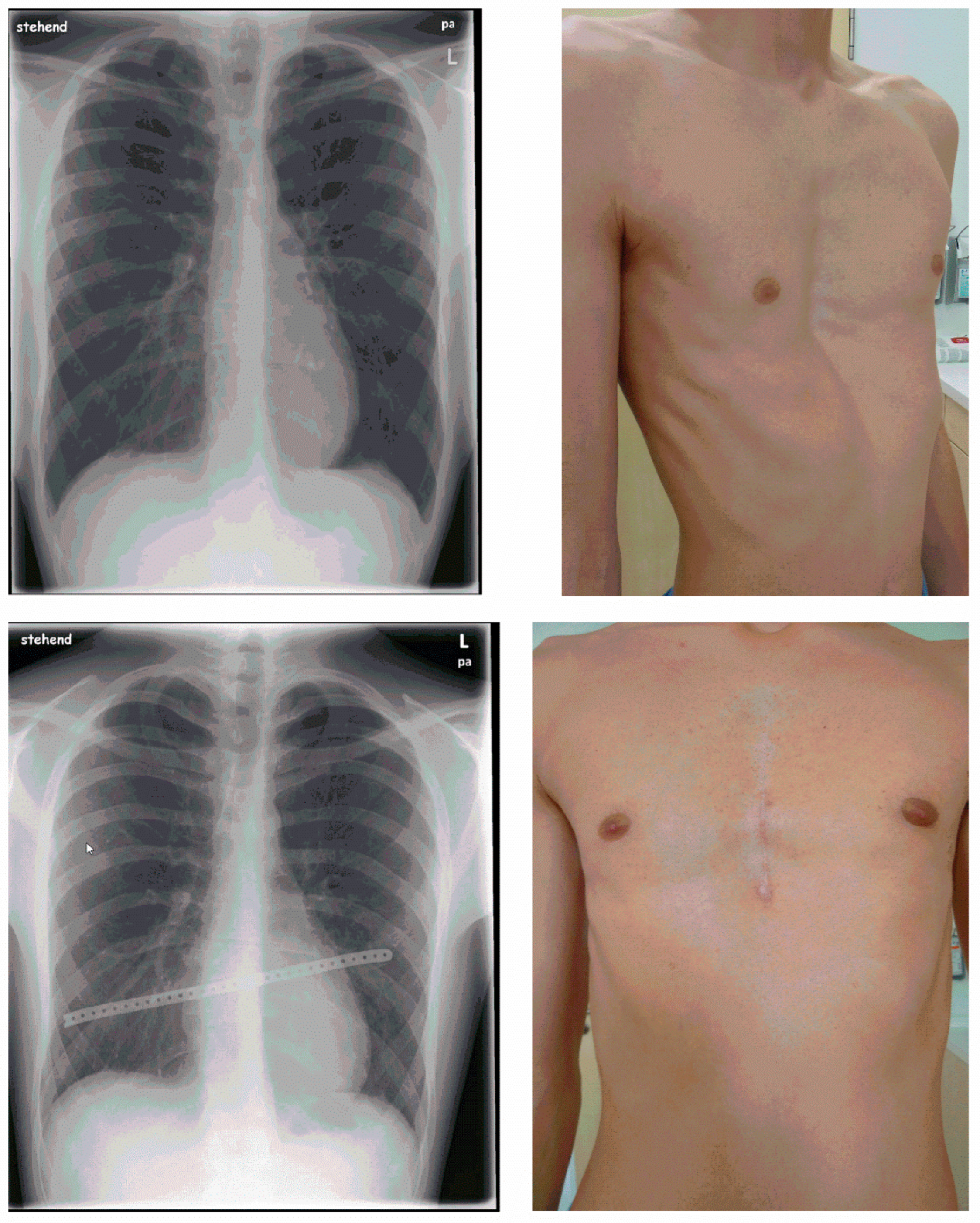
Figure 1. Preoperative (upper images) and postoperative (lower images) findings after MEK.
Applsci 13 10009 g001
Table 1. Patient data.
Sex
 Male255 patients (84.7%)
 Female46 patients (15.3%)
Age20.3 ± 9.3 years (12.8–29.4 years)
Body Mass Index (292 patients)20.3 ± 2.9 kg/m2 (12.8–29.4 kg/m2)
Morphology
 Asymmetrical143 patients (47.5%)
 Symmetrical107 patients (35.5%)
 Other51 patients (16.9%)
Hümmer Pectus Index
 131 patients104 ± 13.8 (61–142)
 170 patientsNo data
ECG
 Abnormal67 patients (22.3%)
 Normal107 patients (35.5%)
 No data127 patients (42.2%)
Lung function tests
 Abnormal82 patients (27.2%)
 Normal142 patients (47.2%)
 No data77 patients (25.6%)
Table 2. Operation.
Table 2. Operation.
Length of incision8.9 ± 1.6 cm (4.0–15.5 cm)
 Female patients8.7 ± 1.5 cm
 Male patients9.0 ± 1.6 cm
Operative procedures
 Chondrotomy301 patients (100%)
 Retrosternal dissection298 patients (99.0%)
 Tensiometry
  Before mobilization190 ± 36 N
  After median chondrotomy100 ± 38 N
  After retrosternal dissection36 ± 16 N
Number of metal implants
 1 implant228 patients (75.7%)
 2 implants71 patients (23.6%)
 3 implants2 patients (0.7%)
Implant positioning
 Transsternal286 patients (95.0%)
 Episternal1 patient (0.3%)
 Trans- and intrasternal11 patients (3.7%)
 Trans- and parasternal 3 patients (1.0%)
Duration of operation89.5 ± 19.7 min (45–175 min)
 Female patients100.3 ± 32.4 min
 Male patients87.9 ± 17.7 min
Table 3. Post-operative course.
Drainage
Chest drains
 03 patients (0.4%)
 1297 patients (99.3%)
 21 patients (0.3%)
Redon drains
 19 patients (3.0%)
 2292 patients (97.0%)
Complications
Pneumothorax54 patients (17.9%)
 Drainage5 patients (1.7%)
Pleural effusion51 patients (17.0%)
 Drainage9 patients (3.0%)
 Aspiration6 patients (2.0%)
Wound healing
 Normal182 patients (62.1%)
 Redness72 patients (25.9%)
 Swelling9 patients (3.1%)
 Secretion8 patients (2.7%)
 Dehiscence2 patients (0.7%)
Mobilization3 ± 0.6 days after surgery (3–7 days)
Length of hospitalization9 ± 1.7 days after surgery (5–21 days)
Disclaimer/Publisher’s Note: The statements, opinions and data contained in all publications are solely those of the individual author(s) and contributor(s) and not of MDPI and/or the editor(s). MDPI and/or the editor(s) disclaim responsibility for any injury to people or property resulting from any ideas, methods, instructions or products referred to in the content.