Article

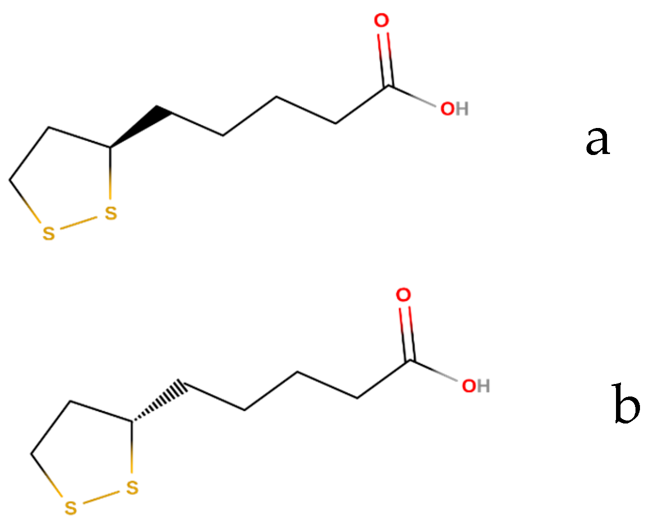
Encapsulation of α-Lipoic Acid in Halloysite Nanotubes

1
Physical Chemistry Department, National Technical University of Ukraine “Igor Sikorsky Kyiv Polytechnic Institute”, 03056 Kyiv, Ukraine
2
Department of Physics and Chemistry, University of Palermo, 90128 Palermo, Italy
*
Author to whom correspondence should be addressed.
Appl. Sci. 2023, 13(18), 10214; https://doi.org/10.3390/app131810214
Submission received: 9 August 2023 / Revised: 1 September 2023 / Accepted: 6 September 2023 / Published: 11 September 2023
(This article belongs to the Section Nanotechnology and Applied Nanosciences)

Abstract

:
A nanocomposite material based on halloysite and α-lipoic acid was studied. The kinetics of the degradation process of α-lipoic acid under the influence of ultraviolet radiation and thermal stress in its native state and in the composition of a halloysite-based nanocomposite were studied. The concentration of undegraded α-lipoic acid and the effect of the nanocomposite composition were determined by the HPLC method. It has been shown that adding α-lipoic acid to halloysite using a vacuum method allows for an increase in its resistance toward UV light of 84.4%, and the thermal resistance was also significantly improved. The composite based on halloysite and α-lipoic acid can be used to improve the resistance to photodegradation of pharmaceutical drugs or sunscreen cosmetics because this strategy preserves the biological active properties and shelf life of the α-lipoic acid.

1. Introduction

One of the well-known natural antioxidants is α-lipoic acid (ALA), which is also known as thioctic acid, 6,8-dithiooctic acid, or 1,2-dithiolane–3-pentanoic acid.
α-lipoic acid exists in the form of two enantiomers (Figure 1): R-(+)-ALA and S-(−)–ALA. But in nature, only the R-form of α-lipoic acid is found in organisms, both in free form and in conjugated forms with lysine residues [direct and indirect antioxidant properties of]. Commercial sources contain only the racemic form of this compound (rac-ALA).
Having a special molecular structure, α-lipoic acid easily transfers electrons or acyl groups to acceptor compounds, which counteracts redox reactions. Together with its reduced form (dihydrolipoic acid, DHLA), α-lipoic acid is one of the most powerful antioxidant pairs found in nature and is essential for cellular energy metabolism. The ALA/DHLA redox couple is capable of regenerating other endogenous antioxidants, such as vitamins C and E and glutathione [1,2].
The influence of α-lipoic acid on the cellular energy metabolism of most living organisms, from bacteria to humans, is possible due to the fact that the molecule of α-lipoic acid is very small and easily absorbed through the cell membrane. Due to its amphiphilic nature, α-lipoic acid is located in the lipid compartment of cells; therefore, it is widely distributed in living organisms [3,4].
Due to its unique properties, mainly antioxidant, α-lipoic acid is widely used in the practice of treating a number of diseases in people, such as slowed metabolism, states of intoxication, disorders of lipid and carbohydrate metabolism, and reduced liver function. Contributes to a decrease in blood sugar and an increase in the amount of glycogen in the liver. Its action is similar to the pharmacological properties of B vitamins. Therefore, α-lipoic acid is one of the most widely used substances in pharmaceuticals [5].
However, the disadvantages of this chemical are its instability to thermal exposure, oxidation, and degradation under the influence of daylight, which significantly reduces its antioxidant efficiency and ability to perform other important functions in human treatment. This is also challenging from an analytical point of view [6].
One of the ways to protect α-lipoic acid in the composition of medicinal products is through the use of delivery systems or encapsulation in various carriers, which has become the subject of recent research by a wide range of scientists.
Recent studies indicate that α-lipoic acid has been studied in a wide range of delivery systems, in particular using chitosan microspheres, polysaccharide gel beads, and a number of other polymer-based carriers [7,8,9].
The study of the antioxidant capacity of α-lipoic acid using the 2,2-diphenyl–1-picrylhydrazyl (DPPH) radical showed that microencapsulation in various polymer matrices and carriers allows it to almost completely preserve its ability to absorb free radicals in an in vitro model. These data are confirmed by numerous literature studies on the use of colloid carriers in other systems as well [7].
Loading into structural cavities created by polymeric carriers is a widely practiced approach to stabilizing unstable ingredients. In addition to the stabilizing effect, the release or delivery of captured ingredients can be controlled, improving their bioavailability for humans [8].
An important issue in the development of effective ways to protect unstable ingredients for oral use is their solubility in water, which determines their biological half-life and bioavailability. An increase in bioavailability is also achieved due to the dispersion and reduction of the particle size of the insoluble substance, which improves the speed of its removal and stability during storage. Therefore, encapsulation in nano-sized particles was proposed as an effective method for increasing the dispersion in water and bioavailability of various non-polar bioactive substances [9].
Nanoscale encapsulation in cyclodextrin and amylose was proposed as one of the effective processes in the preparation of aqueous dispersions of hydrophobic substances [10,11]. The level of protection of α-lipoic acid against thermal degradation and oxidation during encapsulation in starch with a high amylose content, both in the native and octenylsuccinylated states, was investigated by modeling the digestive tract [12]. Octenylsuccinylated starch was found to be more effective in stabilizing α-lipoic acid than native starch, probably due to the amphiphilic and hydrophobic nature of the substituents in the modified starch. Moreover, the stability during gastric and intestinal digestion in vitro and the subsequent slow release of α-lipoic acid during digestion further suggest that high-amylose octenylsuccinylated starch is a possible digestive carrier of controlled-release α-lipoic acid.
The photochemical stability of α-lipoic acid under the influence of natural daylight at room temperature was investigated as part of an aqueous dispersion of a nanostructured lipid carrier (NLC), which is a known effective delivery system for drugs and other biologically active substances. Encapsulation in NLCs was found to be an effective process that retained 88.5% of α-lipoic acid after 120 days of exposure to daylight, while unencapsulated acid degraded by 99% during this time. It was shown that the photodegradation of α-lipoic acid occurs due to the breaking of the S-S bond of the 1,2-dithiolane ring in the molecule, which is accompanied by an unpleasant odor [13].
Halloysite nanotubes are one of the best-known transport systems for the delivery of organic substances. It is a natural material with a special tubular nanostructure (HNTs) with the chemical formula Al2Si2O5 (OH)4, which makes it an inexpensive and valuable alternative to the most common carbon nanotubes. The length of nanotubes is mainly from 200 nm to 2 μm, their inner diameter is 15–50 nm, and the outer diameter is 50–200 nm [14,15]. One of the main limitations of this nanomaterial is its wide polydispersion in size, and therefore the separation and purification procedures are challenging [16,17]. However, its unique structure can be used to modify the surface with organic molecules (for example, polymers, biopolymers, or surfactants) using both electrostatic and van der Waals interactions [18]. In addition, the outer and inner surfaces have opposite charges due to different chemical compositions (Al-OH and Si-O-Si groups, respectively). Thus, the inner (Al) surface is positively charged and the outer (Si) surface is negatively charged, and targeted electrostatic interactions are envisaged [19,20].
Based on their structure, halloysite nanotubes can be loaded with negatively charged substances, such as biologically active molecules, antioxidants, etc. Biologically active substances loaded inside the inner lumen of nanotubes can receive additional protection from external factors [21,22,23,24]. This is a very important characteristic that ensures the creation of new smart nanocontainers for loading, storage, and long-term release of chemical agents of various effects. Equally important is the fact that halloysite, being completely non-toxic, creates additional opportunities for the wide application of nanocomposites based on it with the use of active ingredients [25] and for the delivery and controlled release of pharmaceuticals [25,26,27,28].
The biocompatibility of halloysite nanoutubes has been tested on different living organisms, providing low toxicity even for oral administration [29,30,31]. Besides medical applications, including tissue engineering, cosmetic formulations for hair treatments have been recently reported [32,33,34].
Therefore, a promising route for the development of cosmetic formulations based on α-lipoic acid is the encopsulation of this active ingredient into the cavity of the halloysite nanotubes. The obtained nanocomposite will be checked for photosensitivity, resistance to the influence of ultraviolet light, and heat resistance as the protection effect of the nanotubular clay is predicted.

2. Materials and Methods

2.1. Reagents

Sigma Aldrich halloysite was used for research, α-lipoic acid was purchased from USP Reference Standards.
A transmission scanning electron microscope (S/TEM) Themis from FEI (Thermo Scientific, Waltham, MA, USA) was used for microscopic studies in STEM and TEM modes of operation. An electron gun with a Schottky field emission cathode. Resolution limit: 0.7 Å (angstroms) in TEM/STEM modes when using image corrector and probe. The accelerating voltage range is from 60 to 300 kV. HAADF detector. Triple detectors DF1/DF2/BF are located on the axis. Camera Ceta 16 M; Gatan US1000/US4000 cameras. A series of energy filters, Gatan. Super-X: highly sensitive windowless EDX detector system (patented technology). EDS detector: 0.13 srad solid angle.
A Hitachi S–4800 field emission scanning electron microscope at 3.0 kV was used for microscopic studies.
An Agilent 1290 Infinity II liquid chromatograph was used to identify the concentration of α-lipoic acid. Multisampler 1290 Infinity II. Agilent (Kiev, Ukraine) Max-Light cuvette with an optical path length of 60 mm. DAD detector, pump 1290 Infinity II Flexible Pump (Kiev, Ukraine).
The ultraviolet lamp was from Fisher Bioblock Scientific (Brovary, Ukraine) (model VL–215.G 2 × 15 W—254 nm Tube POWER: 60 W).
A UNB200 Memmert c thermal cabinet was used for thermal stress tests.

2.2. Loading Halloysite Nanotubes

In this work, the vacuum method is used as the most widely used method of loading nanotubes with the substance under study. Previously, two methods were tested in the work, in the first of which loading occurs from a solution where the active pharmaceutical ingredient (API) is in excess, according to literature [35], and in the second, α-lipoic acid and HNTs were mixed in a mass ratio of 2:1.
According to Method 1, crushed, sieved, and dried HNTs were added to the solution with an excess of API. A vacuum was applied to the formed suspension and, after the appearance of bubbles on the surface of the suspension,
The use of vacuum to improve the loading efficacy of the halloysite nanotubes is well established experimentally, but its interpretation has been reported recently [36,37]. Of course, due to capillarity, the nanotubes are quickly filled, and there is no need for vacuum to remove air from inside. This process was repeated at least two to three times to completely fill the halloysite tubes with the α-lipoic acid solution. This filling protocol ensures a slow release of the active molecules from the composite [37]. After completion of the vacuum cycle, the mixture was centrifuged, decanted, and dried under vacuum. According to the second method, a mixture of solution and halloysite was obtained, which was a thick paste. Further processing in a vacuum with periodic returns to atmospheric pressure was carried out three times according to the first method. Testing both methods with α-lipoic acid showed that the second method is more rational and accurate because the amount of drug added to HNTs can be determined directly without analyzing the nanocomposite, which makes it possible to more accurately establish the concentration of α-lipoic acid in the solution [38]. Therefore, all further studies on the loading of α-lipoic acid were carried out according to the second method.

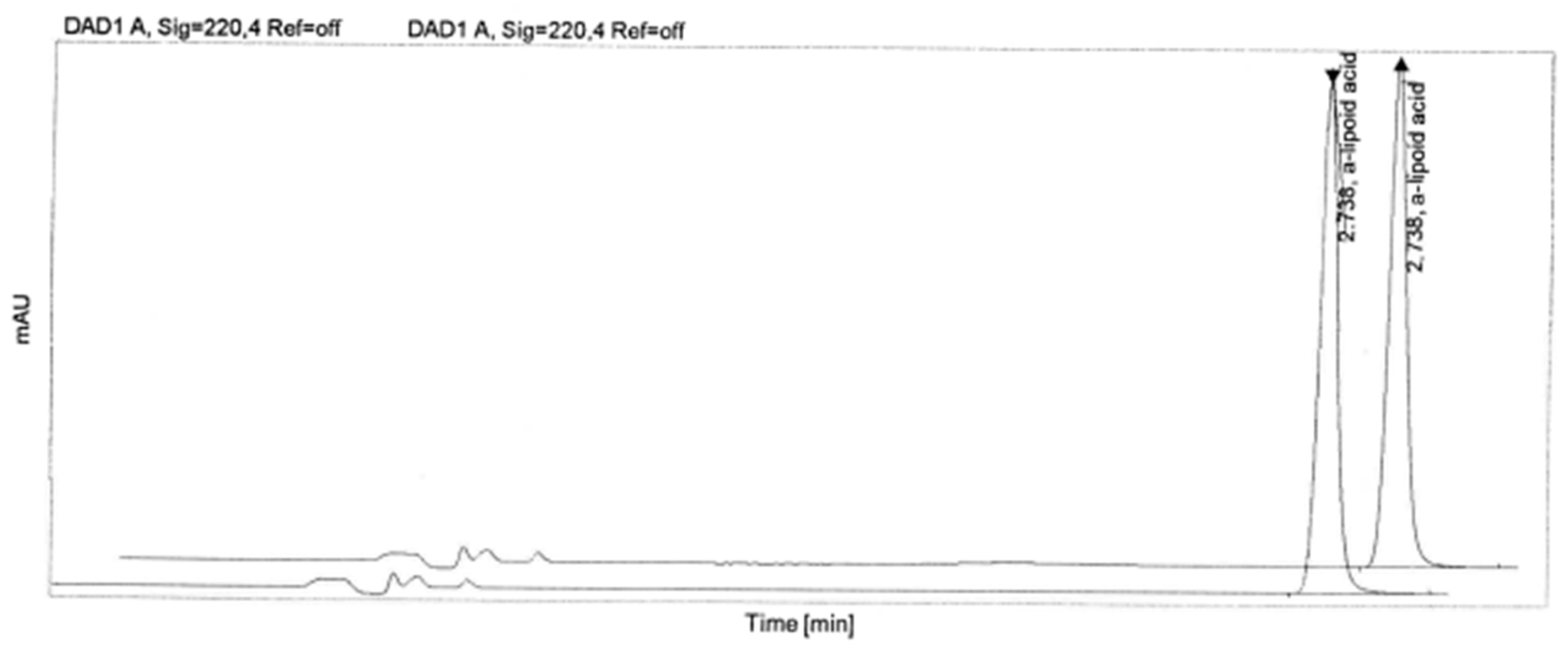
2.3. Methodology for Determining the Concentration of α-Lipoic Acid

For the analytical control of α-lipoic acid content, a chromatographic method of analysis was used, which has several advantages. Namely, the substance practically does not change during separation, which is very important for further chemical research. This method is quite suitable for the separation of liquid and gaseous compounds and can be used for the fractionation of mixtures of substances that are similar in chemical composition, properties, and structure. In addition, chromatography has high accuracy in the separation of substances and is relatively easy to use.
Reverse-phase liquid chromatography was chosen for the study. During reverse phase chromatography (RPC), hydrophobic substances dissolved in a polar solvent selectively interact with the stationary phase. The liquid phase consists of an aqueous buffer containing an organic modifier dissolved in water, and this modifier forms a liquid interface between the two phases, the mobile being polar and the immobile being hydrophobic.
The sorbent for this method of identifying α-lipoic acid in chromatography consists of a basic matrix to which organic ligands, which are alkylated hydrocarbon chains, are “sewn”.
The lipophilic medium of the stationary part of the chromatographic system was selected in such a way as to attract molecules of the organic modifier to form a liquid adsorption phase. Octadecyl groups—C18—were used as matrix coatings.
Since there is no method of identification of α-lipoic acid in the literature, this study developed a method of identification and determination of the quantitative content of this active pharmaceutical ingredient by liquid chromatography.
A mixture of distilled water and methanol in a 1:1 ratio was used as a solvent. To prepare the comparison solution, 15 mg of a-lipoic acid was dissolved in 5 mL of methanol and brought to a volume of 50.0 mL with the solvent. For the investigated solutions, an aliquot of 5.0 mL of the sample was taken and adjusted to a volume of 20.0 mL with the solvent, then 2.0 mL of the obtained solution was adjusted to a volume of 20.0 mL with the solvent.
Chromatography was performed on a liquid chromatograph with a spectrophotometric detector. The α-lipoic acid peak was identified using a Zorbax C-18 chromatographic column (Agilent, Kiev, Ukraine). Acetonitrile and 0.2 M potassium dihydrogen phosphate solution were used as mobile phases. The flow rate of the mobile phase was 1.7 mL/min. The temperature of the chromatographic column is 30 °C. The optical absorption density is 220 nm. The volume of the injected sample was 10 μL. The concentration of lipoic acid was determined by the response (in mAU—milli Absorbance Unit) of the chromatograph detector.
In this study, tests were performed on the separation of α-lipoic acid from halloysite using different types of Agilent membrane filters. Two-layer membrane filters are used to purify the sample solution. Such a filter consists of two parts: a preliminary coarse glass fiber filter and, in fact, a membrane filter. The prefilter consists of borosilicate glass fibers. It is chemically inert and resistant to most solvents. The presence of a prefilter with a large surface area in the design ensures its large capacity and the possibility of repeated use.
This study is necessary to establish which of the filter types is best suited for the separation of α-lipoic acid from halloysite. In the course of laboratory studies, it was determined that the membrane filter Agilent PES 0.1 mkm is best suited for the performance of the tasks. The accuracy of the results is greater than 99.74%.

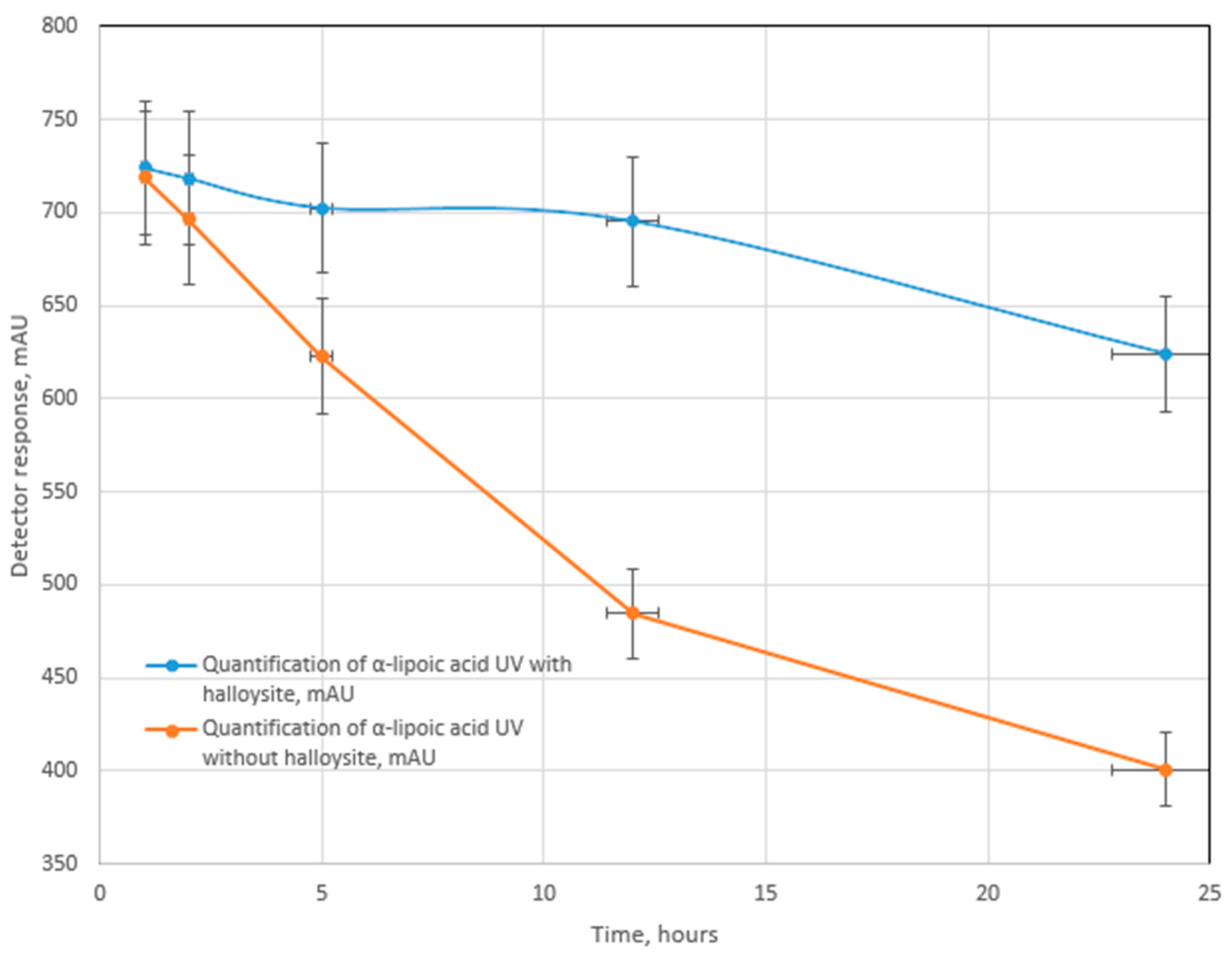
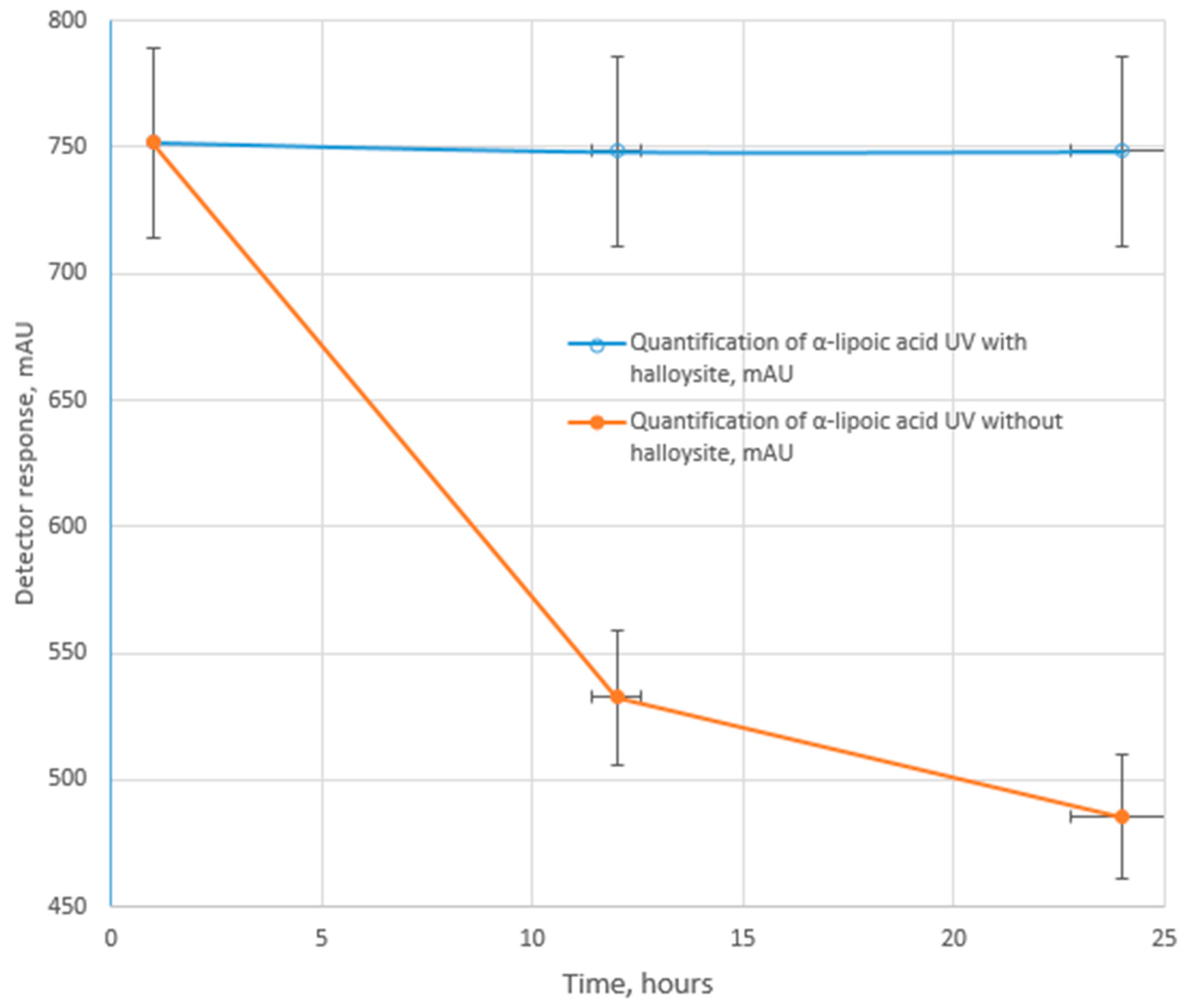
2.4. Testing for UV-, Photo-, and Thermal Stability

The UV test was performed by irradiating α-lipoic acid solutions with an ultraviolet lamp.
Photostability testing was performed in accordance with stability testing: photostability testing of new drug substances and products Q1B [38,39].
The thermal stability test was performed by keeping powdered α-lipoic acid and its composite with halloysite in a thermal cabinet at a temperature of 60 °C.

3. Results

The morphology of the halloysite sample used in this work was imaged by electron microscopy (Figure 2). The length of the nanotubes ranges between 0.3 and 5 μm. The hollow cavity is clearly visible in the TEM image (Figure 2). A proper statistical treatment evidencing the average size of the nanotubes was provided in a recent report [14].
Research activities were used to identify α-lipoic acid. High-performance liquid chromatography with UV detection is the method used in this study. A chromatogram with α-lipoic acid is shown in Figure 3, with a peak time of approximately 2.7 min. The peak parameters are shown in Table 1. The optimal chromatographic conditions were selected to ensure effective separation of the component. The suitability of the chromatographic system for the characteristics of the identified peak meets the requirements of the European Pharmacopoeia 11.0, which indicates that the method developed by us gives correct values.
α-lipoic acid was irradiated with UV light in its native form in a solution and in a suspension of halloysite nanocomposite with α-lipoic acid. The total exposure time was 24 h. Control of the concentration of α-lipoic acid was carried out after 1, 2, 5, 12 and 24 h of testing.
Studies show that α-lipoic acid degrades quite quickly under the influence of ultraviolet radiation. After a day of irradiation, its concentration in the solution decreased by 54.2%. The most significant degradation occurs in the first 12 h of exposure, when the amount of destroyed substance is reduced by more than half.
The degradation of α-lipoic acid in halloysite under the influence of UV light is much slower. Up to 12 h of irradiation tests, the concentration of α-lipoic acid in the solution did not change significantly. It should be noted that after UV irradiation for 24 h, 84.4% of α-lipoic acid is preserved (Figure 4).
A photostability test of α-lipoic acid in its native state without loading into nanotubes for a day shows that it degrades, and the residual content in the suspension after the tests is 82.0% (Figure 5, Table 2).
Tests of α-lipoic acid loaded into halloysite showed that halloysite is a good protective material against photodegradation and can protect it with an efficacy of 98.5% (Figure 6, Table 2).
The powder-like mass of α-lipoic acid was subjected to temperature stress at a temperature of 60 °C for 1, 12, and 24 h (Figure 7). The results of the study show that without loading into halloysite, α-lipoic acid quickly degrades, and after tests for a day, its residual content is 65.7%. When loaded into halloysite, α-lipoic acid does not degrade, which is evidenced by the fact that its concentration does not change after temperature tests.
According to the scientific literature, mineral clays provide good protection against the photodegradation of photolabile organic substances used in pharmaceuticals and cosmetics.
Thus, Ambrogi, in co-authorship, investigated the role of montmorillonite and halloysite in the photostability of piroxicam, which is a non-steroidal anti-inflammatory drug [39]. Piroxicam was introduced into clays by mechanical mixing followed by intercalation in solution. Photostability tests using a xenon lamp showed that hybrids based on montmorillonite showed greater photostability than hybrids based on halloysite. The authors claim that in montmorillonite there were “specific interactions” between organic molecules and clay layers, whereas in halloysite there were none.
The occurrence of specific bonds in montmorillonite during the encapsulation of photounstable tetracycline is evidenced by the authors’ research [40]. It was shown that the effect of increasing photostability is provided by the lamellar structure of montmorillonite, which ensures the placement of organic molecules in the interlayer space as well as a certain chemical composition with the ability for cation exchange. Other clays, such as palygorskite and sepiolite, which have a fibrous structure, did not provide effective photoprotection.
Ambrogi, in co-authorship [41], obtained successful results on the protection against photodegradation of promethazine (a phenothiazine drug) intercalated in montmorillonite. Ultraviolet irradiation testing of a hybrid based on montmorillonite and promethazine in solution indicates effective protection of this organic drug against photodestruction.
There are known studies on the successful loading of nifedipine into clay minerals, which undergo photodegradation under the influence of light in other drug delivery systems and turn into compounds that have no pharmacological effect. A high photoprotective effect of photolabile nifedipine was obtained by intercalation in montmorillonite [41] and halloysite [42].
It was proven that nifedipine in the composition obtained by encapsulation into halloysite has high photostability [42]. Thus, after testing for photodestruction under conditions of irradiation for 1000 h, about 58% of nifedipine remained in the composite and only 18% in the micronized state.
Thus, the results of research in the publications indicate that organic substances intercalated in natural mineral clays have increased photostability. One of the effective clays is montmorillonite, whose protective effect is based on the intercalation of photolabile molecules in the internal cavities and the formation of certain interactions with them, including the flow of cation exchange reactions. This ensures the formation of stable hybrid materials. Halloysite, which consists of nanotubes, is an equally effective nanocarrier that also provides protection against photodegradation. The photoprotective effect of halloysite is ensured by the fact that the organic molecules intercalated in the middle of the nanotubes are protected by multilayered tubes of the mineral.
The results of the research presented in this paper indicate that the obtained effect of increasing the photostability of α-lipoic acid in the composition of halloysite is commensurate in terms of protection with other hybrid materials based on halloysite and montmorillonite.

4. Conclusions

The studies indicate that halloysite nanotubes can be used as effective biocompatible nanocontainers for the preservation and delivery of α-lipoic acid.
Tests on UV light irradiation showed that α-lipoic acid in native form degrades by 54.2% within a day. More than half of this amount is degraded in the first 12 h of UV exposure. After being loaded into halloysite nanotubes, α-lipoic acid is more resistant to UV exposure. During the first 12 h of testing, the acid concentration did not change much. After a day of ultraviolet irradiation, the amount of undegraded acid was 84.4%.
Tests of α-lipoic acid in halloysite have shown that this pharmaceutical ingredient has an extended photostability period. After a day of testing, 98.5% of the substance was preserved, in contrast to the native substance, which during this time remained unaffected by only 82%.
Testing for heat resistance shows that α-lipoic acid practically does not degrade in the composite after encapsulation into the halloysite cavity, and only 65.7% of the acid remains in a non-degraded form in its native form after a day of testing. The obtained results suggest that the use of α-lipoic acid in cosmetic formulations can be improved by adding halloysite nanotubes to the formulations.

Author Contributions

Conceptualization, G.L. and O.C.; investigation, A.M.; resources, O.C. and G.L.; data curation, A.M.; writing—original draft preparation, A.M.; supervision, O.C. All authors have read and agreed to the published version of the manuscript.

Funding

This research received no external funding.

Data Availability Statement

Original data are available upon request to the corresponding author.

Acknowledgments

The R & D laboratory of pharmaceutical company “Darnytsia” (Kiev, Uckraine) is acknowledged for its help in LC measurements.

Conflicts of Interest

The authors declare no conflict of interest.

References

  1. Bast, A.; Haenen, G.R.M.M. Lipoic Acid: A Multifunctional Antioxidant. BioFactors 2003, 17, 207–213. [Google Scholar] [CrossRef]
  2. Moura, F.; De Andrade, K.; Farias Dos Santos, J.; Fonseca Goulart, M. Lipoic Acid: Its Antioxidant and Anti-Inflammatory Role and Clinical Applications. Curr. Top. Med. Chem. 2015, 15, 458–483. [Google Scholar] [CrossRef] [PubMed]
  3. Pop, A.L.; Crișan, S.; Bârcă, M.; Ciobanu, A.-M.; Varlas, V.N.; Pop, C.; Pali, M.-A.; Cauni, D.; Ozon, E.A.; Udeanu, D.; et al. Evaluation of Dissolution Profiles of a Newly Developed Solid Oral Immediate-Release Formula Containing Alpha-Lipoic Acid. Processes 2021, 9, 176. [Google Scholar] [CrossRef]
  4. Tripathi, A.K.; Ray, A.K.; Mishra, S.K.; Bishen, S.M.; Mishra, H.; Khurana, A. Molecular and Therapeutic Insights of Alpha-Lipoic Acid as a Potential Molecule for Disease Prevention. Rev. Bras. Farmacogn. 2023, 33, 272–287. [Google Scholar] [CrossRef] [PubMed]
  5. He, M.; Wu, Y.; Hong, M.; Yun, Z.; Li, T.; Jiang, Y. α-Lipoic Acid Treatment Alleviates Postharvest Pericarp Browning of Litchi Fruit by Regulating Antioxidant Ability and Energy Metabolism. Postharvest Biol. Technol. 2021, 180, 111629. [Google Scholar] [CrossRef]
  6. Srivastava, S.; Dhaneshwar, S.; Kawathekar, N. Stress Degradation Studies and Development of Validated Stability Indicating Densitometric Method for Estimation of Alpha Lipoic Acid in Bulk and Capsule Dosage Form. Acta Chromatogr. 2023, 35, 180–186. [Google Scholar] [CrossRef]
  7. Ruktanonchai, U.; Bejrapha, P.; Sakulkhu, U.; Opanasopit, P.; Bunyapraphatsara, N.; Junyaprasert, V.; Puttipipatkhachorn, S. Physicochemical Characteristics, Cytotoxicity, and Antioxidant Activity of Three Lipid Nanoparticulate Formulations of Alpha-Lipoic Acid. AAPS PharmSciTech 2009, 10, 227–234. [Google Scholar] [CrossRef] [PubMed]
  8. Dima, Ş.; Dima, C.; Iordăchescu, G. Encapsulation of Functional Lipophilic Food and Drug Biocomponents. Food Eng. Rev. 2015, 7, 417–438. [Google Scholar] [CrossRef]
  9. Lei, X.; Zhou, Y.; Liu, X.; Kong, L.; Liao, L.; Li, Y.; Liu, M.; Tian, L.; Rao, W.; Lv, G. Effective PH-Responsive Nanocarrier Based on the Anisotropic Surfaces of Halloysite Nanotubes for Controlled Drug Release. Appl. Clay Sci. 2023, 232, 106799. [Google Scholar] [CrossRef]
  10. Calvino, M.M.; Lazzara, G.; Cavallaro, G.; Milioto, S. Inclusion Complexes of Triblock L35 Copolymer and Hydroxyl Propyl Cyclodextrins: A Physico-Chemical Study. New J. Chem. 2022, 46, 6114–6120. [Google Scholar] [CrossRef]
  11. Kim, J.-Y.; Seo, T.-R.; Lim, S.-T. Preparation of Aqueous Dispersion of β-Carotene Nano-Composites through Complex Formation with Starch Dextrin. Food Hydrocoll. 2013, 33, 256–263. [Google Scholar] [CrossRef]
  12. Li, Y.-X.; Park, E.Y.; Lim, S.-T. Stabilization of Alpha-Lipoic Acid by Complex Formation with Octenylsuccinylated High Amylose Starch. Food Chem. 2018, 242, 389–394. [Google Scholar] [CrossRef] [PubMed]
  13. Wang, J.; Tang, J.; Zhou, X.; Xia, Q. Physicochemical Characterization, Identification and Improved Photo-Stability of Alpha-Lipoic Acid-Loaded Nanostructured Lipid Carrier. Drug Dev. Ind. Pharm. 2014, 40, 201–210. [Google Scholar] [CrossRef]
  14. Cavallaro, G.; Chiappisi, L.; Pasbakhsh, P.; Gradzielski, M.; Lazzara, G. A Structural Comparison of Halloysite Nanotubes of Different Origin by Small-Angle Neutron Scattering (SANS) and Electric Birefringence. Appl. Clay Sci. 2018, 160, 71–80. [Google Scholar] [CrossRef]
  15. Pasbakhsh, P.; Churchman, G.J.; Keeling, J.L. Characterisation of Properties of Various Halloysites Relevant to Their Use as Nanotubes and Microfibre Fillers. Appl. Clay Sci. 2013, 74, 47–57. [Google Scholar] [CrossRef]
  16. Durgut, E.; Cinar, M.; Terzi, M.; Kursun Unver, I.; Yildirim, Y.; Ozdemir, O. Evaluation of Different Dispersants on the Dispersion/Sedimentation Behavior of Halloysite, Kaolinite, and Quartz Suspensions in the Enrichment of Halloysite Ore by Mechanical Dispersion. Minerals 2022, 12, 1426. [Google Scholar] [CrossRef]
  17. Calvino, M.M.; Cavallaro, G.; Lisuzzo, L.; Milioto, S.; Lazzara, G. Separation of Halloysite/Kaolinite Mixtures in Water Controlled by Sucrose Addition: The Influence of the Attractive Forces on the Sedimentation Behavior. Colloids Surf. A Physicochem. Eng. Asp. 2022, 641, 128530. [Google Scholar] [CrossRef]
  18. Liao, J.; Wang, H.; Liu, N.; Yang, H. Functionally Modified Halloysite Nanotubes for Personalized Bioapplications. Adv. Colloid Interface Sci. 2023, 311, 102812. [Google Scholar] [CrossRef]
  19. Glotov, A.; Novikov, A.; Stavitskaya, A.; Nedolivko, V.; Kopitsyn, D.; Kuchierskaya, A.; Ivanov, E.; Stytsenko, V.; Vinokurov, V.; Lvov, Y. Nanoreactors Based on Hydrophobized Tubular Aluminosilicates Decorated with Ruthenium: Highly Active and Stable Catalysts for Aromatics Hydrogenation. Catal. Today 2021, 378, 33–42. [Google Scholar] [CrossRef]
  20. Farinmade, A.; Ojo, O.F.; Trout, J.; He, J.; John, V.; Blake, D.A.; Lvov, Y.M.; Zhang, D.; Nguyen, D.; Bose, A. Targeted and Stimulus-Responsive Delivery of Surfactant to the Oil–Water Interface for Applications in Oil Spill Remediation. ACS Appl. Mater. Interfaces 2020, 12, 1840–1849. [Google Scholar] [CrossRef]
  21. Lisuzzo, L.; Cavallaro, G.; Milioto, S.; Lazzara, G. Halloysite Nanotubes Filled with MgO for Paper Reinforcement and Deacidification. Appl. Clay Sci. 2021, 213, 106231. [Google Scholar] [CrossRef]
  22. Lvov, Y.; Aerov, A.; Fakhrullin, R. Clay Nanotube Encapsulation for Functional Biocomposites. Adv. Colloid Interface Sci. 2014, 207, 189–198. [Google Scholar] [CrossRef] [PubMed]
  23. Dzamukova, M.R.; Naumenko, E.A.; Lvov, Y.M.; Fakhrullin, R.F. Enzyme-Activated Intracellular Drug Delivery with Tubule Clay Nanoformulation. Sci. Rep. 2015, 5, 10560. [Google Scholar] [CrossRef]
  24. Qin, Y.; Su, W.; Meng, G.; Cui, L.; Wu, J.; Yang, S.; Liu, Z.; Liu, J.; Guo, X. Polymer-Modified Halloysite Nanotubes with High Adhesion and UV-Shielding Properties for Chlopyrifos Application on Cotton Leaves. Appl. Clay Sci. 2023, 234, 106811. [Google Scholar] [CrossRef]
  25. Cavallaro, G.; Lazzara, G.; Milioto, S. Nanocomposites Based on Halloysite Nanotubes and Sulphated Galactan from Red Seaweed Gloiopeltis: Properties and Delivery Capacity of Sodium Diclofenac. Int. J. Biol. Macromol. 2023, 234, 123645. [Google Scholar] [CrossRef]
  26. Caruso, M.R.; Cavallaro, G.; Milioto, S.; Lazzara, G. Halloysite Nanotubes/Keratin Composites for Wool Treatment. Appl. Clay Sci. 2023, 238, 106930. [Google Scholar] [CrossRef]
  27. Cavallaro, G.; Milioto, S.; Parisi, F.; Lazzara, G. Halloysite Nanotubes Loaded with Calcium Hydroxide: Alkaline Fillers for the Deacidification of Waterlogged Archeological Woods. ACS Appl. Mater. Interfaces 2018, 10, 27355–27364. [Google Scholar] [CrossRef] [PubMed]
  28. Yendluri, R.; Lvov, Y.; de Villiers, M.M.; Vinokurov, V.; Naumenko, E.; Tarasova, E.; Fakhrullin, R. Paclitaxel Encapsulated in Halloysite Clay Nanotubes for Intestinal and Intracellular Delivery. J. Pharm. Sci. 2017, 106, 3131–3139. [Google Scholar] [CrossRef]
  29. Kryuchkova, M.; Danilushkina, A.; Lvov, Y.; Fakhrullin, R. Evaluation of Toxicity of Nanoclays and Graphene Oxide in Vivo: A Paramecium caudatum Study. Environ. Sci. Nano 2016, 3, 442–452. [Google Scholar] [CrossRef]
  30. Rozhina, E.; Batasheva, S.; Miftakhova, R.; Yan, X.; Vikulina, A.; Volodkin, D.; Fakhrullin, R. Comparative Cytotoxicity of Kaolinite, Halloysite, Multiwalled Carbon Nanotubes and Graphene Oxide. Appl. Clay Sci. 2021, 205, 106041. [Google Scholar] [CrossRef]
  31. Wang, X.; Gong, J.; Rong, R.; Gui, Z.; Hu, T.; Xu, X. Halloysite Nanotubes-Induced Al Accumulation and Fibrotic Response in Lung of Mice after 30-Day Repeated Oral Administration. J. Agric. Food Chem. 2018, 66, 2925–2933. [Google Scholar] [CrossRef] [PubMed]
  32. Cavallaro, G.; Caruso, M.R.; Milioto, S.; Fakhrullin, R.; Lazzara, G. Keratin/Alginate Hybrid Hydrogels Filled with Halloysite Clay Nanotubes for Protective Treatment of Human Hair. Int. J. Biol. Macromol. 2022, 222, 228–238. [Google Scholar] [CrossRef] [PubMed]
  33. Panchal, A.; Fakhrullina, G.; Fakhrullin, R.; Lvov, Y. Self-Assembly of Clay Nanotubes on Hair Surface for Medical and Cosmetic Formulations. Nanoscale 2018, 10, 18205–18216. [Google Scholar] [CrossRef] [PubMed]
  34. Naumenko, E.; Fakhrullin, R. Halloysite Nanoclay/Biopolymers Composite Materials in Tissue Engineering. Biotechnol. J. 2019, 14, 1900055. [Google Scholar] [CrossRef] [PubMed]
  35. Kelly, H.M.; Deasy, P.B.; Ziaka, E.; Claffey, N. Formulation and Preliminary in Vivo Dog Studies of a Novel Drug Delivery System for the Treatment of Periodontitis. Int. J. Pharm. 2004, 274, 167–183. [Google Scholar] [CrossRef] [PubMed]
  36. Lisuzzo, L.; Cavallaro, G.; Pasbakhsh, P.; Milioto, S.; Lazzara, G. Why Does Vacuum Drive to the Loading of Halloysite Nanotubes? The Key Role of Water Confinement. J. Colloid Interface Sci. 2019, 547, 361–369. [Google Scholar] [CrossRef]
  37. Lisuzzo, L.; Cavallaro, G.; Milioto, S.; Lazzara, G. Halloysite Nanotubes Filled with Salicylic Acid and Sodium Diclofenac: Effects of Vacuum Pumping on Loading and Release Properties. J. Nanostructure Chem. 2021, 11, 663–673. [Google Scholar] [CrossRef]
  38. Tan, D.; Yuan, P.; Annabi-Bergaya, F.; Liu, D.; Wang, L.; Liu, H.; He, H. Loading and in Vitro Release of Ibuprofen in Tubular Halloysite. Appl. Clay Sci. 2014, 96, 50–55. [Google Scholar] [CrossRef]
  39. Ambrogi, V.; Latterini, L.; Nocchetti, M.; Pagano, C.; Ricci, M. Montmorillonite as an Agent for Drug Photostability. J. Mater. Chem. 2012, 22, 22743–22749. [Google Scholar] [CrossRef]
  40. Da Rocha, M.C.; Braz, E.M.D.A.; Honório, L.M.C.; Trigueiro, P.; Fonseca, M.G.; Silva-Filho, E.C.; Carrasco, S.M.; Polo, M.S.; Iborra, C.V.; Osajima, J.A. Understanding the Effect of UV Light in Systems Containing Clay Minerals and Tetracycline. Appl. Clay Sci. 2019, 183, 105311. [Google Scholar] [CrossRef]
  41. Ambrogi, V.; Nocchetti, M.; Latterini, L. Promethazine–Montmorillonite Inclusion Complex To Enhance Drug Photostability. Langmuir 2014, 30, 14612–14620. [Google Scholar] [CrossRef]
  42. Yendluri, R.; Otto, D.P.; De Villiers, M.M.; Vinokurov, V.; Lvov, Y.M. Application of Halloysite Clay Nanotubes as a Pharmaceutical Excipient. Int. J. Pharm. 2017, 521, 267–273. [Google Scholar] [CrossRef]
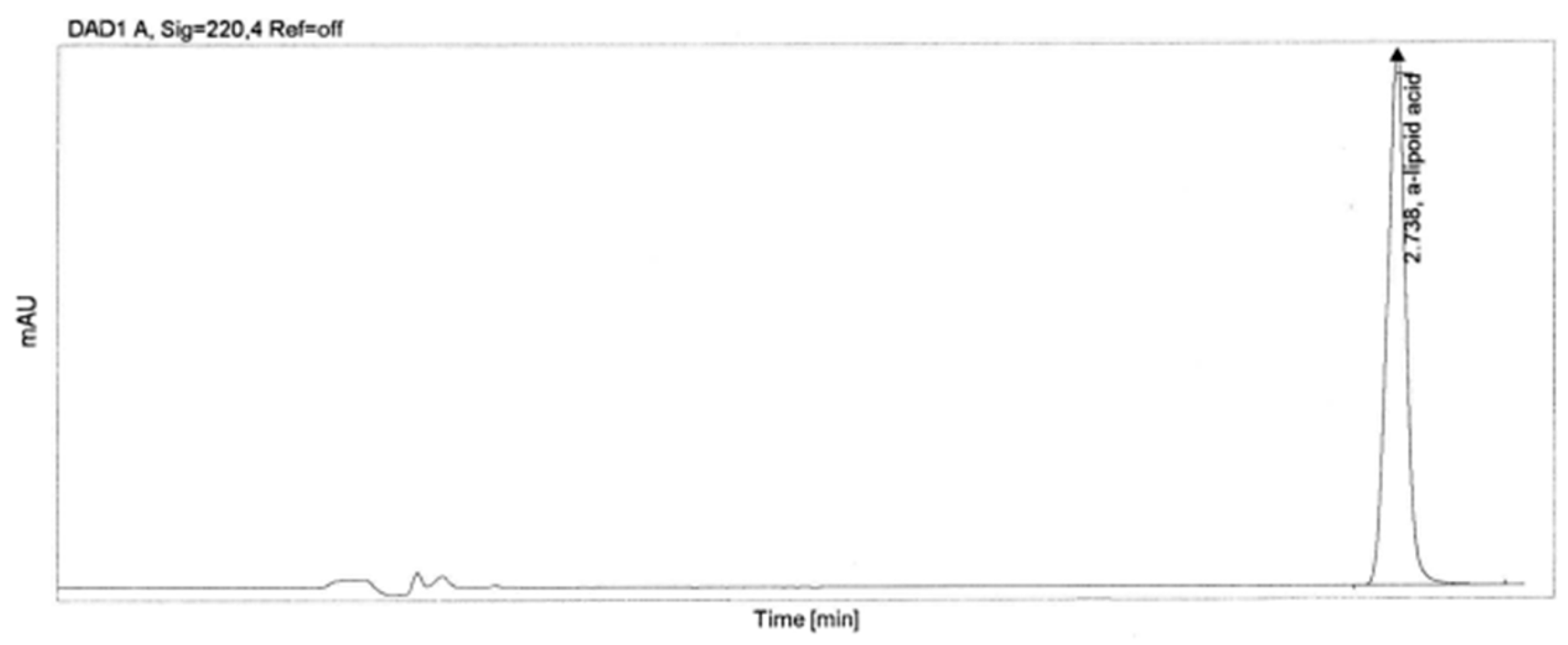
Figure 1. Structural formula of α-lipoic acid in two forms: (a)—R form, (b)—S form.
Applsci 13 10214 g001
Figure 2. Microscopic images of halloysite: SEM (left hand side), TEM (right hand side).
Applsci 13 10214 g002
Figure 3. Chromatogram of α-lipoic acid identification.
Applsci 13 10214 g003
Figure 4. Degradation of α-lipoic acid under the influence of UV radiation.
Applsci 13 10214 g004
Figure 5. Chromatogram of the photostability test of α-lipoic acid without halloysite.
Applsci 13 10214 g005
Figure 6. Chromatogram of the photostability test of α-lipoic acid with halloysite.
Applsci 13 10214 g006
Figure 7. Degradation of α-lipoic acid under temperature stress.
Applsci 13 10214 g007
Table 1. Peak characteristics of α-lipoic acid from HPLC analysis.
SampleRT (min)AreaPeak HeightTail FactorTheoretical Plates EP
α-lipoic acid2.738718.83270.8291.025,769
Table 2. Peak characteristics of α-lipoic acid with halloysite of the photostability test.
SampleSampleRT (min)AreaPeak HeightTail FactorTheoretical Plates EP
photostability test of α-lipoic acid 1st injection2.736606.63224.6641.025,569
2nd injection2.736607.53225.0211.125,580
Mean2.736607.08 1.125,574
RSD0.000.10
photostability test of α-lipoic acid with HNTs1st injection2.738727.31273.2131.025,732
2nd injection2.738728.72273.5071.025,672
Mean2.738728.02 1.025,702
RSD0.010.14
Disclaimer/Publisher’s Note: The statements, opinions and data contained in all publications are solely those of the individual author(s) and contributor(s) and not of MDPI and/or the editor(s). MDPI and/or the editor(s) disclaim responsibility for any injury to people or property resulting from any ideas, methods, instructions or products referred to in the content.

