

Misperception of Visual Verticality Measured with a Virtual Reality Device in Patients with Fibromyalgia Syndrome: A Cross-Sectional Study

 ,
,  ,
,  , ,
, ,  and
and
Abstract
:1. Introduction
2. Materials and Methods
2.1. Study Design
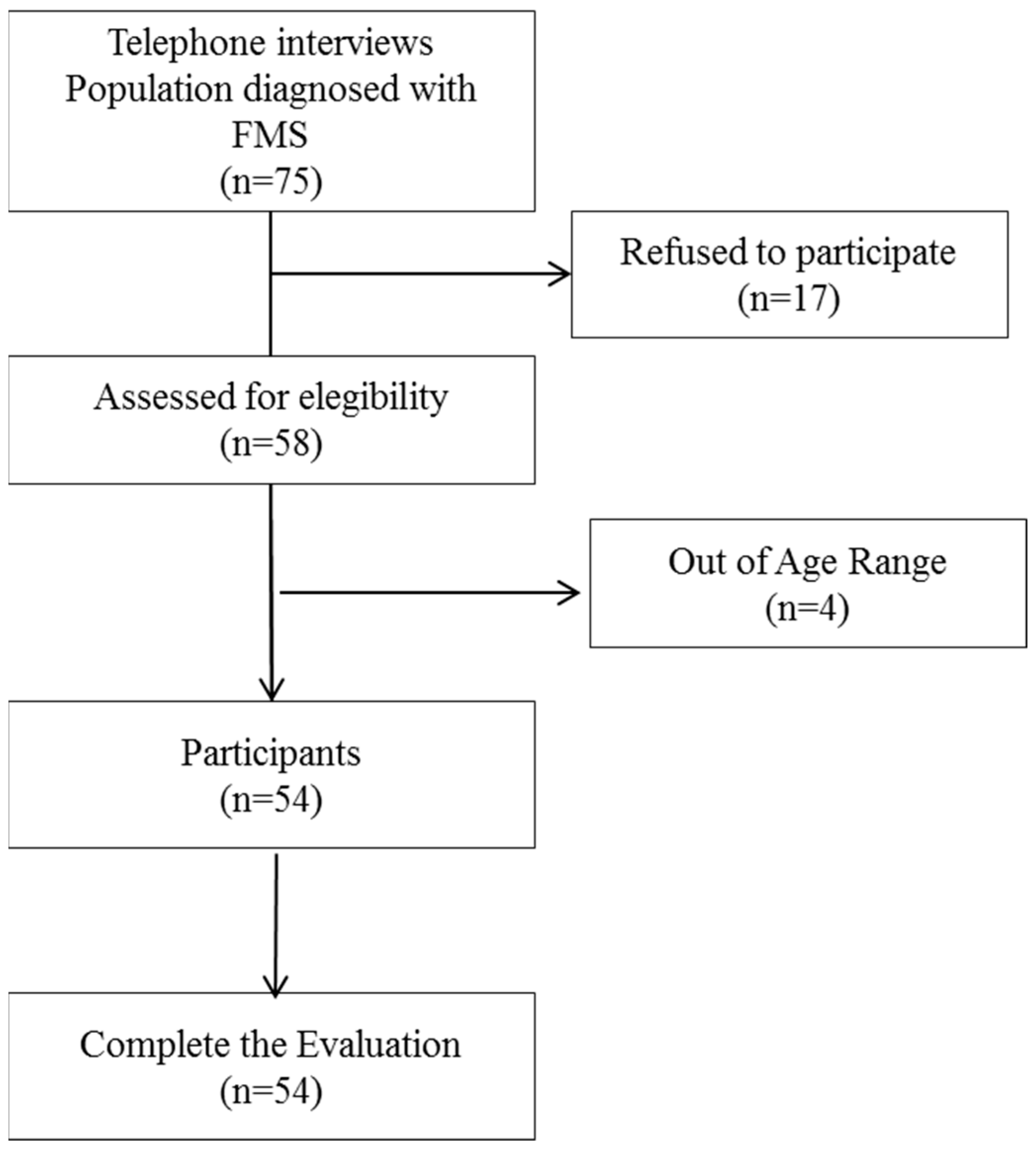
2.2. Participants
2.3. Sample Size Calculation
2.4. Measurements
2.4.1. Primary Outcome Measures
2.4.2. Patient-Reported Outcome Measures (PROMs)
2.4.3. Static and Dynamic Balance Measures
2.5. Data Analysis
3. Results
4. Discussion
5. Conclusions
Author Contributions
Funding
Institutional Review Board Statement
Informed Consent Statement
Data Availability Statement
Acknowledgments
Conflicts of Interest
References
- Wolfe, F.; Walitt, B. Culture, science and the changing nature of fibromyalgia. Nat. Rev. Rheumatol. 2013, 9, 751–755. [Google Scholar] [CrossRef] [PubMed]
- Sarzi-Puttini, P.; Giorgi, V.; Marotto, D.; Atzeni, F. Fibromyalgia: An update on clinical characteristics, etiopathogenesis and treatment. Nat. Rev. Rheumatol. 2020, 16, 645–660. [Google Scholar] [CrossRef] [PubMed]
- Siracusa, R.; Paola, R.D.; Cuzzocrea, S.; Impellizzeri, D. Fibromyalgia: Pathogenesis, Mechanisms, Diagnosis and Treatment Options Update. Int. J. Mol. Sci. 2021, 22, 3891. [Google Scholar] [CrossRef] [PubMed]
- Costa, I.D.S.; Gamundí, A.; Miranda, J.G.V.; França, L.G.S.; De Santana, C.N.; Montoya, P. Altered Functional Performance in Patients with Fibromyalgia. Front. Hum. Neurosci. 2017, 11, 14. [Google Scholar] [CrossRef] [PubMed]
- Cabo-Meseguer, A.; Cerdá-Olmedo, G.; Trillo-Mata, J.L. Fibromyalgia: Prevalence, epidemiologic profiles and economic costs. Med. Clin. 2017, 149, 441–448. [Google Scholar] [CrossRef] [PubMed]
- Heidari, F.; Afshari, M.; Moosazadeh, M. Prevalence of fibromyalgia in general population and patients, a systematic review and meta-analysis. Rheumatol. Int. 2017, 37, 1527–1539. [Google Scholar] [CrossRef] [PubMed]
- Gyorfi, M.; Rupp, A.; Abd-Elsayed, A. Fibromyalgia Pathophysiology. Biomedicines 2022, 10, 3070. [Google Scholar] [CrossRef]
- Boer, C.D.; Dries, L.; Terluin, B.; Van Der Wouden, J.C.; Blankenstein, A.H.; Van Wilgen, C.P.; Lucassen, P.J.; Van Der Horst, H.E. Central Sensitization in Chronic Pain and Medically Unexplained Symptom Research: A Systematic Review of Definitions, Operationalizations and Measurement Instruments. J. Psychosom. Res. 2019, 117, 32–40. [Google Scholar] [CrossRef]
- Hashimoto, K.; Takeuchi, T.; Ueno, T.; Suka, S.; Hiiragi, M.; Yamada, M.; Koyama, A.; Nakamura, Y.; Miyakoda, J.; Hashizume, M. Effect of Central Sensitization on Dizziness-Related Symptoms of Persistent Postural-Perceptual Dizziness. Biopsychosoc. Med. 2022, 16, 7. [Google Scholar] [CrossRef]
- Mucci, V.; Demori, I.; Rapallo, F.; Molinari, E.; Losacco, S.; Marinelli, L.; Browne, C.J.; Burlando, B. Vestibular Disability/Handicap in Fibromyalgia: A Questionnaire Study. J. Clin. Med. 2022, 11, 4017. [Google Scholar] [CrossRef]
- Klein, A.; Schankin, C.J. Visual snow syndrome, the spectrum of perceptual disorders, and migraine as a common risk factor: A narrative review. Headache 2021, 61, 1306–1313. [Google Scholar] [CrossRef] [PubMed]
- Funabashi, M.; Santos-Pontelli, T.; Colafemina, J.; Pavan, T.; Carneiro, A.; Takayanagui, O. A new method to analyze the subjective visual vertical in patients with bilateral vestibular dysfunction. Clinics 2012, 67, 1127–1131. [Google Scholar] [CrossRef]
- De Winkel, K.N.; Edel, E.; Happee, R.; Bülthoff, H.H. Multisensory Interactions in Head and Body Centered Perception of Verticality. Front. Neurosci. 2021, 14, 599226. [Google Scholar] [CrossRef] [PubMed]
- Piscicelli, C.; Nadeau, S.; Barra, J.; Pérennou, D. Assessing the Visual Vertical: How Many Trials Are Required? BMC Neurol. 2015, 15, 215. [Google Scholar] [CrossRef] [PubMed]
- Isableu, B.; Gueguen, M.; Fourré, B.; Giraudet, G.; Amorim, M.-A. Assessment of visual field dependence: Comparison between the mechanical 3D rod-and-frame test developed by Oltman in 1968 with a 2D computer-based version. J. Vestib. Res. 2009, 18, 239–247. [Google Scholar] [CrossRef]
- Lomas-Vega, R.; Rodríguez-Almagro, D.; Peinado-Rubia, A.B.; Zagalaz-Anula, N.; Molina, F.; Obrero-Gaitán, E.; Ibáñez-Vera, A.J.; Osuna-Pérez, M.C. Joint Assessment of Equilibrium and Neuromotor Function: A Validation Study in Patients with Fibromyalgia. Diagnostics 2020, 10, 1057. [Google Scholar] [CrossRef]
- Manera, V.; Rovini, E.; Wais, P.E. Editorial: Early Detection of Neurodegenerative Disorders Using Behavioral Markers and New Technologies: New Methods and Perspectives. Front. Aging Neurosci. 2023, 15, 1149886. [Google Scholar] [CrossRef]
- Daikhes, N.A. The Interdisciplinary Approach and New Technologies in the Scientific and Clinical Development of Otorhinolaryngology. Her. Russ. Acad. Sci. 2021, 91, 438–444. [Google Scholar] [CrossRef]
- Realdon, O.; Adorni, R.; Ginelli, D.; Micucci, D.; Blasi, V.; Bellavia, D.; Schettini, F.; Carradore, R.; Polsinelli, P.; D’Addario, M.; et al. Embedding the Patient-Citizen Perspective into an Operational Framework for the Development and the Introduction of New Technologies in Rehabilitation Care: The Smart&Touch-ID Model. Healthcare 2023, 11, 1604. [Google Scholar] [CrossRef]
- Matamala-Gomez, M.; Stasolla, F.; Seinfeld, S.; Caffò, A.O.; Banakou, D.; Bottiroli, S. Editorial: Neuropsychological and Cognitive-Behavioral Assessment of Neurodegenerative Disease and Rehabilitation Using New Technologies and Virtual Reality. Front. Psychol. 2021, 12, 691909. [Google Scholar] [CrossRef]
- EvaluateMedTech. World Preview 2018, Outlook to 2024. Available online: https://info.evaluategroup.com› (accessed on 16 August 2023).
- Zaleski-King, A.; Pinto, R.; Lee, G.; Brungart, D.S. Use of Commercial Virtual Reality Technology to Assess Verticality Perception in Static and Dynamic Visual Backgrounds. Ear Hear. 2019, 41, 125–135. [Google Scholar] [CrossRef] [PubMed]
- Negrillo-Cárdenas, J.; Rueda-Ruiz, A.J.; Ogayar-Anguita, C.J.; Lomas-Vega, R.; Segura-Sánchez, R.J. A System for the Measurement of the Subjective Visual Vertical using a Virtual Reality Device. J. Med. Syst. 2018, 42, 124. [Google Scholar] [CrossRef] [PubMed]
- Rodríguez-Almagro, D.; Obrero-Gaitán, E.; Lomas-Vega, R.; Zagalaz-Anula, N.; Osuna-Pérez, M.C.; Achalandabaso-Ochoa, A. New Mobile Device to Measure Verticality Perception: Results in Young Subjects with Headaches. Diagnostics 2020, 10, 796. [Google Scholar] [CrossRef] [PubMed]
- Piscicelli, C.; Pérennou, D. Visual verticality perception after stroke: A systematic review of methodological approaches and suggestions for standardization. Ann. Phys. Rehabil. Med. 2017, 60, 208–216. [Google Scholar] [CrossRef] [PubMed]
- Bagust, J.; Docherty, S.; Haynes, W.; Telford, R.; Isableu, B. Changes in Rod and Frame Test Scores Recorded in Schoolchildren during Development–A Longitudinal Study. PLoS ONE 2013, 8, e65321. [Google Scholar] [CrossRef] [PubMed]
- Meireles, S.A.; Antero, D.C.; Kulczycki, M.M.; Skare, T.L. Prevalence of Falls in Fibromyalgia Patients. Acta Ortop. Bras. 2014, 22, 163–166. [Google Scholar] [CrossRef] [PubMed]
- Rivera, J.; Gonzalez, T. The Fibromyalgia Impact Questionnaire: A Validated Spanish Version to Assess the Health Status in Women with Fibromyalgia. Clin. Exp. Rheumatol. 2004, 22, 554–560. [Google Scholar]
- Ferreira-Valente, M.A.; Pais-Ribeiro, J.L.; Jensen, M.P. Validity of Four Pain Intensity Rating Scales. Pain 2011, 152, 2399–2404. [Google Scholar] [CrossRef]
- Cuesta-Vargas, A.I.; Neblett, R.; Gatchel, R.J.; Roldán-Jiménez, C. Cross-Cultural Adaptation and Validity of the Spanish Fear-Avoidance Components Scale and Clinical Implications in Primary Care. BMC Fam. Pract. 2020, 21, 44. [Google Scholar] [CrossRef]
- García Campayo, J.; Rodero, B.; Alda, M.; Sobradiel, N.; Montero, J.; Moreno, S. Validación de La Versión Española de La Escala de La Catastrofización Ante El Dolor (Pain Catastrophizing Scale) En La Fibromialgia. Med. Clín. 2008, 131, 487–492. [Google Scholar] [CrossRef]
- Gómez-Pérez, L.; López-Martínez, A.E.; Ruiz-Párraga, G.T. Psychometric Properties of the Spanish Version of the Tampa Scale for Kinesiophobia (TSK). J. Pain. 2011, 12, 425–435. [Google Scholar] [CrossRef] [PubMed]
- Pérez, N.; Garmendia, I.; Martín, E.L.; García-Tapia, R. Cultural Adaptation of 2 Questionnaires for Health Measurement in Patients with Vertigo. Acta Otorrinolaringol. Esp. 2000, 51, 572–580. [Google Scholar] [PubMed]
- Montilla-Ibáñez, A.; Martínez-Amat, A.; Lomas-Vega, R.; Cruz-Díaz, D.; la Torre-Cruz, M.J.D.; Casuso-Pérez, R.; Hita-Contreras, F. The Activities-Specific Balance Confidence Scale: Reliability and Validity in Spanish Patients with Vestibular Disorders. Disabil. Rehabil. 2016, 39, 697–703. [Google Scholar] [CrossRef] [PubMed]
- Lomas-Vega, R.; Hita-Contreras, F.; Mendoza, N.; Martínez-Amat, A. Cross-Cultural Adaptation and Validation of the Falls Efficacy Scale International in Spanish Postmenopausal Women. Menopause 2012, 19, 904–908. [Google Scholar] [CrossRef] [PubMed]
- Téllez, N.; Río, J.; Tintoré, M.; Nos, C.; Galán, I.; Montalban, X. Does the Modified Fatigue Impact Scale Offer a More Comprehensive Assessment of Fatigue in MS? Mult. Scler. 2005, 11, 198–202. [Google Scholar] [CrossRef] [PubMed]
- Romero-Franco, N.; Martínez-López, E.J.; Lomas-Vega, R.; Hita-Contreras, F.; Osuna-Pérez, M.C.; Martínez-Amat, A. Short-Term Effects of Proprioceptive Training with Unstable Platform on Athletes’ Stabilometry. J. Strength. Cond. Res. 2013, 27, 2189–2197. [Google Scholar] [CrossRef] [PubMed]
- Embrechts, E.; Van Der Waal, C.; Anseeuw, D.; Van Buijnderen, J.; Leroij, A.; Lafosse, C.; Nijboer, T.C.W.; Truijen, S.; Saeys, W. Association between Spatial Neglect and Impaired Verticality Perception after Stroke: A Systematic Review. Ann. Phys. Rehabil. Med. 2023, 66, 101700. [Google Scholar] [CrossRef] [PubMed]
- Ferreira, M.M.; De Carvalho Lopes, K.; De Abreu E Silva Grigol, T.A.; Ganança, M.M.; Caovilla, H.H. Subjective Visual Vertical and Vestibular Evoked Myogenic Potential in Meniere’s Disease. Braz. J. Otorhinolaryngol. 2023, 89, 485–493. [Google Scholar] [CrossRef]
- Al-Sharif, D.S.; Roehm, P.; Lindemann, T.L.; Dumenci, L.; Keshner, E.A. Visual-Vestibular Mismatch Correlates with Headache. J. Vestib. Res. 2021, 31, 173–180. [Google Scholar] [CrossRef]
- Lord, S.R.; Webster, I. Visual Field Dependence in Elderly Fallers and Non-Fallers. Int. J. Aging Hum. Dev. 1990, 31, 267–277. [Google Scholar] [CrossRef]
- Hafström, A.; Modig, F.; Karlberg, M.; Fransson, P.-A. Increased Visual Dependence and Otolith Dysfunction with Alcohol Intoxication. Neuroreport 2007, 18, 391–394. [Google Scholar] [CrossRef] [PubMed]
- Willey, C.R.; Liu, Z. Re-Assessing the Role of Culture on the Visual Orientation Perception of the Rod and Frame Test. PLoS ONE 2022, 17, e0276393. [Google Scholar] [CrossRef] [PubMed]
- Abdul-Razzak, R.; Bagust, J. Perceptual Lateralization on the Rod-And-Frame Test in Young and Older Adults. Appl. Neuropsychol. Adult 2022, 1–7. [Google Scholar] [CrossRef] [PubMed]
- De Tommaso, M.; Federici, A.; Serpino, C.; Vecchio, E.; Franco, G.; Sardaro, M.; Delussi, M.; Livrea, P. Clinical Features of Headache Patients with Fibromyalgia Comorbidity. J. Headache Pain. 2011, 12, 629–638. [Google Scholar] [CrossRef] [PubMed]
- Jeong, S.-H.; Oh, S.-Y.; Kim, H.; Koo, J.-W.; Kim, J.S. Vestibular Dysfunction in Migraine: Effects of Associated Vertigo and Motion Sickness. J. Neurol. 2009, 257, 905–912. [Google Scholar] [CrossRef] [PubMed]
- Nair, M.A.; Mulavara, A.P.; Bloomberg, J.J.; Sangi-Haghpeykar, H.; Cohen, H.S. Visual Dependence and Spatial Orientation in Benign Paroxysmal Positional Vertigo. J. Vestib. Res. 2018, 27, 279–286. [Google Scholar] [CrossRef] [PubMed]
- Bauer, M.; Benito-Orejas, J.I.; Ramírez-Salas, J.E. Rehabilitación vestibular en la dependencia visual y somatosensorial. Rev. ORL 2019, 11, 79. [Google Scholar] [CrossRef]
- Toprak Celenay, S.; Mete, O.; Coban, O.; Oskay, D.; Erten, S. Trunk Position Sense, Postural Stability, and Spine Posture in Fibromyalgia. Rheumatol. Int. 2019, 39, 2087–2094. [Google Scholar] [CrossRef]
- Reddy, R.S.; Meziat-Filho, N.; De Sá Ferreira, A.; Tedla, J.S.; Kandakurti, P.K.; Kakaraparthi, V.N. Comparison of Neck Extensor Muscle Endurance and Cervical Proprioception between Asymptomatic Individuals and Patients with Chronic Neck Pain. J. Bodyw. Mov. Ther. 2021, 26, 180–186. [Google Scholar] [CrossRef]
- Yu, Y.; Lauer, R.T.; Tucker, C.A.; Thompson, E.D.; Keshner, E.A. Visual Dependence Affects Postural Sway Responses to Continuous Visual Field Motion in Individuals with Cerebral Palsy. Dev. Neurorehabil 2018, 21, 531–541. [Google Scholar] [CrossRef]
- Gosselin, G.; Fagan, M.J. Effects of Cervical Muscle Fatigue on the Perception of the Subjective Vertical and Horizontal. SpringerPlus 2014, 3, 78. [Google Scholar] [CrossRef]


| Frequency | % | Mean | SD | ||
|---|---|---|---|---|---|
| Gender | Female | 51 | 94.40 | ||
| Male | 3 | 5.60 | |||
| Civil Status | Single | 2 | 3.70 | ||
| Married | 39 | 72.20 | |||
| Divorced | 10 | 18.50 | |||
| Widower | 3 | 5.60 | |||
| Education level | No Education | 2 | 3.70 | ||
| Primary | 17 | 31.50 | |||
| Secondary | 24 | 44.40 | |||
| University | 11 | 20.40 | |||
| Employment status | Active | 18 | 33.30 | ||
| Sick Leave | 10 | 18.50 | |||
| Unemployed | 7 | 13.00 | |||
| Housewife | 4 | 7.40 | |||
| Retired | 15 | 27.80 | |||
| Fibromyalgia Impact Questionnaire FIQ (0–100) | Mild (<50) | 7 | 13 | ||
| Moderate (50–75) | 29 | 53.7 | |||
| Severe (>75) | 18 | 33.3 | |||
| SVV category | Healthy | 35 | 64.8 | ||
| Altered SVV (>2.5 degrees) | 19 | 35.2 | |||
| RFT category | Healthy | 22 | 40.7 | ||
| Altered RFT (>4.5 degrees) | 32 | 59.3 | |||
| Age (years) | 53.00 | 6.00 | |||
| Weight (kg) | 77.00 | 16.00 | |||
| Height (cm) | 161.00 | 6.00 | |||
| Body Mass Index (kg/m2) | 30.00 | 7.00 |
| Patient-Reported Outcome Measures | MAE in SVV | MAE in RFT | ||||
|---|---|---|---|---|---|---|
| Mean | SD | Correlation (Rho) | p-Value | Correlation (Rho) | p-Value | |
| Falls in last 3 months | 1.31 | 1.46 | 0.168 | 0.226 | 0.033 | 0.813 |
| Fibromyalgia Impact Questionnaire FIQ (0–100) | 67.94 | 14.02 | 0.399 | 0.003 ** | 0.147 | 0.289 |
| Numeric Pain Rating Scale (0–10) | 6.20 | 1.96 | 0.291 | 0.033 * | 0.235 | 0.087 |
| Central Sensitization Inventory CSI (0–100) | 64.96 | 11.3 | 0.115 | 0.408 | 0.091 | 0.514 |
| Pain Catastrophizing Scale PCS (0–52) | 25.8 | 13.43 | 0.417 | 0.002 ** | 0.147 | 0.290 |
| Pain Catastrophizing Scale Rumination (0–16) | 8.61 | 4.61 | 0.347 | 0.010 * | 0.176 | 0.202 |
| Pain Catastrophizing Scale Magnification (0–12) | 5.17 | 3.52 | 0.346 | 0.010 * | 0.032 | 0.820 |
| Pain Catastrophizing Scale Hopelessness (0–24) | 12.02 | 6.14 | 0.440 | 0.001 ** | 0.136 | 0.326 |
| Tampa Scale of Kinesiophobia TSK (11–44) | 26.78 | 6.77 | 0.113 | 0.417 | 0.148 | 0.285 |
| Dizziness Handicap Inventory Total DHI (0–100) | 57.19 | 20.86 | 0.376 | 0.005 ** | 0.069 | 0.620 |
| Dizziness Handicap Inventory Emotional (0–36) | 16.19 | 9.6 | 0.319 | 0.019 * | 0.075 | 0.588 |
| Dizziness Handicap Inventory Functional (0–36) | 20.67 | 8.17 | 0.412 | 0.002 ** | 0.051 | 0.715 |
| Dizziness Handicap Inventory Physical (0–28) | 20.33 | 5.23 | 0.291 | 0.033 * | 0.076 | 0.583 |
| Activities-Specific Balance Confidence Scale ABC (0–100) | 48.33 | 20.83 | −0.367 | 0.006 ** | −0.168 | 0.226 |
| Falls Efficacy Scale International FES-I (16–64) | 36.65 | 10.22 | 0.246 | 0.073 | 0.162 | 0.243 |
| Fatigue Severity Scale FSS (9–63) | 52.37 | 8.2 | 0.280 | 0.040 * | 0.150 | 0.278 |
| MAE in SVV Test | MAE in RFT | |||||
|---|---|---|---|---|---|---|
| Static and Dynamic Balance Measures | Mean | SD | Correlation (Rho) | p-Value | Correlation (Rho) | p-Value |
| Sway Area Eyes Open (mm2) | 524.08 | 1096.97 | −0.007 | 0.960 | 0.042 | 0.764 |
| Mean Velocity Eyes Open (mm/s) | 20.52 | 5.66 | 0.062 | 0.656 | 0.080 | 0.563 |
| Medial–Lateral Root Mean Squared Eyes Open (mm) | 0.39 | 0.11 | 0.023 | 0.868 | 0.088 | 0.528 |
| Posterior–Anterior Root Mean Squared Eyes Open (mm) | 0.43 | 0.14 | 0.033 | 0.811 | 0.126 | 0.366 |
| Mean Medial–Lateral Oscillation Eyes Open (mm) | −1.44 | 6.34 | −0.173 | 0.210 | −0.046 | 0.739 |
| Mean Posterior–Anterior Oscillation Eyes Open (mm) | −19.47 | 11.92 | −0.057 | 0.684 | −0.283 | 0.038 * |
| Sway Area Eyes Closed (mm2) | 623.72 | 839.33 | 0.064 | 0.654 | 0.064 | 0.651 |
| Mean Velocity Eyes Closed (mm/s) | 23.31 | 6.41 | 0.115 | 0.418 | 0.077 | 0.587 |
| Medial-Lateral Root Mean Squared Eyes Closed (mm) | 0.45 | 0.16 | 0.065 | 0.649 | 0.095 | 0.504 |
| Postero-anterior Root Mean Squared Eyes Closed (mm) | 0.54 | 0.17 | 0.162 | 0.253 | 0.022 | 0.874 |
| Mean Medial–Lateral Oscillation Eyes Closed (mm) | −1.04 | 6.42 | −0.116 | 0.412 | −0.132 | 0.349 |
| Mean Posterior–Anterior Oscillation Eyes Closed (mm) | −17.86 | 13.68 | −0.158 | 0.264 | −0.207 | 0.142 |
| JAEN Total Score (0–80) | 36.85 | 10.58 | 0.224 | 0.103 | 0.268 | 0.050 |
| JAEN Instability Head Movements (0–24) | 7.3 | 4.72 | 0.133 | 0.339 | −0.044 | 0.751 |
| JAEN Instability Support Reduced (0–32) | 21.54 | 5.5 | 0.251 | 0.067 | 0.327 | 0.016 * |
| JAEN Instability Gait Eyes Open (0–12) | 4.24 | 2.27 | 0.210 | 0.127 | 0.312 | 0.022 * |
| JAEN Instability Standing and Walking Eyes Closed (0–12) | 3.78 | 1.9 | 0.050 | 0.721 | 0.184 | 0.184 |
Disclaimer/Publisher’s Note: The statements, opinions and data contained in all publications are solely those of the individual author(s) and contributor(s) and not of MDPI and/or the editor(s). MDPI and/or the editor(s) disclaim responsibility for any injury to people or property resulting from any ideas, methods, instructions or products referred to in the content. |
© 2023 by the authors. Licensee MDPI, Basel, Switzerland. This article is an open access article distributed under the terms and conditions of the Creative Commons Attribution (CC BY) license (https://creativecommons.org/licenses/by/4.0/).
Share and Cite
Lomas-Vega, R.; Ogáyar-Anguita, C.J.; Segura-Sánchez, R.J.; Rueda-Ruiz, A.J.; Osuna-Pérez, M.C.; Peinado-Rubia, A.B. Misperception of Visual Verticality Measured with a Virtual Reality Device in Patients with Fibromyalgia Syndrome: A Cross-Sectional Study. Appl. Sci. 2023, 13, 10579. https://doi.org/10.3390/app131910579
Lomas-Vega R, Ogáyar-Anguita CJ, Segura-Sánchez RJ, Rueda-Ruiz AJ, Osuna-Pérez MC, Peinado-Rubia AB. Misperception of Visual Verticality Measured with a Virtual Reality Device in Patients with Fibromyalgia Syndrome: A Cross-Sectional Study. Applied Sciences. 2023; 13(19):10579. https://doi.org/10.3390/app131910579
Chicago/Turabian StyleLomas-Vega, Rafael, Carlos Javier Ogáyar-Anguita, Rafael J. Segura-Sánchez, Antonio Jesús Rueda-Ruiz, María Catalina Osuna-Pérez, and Ana Belén Peinado-Rubia. 2023. "Misperception of Visual Verticality Measured with a Virtual Reality Device in Patients with Fibromyalgia Syndrome: A Cross-Sectional Study" Applied Sciences 13, no. 19: 10579. https://doi.org/10.3390/app131910579
APA StyleLomas-Vega, R., Ogáyar-Anguita, C. J., Segura-Sánchez, R. J., Rueda-Ruiz, A. J., Osuna-Pérez, M. C., & Peinado-Rubia, A. B. (2023). Misperception of Visual Verticality Measured with a Virtual Reality Device in Patients with Fibromyalgia Syndrome: A Cross-Sectional Study. Applied Sciences, 13(19), 10579. https://doi.org/10.3390/app131910579