Article

4-Hexylresorcinol and Its Effects on Circumvallate Papillae Taste Buds in Diabetic and Healthy Rats: An Initial Investigation

Department of Oral and Maxillofacial Surgery, College of Dentistry, Gangneung-Wonju National University, Jukheon gil 7, Gangneung 25457, Republic of Korea
*
Author to whom correspondence should be addressed.
Appl. Sci. 2023, 13(21), 11617; https://doi.org/10.3390/app132111617
Submission received: 12 October 2023 / Revised: 20 October 2023 / Accepted: 23 October 2023 / Published: 24 October 2023
(This article belongs to the Section Applied Dentistry and Oral Sciences)

Abstract

:
Taste perception plays a crucial role in health and well-being but can be compromised by conditions such as diabetes mellitus (DM). This study delved into the efficacy of 4-hexylresorcinol (4-HR) in mitigating taste bud apoptosis, particularly in relation to DM-induced taste issues. Two primary rat groups were investigated: healthy rats and streptozotocin (STZ)-induced diabetic rats. Each group was further divided into control and experimental subsets, with the experimental group receiving 4-HR injections. A histological analysis of the circumvallate papillae (CVP) highlighted significant taste bud deterioration in the STZ subgroup, including loss of cellular content and a deviation from their typical morphology. Quantitatively, the control group had a mean of 39.6 ± 14.3 taste bud pores/mm2, contrasting with the 4HR, STZ, and STZ/4HR groups, which had means of 33.7 ± 14.2, 20.3 ± 6.1, and 28.0 ± 8.3, respectively. Additionally, a TUNEL assay and IHC staining for c-caspase-3 both identified increased apoptotic cells in the STZ subgroup, with the mean number of apoptotic cells per taste bud profile being notably higher in the STZ group at 3.2 ± 1.6. This study underscores the profound impact of diabetes on taste bud physiology and the potential therapeutic benefits of 4-HR. Further research is essential to delve deeper into its mechanisms and to ascertain optimal dosages, with the aim of enhancing the quality of life of diabetic patients.

1. Introduction

Taste perception, an essential sensory function, significantly influences systemic health, nutritional status, and overall quality of life. It is known that factors such as aging, smoking, and certain medications can impair this sense [1]. Additionally, a variety of diseases can lead to gustatory disorders, detrimentally affecting patients’ well-being. Among these conditions is diabetes mellitus (DM), a metabolic disorder that can bring about sensory deficits, including in taste perception [2,3].
Recent research has highlighted the increasing association of DM with taste impairments [4]. Specifically, DM has been shown to negatively influence the perception of sweet, salty, and bitter tastes [5,6]. Electrogustometric thresholds, for instance, tend to increase notably in patients with diabetes [3,7]. Yet, despite the evident link between DM and taste disorders, the fundamental mechanisms at play are not entirely clear.
A deeper exploration into the histological and histopathological aspects of taste buds can provide invaluable insights into the physiopathology of taste impairment in DM. Histologically, taste buds are complex structures housing specialized taste receptor cells (TRCs) that detect different taste qualities [8]. These TRCs are nestled within the epithelial cells in a well-organized manner, forming onion-like structures with a central taste pore opening to the oral cavity [9]. Each taste bud comprises approximately 50–100 TRCs [8] that are continuously renewed, with a lifespan of about 10–20 days [9]. The turnover of TRCs is a tightly regulated process, ensuring the integrity and functionality of taste buds [10].
Histopathologically, diabetes has been observed to induce changes in the structural and cellular integrity of taste buds [11]. Elevated glucose levels, characteristic of DM, can lead to oxidative stress and inflammatory responses within the taste buds, adversely affecting their morphology and function [11]. Such alterations include a reduction in the number of TRCs, changes in their ultrastructural appearance, and disruptions in the signaling pathways crucial for taste transduction [12]. Moreover, the supportive cells surrounding TRCs may also be compromised, further impairing taste perception. The resultant taste disorders could potentially exacerbate the dietary challenges faced by individuals with DM, underscoring the necessity of understanding and addressing the histopathological alterations in taste buds that are induced by DM [6,13].
A closer look at the anatomy and physiology of taste perception reveals the significance of circumvallate papillae (CVP). Rats, interestingly, have only one CVP, positioned at the posterior midline of their tongues. This papilla is surrounded by a furrow, or trench, that contains a considerable number of taste buds [14]. A key process underpinning taste perception is apoptosis, the programmed death of cells. In taste buds, apoptosis is crucial for controlling the balance between the birth and death of taste cells, which inherently have a short lifespan. This balance is vital for the preservation of functional taste buds. If this equilibrium is disrupted, it could potentially result in taste disorders [15]. Although research has delved into apoptosis’s role in taste biology, especially concerning age-related taste deterioration [1], the connection between apoptosis and taste dysfunctions in DM has not been extensively explored. Some initial research on diabetic rat models points to changes in the structure of vallate taste buds but stops short of probing into apoptosis [11,16].
Enter 4-hexylresorcinol (4-HR), a plant-derived compound found in the extract of Quercus cortex, known for its diverse biological activities [17]. Current studies suggest that 4-HR can modulate cellular stress responses [18], boasting antioxidant properties [19,20]. High glucose levels, a characteristic of DM, can induce oxidative stress, which may, in turn, initiate apoptosis in various tissues [21,22]. 4-HR emerges as a potential solution to counteract the adverse effects of high glucose on taste cells. A recent study on the long-term effects of 4-HR on facial skeletal growth in growing male rats, especially in diabetic models, revealed significant improvements in bone metrics after 4-HR administration, suggesting that the compound has beneficial effects, potentially via mitochondrial respiration modulation [23]. This finding hints at the broader cellular benefits of 4-HR, which may extend to modulating oxidative stress and apoptosis, pivotal factors affecting taste bud health in DM. Considering apoptosis’s role in taste bud health and DM’s influence on taste, 4-HR may be pivotal in thwarting taste bud apoptosis due to heightened serum glucose levels.
The existing literature provides a wealth of information regarding the effects of DM on taste perception [11,16] and the medicinal attributes of 4-HR [19,20], largely derived from studies conducted on animal models. While these studies offer invaluable insights, they also highlight a notable limitation—the potential disparity between the responses observed in animal models and humans. Despite this limitation, the current body of research serves as a foundation upon which further inquiries can be built. There is a conspicuous absence of research investigating the combined effects of DM and 4-HR on taste bud apoptosis, emphasizing the imperative for focused research in this area. In line with this, our study, albeit conducted on an animal model, seeks to delve into this unexplored domain. The primary objective is to ascertain the impact of 4-HR administration in preventing taste bud apoptosis triggered by diabetes, thus paving the way for subsequent human-centric studies that may provide a more direct translational value to the findings. By exploring this avenue, this current study aims to contribute a nuanced understanding of 4-HR’s potential in mitigating taste impairments in DM, creating a foundation for future research that could further elucidate this interaction in human subjects.

2. Materials and Methods

2.1. Study Design

Sprague Dawley outbred rats were purchased from Samtako (Osan, Republic of Korea) for this experiment. A total of 40, 7-weeks-old male rats, were utilized. This study involved two primary groups of rats: those that were healthy, and those induced with diabetes using streptozotocin (STZ). When the rats received the STZ injection, their body weight ranged from 270 to 290 g. Each primary group was further stratified into two subgroups, control and experimental, based on the type of injected substance. The procedures abided by the ethical guidelines established by the Institutional Animal Care and Use Committee of Gangneung-Wonju National University, Gangneung, Republic of Korea (GWNU-2021-21 and GWNU-2021-22 and were approved on 23 November 2021).

2.2. Sample Collection and Preparation

The rat tongue features four distinct papillae types: filiform (FI), foliate (FO), fungiform (FU), and CVP (Figure 1a). While the filiform papillae serve a mechanical role, the others have gustatory functions [24]. Samples were obtained from the heads of male Sprague Dawley rats used in previous studies [23].
For this study, the chosen dosage of 4-HR at 10 mg/kg/week was based on previous research. The dosage determination considered safety data, including the LD50 for rats and the observed effects in cats. In addition, prior work demonstrated the efficacy of 4-HR at dosages close to the one chosen for this study, with notable effects such as decreased blood sugar levels [25,26]. For practicality in dosage calculations and to minimize potential errors, a dose of 10 mg/kg weekly was selected. This decision also considered potential complications that might arise from higher dosages and the dose-dependent nature of the drug’s effects. Initially, the rats were categorized into two primary groups: 20 healthy and 18 STZ-induced diabetic rats. These groups were then subdivided into control and experimental subgroups, with the latter receiving a subcutaneous injection of 4-HR at 10 mg/kg/week. After harvesting the tongues from the rat heads, which were preserved in 10% formalin, the posterior tongue tissue containing CVP was incised in a rectangular shape (Figure 1b). The extracted region, located near the tongue base, was embedded in paraffin blocks. Sectioning was performed parallel to the horizontal plane, producing sections with a thickness of 5-µm.

2.3. Histologic Examination

2.3.1. Hematoxylin and Eosin (H&E) Staining

The staining process began with the deparaffinization of the slides in xylene, followed by hydration using a graded ethanol sequence. After rinsing with distilled water, Mayer hematoxylin was applied, and the slides were then washed with tap water. A quick decolorization with acid alcohol was performed, followed by another rinse. After the eosin counterstaining, the slides underwent dehydration with sequential ethanol concentrations and a clearing process using xylene. The number of CVP was then counted to compare between the groups.

2.3.2. Immunohistochemical (IHC) Staining

For the histological examination, a primary antibody targeting cleaved caspase 3 (c-casp3) from Santa Cruz Biotechnology was utilized. The activation of caspase 3, which results in its cleaved form, serves as a hallmark of apoptosis, thus rendering the c-casp3 antibody a reliable indicator of apoptotic activity within the examined tissues [27,28]. This methodological choice was aimed at offering a robust assessment of apoptosis, which in turn would elucidate the potential protective effects of 4-HR on taste bud cells amidst the oxidative stress induced by DM. Once the slides were prepped through deparaffinization, hydration, and washing, a peroxidase-blocking solution was applied for antigen retrieval. After a 7 min incubation, the slides were rinsed with 1X PBS. A protein block serum-free solution was applied, and the slides were set aside for an hour. Primary antibodies, diluted in PBS (1:100 ratio), were then added to the slides, which were stored overnight at 4 °C. The next day, the slides underwent a thorough 1X PBS wash before the application of the secondary antibodies. Following 30 min of incubation at room temperature, the slides were washed and then exposed to a 3,3′-diaminobenzidine (DAB) solution for color development. The sections were examined under a microscope (×400 magnification). The assessment focused on both cytoplasmic and nuclear staining in the taste bud cells. The percentage of stained cells (identifiable by a brown hue) relative to the overall taste bud area was compared across the groups.

2.3.3. TUNEL Assay

Post-deparaffinization and hydration, the slides were treated with a trypsin solution for 60 min at 37 °C. This was followed by a rinse and the application of the TUNEL agent. The slides were then incubated in an oven at 37 °C for an hour and subsequently washed. Immediate examination was crucial to prevent fluorescence signal fading.
For quantification, random sections from each subgroup were selected and TUNEL-positive (apoptotic) cells were counted at a ×200 magnification.

2.3.4. Immunoprecipitation High-Performance Liquid Chromatography (IP-HPLC)

Arterial blood plasma was obtained from the heart of each animal in the STZ and STZ/4HR groups. Each 20 μL of plasma was immunoprecipitated using antisera of GAPDH, mitochondrial hexokinase II (HKII), insulin, leptin, SIRT1, SIRT3, SIRT6, SIRT7, leucine-rich repeat-containing G protein-coupled receptor 4 (LGR4), forkhead box protein O1 (FOXO1), FOXO3b, and klotho, followed by an HPLC analysis. Proportional data (%) were plotted on a line graph and a star plot. The expression of housekeeping proteins, i.e., β-actin and α-tubulin, was compared to that of the non-responsive control (≤5%).

2.4. Statistical Methods

Data sourced from the CVP count, TUNEL assay, and c-caspase-3 positive area underwent a one-way analysis of variance (ANOVA). This was complemented by a Bonferroni test, with a significance level established at p < 0.05. All analyses were executed using SPSS 25.0 software (IBM, Armonk, NY, USA).

3. Results

The examination of horizontal sections of CVP stained with H&E revealed notable findings in the taste buds of the STZ subgroup. Specifically, these taste buds showed a relative loss of cellular content, a deviation from their usual morphology (inverted cone), and a scarcity of taste pores, as evident in Figure 2a. In contrast, the taste buds of both subgroups within the healthy group exhibited a dense packing of cells (Figure 2c,d), maintaining their typical inverted cone shape, with a clear delineation of taste pores and an organized arrangement of TRCs, supporting cells, and basal cells. These are considered the normal features of healthy taste buds, and are crucial for proper taste sensation. Furthermore, it is worth mentioning that the taste buds in the STZ/4HR subgroup (Figure 2b) displayed relatively similar characteristics to those observed in the healthy group, suggesting 4-HR has a protective effect on the structural integrity of taste buds amidst DM-induced challenges.
In this study, the researchers investigated the number of taste bud pores and the presence of apoptotic taste bud cells in different experimental groups. The control group exhibited a mean of 39.6 ± 14.3 taste bud pores per square millimeter, which was significantly different from the 4HR, STZ, and STZ/4HR groups, with means of 33.7 ± 14.2, 20.3 ± 6.1, and 28.0 ± 8.3 taste bud pores per square millimeter, respectively (Figure 3). This observation indicates a significant variation among the groups (p = 0.007). Specifically, the difference between the control and STZ groups was found to be statistically significant (p = 0.005) through a post hoc test.
This study also examined the presence of TUNEL+ cells, which are indicative of apoptotic taste bud cells, in the different groups. The diabetic group, particularly the STZ subgroup, exhibited a higher number of TUNEL+ cells, especially in the epithelium surrounding the trench (Figure 4). The quantitative analysis revealed that the mean number of apoptotic taste bud cells per taste bud profile was 1.8 ± 0.9, 1.3 ± 0.9, 3.2 ± 1.6, and 1.1 ± 0.7 in the control, 4HR, STZ, and STZ/4HR subgroups, respectively. The difference among these groups was statistically significant (p = 0.001). Furthermore, the post hoc test revealed that the STZ group exhibited significantly higher levels of TUNEL+ cells compared to the control, 4HR, and STZ/4HR groups (p = 0.049, 0.003, and 0.001, respectively).
Overall, the findings of this study suggest that both the number of taste bud pores and the presence of apoptotic taste bud cells vary significantly across the different experimental groups. These differences were particularly notable in the STZ subgroup of the diabetic group. These results contribute to our understanding of the impact of diabetes on taste bud physiology and emphasize the importance of further investigation into the potential effects of diabetes on taste perception and gustatory function. Additional research on the underlying mechanisms of these observations is warranted to establish a clearer understanding of these phenomena.
The TUNEL assay and the IHC staining for c-caspase-3 had the same purpose, which was the determination and comparison of the frequency of apoptotic cells in taste buds. The horizontal sections of CVP from each group were immune-stained with an antibody against c-casp3, which is accountable for alterations in shape and biochemical composition during apoptosis. The IHC staining results showed relatively more c-casp3-positive taste cells in the STZ subgroup, as well as in the STZ/4HR subgroup (Figure 5). The results of calculating the percentage of taste buds with stained (positive) cells in relation to the total number of taste buds showed that there was a statistical difference among the groups (Figure 6; p < 0.001). In the post hoc test, the STZ subgroup had a significantly higher number of c-casp3-positive taste cells compared to the control, 4HR, and STZ/4HR subgroups (p = 0.010, <0.001, and 0.007, respectively).
The results of the IP-HPLC analysis using blood serum are presented in Figure 7. In terms of 4HR’s effect on DM-induced rats, it up-regulated HKII by 27.8%, sirtuin 1 by 13.4%, and FOXO3 by 15.4%.

4. Discussion

Taste disturbances, a prevalent challenge among individuals with DM, are known to impinge significantly on patients’ quality of life and dietary choices [4,5]. This study set out to understand the potential mitigative role of 4-HR in reducing taste bud apoptosis in diabetic rats, thereby potentially alleviating the taste disturbances commonly associated with DM (Figure 4 and Figure 6). Our findings highlighted marked alterations in the taste buds of diabetic rats (Figure 2). There was a noticeable reduction in the cellular content of taste buds in these rats, echoing outcomes from earlier studies [11,16]. Intriguingly, these alterations were more pronounced in the STZ-induced diabetic rats that did not receive 4-HR treatment compared to those that did, indicating that 4-HR plays a protective role against the taste disturbances seen in diabetes.
Hevér et al.’s research [10] resonates with our findings. They noted a significant decline in specific nerve fibers in the taste buds of diabetic rats, bringing to the forefront the clinical implications of such observations. As neurotrophins, such as brain-derived neurotrophic factor and nerve growth factor, are central to the development and sustenance of taste buds [10,29], DM-induced neuropathy might be a pivotal factor behind the taste disturbances [30]. These findings are consistent with similar research on taste bud degeneration in response to the removal of taste axons [31,32].
The impact of neuropathy-induced taste disturbances extends beyond a mere alteration in taste perception. It holds substantial clinical significance as taste disturbances can adversely affect patients’ quality of life and dietary choices [33]. For individuals with DM, taste disturbances might lead to poor dietary choices, contributing to a vicious cycle of worsening glycemic control and further exacerbating their metabolic disorder [5]. Moreover, altered taste perception can lead to inadequate nutrition, as individuals may avoid certain foods or consume others in excess, potentially resulting in malnutrition or the exacerbation of obesity, which are both common challenges in diabetic care [34]. Furthermore, taste disturbances may deter patients from adhering to dietary recommendations, which is critical for managing DM and preventing its associated complications [13]. Hence, understanding and addressing neuropathy-induced taste disturbances in individuals with DM not only elucidates a lesser-known complication of this metabolic disorder, but also opens avenues for improving dietary management and overall quality of life for these patients.
While the primary focus of this study revolves around assessing the potential of 4-HR in alleviating DM-induced taste disorders, it is pivotal to acknowledge that the findings could contribute to a broader understanding of 4-HR’s therapeutic potential. For instance, various compounds and interventions have been explored for managing taste disorders and improving taste function in individuals with DM and other conditions. A systematic review sheds light on numerous pharmacological, surgical, and physical treatments proposed for taste function recovery [35]. Additionally, bioactive compounds derived from microalgae have shown promise in improving insulin sensitivity, which might indirectly impact the taste disorders associated with DM [36]. Another compound, allicin, has been probed for its potential in alleviating diabetes mellitus by combating oxidative stress and inflammation—key factors in the development and progression of DM and its associated complications, including taste disorders [37].
The selection of 4-HR for this study was motivated by its multifarious beneficial properties, particularly its robust antioxidant activity [19,20], which could potentially counteract the oxidative stress associated with DM-induced taste disorders. Furthermore, a study by Song et al. [38] underscored 4-HR as a novel inhibitor of α-glucosidase and non-enzymatic glycation, indicating its potential as a drug candidate for the prevention and treatment of type 2 diabetes. In sum, the rationale for selecting 4-HR was firmly rooted in its diverse biological activities and potential therapeutic benefits, not only in alleviating the taste impairments associated with DM, but also in the broader context of managing DM and its complications [23]. This underlines the relevance and potential impact of this study in advancing the understanding and management of DM-induced taste disorders.
A significant finding of this study was the enhanced apoptotic activity within the taste buds of diabetic rats, especially those in the STZ subgroup (Figure 4 and Figure 6). This was deduced through the TUNEL assay (Figure 4). Although taste bud cells have a known turnover cycle, the amplified apoptotic activity observed in diabetic rats, particularly those not treated with 4-HR, solidifies the link between DM and taste impairments (Figure 2). The protective capacity of 4-HR was further endorsed by IHC staining, which demonstrated the changes in taste bud composition attributable to diabetes (Figure 6).
The transcription factor FOXO3a, upon interaction with SIRT1, has been demonstrated to induce the expression of antioxidant genes, a crucial interaction that aids in combating oxidative stress, which is often exacerbated in DM due to elevated glucose levels [39]. FOXO3 plays a pivotal role in up-regulating mitochondrial antioxidant enzymes under stress conditions. The up-regulation of these enzymes facilitates reactive oxygen species detoxification, which is indispensable for managing oxidative stress in DM, a condition frequently associated with heightened levels of oxidative stress that contribute to complications [40]. In the realm of taste bud health, the up-regulation of mitochondrial antioxidant enzymes holds significant merit. The oxidative stress mitigation afforded by these enzymes can play a crucial role in preserving the structural and functional integrity of taste buds [41]. Elevated oxidative stress levels, as seen in DM, can potentially lead to cellular damage within the taste buds, disrupting their normal function and possibly leading to taste disorders [42]. The mitochondrial antioxidant enzymes, by counteracting oxidative stress, help maintain a conducive cellular environment within the taste buds, thus preserving taste perception [43]. This is particularly pertinent considering the adverse impact of taste disorders on nutritional intake and the overall quality of life of individuals with DM [13,35]. Our serum findings further enriched our understanding of this mechanism. It was observed that FOXO3a and SIRT1 were markedly expressed in the STZ/4HR group compared to the STZ group (Figure 7), hinting at the potential protective effect of 4-HR in preserving taste bud function amid DM-induced oxidative stress through the up-regulation of mitochondrial antioxidant enzymes. This suggests that the antioxidant activity of 4-HR might be instrumental in curbing apoptotic stress in DM, lending credence to its potential therapeutic role.
Another study focusing on quercetin’s impact on rat papillae structure in STZ-induced diabetes illuminated the therapeutic merits of compounds with antioxidant and anti-inflammatory characteristics in counteracting the adverse effects of DM on taste [44]. Both quercetin and 4-HR possess beneficial properties, which rationalizes the observed protective effects of 4-HR on taste disturbances in diabetic rats [45]. In summary, the most striking observation in this study was the statistically significant increase in apoptotic activity among diabetic rats in the control subgroup (Figure 4). This finding underscores the well-documented association between diabetes and taste disturbances [46]. Moreover, the administration of 4-HR demonstrated the potential to alleviate taste bud apoptosis (Figure 4). This suggests that 4-HR may serve as a protective agent against the harmful effects of diabetes on taste perception. The findings from this study lay a preliminary foundation for exploring therapeutic interventions aimed at ameliorating taste disturbances in individuals with diabetes. By showcasing the potential of 4-HR in alleviating taste bud apoptosis (Figure 4) and possibly restoring taste function, this research highlights the promising avenue of employing antioxidant and anti-inflammatory compounds like 4-HR [19,20] as part of a comprehensive approach to manage DM-induced complications. Such interventions could significantly enhance the quality of life of individuals with diabetes by improving their taste perception, which, in turn, could promote better dietary choices and nutritional intake. The demonstrated potential of 4-HR in mitigating taste disturbances invites further exploration and validation in clinical settings, which could eventually translate into novel therapeutic strategies to address this lesser-known, but impactful, aspect of diabetes management.
This study, while insightful, presents certain limitations that could affect the generalizability and robustness of the conclusions drawn. One notable limitation is the relatively small sample size employed, which may not capture the full diversity and spectrum of responses to DM and 4-HR treatment. Moreover, the use of a rat model, although invaluable, may not fully encapsulate the intricacies and variances inherent in human taste perception, thereby potentially limiting the direct translatability of the findings to human subjects. Furthermore, the distribution of rats across different groups was uneven, and the reutilization of samples from previous research could have introduced inherent biases, impacting the reliability of the results. This study also did not explore the optimal dosage of 4-HR for preventing taste bud apoptosis, which is a crucial aspect for translating these findings into practical therapeutic interventions. In this regard, it is pertinent to address the decision to employ a single-dosage model. The chosen dosage of 4-HR for rat experiments was predicated on our previous research [25,26], and the selected dose aimed to elicit potential therapeutic effects without inducing significant morbidity or mortality. While this study serves as a preliminary investigation, it is acknowledged that employing a single-dosage model limits the understanding of the dose–response relationship and the potential efficacy of varied dosages of 4-HR. The rationale was to minimize the use of animals in the absence of prior evidence regarding 4-HR’s effect on taste bud health, adhering to ethical considerations. Future studies, predicated on the foundational findings of this research, may necessitate exploring multiple dosage models to ascertain the optimal therapeutic dose of 4-HR. For a more robust and generalizable understanding, future studies should consider employing larger and more diverse sample sizes, possibly extending to different animal models or even preliminary human trials. Also, exploring the optimal dosage of 4-HR and its potential side effects will be essential to ascertain the practicality and efficacy of 4-HR as a therapeutic agent for DM-induced taste disorders. Comprehensive histological evaluations and other relevant metrics should be incorporated to provide a more nuanced understanding of the interplay between DM, 4-HR, and taste bud health. These considerations are essential for refining the experimental design in future endeavors, which, in turn, would contribute to a more precise and reliable elucidation of the therapeutic potential of 4-HR in alleviating taste disturbances associated with DM.

5. Conclusions

In conclusion, these results underscore the heightened apoptotic activity in diabetic rats, especially those not treated with 4-HR. This highlights the existing correlation between diabetes and taste disturbances, and puts forth 4-HR as a potential therapeutic agent against diabetes-induced taste impairments. A deeper grasp of the underlying molecular mechanisms can provide critical insights, possibly paving the way for innovative therapeutic interventions to improve the quality of life of individuals with DM. Given the implications of taste disturbances on dietary decisions and diabetes management, delving deeper into this domain is of the utmost importance, especially given the promising attributes of 4-HR.
However, this research presents certain limitations. Employing a rat model may not fully encapsulate the complexities inherent to human taste perception. Additionally, potential biases could arise from the uneven distribution of rats across the groups and the reuse of samples from previous research. For a more comprehensive understanding, future studies should consider broader experimental approaches, including metrics such as tongue weight measurements and a detailed analysis of the frontal sections of the CVP. Addressing these limitations will provide a more holistic perspective, further enhancing the potential therapeutic application of 4-HR in managing taste disturbances.

Author Contributions

Conceptualization, D.G. and S.-G.K.; methodology, S.-G.K.; software, D.G.; validation, D.G. and S.-G.K.; formal analysis, D.G.; investigation D.G. and S.-G.K.; resources, S.-G.K.; data curation, D.G.; writing—original draft preparation, D.G.; writing—review and editing, S.-G.K.; visualization S.-G.K.; supervision, Y.-W.P.; project administration, S.-G.K.; funding acquisition, S.-G.K. All authors have read and agreed to the published version of the manuscript.

Funding

This research received no external funding.

Institutional Review Board Statement

The animal study protocol was approved by the Institutional Animal Care and Use Committee of Gangneung-Wonju National University, Gangneung, Republic of Korea (GWNU-2021-2-1).

Informed Consent Statement

Not applicable.

Data Availability Statement

Data sharing not applicable.

Acknowledgments

IP-HPLC was performed with the help of Suk Keun Lee.

Conflicts of Interest

The authors declare no conflict of interest.

References

  1. Risso, D.; Drayna, D.; Morini, G. Alteration, reduction and taste loss: Main causes and potential implications on dietary habits. Nutrients 2020, 12, 3284. [Google Scholar] [CrossRef] [PubMed]
  2. Maheswaran, T.; Abikshyeet, P.; Sitra, G.; Gokulanathan, S.; Vaithiyanadane, V.; Jeelani, S. Gustatory dysfunction. J. Pharm. Bioallied Sci. 2014, 6, S30. [Google Scholar] [CrossRef] [PubMed]
  3. Bhat, A.R.; Meghana, I.S.S.; Thomas, B.; Shenoy, N.; Bhandary, R. Diabetes mellitus and potential oral complications—A review. Rom. J. Diabetes Nutr. Metab. Dis. 2021, 28, 453–462. [Google Scholar]
  4. Catamo, E.; Tornese, G.; Concas, M.P.; Gasparini, P.; Robino, A. Differences in taste and smell perception between type 2 diabetes mellitus patients and healthy controls. Nutr. Metab. Cardiovasc. Dis. 2021, 31, 193–200. [Google Scholar] [CrossRef]
  5. Pugnaloni, S.; Alia, S.; Mancini, M.; Santoro, V.; Di Paolo, A.; Rabini, R.A.; Fiorini, R.; Sabbatinelli, J.; Fabri, M.; Mazzanti, L.; et al. A study on the relationship between type 2 diabetes and taste function in patients with good glycemic control. Nutrients 2020, 12, 1112. [Google Scholar] [CrossRef] [PubMed]
  6. Tan, S.Y.; Hack, C.; Yu, C.; Rennick, I.; Ohanian, J.; Dezan, M.; Mott, N.; Manibo, R.; Tucker, R.M. Alterations in sweet taste function in adults with diabetes mellitus: A systematic review and potential implications. Crit. Rev. Food Sci. Nutr. 2023, 63, 2613–2625. [Google Scholar] [CrossRef] [PubMed]
  7. Pavlidis, P.; Gouveris, H.; Kekes, G.; Maurer, J. Electrogustometry thresholds, tongue tip vascularization, and density and morphology of the fungiform papillae in diabetes. B-ENT 2014, 10, 271–278. [Google Scholar]
  8. Liang, Z.; Wilson, C.E.; Teng, B.; Kinnamon, S.C.; Liman, E.R. The proton channel OTOP1 is a sensor for the taste of ammonium chloride. Nat. Commun. 2023, 14, 6194. [Google Scholar] [CrossRef] [PubMed]
  9. Lee, H.; Macpherson, L.J.; Parada, C.A.; Zuker, C.S.; Ryba, N.J.P. Rewiring the taste system. Nature 2017, 548, 330–333. [Google Scholar] [CrossRef] [PubMed]
  10. Golden, E.J.; Larson, E.D.; Shechtman, L.A.; Trahan, G.D.; Gaillard, D.; Fellin, T.J.; Scott, J.K.; Jones, K.L.; Barlow, L.A. Onset of taste bud cell renewal starts at birth and coincides with a shift in SHH function. eLife 2021, 10, e64013. [Google Scholar] [CrossRef]
  11. Cheng, B.; Pan, S.; Liu, X.; Zhang, S.; Sun, X. Cell apoptosis of taste buds in circumvallate papillae in diabetic rats. Exp. Clin. Endocrinol. Diabetes 2011, 119, 480–483. [Google Scholar] [CrossRef]
  12. Lee, A.A.; Owyang, C. Sugars, sweet taste receptors, and brain responses. Nutrients 2017, 9, 653. [Google Scholar] [CrossRef] [PubMed]
  13. Catamo, E.; Robino, A.; Tinti, D.; Dovc, K.; Franceschi, R.; Giangreco, M.; Gasparini, P.; Barbi, E.; Cauvin, V.; Rabbone, I.; et al. Altered taste function in young individuals with type 1 diabetes. Front. Nutr. 2022, 8, 797920. [Google Scholar] [CrossRef] [PubMed]
  14. El-Haddad, K.; El-Faramawy, N. Effects of dose-dependent response to gamma radiation on circumvallate papilla by expression of caspase-3 in vivo. Saudi Dent. J. 2021, 33, 869–876. [Google Scholar] [CrossRef]
  15. Ichimori, Y.; Ueda, K.; Okada, H.; Honma, S.; Wakisaka, S. Histochemical changes and apoptosis in degenerating taste buds of the rat circumvallate papilla. Arch. Histol. Cytol. 2009, 72, 91–100. [Google Scholar] [CrossRef] [PubMed]
  16. Hevér, H.; Altdorfer, K.; Zelles, T.; Batbayar, B.; Fehér, E. Innervation of papilla vallata in diabetic rats. Orvosi Hetilap 2013, 154, 443–448. [Google Scholar] [CrossRef]
  17. Zavyalov, O.; Galimzhan, D.; Marina, K. Effect of feeding bioactive compounds identified from plant extracts (4-hexylresorcinol, 7-hydroxycoumarin, and gamma-octalactone) on the productivity and quality of broiler meat. Vet. World 2022, 15, 2986–2996. [Google Scholar] [CrossRef] [PubMed]
  18. Kim, S.-G. 4-Hexylresorcinol: Pharmacologic chaperone and its application for wound healing. Maxillofac. Plast. Reconstr. Surg. 2022, 44, 5. [Google Scholar] [CrossRef]
  19. Lee, I.S.; Chang, J.H.; Kim, D.W.; Kim, S.G.; Kim, T.W. The effect of 4-hexylresorinol administration on NAD+ level and SIRT activity in Saos-2 cells. Maxillofac. Plast. Reconstr. Surg. 2021, 43, 39. [Google Scholar] [CrossRef] [PubMed]
  20. Fan, Y.; Yu, M.; Li, D.; Zhao, G.; Zhang, M.; Wang, Z.; Liu, Y.; Zhou, D. Effects of non-enzymatic browning and lipid oxidation on color of ready-to-eat abalone during accelerated storage and its control. Foods 2023, 12, 1514. [Google Scholar] [CrossRef] [PubMed]
  21. Giacco, F.; Brownlee, M. Oxidative stress and diabetic complications. Circ. Res. 2010, 107, 1058–1070. [Google Scholar] [CrossRef]
  22. Cheng, X.; Ni, B.; Zhang, F.; Hu, Y.; Zhao, J. High glucose-induced oxidative stress mediates apoptosis and extracellular matrix metabolic imbalances possibly via p38 MAPK activation in rat nucleus pulposus cells. J. Diabetes Res. 2016, 2016, 3765173. [Google Scholar] [CrossRef]
  23. Jeong, H.; Kim, J.; Che, X.; Choi, J.; Jang, I.; Kim, S. Effects of 4-hexylresorcinol on Facial Skeletal Development in Growing Rats: Considerations for Diabetes. Korean J. Orthod. 2023, in press. Available online: https://e-kjo.org/journal/view.html?uid=2101&vmd=Full (accessed on 22 October 2023).
  24. Davydova, L.; Tkach, G.; Tymoshenko, A.; Moskalenko, A.; Sikora, V.; Kyptenko, L.; Lyndin, M.; Muravskyi, D.; Maksymova, O.; Suchonos, O. Anatomical and morphological aspects of papillae, epithelium, muscles, and glands of rats’ tongue: Light, scanning, and transmission electron microscopic study. Interv. Med. Appl. Sci. 2017, 9, 168–177. [Google Scholar] [CrossRef] [PubMed]
  25. Lee, I.-S.; Kim, D.-W.; Oh, J.-H.; Lee, S.K.; Choi, J.-Y.; Kim, S.-G.; Kim, T.-W. Effects of 4-hexylresorcinol on craniofacial growth in rats. Int. J. Mol. Sci. 2021, 22, 8935. [Google Scholar] [CrossRef] [PubMed]
  26. Yoon, J.-H.; Kim, D.-W.; Lee, S.K.; Kim, S.-G. Effects of 4-hexylresorcinol administration on the submandibular glands in a growing rat model. Head Face Med. 2022, 18, 16. [Google Scholar] [CrossRef] [PubMed]
  27. Silva, F.F.V.E.; Padín-Iruegas, M.E.; Caponio, V.C.A.; Lorenzo-Pouso, A.I.; Saavedra-Nieves, P.; Chamorro-Petronacci, C.M.; Suaréz-Peñaranda, J.; Pérez-Sayáns, M. Caspase 3 and cleaved caspase 3 expression in tumorogenesis and its correlations with prognosis in head and neck cancer: A systematic review and meta-analysis. Int. J. Mol. Sci. 2022, 23, 11937. [Google Scholar] [CrossRef] [PubMed]
  28. Asadi, M.; Taghizadeh, S.; Kaviani, E.; Vakili, O.; Taheri-Anganeh, M.; Tahamtan, M.; Savardashtaki, A. Caspase-3: Structure, function, and biotechnological aspects. Biotechnol. Appl. Biochem. 2022, 69, 1633–1645. [Google Scholar] [CrossRef] [PubMed]
  29. Nishida, K.; Bansho, S.; Ikukawa, A.; Kubota, T.; Ohishi, A.; Nagasawa, K. Expression profile of the zinc transporter ZnT3 in taste cells of rat circumvallate papillae and its role in zinc release, a potential mechanism for taste stimulation. Eur. J. Histochem. 2022, 66, PMC9693774. [Google Scholar] [CrossRef]
  30. De Carli, L.; Gambino, R.; Lubrano, C.; Rosato, R.; Bongiovanni, D.; Lanfranco, F.; Broglio, F.; Ghigo, E.; Bo, S. Impaired taste sensation in type 2 diabetic patients without chronic complications: A case-control study. J. Endocrinol. Investig. 2018, 41, 765–772. [Google Scholar] [CrossRef] [PubMed]
  31. Miura, H.; Kusakabe, Y.; Hashido, K.; Hino, A.; Ooki, M.; Harada, S. The glossopharyngeal nerve controls epithelial expression of Sprr2a and Krt13 around taste buds in the circumvallate papilla. Neurosci. Lett. 2014, 580, 147–152. [Google Scholar] [CrossRef]
  32. Meng, L.; Jiang, X.; Ji, R. Role of neurotrophin in the taste system following gustatory nerve injury. Metab. Brain Dis. 2015, 30, 605–613. [Google Scholar] [CrossRef]
  33. Hiroyuki, O.; Amano, K.; Morita, T.; Miura, T.; Mori, N.; Tatara, R.; Kessoku, T.; Matsuda, Y.; Tagami, K.; Mori, M.; et al. Impact of taste/smell disturbances on dietary intakes and cachexia-related quality of life in patients with advanced cancer. Support. Care Cancer 2023, 31, 141. [Google Scholar] [CrossRef] [PubMed]
  34. Ahmad, R.; Haque, M. Oral health messiers: Diabetes mellitus relevance. Diabetes Metab. Syndr. Obes. 2021, 14, 3001–3015. [Google Scholar] [CrossRef] [PubMed]
  35. Braud, A.; Boucher, Y. Taste disorder’s management: A systematic review. Clin. Oral Investig. 2020, 24, 1889–1908. [Google Scholar] [CrossRef]
  36. Tamel Selvan, K.; Goon, J.A.; Makpol, S.; Tan, J.K. Therapeutic potentials of microalgae and their bioactive compounds on diabetes mellitus. Mar. Drugs 2023, 21, 462. [Google Scholar] [CrossRef] [PubMed]
  37. Li, L.; Song, Q.; Zhang, X.; Yan, Y.; Wang, X. Allicin alleviates diabetes mellitus by inhibiting the formation of advanced glycation end products. Molecules 2022, 27, 8793. [Google Scholar] [CrossRef] [PubMed]
  38. Song, S.; Liu, Q.; Chai, W.-M.; Xia, S.-S.; Yu, Z.-Y.; Wei, Q.-M. Inhibitory potential of 4-hexylresorcinol against α-glucosidase and non-enzymatic glycation: Activity and mechanism. J. Biosci. Bioeng. 2021, 131, 241–249. [Google Scholar] [CrossRef]
  39. Olmos, Y.; Sánchez-Gómez, F.J.; Wild, B.; García-Quintans, N.; Cabezudo, S.; Lamas, S.; Monsalve, M. SirT1 regulation of antioxidant genes is dependent on the formation of a FoxO3a/PGC-1α complex. Antioxid. Redox Signal 2013, 19, 1507–1521. [Google Scholar] [CrossRef]
  40. Tseng, A.H.; Wu, L.H.; Shieh, S.S.; Wang, D.L. SIRT3 interactions with FOXO3 acetylation, phosphorylation and ubiquitinylation mediate endothelial cell responses to hypoxia. Biochem. J. 2014, 464, 157–168. [Google Scholar] [CrossRef] [PubMed]
  41. Khalil, N.M.; Nagui, D.A. Effect of Quercetin on filiform and fungiform papillae of the tongue of albino rats with induced diabetes. Egypt. Dent. J. 2020, 66, 197–211. [Google Scholar] [CrossRef]
  42. Welcome, M.O.; Dogo, D.; Mastorakis, N.E. Cellular mechanisms and molecular pathways linking bitter taste receptor signalling to cardiac inflammation, oxidative stress, arrhythmia and contractile dysfunction in heart diseases. Inflammopharmacology 2023, 31, 89–117. [Google Scholar] [CrossRef]
  43. Kayode, O.T.; Bello, J.A.; Oguntola, J.A.; Kayode, A.A.A.; Olukoya, D.K. The interplay between monosodium glutamate (MSG) consumption and metabolic disorders. Heliyon 2023, 9, e19675. [Google Scholar] [CrossRef] [PubMed]
  44. Bravo-Sánchez, E.; Peña-Montes, D.; Sánchez-Duarte, S.; Saavedra-Molina, A.; Sánchez-Duarte, E.; Montoya-Pérez, R. Effects of apocynin on heart muscle oxidative stress of rats with experimental diabetes: Implications for mitochondria. Antioxidants (Basel) 2021, 10, 335. [Google Scholar] [CrossRef] [PubMed]
  45. Panchal, S.K.; Poudyal, H.; Brown, L. Quercetin ameliorates cardiovascular, hepatic, and metabolic changes in diet-induced metabolic syndrome in rats. J. Nutr. 2012, 142, 1026–1032. [Google Scholar] [CrossRef]
  46. Yu, J.H.; Shin, M.-S.; Lee, J.R.; Choi, J.H.; Koh, E.H.; Lee, W.J.; Park, J.-Y.; Kim, M.-S. Decreased sucrose preference in patients with type 2 diabetes mellitus. Diabetes Res. Clin. Pract. 2014, 104, 214–219. [Google Scholar] [CrossRef] [PubMed]
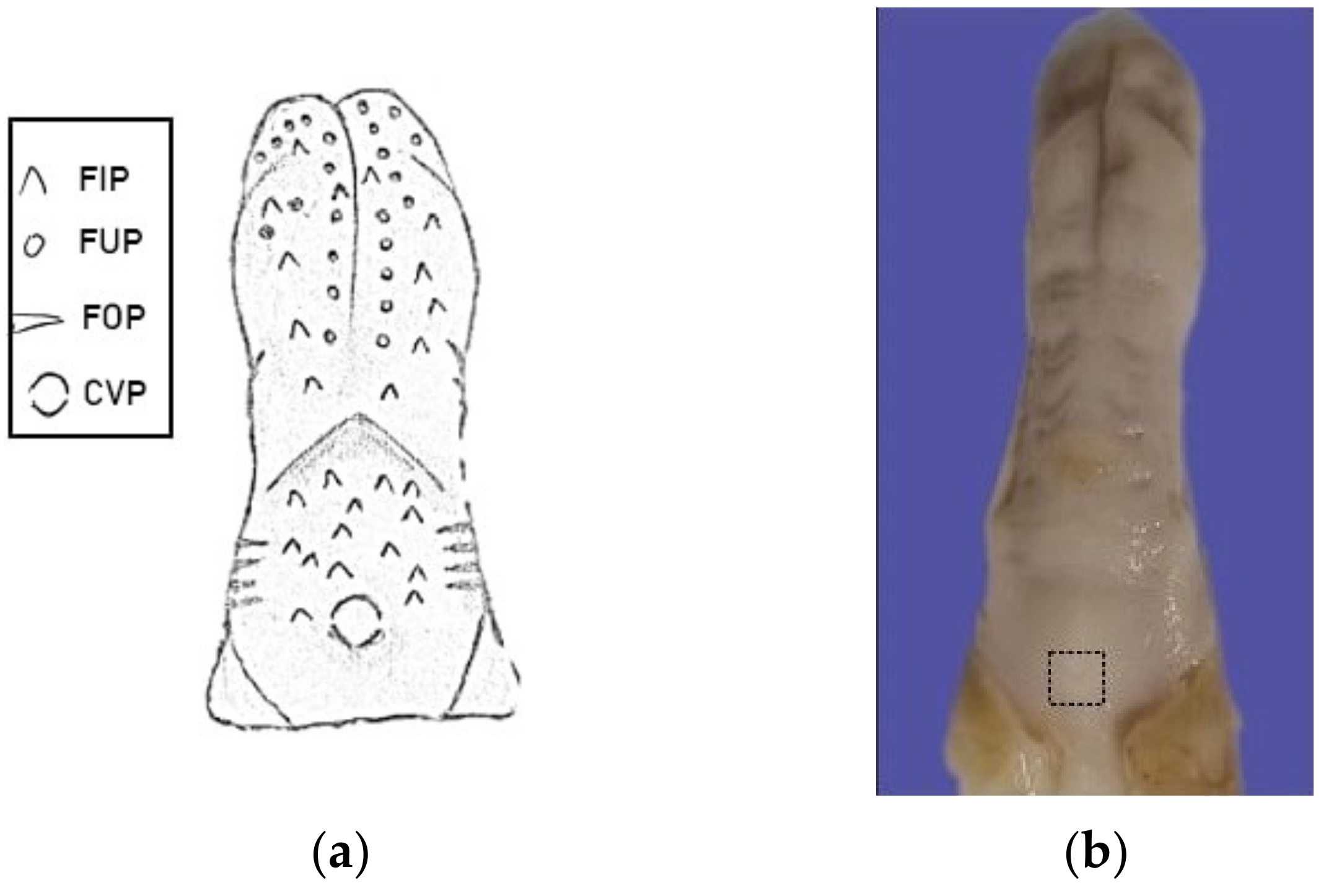
Figure 1. (a) Diagram illustrating the four distinct types of papillae on a rat’s tongue. FIP: filiform papillae, FOP: foliate papillae, FUP: fungiform papillae, and CVP: circumvallate papillae. (b) Photograph of a rat tongue highlighting the sample collection area, indicated by the dotted region. While the sizes of the collected samples slightly varied, they typically measured 5 mm × 5 mm.
Applsci 13 11617 g001
Figure 2. Photomicrographs of H&E-stained taste buds in the circumvallate papillae. In image (a), degenerative changes can be observed, along with a scarcity of taste pores (arrowhead). Conversely, images (bd) showcase taste buds that are densely packed with cells (yellow arrows) and exhibit multiple taste pores (arrows). (a) Diabetic control subgroup (STZ); (b) diabetic experimental subgroup (STZ/4HR); (c) healthy control subgroup (control) and (d) healthy experimental subgroup (4HR). (Original magnification, ×200, bar = 50 µm).
Applsci 13 11617 g002
Figure 3. Comparison of the number of taste bud pores across different experimental groups. The control group showed a mean of 39.6 ± 14.3 pores/mm2. In contrast, the 4HR, STZ, and STZ/4HR groups exhibited means of 33.7 ± 14.2, 20.3 ± 6.1, and 28.0 ± 8.3 pores/mm2, respectively. A statistically significant difference was observed between the control and STZ groups (* p = 0.005).
Applsci 13 11617 g003
Figure 4. Circumvallate papillae TUNEL staining. TUNEL-positive nuclei (indicated by arrows) are observable both within the taste bud profile and the epithelium. The diabetic experimental subgroup (STZ/4HR), healthy control subgroup (control), and healthy experimental subgroup (4HR) all showed fewer TUNEL-positive taste bud cells compared to the diabetic control subgroup (STZ) (Original magnification, ×200 without counterstaining). Quantitative analysis highlighted a significantly greater number of TUNEL+ cells in the STZ group in contrast to the control, 4HR, and STZ/4HR groups (* p < 0.05).
Applsci 13 11617 g004
Figure 5. Immunohistochemical staining depicting the presence of cleaved caspase-3 (c-casp3) in different subgroups. The brown coloration, marked by arrows, indicates c-casp3 positive cells. (a) Diabetic control subgroup (STZ); (b) diabetic experimental subgroup (STZ/4HR); (c) healthy control subgroup (Control); and (d) healthy experimental subgroup (4HR). A notably higher quantity of c-casp3 positive cells was observed in the STZ group compared to the other subgroups (Original magnification, ×400 without counterstaining, bar = 20 µm).
Applsci 13 11617 g005
Figure 6. The percentage of cleaved caspase-3 positive cells in the taste bud. There was a statistically significant difference between the STZ subgroup and the other subgroups (* p = 0.005).
Applsci 13 11617 g006
Figure 7. IP-HPLC analysis of DM-related protein expression in blood serum. The results indicate that in DM-induced rats treated with 4HR, there was an up-regulation of HKII by 27.8%, sirtuin 1 by 13.4%, and FOXO3 by 15.4%.
Applsci 13 11617 g007
Disclaimer/Publisher’s Note: The statements, opinions and data contained in all publications are solely those of the individual author(s) and contributor(s) and not of MDPI and/or the editor(s). MDPI and/or the editor(s) disclaim responsibility for any injury to people or property resulting from any ideas, methods, instructions or products referred to in the content.