Article

Protective Effect of Iris germanica L. Rhizome-Derived Exosome against Oxidative-Stress-Induced Cellular Senescence in Human Epidermal Keratinocytes

1
Department of Cosmetics Engineering, Konkuk University, 120 Neungdong-ro, Gwangjin-gu, Seoul 05029, Republic of Korea
2
Advanced Actives of Plants & Life Science Institute, ABio Materials Co., Ltd., Cheonan 31005, Republic of Korea
3
Shinsegae International Inc., Seoul 06015, Republic of Korea
*
Author to whom correspondence should be addressed.
Appl. Sci. 2023, 13(21), 11681; https://doi.org/10.3390/app132111681
Submission received: 30 September 2023 / Revised: 19 October 2023 / Accepted: 23 October 2023 / Published: 25 October 2023

Abstract

:
Plant-derived exosomes can exert therapeutic effects against various dermatological conditions. Several studies have demonstrated that plant-derived exosomes can have positive effects on the skin, preventing aging, hyperpigmentation, and hair loss. In this study, the protective effects of Iris germanica L. rhizome-derived exosomes (Iris-exosomes) on oxidative-stress-induced cellular dysfunction were investigated in human epidermal keratinocytes (nHEKs). Iris-exosomes with a diameter range of 100–300 nm were detected. In the cytotoxicity assay, Iris-exosomes with up to 107 particles per milliliter were found to possess no cytotoxicity, and we recovered H2O2-induced cell viability loss. In nHEKs, H2O2-induced ROS levels were significantly reduced using Iris-exosomes and additionally associated with increases in antioxidant enzyme transcription. The H2O2-induced SA-β-gal-positive nHEKs were decreased using Iris-exosomes; these effects correlate with the changed levels of cell cycle arrest marker p21. Furthermore, the H2O2-induced loss of in vitro wound-healing properties and early detection of keratin 1 and 10—keratinization markers—were restored to control levels using Iris-exosomes. Altogether, these results indicate the possibility that Iris-exosomes exert antioxidant and anti-senescence effects in order to protect against oxidative-stress-induced cellular dysfunction in nHEKs.

1. Introduction

Exosomes are among the extracellular vesicles produced by eukaryotic cells, a category of cell that includes plant cells [1]. Possessing a lipid bilayer structure, exosomes are between 30 and 200 nm in diameter, with an average diameter of 100 nm [2,3]. A variety of investigations have found that exosomes can contain various cellular constituents including DNA, RNA, lipids, metabolites, and cytosolic and cell-surface proteins, depending on the origin of the cells [3]. The physiological roles of exosomes are based on maintaining cellular homeostasis by removing unnecessary constituents from cells [4]. However, recent studies have found that exosomes play a large part in intercellular communication and can alter biological responses, including immune responses and cancer progression [5,6,7,8]. Therefore, these properties can be used in the treatment and diagnosis of various diseases, including cancer [3,9]. In addition, recent studies have shown that exosomes have low immunogenicity and high hemocompatibility and are effective in treating skin problems such as aging, atopic dermatitis, and wounds [10]. Moreover, exosomes are being studied for their potential use as ingredients in skin care products. One study found that exosomes secreted by dermal papilla cells helped hair follicle development [11]. Another found that cardiac progenitor cell-derived exosomes protected cardiomyocytes against oxidative-stress-induced apoptosis [12]. Moreover, treatment with blueberry-derived exosomes protected human endothelial cells from tumor necrosis factor-α (TNF-α)-induced reactive oxygen species (ROS) production and viability loss [13]. Recent studies have indicated the functional therapeutic roles of plant-derived exosomes originating from cabbage, ginseng (Panax ginseng), and green tea (Camellia sinensis) [14,15,16,17,18,19]. According to research, exosomes can be absorbed effectively into keratinocytes owing to their biocompatibility. Additionally, comparative gene expression analysis has shown that exosome treatment contributes to maintaining skin condition better than several plant extract treatment groups in genes related to skin aging, regeneration, skin barrier, and moisturization. These findings indicate that plant-derived exosomes affect human cells, including skin cells, differently than the extracts, offering a new perspective on their potential application as cosmeceutical ingredients [18].
Oxygen molecules with unpaired electrons, known as free radicals, are produced by extrinsic factors, including UV light, pollution, stress, and smoking, as well as endogenous factors inside the body. Antioxidant defense systems within cells counteract ROS and maintain a balance between ROS generation and neutralization [20]. The inability of the body to scavenge free radicals, as occurs when an imbalance between ROS and the antioxidative defense system emerges, results in skin aging and diseases such as psoriasis and atopic dermatitis [21,22,23]. Skin aging and such diseases are closely associated with keratinocyte functioning. Keratinocytes play an important role in skin regeneration and barrier functions [24,25,26]. When oxidative stress is excessive, keratinocyte differentiation becomes abnormal [25,27], resulting in an abnormal skin barrier that causes skin diseases and accelerates the overall aging of the skin due to excessive moisture evaporation [28,29]. Therefore, if one is to maintain good skin health, it is crucial to protect keratinocytes from oxidative stress.
The skin is the largest organ in the body [30] and comprises the following three layers: epidermis, dermis, and subcutaneous fat [31]. Multiple layers of keratinocytes are present in the epidermis, each with a different degree of differentiation [32,33]. As keratinocytes differentiate, they migrate toward the surface and become fully differentiated corneocytes [33,34]. Located in the outermost layer of the skin, keratinocytes are continually exposed to various kinds of stresses, as well as UV irradiation [35,36]. UV is one of the major factors contributing to ROS generation [37,38], and UV-induced ROS generation reduces physiological antioxidant levels in the epidermis, causing oxidative stress [39]. Keratinocytes are susceptible to oxidative stress, which can cause serious skin diseases [40,41]. Significant ROS accumulation in keratinocytes is associated with oxidative modifications of nucleic acids, lipids, proteins, and other intracellular molecules, leading to cellular dysfunction such as cell viability loss, cellular senescence, and apoptosis [42,43,44,45,46]. The epidermis is the first barrier against pathogens, pollutants, toxic chemicals, and UV irradiation—all factors that contribute to oxidative stress [47]. Therefore, the identification of bioactive molecules that protect keratinocytes from oxidative stress can be a promising therapeutic and/or cosmeceutical strategy in preventing oxidative-stress-related skin diseases.
Various novel bioactive molecules are produced by plants and used to prepare a variety of medicines [48]. Iris species, members of the Iridaceae family, are valuable traditional medicines with a variety of applications, such as relieving joint pain and cough symptoms [49,50,51]. Several studies have shown that Iris extracts have anti-inflammatory, anti-cancer, antioxidant, and antimicrobial activities [52,53,54,55,56]. In addition, Iris germanica L. contains many secondary metabolites with strong antioxidant potential, including flavonoids and triterpenes [57,58,59]. Specifically, methanolic extracts of Iris germanica L. rhizomes possess antioxidant and anti-inflammatory properties due to their high content of isoflavonoids, such as tectoridin, iridin, 8-hydroxyirilone, and tectorigenin [60]. There are also bioactive molecules in Iris germanica L., including isoflavones such as irigenin, iristectoirigenin A, nigricin, irisflorentin, irilone, and irisolidone [59]. Furthermore, previous phytochemical studies have demonstrated that Iris germanica L. includes a variety of chemical compounds [61,62], including flavonoids, triterpenes, sterols, phenolic compounds, ceramides, and benzoquinone derivatives. Therefore, it is reasonable to assume that exosomes derived from Iris rhizomes are an optimum source of antiaging agents for action against intracellular ROS. However, there are limited studies on how exosomes derived from the rhizome of Iris germanica L. affect the antioxidant activity of keratinocytes.
In this study, we investigate whether Iris-exosomes possess antioxidant properties and protect against oxidative stress. H2O2 is a common oxidizing agent that accumulates mostly in the epidermis and can move into and out of cells and tissues [63,64,65]; therefore, we used H2O2 in order to induce oxidative stress in keratinocytes.

2. Materials and Methods

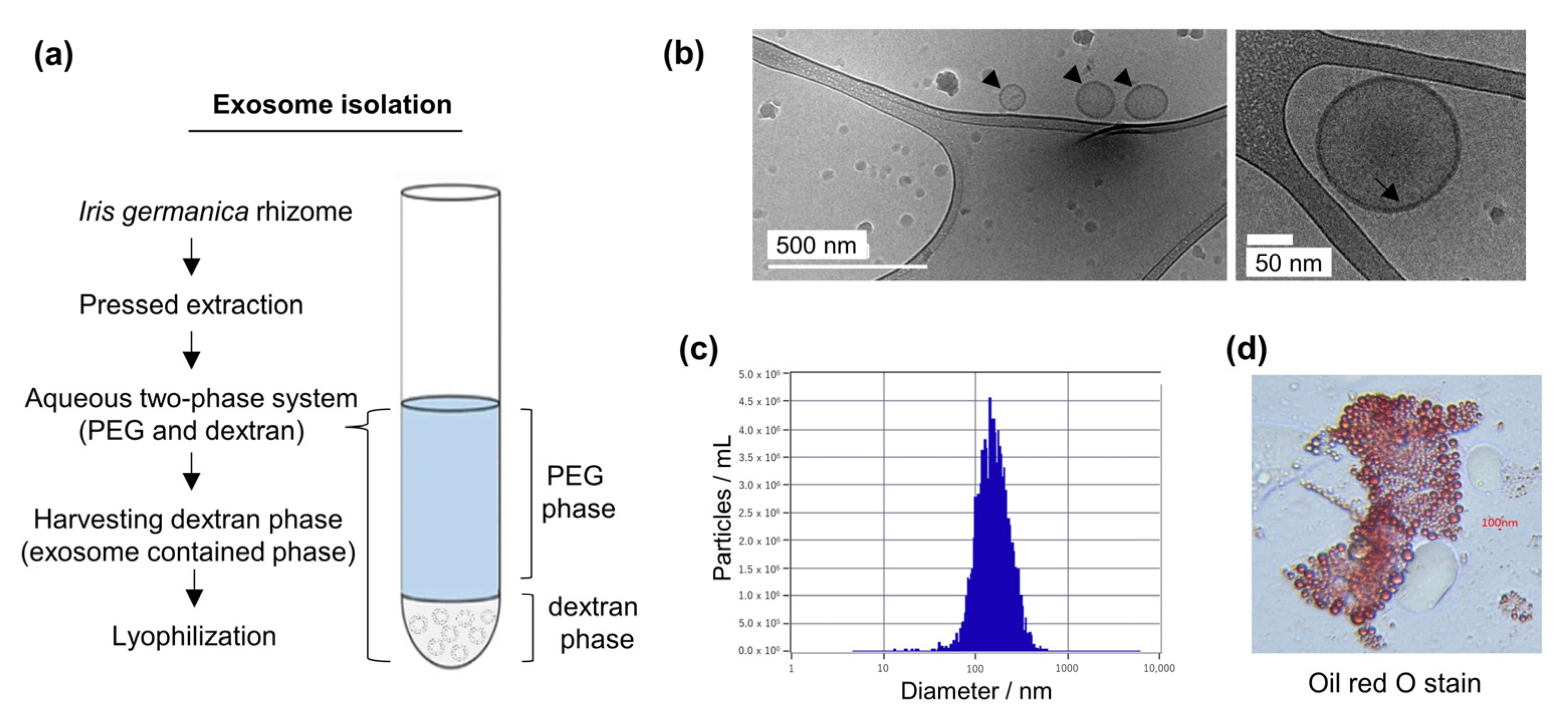
2.1. Isolation and Characterization of Iris germanica L. Rhizome-Derived Exosomes (Iris-Exosomes)

Iris germanica L. rhizomes were purchased from Produits Prestiges S.A. (Montreux, Switzerland). The dry Iris rhizomes (Iris germanica L./100 g) were packed in a plastic pack with distilled water and sealed well to prevent air infiltration. Then, ultra-high-pressure treatment was performed for 30 sec at 25 °C at a pressure of 200 Mpa. Iris rhizomes treated with ultrahigh pressure were then extracted with a general juicer using a low-speed screw at 30 rpm, and the obtained Iris rhizome extracts were filtered using a mesh net. The Iris rhizome extracts were stored at −80 °C until purification could be performed. For exosome purification, the Iris rhizome extracts were centrifugated at 10,000 × g for 10 min at 4 °C to remove large contaminants. The supernatant was collected, frozen at −80 °C for 20 h, and then dried in a freeze dryer for 100 h under vacuum conditions. The lyophilized supernatant was mixed with distilled water (DW) and an aqueous two-phase system (ATPS). The ATPS was prepared by adding 3.3% PEG (Sigma-Aldrich, St. Louis, MO, USA) with a molecular weight of 10,000–35,000 and 1.7% dextran (Sigma-Aldrich, St. Louis, MO, USA) with a molecular weight of 300,000–650,000. After mixing the lyophilized supernatant and PEG/dextran solution, centrifugation at 1000× g for 10 min was performed at 4 °C. Exosomes were recovered via centrifugation after the supernatant was removed. To increase the purity, the recovered lower-layer solution was washed with an additional aqueous two-phase system at the same concentration. The final exosome-concentrated layer was recovered after repeating the same washing process three times (Figure 1a). As the exosomes purified from Iris germanica L. rhizome are in the form of an aqueous solution of nanoparticles with an inherent tendency to sediment, they were carefully treated with cells using gentle pipetting to prevent the destabilization of the exosomes.

2.1.1. Cryo-TEM Analysis

We placed 5 μL of the Iris-exosomes sample on a Lacey carbon grid. This was settled on a cryoholder under conditions of a wait time of 30 s and blot time of 5 s in Vitrobot Mark IV (FEI Company, Hillsboro, OR, USA) using cryogenic transmission electron microscopy (Cryo-TEM).

2.1.2. Nanoparticle Tracking Analysis (NTA)

Exosomes separated from Iris germanica L. rhizome were analyzed using nanoparticle tracking analysis (NTA) to verify particle size distributions and particle numbers per unit volume. ZetaView (Particle Metrix GmbH, Meerbusch, Germany) was used to conduct nanoparticle tracking analysis (NTA) measurements. After diluting the sample to an appropriate concentration where the number of detected particles was between 50 and 300, about 1 mL of the sample was injected into the cell in the device. We then examined the product at a wavelength of 488 nm in scatter mode.

2.1.3. Oil Red O Staining

In total, 1 mL of oil red O solution (Sigma Aldrich) was added to the Iris-exosomes. After 10 min, the Iris-exosomes were centrifuged again to remove any remaining oil red O. Afterward, 1 mL of distilled water was added, and centrifugation was repeated twice to eliminate any remaining oil red O. Iris-exosomes stained with oil red O were observed under a microscope.

2.2. Cell Culture and Reagents

Primary normal human epidermal keratinocytes (nHEKs) provided by CELLnTEC (Bern, Switzerland) were maintained in Keratinocyte Basal Medium-2 (Lonza, Basel, Switzerland) supplemented with bovine pituitary extract (BPE), human epidermal growth factor (hEGF), insulin, hydrocortisone, transferrin, epinephrine, and gentamicin sulfate-amphotericin (GA-1000). nHEKs were cultured in collagen-coated plates (SPL Life Sciences, Pocheon city, Republic of Korea) and incubated at 5% CO2 and 37 °C. The cells were washed with HEPES-buffered saline solution (HSS) (Promocell, Heidelberg, Germany) and subcultured using trypsin/ethylenediaminetetraacetic acid (EDTA) at a ratio of 0.04%/0.03% (Promocell). Passages 4–6 of nHEKs were used in this experiment.
Human dermal fibroblast (nHDF) purchased from Promocell (Heidelberg, Germany) were maintained in Dulbecco’s Modified Eagle’s medium (DMEM) supplemented with 10% fetal bovine serum (FBS), 1% streptomycin (100 mg/mL), and penicillin (100 U/mL) at 37 °C and 5% CO2. Passages 12–14 of nHDFs were used in this study. N-acetyl-l-cysteine (NAC) (Sigma-Aldrich, St. Louis, MO, USA) was used as an antioxidant control.

2.3. In Vitro DPPH and ABTS Radical Scavenging Assays

1,1-Diphenyl-2-picrylhydrazyl (DPPH) was used to measure radical scavenging activity via a modification of the Blois method (Blois, 1958) [66]. The DPPH reagent (0.2 mM) and various concentrations of Iris-exosomes were mixed in a 96-well plate and incubated for 15 min. The absorbance was measured at 517 nm using a microplate reader.
2,2′-Azino-bis (3-ethylbenzothiazoline-6-sulfonic acid) (ABTS) radical cations were prepared by reacting 7 mM ABTS solution (MedChemExpress, Monmouth Junction, NJ, USA) with potassium peroxydisulfate (Supelco, Bellefonte, PA, USA) to a final concentration of 2.45 mM. The mixture was then incubated overnight in the dark at room temperature. After incubation, the solution was diluted until the absorbance at 734 nm was 0.70 (±0.02). L-ascorbic acid (Sigma-Aldrich) was used as a positive control. Each concentration of Iris-exosomes and L-ascorbic acid were mixed with ABTS solution in 96 wells. Then, absorbance was measured at 734 nm using a microplate reader after shaking in the dark for 10 min.

2.4. Cell Viability Assay

To evaluate cell viability, the nHEKs and nHDFs were subjected to a Water-Soluble Tetrazolium-1 (WST-1) assay. The cells were seeded in 96-well plates and treated with various concentrations of Iris-exosomes or H2O2 and incubated at 37 °C for 24 h. Subsequently, EZ-Cytox solution (DoGenBio, Seoul, Republic of Korea) was added to the plate, and the cells were incubated at 37 °C with 5% CO2 for 30 min. Cell viability was analyzed using an iMark microplate reader measuring absorbance at 450 nm.
For the 3-(4,5-dimethylthiazol-2-yl)-2,5-diphenyl tetrazolium bromide (MTT) assay, cells were seeded in a 12-well plate and incubated at 37 °C for 24 h. Subsequently, the cells were pretreated with Iris-exosomes or NAC for 4 h, followed by exposure to 200 μM H2O2 for another 24 h. Next, 500 μg/mL of MTT solution was added to the cells, which were incubated at 37 °C with 5% CO2 for 3 h in the dark. The MTT solution was carefully removed, and 1 mL of DMSO (Sigma-Aldrich) was added to each well. The plates were then shaken for 5 min to ensure proper dissolution of the formazan crystals. Finally, the absorbance was measured at 595 nm using a microplate reader.

2.5. Intracellular ROS Measurement Assay

Intracellular ROS levels were evaluated using an H2DCFDA assay. nHEKs were seeded in 6-well plates and incubated for 24 h. After pretreatment with Iris-exosomes for 4 h, DCF-DA solution (10 μM) was added and incubated for 30 min in the dark. The cells were then treated with 500 μM H2O2 to induce intracellular ROS production and incubated for 30 min in the dark. The intracellular ROS levels were determined using fluorescence microplate reader at 485 nm for excitation and 520 nm for emission.

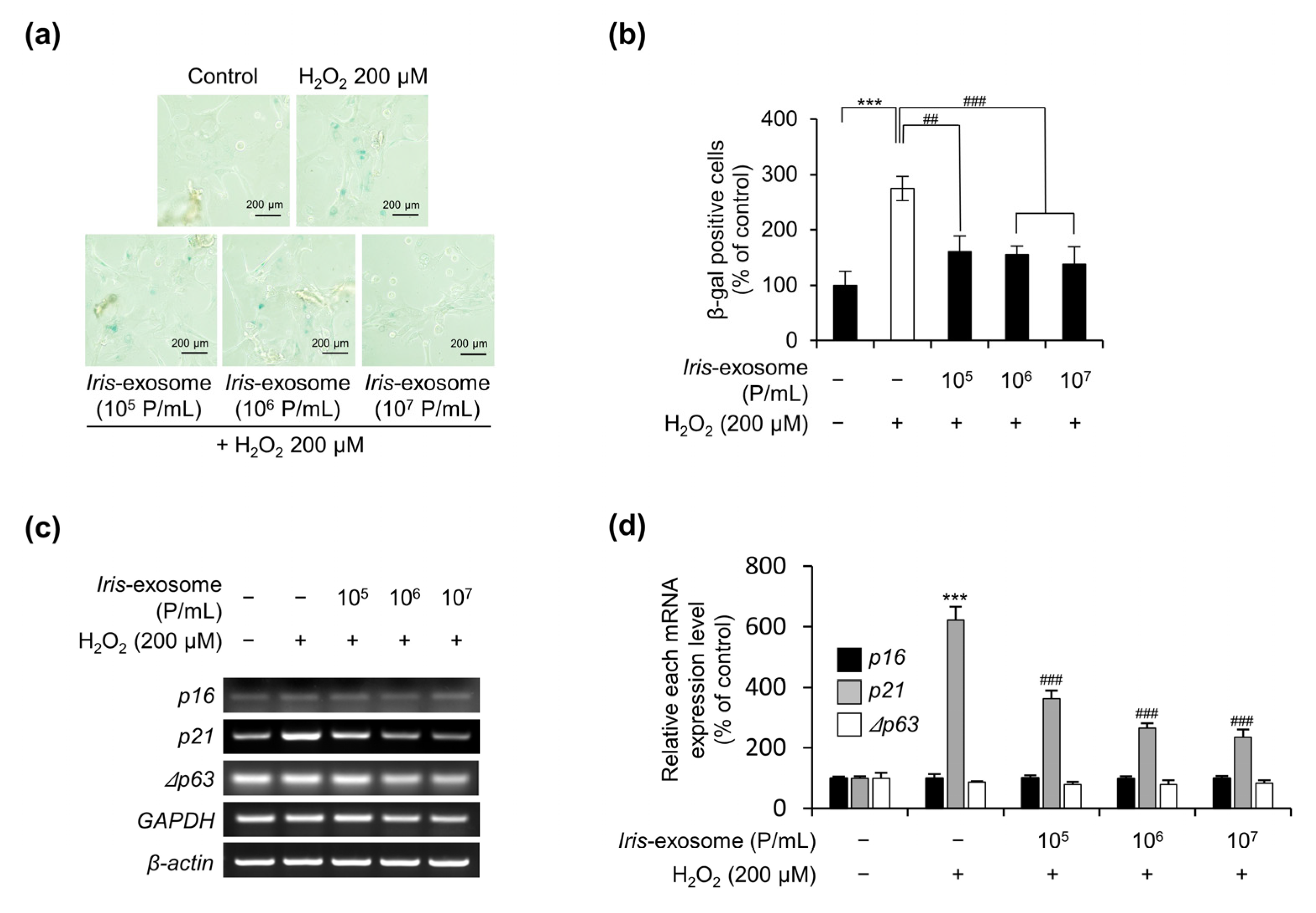
2.6. Cellular-Senescence-Analysis-Associated SA-β-Galactosidase Activity

nHEKs were treated with 200 μM H2O2 for 4 h after the treatment of Iris-exosomes for 4 h. Using a senescence β-galactosidase staining kit (Cell Signaling Technology, Danvers, MA, USA), the cells were fixed for 5 min and incubated in a staining solution at 37 °C for 8 h. Cellular senescence levels were assessed by counting senescent cells using a bright-field microscope.

2.7. Polymerase Chain Reaction (PCR) and Quantitative Reverse Transcription PCR (RT-qPCR)

nHEKs were pretreated with Iris-exosomes for 4 h, followed by treatment with 200 μM H2O2 for 4 h. The cells were further incubated for 40 h, and total RNAs were extracted using RiboEx reagent (GeneAll Biotechnology, Seoul, Korea). cDNA was synthesized using Moloney murine leukemia virus (M-MLV) reverse transcriptase (Thermo Fisher Scientific, Waltham, MA, USA). RT-qPCR was conducted using a reaction mixture containing each primer and the Evagreen qPCR master mix (Solis BioDyne, Tartu, Estonia). Gene expression was normalized to that of GAPDH. The primer sequences for specific genes used in the analysis were as follows: p16 forward, 5′-TGCCTTTTCACTGTGTTGGA-3′ and p16 reverse 5′-GCCATTTGCTAGCAGTGTGA-3′; p21 forward, 5′-GAACTTCGACTTTGTCACCGAGAC-3′ and p21 reverse, 5′-TGGAGTGGTAGAAATCTGTCATGCT-3′; p53 forward, 5′-CGGGATCCCGAATGTTGTACCTGGAAAACAATG-3′ and p53 reverse, 5′-GCTCTAGAGCTCACTCCCCCTCCTCTTTGATG-3′; p63 forward, 5′-CGGGATCCCGAATGTTGTACCTGGAAAACAATG-3′ and p63 reverse, 5′-GCTCTAGAGCTCACTCCCCCTCCTCTTTGATG-3′; HO-1 forward, 5′-GCCCTTCAGCATCCTCAGTTCC-3′ and HO-1 reverse, 5′-AGTGGTCAT GGCCGTGTCAAC-3′; NQO-1 forward, 5′-GGGAGACAGCCTCTTACTTGCC-3′ and NQO-1 reverse, 5′-AACACCCAGCCGTCAGCTATTG-3′; GSS forward, 5′-TAGATGCCCCACGTGCTTGT-3′ and GSS reverse, 5′-ATCCTCATGGAGAAGATCGA-3′; SOD forward, 5′-CCAGTGCAGGGCATCATCA-3′ and SOD reverse, 5′-TTGGCCCACCGTGTTTTCT-3′; GPx1 forward, 5′-GCAGCTCGTTCATCTGGGTG-3′ and GPx1 reverse, 5′-ATGTGTGCTGCTCGGCTAGC-3′; Nrf2 forward, 5′-ATAGCTGAGCCCAGTATC-3′ and Nrf2 reverse, 5′-CATGCACGTGAGTGCTCT-3′; GAPDH forward, 5′-TCCAAAATCAAGTGGGGCGATGC-3′, GAPDH reverse, 5′-GCCAGTAGAGGCAGGGATGATGT-3′ and β-actin forward, 5′-CGCTCGGTGAGGATCTTCATG-3′, β-actin reverse, 5′-GGATTCCTATGTGGGCGACGA-3′.

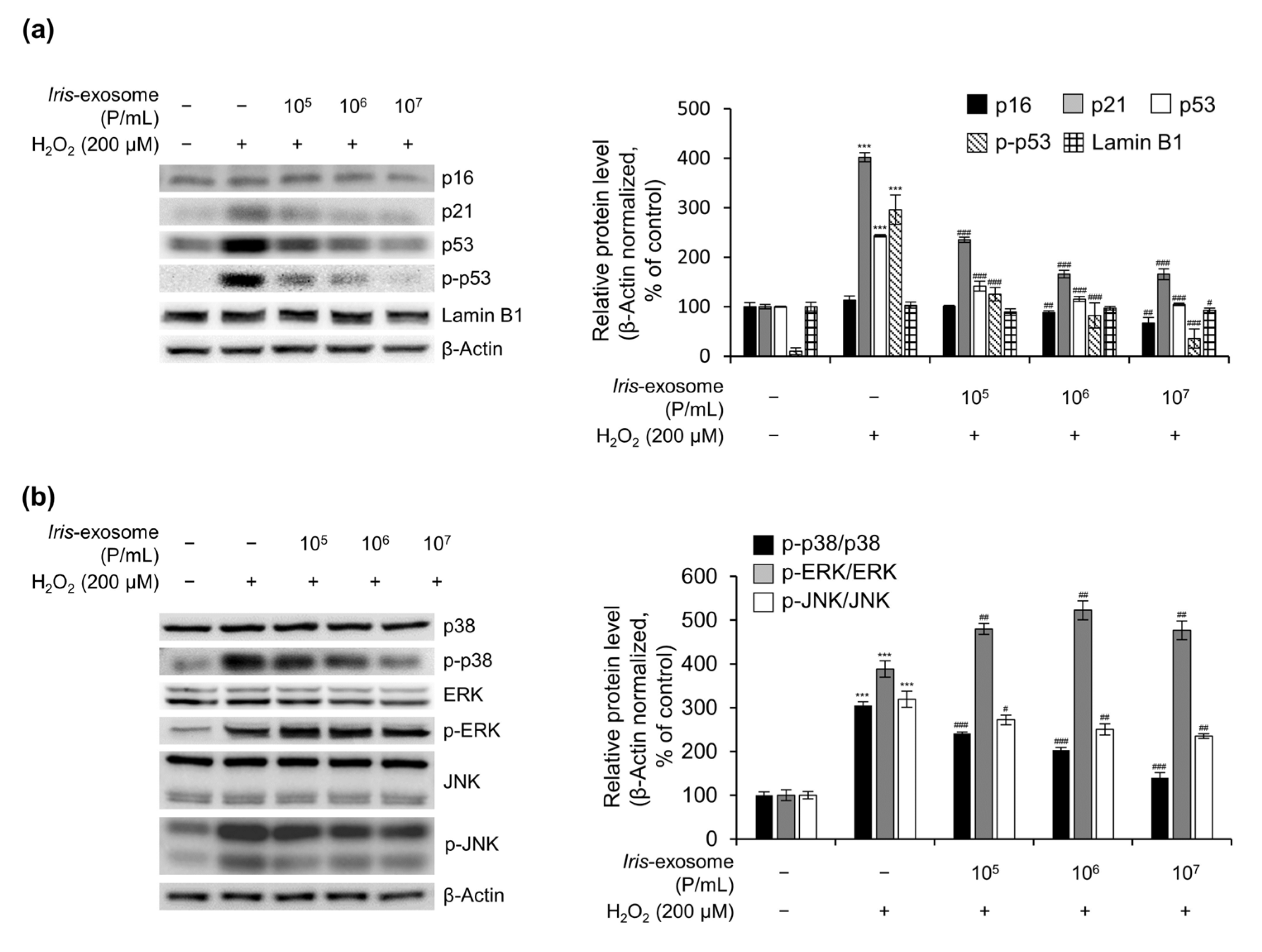
2.8. Immunoblot Analysis

nHEK cells were pretreated using the indicated concentrations of Iris-exosomes for 4 h, after which they were treated with 200 μM H2O2 for another 4 h. After 40 h of further incubation, the cells were harvested, and total proteins were lysed using radioimmunoprecipitation assay (RIPA) buffer containing protease inhibitors. Total protein quantification was performed using a Pierce BCA Protein Assay Kit (Thermo Fisher Scientific). Samples containing 20 µg of protein were separated via sodium dodecyl sulfate (SDS)–polyacrylamide gel electrophoresis, and the proteins were then transferred onto a polyvinylidene fluoride (PVDF) membrane. These membranes were blocked using 2% skim milk in Tris-buffered saline with Tween 20 (TBST) buffer and incubated with the corresponding primary antibodies overnight at 4 °C. Then, the membranes were incubated with a horseradish peroxidase (HRP)-conjugated secondary antibody. The Western blot results were analyzed using enhanced chemiluminescence (ECL) solution (Thermo Fisher Scientific) and visualized using a chemiluminescence detector. The antibodies used were as follows: ERK (1:1000, CST), p-ERK (Thr202/Tyr204)(1:1000, CST), p16 (1:1000, CST), Lamin B1 (1:1000, Abcam), p21 (1:1000, CST), Nrf2 (1:1000, CST), p53 (1:200, Santa Cruz), p-p53 (Ser15), (1:1000, CST), p38 (1:1000, CST), p-p38 (T180/Y182), (1:1000, CST), JNK (1:1000, CST), p-JNK (T183/Y185), (1:1000, CST), Cytokeratin 1 (1:1000, Abcam), Cytokeratin 10 (1:1000, Abcam), Loricrin (1:1000, Abcam), and β-actin (1:1000, Santa Cruz). Quantification of protein level was conducted with ImageJ software (ImageJ 1.52a, National Institutes of Health, Bethesda, MD, USA).

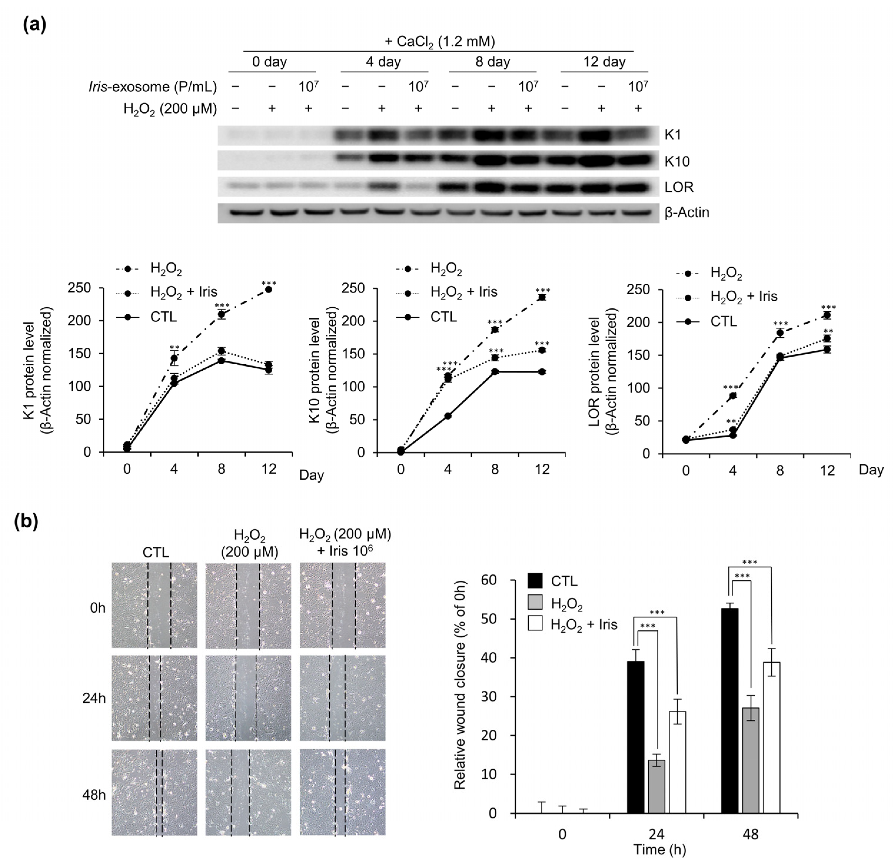
2.9. Keratinocyte Differentiation

nHEKs maintained in a KBM-2 medium containing 0.06 mM CaCl2 solution (Promocell) were seeded on a 100 mm cell culture dish. The seeding date was adjusted so that all cells could be harvested simultaneously on day 12. The cells were pretreated with Iris-exosomes for 4 h before treatment with 200 μM H2O2 for 4 h. To induce differentiation, the cells were incubated in a KBM-2 medium containing 1.2 mM CaCl2 for 0, 4, 8, and 12 days. Media with 1.2 mM CaCl2 were changed once every three days. Cell lysates were used to examine the expression levels of the differentiation markers K1, K10, and LOR via Western blotting. The following antibodies were used: Cytokeratin 1 (1:1000, Abcam), Cytokeratin 10 (1:1000, Abcam), Loricrin (1:1000, Abcam), and β-actin (1:1000, Santa Cruz).

2.10. Wound-Healing Assay

nHEKs were treated with the indicated concentrations of Iris-exosomes and/or 200 μM H2O2 for 4 h. Following incubation for 24 h, the confluent cells were scratched with a pipette tip and then washed using HSS buffer. The wound area was photographed at 24 h intervals while incubating under conditions of 5% CO2 and 37 °C. The gap area was determined and measured using the ImageJ software.

2.11. Statistical Analysis

All experiments were performed independently at least thrice. The treatment groups were assessed as statistically significant at p < 0.05 via one-way analysis of variance (ANOVA). Different group means were compared using Tukey’s test. Data are expressed as mean ± standard deviation (SD).

3. Results

3.1. Purification and Characterization of Iris germanica L. Rhizome-Derived Exosome

An in-depth description of Iris-exosome isolation is presented in the Materials and Methods section. Briefly, an aqueous two-phase solution was prepared via a process that used PEG and dextran to isolate Iris-exosomes from the Iris rhizome (Figure 1a). By analyzing the total protein in the Iris-exosomes, it was determined that there was approximately 21.08 μg/mL of protein in the solution. The morphological structure of the exosomes derived from the Iris germanica L. rhizomes was analyzed using cryogenic transmission electron microscopy (Cryo-TEM). The phospholipid particles exhibited a spherical bilayer structure, with an average size of 160.7 ± 3.60 nm (Figure 1b). Nanoparticle tracking analysis (NTA) was conducted using ZetaView to determine the particle size distribution and number per unit volume within the purified exosomes derived from the Iris germanica L. rhizomes. According to the measurements, the average size of the particles was 172.1 ± 66.4 nm, and the concentration was confirmed to be 1.61 × 10⁹ particles/mL (Figure 1c). Next, oil red O staining was conducted to estimate the lipid levels in the Iris-exosomes. This confirmed that the Iris-exosome is composed of a phospholipid bilayer (Figure 1d). The results indicate that exosomes were present in the extracts derived from the Iris germanica L. rhizomes.

3.2. Iris-Exosome Protects nHEKs from the Loss of Cell Viability Caused by Oxidative Stress

Before analyzing the effects of Iris-exosomes on H2O2-induced cellular stress in nHEKs, we assessed the potential cytotoxicity of the nHEKs treated with Iris-exosomes for 24 h. As shown in Figure 2a, nHEKs treated with a concentration of less than 107 (particles/mL) showed no cytotoxicity. In order to evaluate the effect of H2O2 on nHEKs, cell viability was determined via 24 h treatment with various concentrations of H2O2. Consequently, cell viability was reduced by 80% at 200 µM (Figure 2b). Therefore, 105, 106, and 107 Iris-exosomes were used in the following experiments along with 200 µM H2O2. The MTT assay was performed on the nHEKs to determine whether the exosome treatment could recover the reduction in cell viability caused by H2O2. The viability of nHEKs pretreated with Iris-exosomes increased in a concentration-dependent manner compared with that of cells treated with H2O2 alone (Figure 2c). These results indicate that Iris-exosomes have protective effects against the H2O2-induced loss of cell viability in nHEKs.
Furthermore, we conducted the same experiment on nHDFs to determine whether the protective effects observed in nHEKs are also found in other types of primary skin cells. As shown in Supplementary Figure S1a, nHDFs showed no cytotoxicity and increased cell viability at concentrations below 109 (particles/mL). Furthermore, the viability of the nHDFs was reduced by 80% at 300 µM H2O2 (Supplementary Figure S1b). Therefore, we treated 106, 107, and 108 Iris-exosomes with 300 µM H2O2. nHDFs were treated with exosomes to determine whether exosome treatment could restore the viability of the cells post-H2O2 treatment. Compared to the cells treated with H2O2 alone, pretreatment with Iris-exosomes increased the viability of the nHDFs in a concentration-dependent manner (Supplementary Figure S1c). Considering these results, it is evident that Iris-exosomes are protective against the reduction in cell viability caused by H2O2 in not only nHEKs but also nHDFs.

3.3. Iris-Exosome Reduces Intracellular ROS Levels in nHEKs

Our previous results led us to investigate the antioxidant potential of Iris-exosomes. The DPPH and ABTS assays were initially conducted to determine whether Iris-exosomes possess a radical scavenging effect. The Iris-exosomes showed concentration-dependent scavenging activity against DPPH and ABTS radicals but revealed a less potent overall effect than L-ascorbic acid (Figure 3a,b). To determine whether exosomes can scavenge intracellular ROS in nHEKs, a DCF-DA assay was conducted in which nHEKs were pretreated using Iris-exosomes for 4 h and then exposed to 500 μM H2O2 for 30 min. As shown in Figure 3c,d, the increased DCF fluorescence intensity caused by the H2O2 treatment decreased in the Iris-exosome-treated cells in a concentration-dependent manner. To confirm whether these results could be explained by the expression of antioxidant-related genes in the cells, several antioxidant-related genes were measured after Iris-exosome treatment. HO-1, CAT, GSS, GPx1, SOD, and Nrf2 were transcriptionally upregulated in nHEKs following Iris-exosome treatment (Figure 3e). According to these results, it can be said that Iris-exosomes appear to exert a protective effect against cellular oxidative stress caused by H2O2.

3.4. Iris-Exosome Is Capable of Attenuating Cellular Senescence Induced by H2O2

In addition to assessing their radical scavenging activity in the intracellular environment, we investigated whether Iris-exosomes could protect nHEKs against oxidative-stress-induced cellular senescence. nHEKs were treated with various concentrations of Iris-exosomes and/or 200 µM H2O2, and senescence-associated β-galactosidase (SA-β-gal) assay was conducted to examine the SA-β-gal-positive senescent cells. The results indicate that H2O2 treatment only increased the number of senescent cells to 274.78% of the total reached in the untreated control group (100.00%); however, Iris-exosome-pretreated nHEKs reduced the number of senescent cells to 160.31%, 155.16%, and 138.37% (Figure 4a,b). It is well-known that p16 and p21 act as a cell cycle-dependent kinase inhibitor. RT-PCR was used to confirm these results via the analysis of the expression levels of senescent markers (p16 and p21). The results shown in Figure 4c,d indicate that the mRNA expression of p21 decreased concentration-dependently in the Iris-exosome-pretreated cells compared to the H2O2-treated cells. These results suggest that Iris-exosomes protect nHEKs against intracellular ROS production and cellular senescence induced by H2O2.

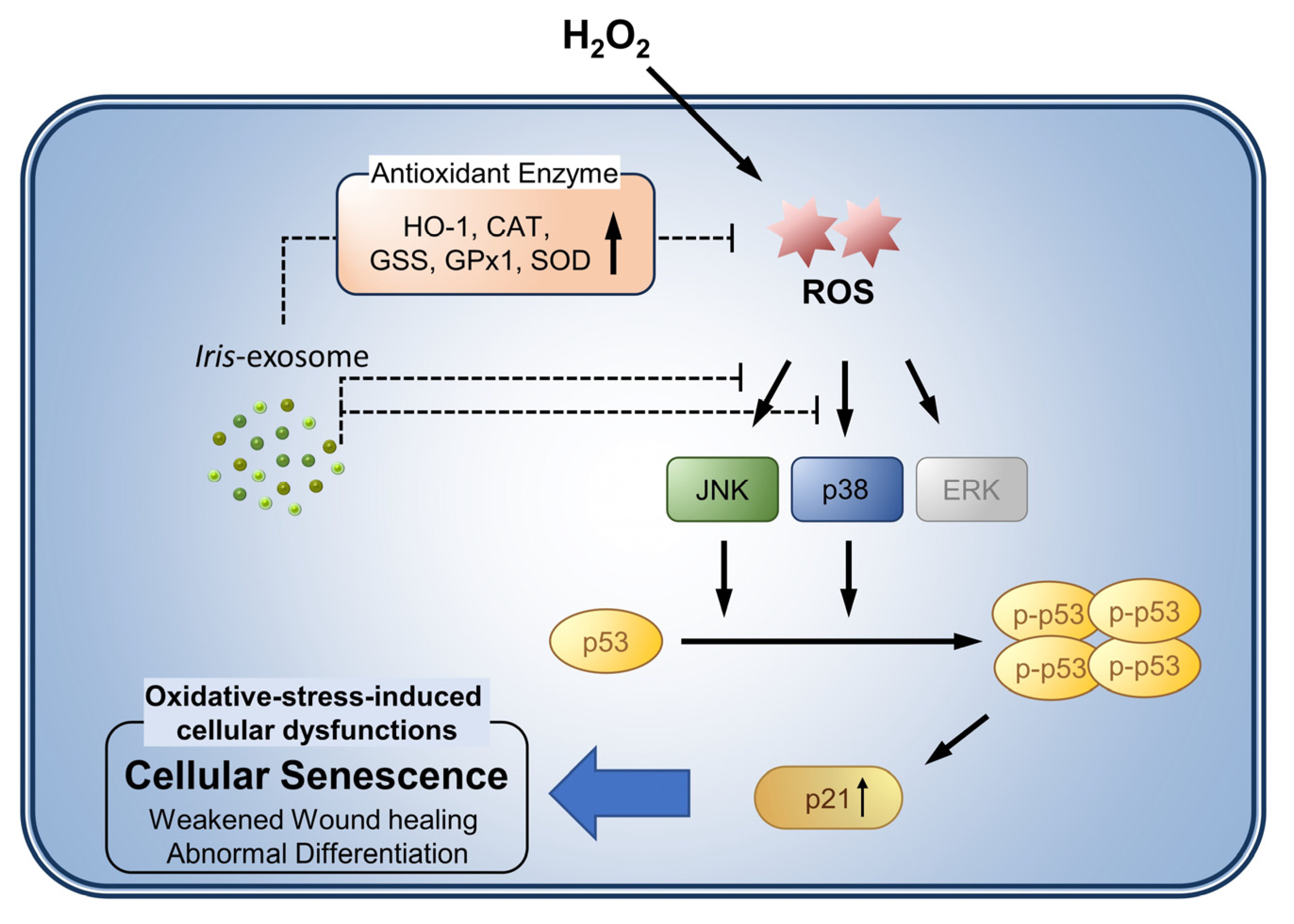
3.5. Iris-Exosome Inhibits H2O2-Induced Cellular Oxidative Stress via p38 Mitogen-Activated Protein Kinase (MAPK) Signaling Pathway

Regulation of p21, a senescence marker and cell cycle-dependent kinase inhibitor, is mediated by p53 in keratinocytes. Therefore, considering the above results, we examined the upstream regulators responsible for p21 regulation. Under the same conditions as the previous experiments, immunoblotting was used to evaluate the expression of p53 and p-p53 (ser15). Similar to the previous RT-qPCR, the expression of p16 and Lamin B1, which are known to be cellular senescence markers, did not change (Figure 5a). Similar to p21, H2O2 treatment upregulated the expression of p53 and p-p53 (Figure 5a). Additionally, the expression of p21, p53, and p-p53 was downregulated by H2O2 after pretreatment with Iris-exosomes in a concentration-dependent manner (Figure 5a). According to these findings, the changes in p21 caused by H2O2 and the Iris-exosome treatment resulted from p53 action. Thus, we determined that the MAPK signaling pathway was involved in the nHEK treatment with H2O2 and Iris-exosomes. H2O2 significantly increased the phosphorylation levels of p38, ERK, and JNK in the nHEKs (Figure 5b). However, the Iris-exosomes markedly inhibited the H2O2-induced phosphorylation of Threonine180 and Tyrosine182 residues in p38 (Figure 5b). Similarly, the phosphorylation of JNK at threonine 183 and tyrosine 185 was also reduced by the Iris-exosomes (Figure 5b). However, the Iris-exosome treatment did not inhibit the ERK activation induced by the H2O2 treatment (Figure 5b). These results indicate the possibility that the Iris-exosomes regulated H2O2-induced cellular senescence responses by decreasing the p38- and JNK-mediated p53–p21 signaling pathways. Altogether, these results indicate that Iris-exosomes may regulate H2O2-induced cellular senescence responses by decreasing JNK- and p38-mediated p53–p21 signaling.

3.6. Iris-Exosome Improves Oxidative-Stress-Induced Impairment of Keratinocyte Barrier Function

Keratinocytes play important roles in epidermal differentiation and skin regeneration. Oxidative stress induced by hydrogen peroxide causes premature keratinocyte senescence and abnormal terminal differentiation [25,27]. According to our findings, Iris-exosomes protect nHEKs from oxidative stress and cellular damage. Therefore, we tested whether Iris-exosome exerts protective effects against H2O2-mediated cell dysfunction during differentiation and regeneration. First, to determine whether Iris-exosomes could restore the abnormal differentiation caused by H2O2 treatment in nHEKs, Western blotting was performed to target differentiation markers in nHEKs treated with CaCl2 (1.2 mM) for 12 days. In the presence of CaCl2, a significant increase in the expression of the intermediate differentiation markers K1, K10, and LOR was observed on days 8 and 12 after treatment with CaCl2 (Figure 6a). Interestingly, compared to the control cells (CaCl2-treated only), we found that the H2O2-treated group showed early signs of upregulated intermediate differentiation markers, K1, K10, and LOR proteins, on day 3 after treatment with CaCl2 (Figure 6a). Iris-exosomes, however, attenuated the early increase in the expression levels of the differentiation markers caused by H2O2 to levels similar to those of the control group (Figure 6a). Then, we tested the protective effects of exosomes against H2O2-induced cellular damage using a wound-healing assay. We treated nHEKs with Iris-exosomes for 4 h, treated them with 200 µM H2O2 for 4 h, and then scratched them with a sterile pipette tip. The change in the gap width after 48 h was used to determine wound-healing ability. The results show that groups treated with Iris-exosomes had higher scratch closure rates than those treated with H2O2 alone (Figure 6b). Altogether, these results indicate that Iris-exosome is a bioactive component found in Iris germanica L. rhizome, displays limited cytotoxicity, and exhibits a protective effect on oxidative-stress-induced cellular dysfunction in nHEKs.

4. Discussion

This study shows that exosomes from the rhizomes of Iris germanica L. suppress oxidative stress in keratinocytes. Interestingly, the exosomes were not very effective at scavenging radicals (Figure 3). Nevertheless, cell viability loss caused by H2O2 treatment was suppressed via treatment with exosomes, suggesting that they are responsible for removing ROS from keratinocytes. The results of the WST-1 and DCF-DA assays show that exosome treatment significantly reduced intracellular ROS generation caused by H2O2 (Figure 2 and Figure 3). Thus, Iris-exosomes are considered to indirectly improve intracellular antioxidant function rather than provide benefits directly.
It is possible to analyze the purity of human-derived exosomes by comparing membrane protein content before and after separation using membrane proteins such as CD81, CD63, and CD9. However, in the case of plant exosomes, no clearly identifiable membrane proteins have yet been identified; even in the case of some membrane proteins that have been identified, significant differences are expected among species. Our group, therefore, conducted additional follow-up studies to overcome this limitation (phospholipid material analysis, unique marker membrane protein analysis, etc.). As a quantitative method, NTA was used to verify plant exosomes, while oil red O and cryo-TEM were used as qualitative methods. In the future, more sophisticated analytical methods should be developed to identify the characteristics of plant-based exosomes in detail.
The results of our study indicate that p21–p53 molecules play a more important role in protecting nHEK cells from H2O2-induced cellular senescence than p16 molecules. ROS production is closely associated with cellular senescence. Antioxidant enzymes, such as CAT and SOD, can alleviate cellular senescence by regulating intracellular ROS levels [67]. Because Iris-exosomes upregulate antioxidant enzymes, we examined whether Iris-exosomes can counteract cellular senescence in H2O2-induced nHEKs. Treatment with Iris-exosomes reduced the activity of SA-β-gal, a marker of senescence (Figure 4). Furthermore, the expression of p21 mRNA decreased in nHEKs treated with Iris-exosomes rather than just H2O2 (Figure 4). Moreover, H2O2 treatment increased the protein levels of p21, p53, and p-p53, whereas Iris-exosome treatment decreased their levels in a concentration-dependent manner (Figure 5). These results indicate that Iris-exosome inhibits cellular senescence caused by H2O2 via the activation of the p53–p21 pathway. Another interesting finding is that the expression of p16 and Lamin B1, other known senescence markers, did not change. The relationship between p16 expression and cellular senescence has been previously reported in many research papers. With age, the epidermis and dermis accumulate p16- and SA-β-gal-positive cells [68,69,70,71,72,73,74]. Sasaki et al. showed that ROS-induced senescence may be triggered by the demethylation of the promoter region of p16 in normal epidermal keratinocytes [75]. These findings indicate that p16 is a marker of cellular senescence in keratinocytes. However, other studies have suggested that p16 is not a reliable senescence marker in keratinocytes. From the in vitro study of skin equivalents, different investigators have reported different results for p16 expression. Some have observed an inverse correlation with the expression of Ki-67, a proliferation marker in skin biopsy, whereas others have reported no change or an increase [76,77]. In addition, nHEKs treated with H2O2 and/or Iris-exosomes did not exhibit any changes in p16 mRNA or protein levels.
A further investigation of the putative mechanism underlying Iris-exosome’s protective effect against oxidative stress revealed that the exosome inhibits p53 activation by inhibiting the expression of p-p38 and p-JNK proteins, which are increased by H2O2, thus reducing the expression of p21. MAPKs (ERK1/2, JNK, and p38) are activated by a variety of stimuli, such as heat shock and hydrogen peroxide [78,79,80,81]. While these MAPK proteins are activated by ROS produced via UV irradiation or H2O2 addition, JNK and p38 appear to be more sensitive to ROS than ERK1/2. It has been reported that excessive ROS production stimulates JNK activity, resulting in pathological conditions such as arthritis and cancer [79]. Additionally, various human cell lines have shown that ROS activates c-Jun N-terminal kinase (JNK) and/or p38 [82,83,84,85]. In human keratinocytes, the same cell lines act independently in response to ROS production. These findings indicate that ROS-mediated MAPKs are important mediators of ROS in the regulation of cellular function and the fate of keratinocytes. Additionally, MAPKs have been reported to regulate p53–p21 pathways upstream. According to one study, oxidative stress induced by H2O2 activates both the p53/p21/Rb and p38MAPK/MAPKAPK-2 pathways involved in the premature senescence of human endometrium-derived mesenchymal stem cells (hMESC) [86]. In other studies, p53 was shown to interact with MAPKs (ERK1/2, JNK, and p38) and phosphorylate and activate p53 in response to stress [87,88,89,90]. According to these results, the MAPK–p53 signaling pathway contributes to the oxidative stress response of keratinocytes. Therefore, we examined how exosomes downregulate these senescence marker proteins. H2O2 treatment increased the phosphorylation of p38 and JNK, whereas Iris-exosomes decreased this phosphorylation in a dose-dependent manner. In line with previous studies, oxidative stress was found to be associated with p38 and JNK mitogen-activated protein kinases (MAPKs). Consequently, it can be stated that Iris-exosome inhibits p38 and JNK protein expression, thus preventing p53 activation.
As keratinocytes differentiate, the following four layers of structure form in the epidermis: the basal layer, spinous layer, granular layer, and stratum corneum. They play crucial roles in maintaining the skin barrier [24,25]. Therefore, it is important to maintain a normal level of keratinocyte differentiation if one is to sustain a healthy epidermal layer. Studies have shown that ROS abnormally promotes keratinocyte differentiation, resulting in an incomplete epidermal layer [25]. Additionally, UV irradiation accumulates ROS levels in nHEKs and improves SA-β-gal activity, and 3D-reconstructed epidermis from UV-irradiated nHEKs shows thinner cross-sectional structures and more extensively differentiated cells [76]. These results indicate that oxidative stress disrupts the balance between the proliferation and differentiation of keratinocytes, resulting in the degeneration of most cells into aged and differentiated cells, as well as the breakdown of the skin barrier and a thinner epidermis. In this study, we found that the Iris-exosome offers recovery from intracellular ROS and investigated whether it protects keratinocytes from oxidative-stress-induced degeneration (differentiation). In our study, early-stage H2O2 treatment resulted in significantly increased levels of intermediate differentiation markers, K1, K10, and LOR (3 days after CaCl2 treatment). However, the changes in the levels of the differentiation markers were close to the control levels in the nHEKs that had undergone pretreatment with Iris-exosomes. These findings suggest that Iris-exosome may exhibit a potential protective effect against H2O2-induced degeneration of nHEKs. In the future, it is expected that a 3D-reconstructed epidermis model can be used to observe the abnormal differentiation caused by H2O2 in the epidermis.
During wound healing, normal keratinocyte function is important for keratinocyte migration and re-epithelialization [91]. Low concentrations of H2O2 can facilitate wound healing, but excessive ROS levels are detrimental [92,93,94]. We tested whether Iris-exosome could protect keratinocytes from wound-healing damage caused by H2O2 using wound-healing assays. Therefore, the scratch on the nHEKs treated with Iris-exosomes closed more than that on the group treated with H2O2 only. These results suggest that Iris-exosome treatment may eventually restore wound-healing function, which is impaired by oxidative damage. Overall, it is concluded that Iris-exosomes are bioactive components found in the rhizome of Iris germanica L., have relatively low cytotoxicity, and protect nHEKs against oxidative-stress-induced cellular dysfunction.

5. Conclusions

Our study shows that Iris-exosomes have antioxidant and anti-senescence properties. Iris-exosomes also possess the potential to reduce oxidative stress and cellular dysfunction caused by H2O2. As shown in Figure 7, Iris-exosomes upregulate the levels of antioxidant enzymes, such as HO-1, CAT, GSS, GPx1, and SOD, which, in turn, regulate intracellular ROS levels. Moreover, Iris-exosomes mitigate oxidative stress induced by H2O2, leading to enhanced wound-healing activity and normal differentiation in skin cells. Iris-exosomes also regulate the p38/JNK MAPK pathway to reduce the levels of p53 and p21, thereby effectively inhibiting cellular senescence. These results may lead to improved skin health and antiaging effects. Iris-exosomes also regulate differentiation markers and accelerate the re-epithelialization of wounds. Therefore, further investigation via human clinical trials is required in order to verify the efficacy of these methods in facilitating skin regeneration.

Supplementary Materials

The following supporting information can be downloaded at: https://www.mdpi.com/article/10.3390/app132111681/s1, Figure S1: Protective effect of Iris-exosome on cell viability against H2O2-induced cytotoxicity in human dermal fibroblast (nHDFs). (a) Cell viabilities pretreated with Iris-exosomes for 24 h and examined by WST-1 assay. (b) Cell viability of nHDFs with H2O2 for 24 h. (c) nHDFs were treated with 300 μM H2O2 for 24 h after Iris-exosomes for 24 h and cell viability was measured by MTT assay. Values are expressed as mean±SD of three independent experiments. ** p < 0.01 and *** p < 0.001 compared with untreated cells; ### p < 0.001 compared with H2O2-treated cells.

Author Contributions

Conceptualization, J.-S.K., H.-J.L., H.L., Y.J., Y.K., S.-J.P., J.K. and S.B.; methodology, J.-S.K., H.-J.L. and S.B.; software, J.-S.K. and H.-J.L.; validation, J.-S.K., H.-J.L. and S.B.; formal analysis, J.-S.K., H.-J.L., E.-J.Y., H.L., Y.J. and Y.K.; investigation, J.-S.K., H.-J.L. and E.-J.Y.; resources, J.-S.K., H.L., S.-J.P. and S.B.; data curation, J.-S.K., H.-J.L. and S.B.; writing—original draft preparation, J.-S.K. and S.B.; writing—review and editing, J.-S.K., Y.J., Y.K., J.K. and S.B.; visualization, J.-S.K. and S.B.; supervision, S.B.; project administration, S.B.; funding acquisition, S.B. All authors have read and agreed to the published version of the manuscript.

Funding

This work was supported by the development of new products subject to purchase conditions (S3303942) funded by the Ministry of SMEs and Startups (MSS, Korea).

Institutional Review Board Statement

Not applicable.

Informed Consent Statement

Not applicable.

Data Availability Statement

Not applicable.

Acknowledgments

The authors are grateful to the Department of Cosmetics Engineering, Konkuk University, for supporting the use of their research facilities.

Conflicts of Interest

The authors declare no conflict of interest.

References

  1. Nemati, M.; Singh, B.; Mir, R.A.; Nemati, M.; Babaei, A.; Ahmadi, M.; Rasmi, Y.; Golezani, A.G.; Rezaie, J. Plant-derived extracellular vesicles: A novel nanomedicine approach with advantages and challenges. Cell. Commun. Signal. 2022, 20, 69. [Google Scholar] [CrossRef] [PubMed]
  2. Pegtel, D.M.; Gould, S.J. Exosomes. Annu. Rev. Biochem. 2019, 88, 487–514. [Google Scholar] [CrossRef] [PubMed]
  3. Kalluri, R.; LeBleu, V.S. The biology, function, and biomedical applications of exosomes. Science 2020, 367, eaau6977. [Google Scholar] [CrossRef] [PubMed]
  4. Takahashi, A.; Okada, R.; Nagao, K.; Kawamata, Y.; Hanyu, A.; Yoshimoto, S.; Takasugi, M.; Watanabe, S.; Kanemaki, M.T.; Obuse, C.; et al. Exosomes maintain cellular homeostasis by excreting harmful DNA from cells. Nat. Commun. 2017, 8, 15287. [Google Scholar] [CrossRef]
  5. Tai, Y.L.; Chen, K.C.; Hsieh, J.T.; Shen, T.L. Exosomes in cancer development and clinical applications. Cancer Sci. 2018, 109, 2364–2374. [Google Scholar] [CrossRef]
  6. Karampoga, A.; Tzaferi, K.; Koutsakis, C.; Kyriakopoulou, K.; Karamanos, N. Exosomes and the extracellular matrix: A dynamic interplay in cancer progression. Int. J. Dev. Biol. 2022, 66, 97–102. [Google Scholar] [CrossRef]
  7. Jella, K.K.; Nasti, T.H.; Li, Z.; Malla, S.R.; Buchwald, Z.S.; Khan, M.K. Exosomes, Their Biogenesis and Role in Inter-Cellular Communication, Tumor Microenvironment and Cancer Immunotherapy. Vaccines 2018, 6, 69. [Google Scholar] [CrossRef]
  8. Gurung, S.; Perocheau, D.; Touramanidou, L.; Baruteau, J. The exosome journey: From biogenesis to uptake and intracellular signalling. Cell. Commun. Signal. 2021, 19, 47. [Google Scholar] [CrossRef]
  9. Vahabi, A.; Rezaie, J.; Hassanpour, M.; Panahi, Y.; Nemati, M.; Rasmi, Y.; Nemati, M. Tumor Cells-derived exosomal CircRNAs: Novel cancer drivers, molecular mechanisms, and clinical opportunities. Biochem. Pharmacol. 2022, 200, 115038. [Google Scholar] [CrossRef]
  10. Yang, G.H.; Lee, Y.B.; Kang, D.; Choi, E.; Nam, Y.; Lee, K.H.; You, H.J.; Kang, H.J.; An, S.H.; Jeon, H. Overcome the barriers of the skin: Exosome therapy. Biomater. Res. 2021, 25, 22. [Google Scholar] [CrossRef]
  11. Kwack, M.H.; Seo, C.H.; Gangadaran, P.; Ahn, B.C.; Kim, M.K.; Kim, J.C.; Sung, Y.K. Exosomes derived from human dermal papilla cells promote hair growth in cultured human hair follicles and augment the hair-inductive capacity of cultured dermal papilla spheres. Exp. Dermatol. 2019, 28, 854–857. [Google Scholar] [CrossRef] [PubMed]
  12. Xiao, J.; Pan, Y.; Li, X.H.; Yang, X.Y.; Feng, Y.L.; Tan, H.H.; Jiang, L.; Feng, J.; Yu, X.Y. Cardiac progenitor cell-derived exosomes prevent cardiomyocytes apoptosis through exosomal miR-21 by targeting PDCD4. Cell Death Dis. 2016, 7, e2277. [Google Scholar] [CrossRef] [PubMed]
  13. De Robertis, M.; Sarra, A.; D’Oria, V.; Mura, F.; Bordi, F.; Postorino, P.; Fratantonio, D. Blueberry-Derived Exosome-Like Nanoparticles Counter the Response to TNF-α-Induced Change on Gene Expression in EA.hy926 Cells. Biomolecules 2020, 10, 742. [Google Scholar] [CrossRef]
  14. You, J.Y.; Kang, S.J.; Rhee, W.J. Isolation of cabbage exosome-like nanovesicles and investigation of their biological activities in human cells. Bioact. Mater. 2021, 6, 4321–4332. [Google Scholar] [CrossRef]
  15. Meetham, P.; Kanlayavattanakul, M.; Lourith, N. Development and clinical efficacy evaluation of anti-greasy green tea tonner on facial skin. Rev. Bras. Farmacogn. 2018, 28, 214–217. [Google Scholar] [CrossRef]
  16. Kim, J.; Zhang, S.; Zhu, Y.; Wang, R.; Wang, J. Amelioration of colitis progression by ginseng-derived exosome-like nanoparticles through suppression of inflammatory cytokines. J. Ginseng Res. 2023, 47, 627–637. [Google Scholar] [CrossRef] [PubMed]
  17. Kang, T.H.; Park, H.M.; Kim, Y.B.; Kim, H.; Kim, N.; Do, J.H.; Kang, C.; Cho, Y.; Kim, S.Y. Effects of red ginseng extract on UVB irradiation-induced skin aging in hairless mice. J. Ethnopharmacol. 2009, 123, 446–451. [Google Scholar] [CrossRef]
  18. Cho, J.H.; Hong, Y.D.; Kim, D.; Park, S.J.; Kim, J.S.; Kim, H.-M.; Yoon, E.J.; Cho, J.-S. Confirmation of plant-derived exosomes as bioactive substances for skin application through comparative analysis of keratinocyte transcriptome. Appl. Biol. Chem. 2022, 65, 8. [Google Scholar] [CrossRef]
  19. Chen, Q.; Li, Q.; Liang, Y.; Zu, M.; Chen, N.; Canup, B.S.; Luo, L.; Wang, C.; Zeng, L.; Xiao, B. Natural exosome-like nanovesicles from edible tea flowers suppress metastatic breast cancer via ROS generation and microbiota modulation. Acta Pharm. Sin. B 2022, 12, 907–923. [Google Scholar] [CrossRef]
  20. Paulsen, C.E.; Carroll, K.S. Cysteine-mediated redox signaling: Chemistry, biology, and tools for discovery. Chem. Rev. 2013, 113, 4633–4679. [Google Scholar] [CrossRef]
  21. Baumann, L. Skin ageing and its treatment. J. Pathol. A J. Pathol. Soc. Great Br. Irel. 2007, 211, 241–251. [Google Scholar] [CrossRef] [PubMed]
  22. Shimamoto, J.; Kurokawa, T.; Tanizaki, H.; Moriwaki, S. The evaluation of oxidative stress in patients with psoriasis vulgaris and atopic dermatitis by measuring the urinary level of 8-hydroxy-2′-deoxyguanosine. J. Cutan. Immunol. Allergy 2019, 2, 163–168. [Google Scholar] [CrossRef]
  23. Sivaranjani, N.; Rao, S.V.; Rajeev, G. Role of reactive oxygen species and antioxidants in atopic dermatitis. J. Clin. Diagn. Res. 2013, 7, 2683. [Google Scholar] [CrossRef] [PubMed]
  24. Kim, B.; Kim, J.E.; Kim, H.-S. Caffeic acid induces keratinocyte differentiation by activation of PPAR-α. J. Pharm. Pharmacol. 2014, 66, 84–92. [Google Scholar] [CrossRef] [PubMed]
  25. Tan, C.Y.R.; Tan, C.L.; Chin, T.; Morenc, M.; Ho, C.Y.; Rovito, H.A.; Quek, L.S.; Soon, A.L.; Lim, J.S.; Dreesen, O. Nicotinamide prevents UVB-and oxidative stress-induced photoaging in human primary keratinocytes. J. Investig. Dermatol. 2022, 142, 1670–1681.e12. [Google Scholar] [CrossRef]
  26. Zhao, F.; Lang, H.; Wang, Z.; Zhang, T.; Zhang, D.; Wang, R.; Lin, X.; Liu, X.; Shi, P.; Pang, X. Human novel microRNA Seq-915_x4024 in keratinocytes contributes to skin regeneration by suppressing scar formation. Mol. Ther. Nucleic Acids 2019, 14, 410–423. [Google Scholar] [CrossRef]
  27. Hamanaka, R.B.; Glasauer, A.; Hoover, P.; Yang, S.; Blatt, H.; Mullen, A.R.; Getsios, S.; Gottardi, C.J.; DeBerardinis, R.J.; Lavker, R.M. Mitochondrial reactive oxygen species promote epidermal differentiation and hair follicle development. Sci. Signal. 2013, 6, ra8. [Google Scholar] [CrossRef]
  28. De Luca, C.; Mikhal’chik, E.V.; Suprun, M.V.; Papacharalambous, M.; Truhanov, A.I.; Korkina, L.G. Skin antiageing and systemic redox effects of supplementation with marine collagen peptides and plant-derived antioxidants: A single-blind case-control clinical study. Oxidative Med. Cell. Longev. 2016, 2016, 4389410. [Google Scholar] [CrossRef]
  29. Jadoon, S.; Karim, S.; Asad, M.H.H.B.; Akram, M.R.; Kalsoom Khan, A.; Malik, A.; Chen, C.; Murtaza, G. Anti-Aging Potential of Phytoextract Loaded-Pharmaceutical Creams for Human Skin Cell Longetivity. Oxidative Med. Cell. Longev. 2015, 2015, 709628. [Google Scholar] [CrossRef]
  30. de Szalay, S.; Wertz, P.W. Protective Barriers Provided by the Epidermis. Int. J. Mol. Sci. 2023, 24, 3145. [Google Scholar] [CrossRef]
  31. Brüggen, M.C.; Stingl, G. Subcutaneous white adipose tissue: The deepest layer of the cutaneous immune barrier. J. Dtsch. Dermatol. Ges. 2020, 18, 1225–1227. [Google Scholar] [CrossRef]
  32. Jiang, Y.; Tsoi, L.C.; Billi, A.C.; Ward, N.L.; Harms, P.W.; Zeng, C.; Maverakis, E.; Kahlenberg, J.M.; Gudjonsson, J.E. Cytokinocytes: The diverse contribution of keratinocytes to immune responses in skin. JCI Insight 2020, 5, e142067. [Google Scholar] [CrossRef] [PubMed]
  33. Zingkou, E.; Pampalakis, G.; Sotiropoulou, G. Keratinocyte differentiation and proteolytic pathways in skin (patho) physiology. Int. J. Dev. Biol. 2021, 66, 269–275. [Google Scholar] [CrossRef] [PubMed]
  34. Woodley, D.T.; Wysong, A.; DeClerck, B.; Chen, M.; Li, W. Keratinocyte Migration and a Hypothetical New Role for Extracellular Heat Shock Protein 90 Alpha in Orchestrating Skin Wound Healing. Adv. Wound Care 2015, 4, 203–212. [Google Scholar] [CrossRef] [PubMed]
  35. Dalmau, N.; Andrieu-Abadie, N.; Tauler, R.; Bedia, C. Phenotypic and lipidomic characterization of primary human epidermal keratinocytes exposed to simulated solar UV radiation. J. Dermatol. Sci. 2018, 92, 97–105. [Google Scholar] [CrossRef] [PubMed]
  36. Yoshida, M.; Shin, K.O.; Muraoka, S.; Choi, Y.; Park, J.H.; Park, S.H.; Hwang, J.T.; Park, K.; Uchida, Y. The Epidermal Environment’s Influence on the Dermal Environment in Response to External Stress. Skin Pharmacol. Physiol. 2023, 36, 149–159. [Google Scholar] [CrossRef]
  37. de Jager, T.L.; Cockrell, A.E.; Du Plessis, S.S. Ultraviolet Light Induced Generation of Reactive Oxygen Species. Adv. Exp. Med. Biol. 2017, 996, 15–23. [Google Scholar] [CrossRef]
  38. Liebel, F.; Kaur, S.; Ruvolo, E.; Kollias, N.; Southall, M.D. Irradiation of skin with visible light induces reactive oxygen species and matrix-degrading enzymes. J. Investig. Dermatol. 2012, 132, 1901–1907. [Google Scholar] [CrossRef]
  39. Papaccio, F.; D’Arino, A.; Caputo, S.; Bellei, B. Focus on the Contribution of Oxidative Stress in Skin Aging. Antioxidants 2022, 11, 1121. [Google Scholar] [CrossRef]
  40. Bickers, D.R.; Athar, M. Oxidative stress in the pathogenesis of skin disease. J. Investig. Dermatol. 2006, 126, 2565–2575. [Google Scholar] [CrossRef]
  41. Liu, H.-M.; Cheng, M.-Y.; Xun, M.-H.; Zhao, Z.-W.; Zhang, Y.; Tang, W.; Cheng, J.; Ni, J.; Wang, W. Possible Mechanisms of Oxidative Stress-Induced Skin Cellular Senescence, Inflammation, and Cancer and the Therapeutic Potential of Plant Polyphenols. Int. J. Mol. Sci. 2023, 24, 3755. [Google Scholar] [CrossRef] [PubMed]
  42. Taverne, Y.J.; Bogers, A.J.; Duncker, D.J.; Merkus, D. Reactive oxygen species and the cardiovascular system. Oxidative Med. Cell. Longev. 2013, 2013, 862423. [Google Scholar] [CrossRef] [PubMed]
  43. Sharifi-Rad, M.; Anil Kumar, N.V.; Zucca, P.; Varoni, E.M.; Dini, L.; Panzarini, E.; Rajkovic, J.; Tsouh Fokou, P.V.; Azzini, E.; Peluso, I.; et al. Lifestyle, Oxidative Stress, and Antioxidants: Back and Forth in the Pathophysiology of Chronic Diseases. Front. Physiol. 2020, 11, 694. [Google Scholar] [CrossRef] [PubMed]
  44. Ray, P.D.; Huang, B.W.; Tsuji, Y. Reactive oxygen species (ROS) homeostasis and redox regulation in cellular signaling. Cell. Signal. 2012, 24, 981–990. [Google Scholar] [CrossRef]
  45. Davalli, P.; Mitic, T.; Caporali, A.; Lauriola, A.; D’Arca, D. ROS, Cell Senescence, and Novel Molecular Mechanisms in Aging and Age-Related Diseases. Oxid. Med. Cell. Longev. 2016, 2016, 3565127. [Google Scholar] [CrossRef]
  46. Ahmed, E.K.; Rogowska-Wrzesinska, A.; Roepstorff, P.; Bulteau, A.L.; Friguet, B. Protein modification and replicative senescence of WI-38 human embryonic fibroblasts. Aging Cell 2010, 9, 252–272. [Google Scholar] [CrossRef]
  47. Schäfer, M.; Werner, S. The cornified envelope: A first line of defense against reactive oxygen species. J. Investig. Dermatol. 2011, 131, 1409–1411. [Google Scholar] [CrossRef]
  48. Bergonzi, M.C.; Heard, C.M.; Garcia-Pardo, J. Bioactive Molecules from Plants: Discovery and Pharmaceutical Applications. Pharmaceutics 2022, 14, 2116. [Google Scholar] [CrossRef]
  49. Kiasi, Y.; Forouzeh, M.R.; Mirdeilami, S.Z.; Niknahad-Gharmakher, H. Ethnobotanical Study on the Medicinal Plants in Khosh Yeilagh Rangeland, Golestan Province, Iran. 2020. Available online: https://www.researchsquare.com/article/rs-103978/v1 (accessed on 18 October 2023).
  50. Nguyen, T.S.; Xia, N.H.; Van Chu, T.; Van Sam, H. Ethnobotanical study on medicinal plants in traditional markets of Son La province, Vietnam. For. Soc. 2019, 3, 171–192. [Google Scholar] [CrossRef]
  51. Rokaya, M.B.; Münzbergová, Z.; Timsina, B. Ethnobotanical study of medicinal plants from the Humla district of western Nepal. J. Ethnopharmacol. 2010, 130, 485–504. [Google Scholar] [CrossRef]
  52. Askin, H.; Yilmaz, B.; Gulcin, I.; Taslimi, P.; Bakirci, S.; Yildiz, M.; Kandemir, N. Antioxidant Activity of the Aqueous Extract of Iris taochia and Identification of its Chemical Constituents. Indian J. Pharm. Sci. 2018, 80, 802–812. [Google Scholar] [CrossRef]
  53. Kostić, A.Ž.; Gašić, U.M.; Pešić, M.B.; Stanojević, S.P.; Barać, M.B.; Mačukanović-Jocić, M.P.; Avramov, S.N.; Tešić, Ž.L. Phytochemical analysis and total antioxidant capacity of rhizome, above-ground vegetative parts and flower of three Iris species. Chem. Biodivers. 2019, 16, e1800565. [Google Scholar] [CrossRef] [PubMed]
  54. Sayyed, K.; Hdayed, I.; Tabcheh, M.; Abdel-Razzak, Z.; El-Bitar, H. Antioxidant properties of the Lebanese plant Iris x germanica L. crude extracts and antagonism of chlorpromazine toxicity on Saccharomyces cerevisiae. Drug Chem. Toxicol. 2022, 45, 1168–1179. [Google Scholar] [CrossRef] [PubMed]
  55. Shin, J.-S.; Hong, S.-W.; Lee, J.-G.; Lee, Y.-M.; Kim, D.-W.; Kim, J.-E.; Jung, D.-J.; An, S.-K.; Hong, N.-J.; Kim, D. An ethanol extract of Iris nertschinskia induces p53-dependent apoptosis in the MCF7 human breast cancer cell line. Int. J. Mol. Med. 2011, 27, 401–405. [Google Scholar] [PubMed]
  56. Uzair, A.; Bakht, J.; Iqbal, A.; Naveed, K.; Ali, N. In vitro antimicrobial activities of different solvent extracted samples from Iris germanica. Pak. J. Pharm. Sci. 2016, 29, 145–150. [Google Scholar] [PubMed]
  57. Amin, H.I.M.; Hussain, F.H.S.; Najmaldin, S.K.; Thu, Z.M.; Ibrahim, M.F.; Gilardoni, G.; Vidari, G. Phytochemistry and Biological Activities of Iris Species Growing in Iraqi Kurdistan and Phenolic Constituents of the Traditional Plant Iris postii. Molecules 2021, 26, 264. [Google Scholar] [CrossRef]
  58. Kukula-Koch, W.; Sieniawska, E.; Widelski, J.; Urjin, O.; Głowniak, P.; Skalicka-Woźniak, K. Major secondary metabolites of Iris spp. Phytochem. Rev. 2015, 14, 51–80. [Google Scholar] [CrossRef]
  59. Roger, B.; Jeannot, V.; Fernandez, X.; Cerantola, S.; Chahboun, J. Characterisation and quantification of flavonoids in Iris germanica L. and Iris pallida Lam. resinoids from Morocco. Phytochem. Anal. 2012, 23, 450–455. [Google Scholar] [CrossRef]
  60. Rahman, A.U.; Nasim, S.; Baig, I.; Jalil, S.; Orhan, I.; Sener, B.; Choudhary, M.I. Anti-inflammatory isoflavonoids from the rhizomes of Iris germanica. J. Ethnopharmacol. 2003, 86, 177–180. [Google Scholar] [CrossRef]
  61. Ibrahim, S.R.; Mohamed, G.A.; Al-Musayeib, N.M. New constituents from the rhizomes of Egyptian Iris germanica L. Molecules 2012, 17, 2587–2598. [Google Scholar] [CrossRef]
  62. Ibrahim, S.R.; Mohamed, G.A.; Zayed, M.F.; Ross, S.A. 8-Hydroxyirilone 5-methyl ether and 8-hydroxyirilone, new antioxidant and α-amylase inhibitors isoflavonoids from Iris germanica rhizomes. Bioorg. Chem. 2017, 70, 192–198. [Google Scholar] [CrossRef] [PubMed]
  63. Bae, S.; Lee, E.-J.; Lee, J.H.; Park, I.-C.; Lee, S.-J.; Hahn, H.J.; Ahn, K.J.; An, S.; An, I.-S.; Cha, H.J. Oridonin protects HaCaT keratinocytes against hydrogen peroxide-induced oxidative stress by altering microRNA expression. Int. J. Mol. Med. 2014, 33, 185–193. [Google Scholar] [CrossRef] [PubMed]
  64. Pelle, E.; Mammone, T.; Maes, D.; Frenkel, K. Keratinocytes act as a source of reactive oxygen species by transferring hydrogen peroxide to melanocytes. J. Investig. Dermatol. 2005, 124, 793–797. [Google Scholar] [CrossRef] [PubMed]
  65. Sritularak, B.; Likhitwitayawuid, K. New bisbibenzyls from Dendrobium falconeri. Helv. Chim. Acta 2009, 92, 740–744. [Google Scholar] [CrossRef]
  66. Blois, M.S. Antioxidant determinations by the use of a stable free radical. Nature 1958, 181, 1199–1200. [Google Scholar] [CrossRef]
  67. Lu, T.; Finkel, T. Free radicals and senescence. Exp. Cell Res. 2008, 314, 1918–1922. [Google Scholar] [CrossRef]
  68. Wang, A.S.; Ong, P.F.; Chojnowski, A.; Clavel, C.; Dreesen, O. Loss of lamin B1 is a biomarker to quantify cellular senescence in photoaged skin. Sci. Rep. 2017, 7, 15678. [Google Scholar] [CrossRef]
  69. Waaijer, M.E.; Parish, W.E.; Strongitharm, B.H.; van Heemst, D.; Slagboom, P.E.; de Craen, A.J.; Sedivy, J.M.; Westendorp, R.G.; Gunn, D.A.; Maier, A.B. The number of p16INK4a positive cells in human skin reflects biological age. Aging Cell 2012, 11, 722–725. [Google Scholar] [CrossRef]
  70. Valieva, Y.; Ivanova, E.; Fayzullin, A.; Kurkov, A.; Igrunkova, A. Senescence-Associated β-Galactosidase Detection in Pathology. Diagnostics 2022, 12, 2309. [Google Scholar] [CrossRef]
  71. Safwan-Zaiter, H.; Wagner, N.; Wagner, K.D. P16INK4A-More Than a Senescence Marker. Life 2022, 12, 1332. [Google Scholar] [CrossRef]
  72. Ressler, S.; Bartkova, J.; Niederegger, H.; Bartek, J.; Scharffetter-Kochanek, K.; Jansen-Dürr, P.; Wlaschek, M. p16INK4A is a robust in vivo biomarker of cellular aging in human skin. Aging Cell 2006, 5, 379–389. [Google Scholar] [CrossRef] [PubMed]
  73. Ravelojaona, V.; Robert, A.M.; Robert, L. Expression of senescence-associated beta-galactosidase (SA-beta-Gal) by human skin fibroblasts, effect of advanced glycation end-products and fucose or rhamnose-rich polysaccharides. Arch. Gerontol. Geriatr. 2009, 48, 151–154. [Google Scholar] [CrossRef] [PubMed]
  74. Kim, J.-S.; Lee, H.L.; Jeong, J.H.; Yoon, Y.E.; Lee, I.-R.; Kim, J.M.; Wu, C.; Lee, S.-J. OR2AT4, an ectopic olfactory receptor, suppresses oxidative stress-induced senescence in human keratinocytes. Antioxidants 2022, 11, 2180. [Google Scholar] [CrossRef]
  75. Sasaki, M.; Kajiya, H.; Ozeki, S.; Okabe, K.; Ikebe, T. Reactive oxygen species promotes cellular senescence in normal human epidermal keratinocytes through epigenetic regulation of p16INK4a. Biochem. Biophys. Res. Commun. 2014, 452, 622–628. [Google Scholar] [CrossRef] [PubMed]
  76. Wattanapitayakul, S.K.; Chularojmontri, L.; Schäfer-Korting, M. Ultraviolet B irradiation-induced keratinocyte senescence and impaired development of 3D epidermal reconstruct. Acta Pharm. 2021, 71, 293–303. [Google Scholar] [CrossRef]
  77. Ido, Y.; Duranton, A.; Lan, F.; Cacicedo, J.M.; Chen, T.C.; Breton, L.; Ruderman, N.B. Acute activation of AMP-activated protein kinase prevents H2O2-induced premature senescence in primary human keratinocytes. PLoS ONE 2012, 7, e35092. [Google Scholar] [CrossRef]
  78. Dorion, S.; Landry, J. Activation of the mitogen-activated protein kinase pathways by heat shock. Cell Stress Chaperones 2002, 7, 200–206. [Google Scholar] [CrossRef]
  79. Kyriakis, J.M.; Banerjee, P.; Nikolakaki, E.; Dai, T.; Rubie, E.A.; Ahmad, M.F.; Avruch, J.; Woodgett, J.R. The stress-activated protein kinase subfamily of c-Jun kinases. Nature 1994, 369, 156–160. [Google Scholar] [CrossRef]
  80. Son, Y.; Kim, S.; Chung, H.-T.; Pae, H.-O. Reactive oxygen species in the activation of MAP kinases. Methods Enzymol. 2013, 528, 27–48. [Google Scholar]
  81. Wang, L.; Xie, Y.; Yang, W.; Yang, Z.; Jiang, S.; Zhang, C.; Zhang, G. Alfalfa polysaccharide prevents H2O2-induced oxidative damage in MEFs by activating MAPK/Nrf2 signaling pathways and suppressing NF-κB signaling pathways. Sci. Rep. 2019, 9, 1782. [Google Scholar] [CrossRef]
  82. Yoon, Y.; Lee, Y.M.; Song, S.; Lee, Y.Y.; Yeum, K.J. Black soybeans protect human keratinocytes from oxidative stress-induced cell death. Food Sci. Nutr. 2018, 6, 2423–2430. [Google Scholar] [CrossRef]
  83. Park, G.B.; Choi, Y.; Kim, Y.S.; Lee, H.-K.; Kim, D.; Hur, D.Y. ROS-mediated JNK/p38-MAPK activation regulates Bax translocation in Sorafenib-induced apoptosis of EBV-transformed B cells. Int. J. Oncol. 2014, 44, 977–985. [Google Scholar] [CrossRef]
  84. Li, T.; Zhang, X.; Jiang, K.; Liu, J.; Liu, Z. Dural effects of oxidative stress on cardiomyogenesis via Gata4 transcription and protein ubiquitination. Cell Death Dis. 2018, 9, 246. [Google Scholar] [CrossRef]
  85. Lee, K.M.; Lee, A.S.; Choi, I. Melandrii Herba extract attenuates H2O2-induced neurotoxicity in human neuroblastoma SH-SY5Y cells and Scopolamine-induced memory impairment in mice. Molecules 2017, 22, 1646. [Google Scholar] [CrossRef] [PubMed]
  86. Deryabin, P.I.; Borodkina, A.V.; Nikolsky, N.N.; Burova, E.B. Relationship between p53/p21/Rb and MAPK signaling pathways in human endometrium-derived stem cells under oxidative stress. Tsitologiia 2015, 57, 788–795. [Google Scholar] [CrossRef] [PubMed]
  87. Wu, G.S. The functional interactions between the p53 and MAPK signaling pathways. Cancer Biol. Ther. 2004, 3, 156–161. [Google Scholar] [CrossRef] [PubMed]
  88. Thornton, T.M.; Rincon, M. Non-classical p38 map kinase functions: Cell cycle checkpoints and survival. Int. J. Biol. Sci. 2009, 5, 44. [Google Scholar] [CrossRef]
  89. Shi, T.; Dansen, T.B. Reactive oxygen species induced p53 activation: DNA damage, redox signaling, or both? Antioxid. Redox Signal. 2020, 33, 839–859. [Google Scholar] [CrossRef]
  90. Hanson, R.L.; Batchelor, E. Coordination of MAPK and p53 dynamics in the cellular responses to DNA damage and oxidative stress. Mol. Syst. Biol. 2022, 18, e11401. [Google Scholar] [CrossRef]
  91. Santoro, M.M.; Gaudino, G. Cellular and molecular facets of keratinocyte reepithelization during wound healing. Exp. Cell Res. 2005, 304, 274–286. [Google Scholar] [CrossRef]
  92. Kanta, J. The role of hydrogen peroxide and other reactive oxygen species in wound healing. Acta Medica 2011, 54, 97–101. [Google Scholar] [CrossRef] [PubMed]
  93. Loo, A.E.K.; Halliwell, B. Effects of hydrogen peroxide in a keratinocyte-fibroblast co-culture model of wound healing. Biochem. Biophys. Res. Commun. 2012, 423, 253–258. [Google Scholar] [CrossRef] [PubMed]
  94. Loo, A.E.K.; Ho, R.; Halliwell, B. Mechanism of hydrogen peroxide-induced keratinocyte migration in a scratch-wound model. Free. Radic. Biol. Med. 2011, 51, 884–892. [Google Scholar] [CrossRef] [PubMed]
Figure 1. Isolation and characterization of Iris germanica L. rhizome-derived exosome. (a) Isolation of Iris germanica L. rhizome-derived exosome. (b) Representative image of Iris germanica L. rhizome-derived exosome visualized using Cryo-TEM (scale bars, 500 nm and 50 nm). Iris-exosomes are pointed by black arrows within the image. (c) A graph showing the results derived with NTA of Iris-exosomes. (d) Iris-exosomes were stained with oil red O and visualized using a microscope (scale bar, 100 nm).
Applsci 13 11681 g001
Figure 2. Protective effect of Iris-exosome on cell viability against H2O2-induced cytotoxicity in human keratinocytes. (a) Cell viabilities pretreated with Iris-exosomes for 24 h and examined using WST-1 assay. (b) Cell viability of nHEKs with H2O2 for 24 h. (c) nHEKs were treated with 200 μM H2O2 for 24 h after Iris-exosomes for 4 h and cell viability was measured via MTT assay. Values are expressed as mean ± SD of three independent experiments. * p < 0.05 and *** p < 0.001 compared with untreated cells; ### p < 0.001 compared with H2O2-treated cells.
Applsci 13 11681 g002
Figure 3. Iris-exosome can reduce oxidative stress in nHEKs. (a) DPPH and (b) ABTS radical scavenging activity of Iris-exosome. Ascorbic acid (5 µg/mL) as a positive control. Cells were pretreated with Iris-exosomes for 4 h, followed by treatment with 200 μM H2O2 for 30 min. After being incubated at 37 °C in the dark for 30 min in culture medium containing 10 μM DCF-DA, intracellular ROS generation was measured using (c) fluorescence microscopy (scale bar, 250 µm) and (d) a fluorescence microplate reader. (e) nHEKs were treated using Iris-exosomes for 4 h and examined to assess the mRNA level of antioxidant-related genes (NQO-1, HO-1, CAT, GSS, GPx1, SOD, and Nrf2) using RT-PCR, normalized with GAPDH and β-actin. Data are presented as mean ± SD of three independent experiments. *** p < 0.001 compared with untreated cells; ### p < 0.001 compared with H2O2-treated cells.
Applsci 13 11681 g003
Figure 4. Anti-senescence effects of Iris-exosome against H2O2-induced cellular stress in nHEKs. (a) Representative images of nHEKs assayed for senescence-associated β-galactosidase (SA-β-gal; blue) (scale bar, 200 µm). nHEKs were treated with Iris-exosomes for 4 h and oxidative stress caused via 4 h treatment of 200 μM H2O2. The data were analyzed after 40 h incubation. (b) Senescent cells were counted within the same cell number at least three times independently. The level of senescent cells compared with total cells was visualized as a graph. (c) RT-PCR was performed as the previous condition to analyze senescence-related genes (p16, p21, and p63). (d) The mRNA levels of the senescence marker were analyzed via real-time qPCR. Each value was normalized to GAPDH and β-actin as a loading control. The result is expressed as the average of the values quantified by GAPDH and β-actin. *** p < 0.001 compared with untreated cells; ## p < 0.01 and ### p < 0.001 compared with H2O2-treated cells.
Applsci 13 11681 g004
Figure 5. Anti-senescence effect of Iris-exosome is associated with p38 and JNK phosphorylation. (a) Protein levels of senescence markers (p16, p21, p53, p-p53(S15), and Lamin B1) analyzed using Western blot. (b) The protein levels of MAPKinase (ERK, JNK, p38) were assessed using Western blot. β-actin was used as a loading control. The results are presented as mean ± SD (n = 3). *** p < 0.001 compared with untreated cells; # p < 0.05, ## p < 0.01 and ### p < 0.001 compared with H2O2-treated cells.
Applsci 13 11681 g005
Figure 6. Protective effects of Iris-exosome on H2O2-induced impairment of keratinocyte barrier function in nHEKs. (a) Protein levels of differentiation markers (K1, K10, and LOR) were assessed using Western blot. nHEKs were treated with Iris-exosomes for 4 h and with 200 μM H2O2 for 4 h. They were then incubated for 0, 4, 8, 12 days in the medium including 1.2 mM CaCl2. (b) Evaluation of scratch wound healing. Cells were treated using Iris-exosomes and/or 200 μM H2O2 for each 4 h and then scratched with a sterile pipette tip after cell confluence. The results were analyzed using a microscope and the ImageJ software. Data are expressed as the mean ± SD (n = 3). ** p < 0.01 and *** p < 0.001 compared with untreated cells.
Applsci 13 11681 g006
Figure 7. In nHEKs, ROS produced by H2O2 activate the intrinsic pathway of oxidative stress. The Iris-exosomes increase the levels of antioxidant enzymes, such as HO-1, CAT, GSS, GPx1, and SOD, which regulate the levels of intracellular ROS. Furthermore, increased wound-healing activity and normal differentiation of skin cells are associated with Iris-exosome treatment, which mitigates oxidative stress induced by H2O2. Additionally, Iris-exosomes inhibit cellular senescence via modulating the p38/JNK MAPK pathway, resulting in reducing the levels of p53 and p21.
Applsci 13 11681 g007
Disclaimer/Publisher’s Note: The statements, opinions and data contained in all publications are solely those of the individual author(s) and contributor(s) and not of MDPI and/or the editor(s). MDPI and/or the editor(s) disclaim responsibility for any injury to people or property resulting from any ideas, methods, instructions or products referred to in the content.

