Article

Complimentary Staining of Caries Detector Dyes in Primary Teeth with or without the Application of a Dentine Bonding Agent

by
AlWaleed Abushanan
1,*,
Rajashekhara B. Sharanesha
1,
Abdulfatah Alazmah
1 and
Mazin Algahtani
2
1
Department of Preventive Dental Sciences, College of Dentistry, Prince Sattam Bin Abdulaziz University, Al-Kharj 11942, Saudi Arabia
2
Department of Preventive Dental Sciences, College of Dentistry, Imam Abdulrahman Bin Faisal University, Dammam 34212, Saudi Arabia
*
Author to whom correspondence should be addressed.
Appl. Sci. 2023, 13(22), 12124; https://doi.org/10.3390/app132212124
Submission received: 23 September 2023 / Revised: 6 November 2023 / Accepted: 6 November 2023 / Published: 8 November 2023
(This article belongs to the Special Issue New Trends in Pediatric Dentistry)

Abstract

:
Background: Caries detector dyes are introduced to aid in identifying infected dentine. Many researchers claim that they are not specific in identifying infected dentine since dyes also stains sound tooth surfaces. The use of a dentine bonding agent could help in avoiding the penetration of caries detector dyes to some extent in less mineralized primary teeth. This study assesses caries detector dyes penetration in primary teeth with or without dentine bonding agent application. Methodology: Ninety primary extracted carious teeth were considered for the study. Teeth were divided in to two groups (45 teeth each group): group I with dye application without dentine bonding agent and group II with dye application after dentine bonding agent; in both groups three different types of dyes were used. All cavity walls and surfaces of samples in group 1 were stained with caries detector dyes for 10 s, subsequently washed with water for another 10 s and finally air-dried. A similar procedure was carried for teeth in group II after the application of a bonding agent on visibly sound tooth surfaces without the use of an etchant. Results: A statistically significant difference between two groups for staining of different tooth surfaces was noted. A Kruskal–Wallis one-way ANOVA revealed a statistically significant difference in stain removal among the different groups either by distilled water or sodium hypochlorite were observed. Conclusion: A dentine bonding agent can help in preventing caries detector dye penetration in noncarious tooth surfaces of deciduous teeth.

1. Introduction

The dental caries diagnosis is completely subjective, less reproducible, and based on the dentin consistency and in its color [1]. The removal of carious tissue is the main purpose of dental treatment that includes preserving the sound tooth structure and vitality of the pulp [2]. The successful restoration of a carious tooth depends upon the excavation of all the affected tissue so that secondary caries can be prevented by providing a sound structural base for the restoration [3]. This procedure is assessed conventionally using a mouth mirror and an explorer but the visual and tactile method of assessing the carious lesion may be skewed so it is difficult to judge when to stop the excavation. Recently, the clinical management of dental caries has followed the concept of minimal intervention (MI) [4,5,6]. The basis of MI is restoring the cavitated carious lesion with minimal intervention and remineralization of the noncavitated lesion as proposed by the FDI (Federation Dentaire Internationale) in 2000 [7]. Dentinal caries is mainly categorized in two layers as inner and outer layer [8,9], and the previous literature has shown that the destruction of collagen fibers in the outer layer cannot be remineralized, but the inner layer can be remineralized with prompt treatment [10,11,12]. However, the caries removal once again caries an individual variation in the dentists based on their previous experience, change in color and dentin hardness [13,14,15,16]. In clinical trials done in the United States [17] and United Kingdom [18] that assess the use of dyes in cavities prepared by dental students and assessed by clinical instructors it was revealed that the judgment of the instructors was often flawed, and that there was still some residual decay. On this background, propylene glycol and acid red were used to develop a caries detector dye as an objective index for the diagnosis and removal of carious dentin [19,20,21]. The most common components of the caries detector dyes are a dye and a solvent like propylene glycol, and even though these caries detector dyes do not stain the bacteria in the carious lesion, they enable the identification of the infected dentin [22]. The solvent propylene glycol penetrates the loose collagen fibers in infected dentin which causes the staining of the degraded collagen fiber by the dyes. Since the caries detection dye only stains degraded collagen not the bacteria that causes the carious lesion, these dyes are used under the assumption that removing the degraded collagen will remove the bacteria causing the carious lesion [22]. Several studies have shown that these caries detection dyes may yield a false-positive staining of healthy cavity walls leading to the unnecessary removal of the intact dentin [23,24,25]. Nevertheless, a few investigations have revealed dyes may be beneficial in detecting caries, and its use during cavity preparation could aid in detecting carious tissues that may go undiagnosed by the clinician [26,27]. It is now noticeably proven that dyes stain the dentin, which has an organic matrix of less mineralized structure, and not the bacteria [28]. Yip and others compared and confirmed the mineral density at the site of dye stainable dentin and the lack of specificity of caries detector dyes [26]. The penetration of these caries dyes in the matrix of less mineralized dentin can result in the unnecessary removal of tooth structure especially in primary teeth which are usually less mineralized than permanent teeth.
The innovation of adhesive resin systems and dentin bonding is among the most essential areas of study in operative dentistry. The resin present in the dentin bonding agent on polymerization flows into the demineralized enamel and dentine structure that forms micro/nano-mechanical interlocking resin tags to shield the dentine tubules. Applying a dentin bonding agent (DBA) prior to white mineral trioxide aggregate (WMTA) or gray mineral trioxide aggregate (GMTA) could prevent tooth discoloration as described in recent studies [29,30]. Furthermore, the adhesive coating on the outer surface of the composite and its color stability have also been evaluated in certain studies [31,32]. Thus, the use of a dentine bonding agent on tooth surfaces can help in preventing the penetration of dyes on these surfaces. The adhesion and bond strength of a restoration may be affected by the residual dye solution on the sound and affected dentin surfaces [32,33]. However, some studies showed a greater amount of microleakage with the use of caries detection dyes [34]. Therefore, it is very essential to know the ideal cleaning solutions for the removal of these complimentary stains or residues of caries detector dyes.
There is a scarcity of data on the use of bonding agent before caries detector dye application on primary teeth, hence the present study was undertaken with the aim of understanding caries detector dye diffusion on deciduous teeth with or without dentine bonding agent application.

2. Materials and Methods

This was an in vitro experimental study conducted on a total of 90 primary extracted teeth. All teeth were indicated for extraction due to caries and were not intentionally extracted for the purpose of this study. The study was carried out following the approval from the research committee, College of Dentistry, Prince Sattam Bin Abdulaziz University (PSAU2021015). Consent was also taken from both patients and their parents to use the extracted teeth for research purposes.
Following the extraction, teeth were immediately immersed in sterile distilled water and kept in the dark at 4 °C according to a previous study [25]. Teeth having discoloration or hypoplasia were excluded from the study.

2.1. Randomization and Blinding

The sample size was calculated with a 95% power, alpha error at 5%, using G Power, and we arrived at a total sample of 45 for each category for detecting the statistically significance between the complementary staining of teeth with the two categories. A total of 90 teeth were considered for the in vitro experiment. In the sample of 90 teeth, 60 were carious (both anterior, and posterior teeth), and the remaining 30 were sound teeth. The sample of 90 teeth were categorized into two main groups (I and II). Each group were further randomly allocated 15 teeth in 3 subgroups using a computerized software available at www.randomizer.org (accessed on 1 April 2023).
To minimize the scoring bias, before assessing the staining of the caries indicator dye on teeth, the two investigators scored separately for the presence/absence of stains. After confirming the interexaminer differences with 95% confidence intervals, the investigator who scored more precisely was considered for the assessment score.

2.2. Sample Preparation

The outer surface of each sample was cleaned by means of a soft bristle toothbrush using a prophylactic paste and water [35]. Using a low-speed handpiece and 008 sterile diamond bur, small access cavities were prepared, and gross carious tissues were removed by a sterile #5 carbide bur. Teeth considered for the experimental group were mainly divided into two categories (45 teeth in each group) as follows:
  • Group I: dye application without a dentine bonding agent;
  • Group II: dye application after being smeared with a dentine bonding agent.
Each group was further divided into 3 subgroups (15 teeth each) receiving 3 different types of dyes. In group II, the bonding agent (Te-Econom Bond, Ivoclar Vivadent, Schaan, Liechtenstein) was applied with a microbrush to all the specimens and photoactivated for 20 s using a light curing unit (WoodpeckerTM, Guilin, Guangxi, China).

Bonding Agent Application

In group II, the bonding agent (Te-Econom Bond, Ivoclar Vivadent, Schaan, Liechtenstein) was applied with a fine microbrush applicator (Microbrush international, Riyadh, Saudi Arabia). Gentle air flowed at a distance of 10 cm for 5 s, and a second layer of bonding agent was applied. It was repeated for all the 45 samples and photoactivated for 20 s using a light curing unit (WoodpeckerTM, Guilin, Guangxi, China). Te-Econom Bond was considered since it is a single-component bonding agent for enamel and dentin. The advantages of the selected bonding agent were a high bond strength, minimal evaporation, availability in one bottle and a less cumbersome application. The etching was not recommended since it causes demineralization and micropores on the enamel, which affects the staining [36].

2.3. Caries Indicator Dye Application and Staining Assessment

The following three caries indicator/detector dyes and dentin bonding agent were used for both the groups (Figure 1).
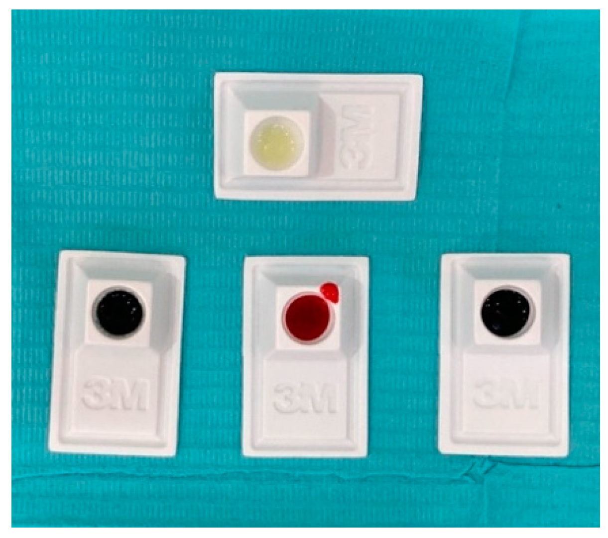
  • Group A Snoop—blue color (SNOOP Caries Detecting Dye—Pulpdent Corporation, Watertown, MA, USA).
  • Group B Sable seek—black color (Ultradent Sable™ Seek™ Caries Indicator, Aam Westhover Berg, Germany).
  • Group C Elements—pink color (Elements, Caries Indicator, Industrial Estate, Digiana, Jammu, India).
All cavity walls and surfaces of samples in group I were coated using a microbrush with caries detector dyes for 10 s, subsequently washed with water for another 10 s and finally air-dried [37]. A similar procedure was carried for teeth in group II after the application of a bonding agent on visibly sound tooth surfaces without the use of an etchant. Both groups were visibly examined for staining by the caries detector dyes and recorded (Figure 2 and Figure 3). The assessment of staining was done by a single operator under the same controlled lighting conditions. Staining was measured using the modified extrinsic stain index, and the teeth scored for staining were considered stained irrespective of the intensity of the stained index score [38].

2.4. Removal of Staining

In a sample of 45 teeth (groups A, B, and C) all the teeth stained by the caries indicator dyes were stored in a glass container with distilled water for 10 min and later cleaned and dried with a cotton gauge. The leftover stained teeth were placed in 3.25% sodium hypochlorite for another 10 min and then cleaned with distilled water [39]. The ability of distilled water and sodium hypochlorite in removing caries indicator dyes from the tooth surface of all the groups were counted using the same index (modified extrinsic stain index), and data were tabulated. The entire experiment was performed by a single operator to minimize the flaws in applying the bonding agent, dyes, recording the complimentary staining and during its removal.
Descriptive statistics were computed, and results were presented as percentages. The statistical analyses were performed using SPSS (IBM Corp. IBM SPSS Statistics for Macintosh, Version 27.0. Armonk, NY, USA: IBM Corp.) and conducted with a confidence interval (CI) of 95% and a significance level of 0.05. A Mann–Whitney U test was used to check the statistical difference between groups. To check the statistical significance in stain removal among the different groups, a Kruskal–Wallis test was used.

3. Results

A total of 90 primary extracted teeth were tested. All the teeth samples used in the experiment were divided into two main groups receiving caries detector dyes, Group I (without dentine bonding agent) and group II (with dentine bonding agent) and again, each group was divided in to three subgroups, groups A, B and C (Table 1).
In group I, among the three different caries detector dyes, all the teeth were stained in group C followed by group A and group B, respectively. When observations were noted in group II with dentine bonding agent application, only four teeth (26.6%) in group C showed the staining (Figure 4). The Mann–Whitney U test showed a statistically significant difference between the two groups for staining of different tooth surfaces (p < 0.05) (Table 2). Table 3 shows the ability of distilled water and sodium hypochlorite to remove caries indicator dyes from the tooth surface. Out of the 15 samples of Snoop caries detector dye, 10 teeth had the stain removed by distilled water (66.6%) and completely with sodium hypochlorite (100%). Whereas in the Sable seek group, in 11 samples, the stain was removed with distilled water (73%), and in all 15 samples, the dye stain was removed when sodium hypochlorite was used (100%). In the Elements group, the stain was not removed in any of the sample when distilled water was used (0%), while only in four samples was sodium hypochlorite able to remove the caries detector dye stain (26.66%) (Figure 5). The Kruskal–Wallis one-way ANOVA revealed a statistically significant difference in stain removal among the different groups either by distilled water or sodium hypochlorite as observed in Table 4 and Figure 6.

4. Discussion

To avoid unnecessary treatment, a diagnostic aid should show a very low level of false positive. This study investigated the use of caries detector dyes with or without the use of a bonding agent to explore the efficacy of caries detector dyes, which in turn will help provide some insight in its clinical applications and significance, as it will aid in providing clinicians with an objective means of diagnosing caries and preserving healthy tooth structure when treating carious teeth. Some studies showed that not all dye-stainable dentin was infected when it was compared to the level of infection of unstained and stained dentin at the amelodentinal junction, after the completion of the cavity preparation [32]. Caries detection dyes are less expensive, available in the market and can be used effectively as a complementary examination at the dental chair. The reason for using a caries detector dye is to make a clear-cut differentiation between the tissue to be removed and the tissue to be kept, ensuring a conservative treatment, and to avoid the possible exposure of the pulp. These nonspecific dyes will stain food debris, enamel pellicle and any other organic matter trapped in substantial amounts in occlusal fissures and possibly demineralized enamel, so false positives are of significant concern [24]. According to Mc Comb none of the caries detector dyes available are caries-specific, and their routine use can lead to overtreatment and pulp exposure [24]. In the present study we also observed similar results in group I, where all three caries detector dyes also stained noncarious part of the tooth surfaces. These results are in accordance with previous studies where authors have stated that dyes that reveal disintegration do not prove to be specific for caries lesions but also stain healthy circumpulpal dentin and healthy dentin at the enamel–dentin junction, which have a lower natural mineral content and are relatively more porous [23,24,25]. Boston et al. found that the use of a caries indicator dye with a polypropylene glycol solvent showed comparatively less penetration than the propylene glycol solvent alone [40].
Also, in the present study, we have applied a dentin bonding agent without applying any etchant, since the etchant can further make enamel more porous in primary teeth, hence the chances of the dye taking up will be increased [41]. Interestingly, we noted that caries detecting dyes were not stained and penetrated in the sound tooth surfaces where the dentine bonding agent was applied on visibly caries-free surfaces. This study is the first of its kind where the effectiveness of the dentine bonding agent was investigated to evaluate the staining ability of caries detector dyes after the application of a dentine bonding agent. Further studies are required with sectioning and scanning the tooth surface to detect the ability of the bonding agent penetration and the avoidance of tooth staining by the caries detector dyes. In their report, Singh et al. found that sound and carious dentin surfaces exhibited less bond strength with the application of caries detection dyes [37]. The study conducted by Emine et al. using different cleaning solutions like distilled water, 3% sodium hypochlorite and 3% hydrogen peroxide for removing the residues of caries detector dyes revealed that sodium hypochlorite was more effective compared to the other agents, and the results were similar to those of the current study [39].
A limitation of the present study Is that although in vitro studies provide a tight control over chemical and physical environments, they fail to replicate the conditions of living organisms which can impede the extrapolation of the results obtained. Hence, future studies should focus on an in vivo model that can provide more reliable results. The findings from this in vitro study could help researchers conduct more investigations on the new bioactive caries-detecting solutions [42,43]. Additionally, different enamel remineralization agents such as fluorides [44], casein phosphopeptide-amorphous calcium phosphate [45] and biomimetic hydroxyapatite [46] have shown promising results. Future studies should focus on investigating caries detector dyes in combination with these agents, as it will provide a more complete overview.

5. Conclusions

The present study evaluated caries detector dyes for staining carious tooth surfaces. The dyes used in the study did not prove to be specific for carious lesions and also stained healthy circumpulpal dentin and healthy dentin at the enamel–dentin junction. Whereas when the same dye was used after the application of a dentine bonding agent on sound tooth surfaces prior to the use of the dye, it spread to many sound tooth surfaces which were caries-free; hence, further studies are required to find the ability of dentine bonding agent to prevent caries detector dye penetration in noncarious tooth surfaces. The dyes’ exact formulations were not mentioned by the manufacturers, which may be a cofactor for complimentary staining of sound tooth surfaces.

Author Contributions

Conceptualization, A.A. (AlWaleed Abushanan) and R.B.S.; methodology, A.A. (AlWaleed Abushanan) and R.B.S.; software, A.A. (AlWaleed Abushanan) and R.B.S.; validation, A.A. (AlWaleed Abushanan) and R.B.S.; formal analysis, A.A. (AlWaleed Abushanan), R.B.S. and M.A.; investigation, A.A. (AlWaleed Abushanan) and R.B.S.; resources, A.A. (AlWaleed Abushanan), M.A. and A.A. (Abdulfatah Alazmah); data curation, M.A. and A.A. (Abdulfatah Alazmah); writing—original draft preparation, A.A. (AlWaleed Abushanan), R.B.S. and A.A. (Abdulfatah Alazmah); writing—review and editing, A.A. (AlWaleed Abushanan) and R.B.S.; visualization, A.A. (Abdulfatah Alazmah) and M.A. All authors have read and agreed to the published version of the manuscript.

Funding

The authors would like to acknowledge the Prince Sattam bin Abdul-Aziz University for their support in funding the study via project number (PSAU/2023/R/1445).

Institutional Review Board Statement

The study was conducted according to the guidelines of the Declaration of Helsinki and approved by the Institutional Review Board of College of Dentistry, at Prince Sattam bin Abdulaziz University (PSAU2021015 and date of approval 18 April 2021).

Informed Consent Statement

Informed consent was obtained from all subjects involved in the study.

Data Availability Statement

The data presented in this study are available on request from the corresponding author. The data are not publicly available due to their containing information that could compromise the privacy of research participants.

Acknowledgments

The authors would like to acknowledge the Prince Sattam bin Abdul-Aziz University for their support in funding the study.

Conflicts of Interest

The authors declare no conflict of interest.

References

  1. Iwami, Y.; Hayashi, N.; Yamamoto, H.; Hayashi, M.; Takeshige, F.; Ebisu, S. Evaluating the objectivity of caries removal with a caries detector dye using color evaluation and PCR. J. Dent. 2007, 35, 749–754. [Google Scholar] [CrossRef] [PubMed]
  2. Roberson, M.; Heymann, O. Sturdevant’s Art and Science of Operative Dentistry, 5th ed.; Mosby: St. Louis, MO, USA, 2006; pp. 308–311. [Google Scholar]
  3. Thompson, V.; Craig, R.G.; Curro, F.A.; Green, W.S.; Ship, J.A. Treatment of deep carious lesions by complete excavation or partial removal: A critical review. J. Am. Dent. Assoc. 2008, 139, 705–712. [Google Scholar] [CrossRef] [PubMed]
  4. Tyas, M.J.; Anusavice, K.J.; Frencken, F.E.; Mount, G.J. Minimal intervention dentistry—A review. FDI Commission Project 1-97. Int. Dent. J. 2000, 50, 1–12. [Google Scholar] [CrossRef]
  5. Murdoch-Kinch, C.A.; McLean, M.E. Minimally invasive dentistry. J. Am. Dent. Assoc. 2003, 134, 87–95. [Google Scholar] [CrossRef] [PubMed]
  6. Mount, G.J.; Ngo, H. Minimal intervention; a new concept for operative dentistry. Quintessence Int. 2000, 31, 527–533. [Google Scholar]
  7. FDI. FDI Policy Statement: Minimal Intervention in the Management of Dental Caries; Adopted by the FDI General Assembly: 1 October 2002-Vienna; FDI: Vienna, Austria, 2002. [Google Scholar]
  8. Fusayama, T.; Terashima, S. Differentiation of two layers of carious dentin by staining. J. Dent. Res. 1972, 51, 866. [Google Scholar] [CrossRef] [PubMed]
  9. Fusayama, T. Two layers of carious dentin; diagnosis and treatment. Oper. Dent. 1979, 4, 63–70. [Google Scholar]
  10. Ohgushi, K.; Fusayama, T. Electron microscopic structure of the two layers of carious dentin. J. Dent. Res. 1975, 54, 1019–1026. [Google Scholar] [CrossRef] [PubMed]
  11. Kuboki, Y.; Ohgushi, K.; Fusayama, T. Collagen biochemistry of two layers of carious dentin. J. Dent. Res. 1977, 56, 1233–1237. [Google Scholar] [CrossRef]
  12. Miyauchi, H.; Iwaku, M.; Fusayama, T. Physiological recalcification of carious dentin. Bull. Tokyo Med. Dent. Univ. 1978, 25, 169–179. [Google Scholar]
  13. Banerjee, A.; Watson, T.F.; Kidd, E.A. Dentine caries: Take it or leave it? Dent. Update 2000, 27, 272–276. [Google Scholar] [CrossRef] [PubMed]
  14. Kidd, E.; Toyston-Bechal, S.; Beighton, D. Microbiological validation of assessments of caries activity during cavity preparation. Caries Res. 1993, 27, 402–408. [Google Scholar] [CrossRef] [PubMed]
  15. Weerheijm, K.L.; de Soet, J.J.; van Amirongen, W.E.; de Graaff, J. The effect of grass-ionomer cement on caries dentin: An in vivo study. Caries Res. 1993, 27, 417–423. [Google Scholar] [CrossRef] [PubMed]
  16. Kidd, E.A.; Fejerskov, O.; Mjor, I. Caries removal and the pulpo-dentinal complex. In Dental Caries: The Disease and Its Clinical Management, 1st ed.; Fejerskov, O., Kidd, E., Eds.; Blackwell Munksgaard: Oxford, UK, 2003; pp. 267–274. [Google Scholar]
  17. Anderson, M.H.; Charbeneau, G.T. A comparison of digital and optical criteria for detecting carious dentin. J. Prosthet. Dent. 1985, 53, 643–646. [Google Scholar] [CrossRef] [PubMed]
  18. Kidd, E.A.; Joyston-Bechal, S.; Beighton, D. The use of a caries detector dye during cavity preparation: A microbiological assessment. Br. Dent. J. 1993, 174, 245–248. [Google Scholar] [CrossRef] [PubMed]
  19. Fusayama, T. Clinical guide removing caries using a caries-detecting solution. Quintessence Int. 1988, 19, 397–401. [Google Scholar] [PubMed]
  20. Zacharia, M.A.; Munshi, A.K. Microbiological assessment of dentin stained with a caries detector dye. J. Clin. Pediatr. Dent. 1995, 19, 111–115. [Google Scholar]
  21. Itoh, K.; Kusunoki, M.; Oikawa, M.; Tani, C.; Hisamitsu, H. In vitro comparison of three caries dyes. Am. J. Dent. 2009, 22, 195–199. [Google Scholar]
  22. Javaheri, M.; Maleki-Kambakhsh, S.; Etemad-Moghadam, S. Efficacy of two caries detector dyes in the diagnosis of dental caries. J. Dent. 2010, 7, 71–76. [Google Scholar]
  23. Banerjee, A.; Kidd, E.A.; Watson, T.F. In vitro validation of carious dentin removed using different excavation criteria. Am. J. Dent. 2003, 16, 228–230. [Google Scholar]
  24. McComb, D. Caries-detector dyes—How accurate and useful are they? J. Can. Dent. Assoc. 2000, 66, 195–198. [Google Scholar] [PubMed]
  25. Yip, H.K.; Stevenson, A.G.; Beeley, J.A. The specificity of caries detector dyes in cavity preparation. Br. Dent. J. 1994, 176, 417–421. [Google Scholar] [CrossRef] [PubMed]
  26. Boston, D.W.; Liao, J. Staining of non-carious human coronal dentin by caries dyes. Oper. Dent. 2004, 29, 280–286. [Google Scholar] [PubMed]
  27. Franco, S.J.; Kelsey, W.P. Caries removal with and without a disclosing solution of basic fuchsin. Oper. Dent. 1981, 6, 46–48. [Google Scholar]
  28. Boston, D.W.; Graver, H.T. Histologicial study of an acid red caries-disclosing dye. Oper. Dent. 1989, 14, 186–192. [Google Scholar]
  29. Akbari, M.; Rouhani, A.; Samiee, S.; Jafarzadeh, H. Effect of dentin bonding agent on the prevention of tooth discoloration produced by mineral trioxide aggregate. Int. J. Dent. 2012, 2012, 563203. [Google Scholar] [CrossRef]
  30. Choi, Y.L.; Jang, Y.E.; Kim, B.S.; Kim, J.W.; Kim, Y. Pre-application of dentin bonding agent prevents discoloration caused by mineral trioxide aggregate. BMC Oral Health 2020, 20, 163. [Google Scholar] [CrossRef]
  31. Melo, A.M.D.S.; Santos, T.J.S.D.; Tertulino, M.D.; dos Medeiros, M.C.S.; da Silva, A.O.; Borges, B.C.D. Degree of conversion, translucency and intrinsic color stability of composites during surface modeling with lubricants. Braz. J. Oral Sci. 2018, 17, e18325. [Google Scholar] [CrossRef]
  32. Araujo, F.S.; Barros, M.C.R.; Santana, M.L.C.; de Jesus Oliveira, L.S.; Silva, P.F.D.; Lima, G.D.S.; Faria-E-Silva, A.L. Effects of adhesive used as modeling liquid on the stability of the color and opacity of composites. J. Esthet. Restor. Dent. 2018, 30, 427–433. [Google Scholar] [CrossRef]
  33. Demacro, F.F.; Matos, A.B.; Matson, E.; Powers, J.M. Dyes for caries detection influence sound dentin bond strength. Oper. Dent. 1998, 23, 294–298. [Google Scholar]
  34. Hui, L.E.; Thomas, M.S.; Jathanna, V.; Lewis, A.J.; Srikant, N. Effect of Caries Detecting Dye on Microleakage of Composite Resin Restorations Bonded with Total-etch and Self-etch Adhesive Systems. J. Clin. Diagn. Res. 2019, 13, ZC01–ZC03. [Google Scholar] [CrossRef]
  35. Singla, T.; Pandit, I.K.; Srivastava, N.; Gugnani, N.; Gupta, M. An evaluation of microleakage of various glass ionomer based restorative materials in deciduous and permanent teeth: An in vitro study. Saudi Dent. J. 2012, 24, 35–42. [Google Scholar] [CrossRef] [PubMed]
  36. Ravichandran, N.K.; Tumkur Lakshmikantha, H.; Park, H.S.; Jeon, M.; Kim, J. Analysis of Enamel Loss by Prophylaxis and Etching Treatment in Human Tooth Using Optical Coherence Tomography: An In Vitro Study. J. Healthc. Eng. 2019, 8973825. [Google Scholar] [CrossRef] [PubMed]
  37. Singh, U.P.; Tikku, A.P.; Chandra, A.; Loomba, K.; Boruah, L.C. Influence of caries detection dye on bond strength of sound and carious affected dentin: An in-vitro study. J. Conserv. Dent. 2011, 14, 32–35. [Google Scholar] [CrossRef] [PubMed]
  38. Marya, C.M.; Taneja, P.; Ahuja, T.K.; Nagpal, R.; Kataria, S. Modified extrinsic tooth stain index and its clinical applicability. J. Indian Soc. Periodontol. 2023, 27, 95–98. [Google Scholar]
  39. Tunc, E.S.; Sahin, Z.; Guler, E.; Bayrak, S.; Tuloglu, N. Effectiveness of different cleaning solutions in removing residual caries detector dyes. J. Esthet. Restor. Dent. 2018, 31, 253–256. [Google Scholar] [CrossRef]
  40. Bostona, D.W.; Jefferiesb, S.R.; Gaughanc, J.P. The Relative Location of the Dye Staining Endpoint Indicated With Polypropylene Glycol-Based Caries Dye versus Conventional Propylene Glycol-Based Caries Dye. Eur. J. Dent. 2008, 2, 29–36. [Google Scholar] [CrossRef]
  41. Mirjanic, V.; Mirjanić, Đ.; Arbutina, A. Changes on dental enamel on acid etching. Contemp. Mater. 2016, 2, 185–189. [Google Scholar] [CrossRef]
  42. Govind, S.; Jena, A.; Kamilla, S.K.; Mohanty, N.; Mallikarjuna, R.M.; Nalawade, T.; Saraf, S.; Khaldi, N.A.; Jahdhami, S.A.; Shivagange, V. Diagnosis and Assessment of Dental Caries Using Novel Bioactive Caries Detecting Dye Solution. Biomedicines 2023, 11, 500. [Google Scholar] [CrossRef]
  43. Shashirekha, G.; Jena, A.; Mohanty, N.; Kamilla, S.K. Novel bioactive caries-detecting dye solution: Cytotoxicity, antimicrobial activity, scanning electron microscope, and stereomicroscopic analysis in diagnosis of dental caries. J. Conserv. Dent. 2020, 23, 79–85. [Google Scholar]
  44. Zampetti, P.; Scribante, A. Historical and bibliometric notes on the use of fluoride in caries prevention. Eur. J. Paediatr. Dent. 2020, 21, 148–152. [Google Scholar] [PubMed]
  45. Fallahzadeh, F.; Heidari, S.; Najafi, F.; Hajihasani, M.; Noshiri, N.; Nazari, N.F. Efficacy of a Novel Bioactive Glass-Polymer Composite for Enamel Remineralization following Erosive Challenge. Int. J. Dent. 2022, 2022, 6539671. [Google Scholar] [CrossRef] [PubMed]
  46. Butera, A.; Pascadopoli, M.; Pellegrini, M.; Trapani, B.; Gallo, S.; Radu, M.; Scribante, A. Biomimetic hydroxyapatite paste for molar-incisor hypomineralization: A randomized clinical trial. Oral Dis. 2023, 29, 2789–2798. [Google Scholar] [CrossRef] [PubMed]
Figure 1. 3 Different caries detector dyes (Snoop—blue color, Sable seek—black color, Elements—pink color) and bonding agent.
Applsci 13 12124 g001
Figure 2. Staining observed in group I.
Applsci 13 12124 g002
Figure 3. Staining observed in group II.
Applsci 13 12124 g003
Figure 4. Staining of the caries detector dyes with/without dentine bonding agent.
Applsci 13 12124 g004
Figure 5. Comparison of distilled water and 3.25% sodium hypochlorite staining removal efficiency amongst the three different groups (Snoop, Sable seek and Elements).
Applsci 13 12124 g005
Figure 6. Mean values of distilled water and 3.25% sodium hypochlorite staining removal amongst the three different groups (Snoop, Sable seek and Elements).
Applsci 13 12124 g006
Table 1. Staining of the caries detector dyes with/without a dentine bonding agent.
Caries Detector DyesWithout Dentine Bonding Agent (Group I)With Dentine Bonding Agent (Group II)
Group ASnoop5 (33.3%)0 (0%)
Group BSable seek4 (26.6%)0 (0%)
Group CElements15 (100%)4 (26.6%)
Table 2. Comparison of different caries detector dyes staining of different tooth surfaces according to the use of a bonding agent.
GroupsSubgroupsMean RankSum of Ranks
Without bonding agent13.00195Mann–Whitney U = 270.00
p value = 0.016
Snoop
With bonding agent18.00270
Without bonding agent17.50210Mann–Whitney U = 82.50
p value = 0.035
Sable seek
With bonding agent13.50255
Without bonding agent17.50210Mann–Whitney U = 82.50
p value = 0.035
Elements
With bonding agent13.50255
Table 3. Stain removal efficiency of distilled water and sodium hypochlorite.
Caries Detector DyesWith Distilled Water With Sodium Hypochlorite 3.25%
Group A-15Snoop10 (66.6%)15 (100%)
Group B-15Sable seek11 (73%)15 (100%)
Group C-15Elements0 (0%)4 (26%)
Table 4. Kruskal–Wallis analysis for comparison of stain removal among the different groups either by distilled water or sodium hypochlorite.
GroupsMean Rank
Distilled waterSnoop15.50Chi Square = 44.00
p < 0.001
Sable seek15.50
Elements38.00
Sodium hypochloriteSnoop15.00Chi Square = 17.25
p < 0.001
Sable seek22.50
Elements31.50
Disclaimer/Publisher’s Note: The statements, opinions and data contained in all publications are solely those of the individual author(s) and contributor(s) and not of MDPI and/or the editor(s). MDPI and/or the editor(s) disclaim responsibility for any injury to people or property resulting from any ideas, methods, instructions or products referred to in the content.

