Review

Oral Cavity Mucocele and Different Surgical Treatment Strategies: Is Laser Excision Effective? A Scoping Review

by
Andrea Scribante
1,*,
Matteo Pellegrini
2,3,
Federica Pulicari
2,3,*,
Francesca De Martino
2,3,
Giacomo Li Vigni
2,3,
Martina Ghizzoni
1 and
Francesco Spadari
2,3
1
Section of Dentistry, Department of Clinical, Surgical, Diagnostic and Pediatric Sciences, University of Pavia, 27100 Pavia, Italy
2
Maxillofacial Surgery and Dental Unit, Fondazione IRCCS Cà Granda Ospedale Maggiore Policlinico, 20122 Milan, Italy
3
Department of Biomedical, Surgical and Dental Sciences, University of Milan, Via della Commenda 10, 20122 Milan, Italy
*
Authors to whom correspondence should be addressed.
Appl. Sci. 2023, 13(22), 12327; https://doi.org/10.3390/app132212327
Submission received: 21 October 2023 / Revised: 3 November 2023 / Accepted: 8 November 2023 / Published: 14 November 2023
(This article belongs to the Section Applied Dentistry and Oral Sciences)

Abstract

:
Mucocele is a common lesion localized in the oral cavity; it originates from the lesion of a salivary duct and the consequent accumulation of mucin in soft tissues. It is a common lesion of young patients and is frequently associated with areas subject to traumas. Surgical treatment is needed especially if it reaches considerable dimensions, as it can cause discomfort in the patient and tends to increase in size if subjected to further traumatism. This scoping review aims to investigate which treatments are used for this type of lesion, which are the most suitable to prevent recurrence, and if laser-assisted surgery is considered more efficient than other methods. An electronic search was performed within the PubMed (MEDLINE) and Scopus databases. Articles published in the years 2010 to 2023 were selected. Techniques like marsupialization and laser-assisted excision effectively treat mucoceles, with no clear prevalence between them. Laser surgery offers speed, precision, and minimal post-operation discomfort, but randomized trials are needed for conclusive comparisons.

1. Introduction

A mucocele is a prevalent oral lesion, with histological findings indicating its occurrence in approximately 11.6% to 24.5% of all pediatric biopsies [1,2]. This lesion can affect individuals of various age groups, including neonates, infants, and adults [1,2]. Clinically, mucoceles typically manifest as dome-shaped, intramucosal growths, most frequently observed on the lower lip. Due to its clinical and histological characteristics, the lesion can be considered a pseudocyst. They can also occur near other salivary gland ducts, such as those in the palate, the ventral part of the tongue, buccal glands, upper labial glands, and the retromolar region [1,2]. Mucoceles can appear in different regions of the oral cavity, including the lower lip, tongue, floor of the mouth, and inner cheeks. Lower lip mucoceles are particularly common due to their increased vulnerability to trauma from accidental biting or repeated friction. These lesions are closely linked to minor salivary glands that are distributed throughout the oral mucosa, responsible for saliva production to aid in digestion and lubrication [2]. Mucoceles are typically rounded, bluish, or translucent in appearance and can vary in size from 1 mm to several centimeters in diameter [2,3]. On a histological level, mucoceles exhibit granulomatous tissue with mucosal liquid content, and in cases of inflammation, neutrophils and macrophages may be present [3]. They are considered benign and asymptomatic growths, with a duration that can extend over years [3,4]. These lesions can arise from two distinct mechanisms: extravasation, resulting from the rupture of a minor salivary gland duct and the accumulation of mucin in the surrounding soft tissue, subsequently surrounded by granulation tissue; or retention cysts, which occur due to ductal obstruction, leading to mucin accumulation and ductal epithelium lining, making it a true cyst [2,3,4]. In the oral cavity, mucoceles located in the floor of the mouth are commonly referred to as “ranula.” Ranulas often involve submandibular glands, originating from either the Wharton’s duct or, in some cases, Rivinus’s duct, associated with the sublingual gland. Trauma, such as accidental biting, repetitive irritation, or minor surgical procedures, can trigger the rupture or blockage of the duct, leading to an inflammatory response and the enlargement of the cystic cavity [5,6].
Differential diagnoses for mucoceles may include fibrous hyperplasia, focal papilloma, lipoma, fibroma, mucoepidermoid carcinoma, bullous lichen planus, pemphigoid, and herpes [5,6].
While certain mucoceles may naturally resolve within a brief period, if they persist chronically, surgical intervention or marsupialization becomes necessary [7]. Numerous therapeutic approaches have been outlined in the medical literature, including surgical excision with a scalpel, carbon dioxide ablation, laser excision, marsupialization, and cryosurgery [7]. In the event of a recurrence, it is recommended to remove the cyst, together with the adjacent salivary glands, down to the muscular layer [8]. Presently, surgical enucleation stands as the most commonly employed technique for this purpose, but non-surgical methods are also described in the literature, such as micro-marsupialization, the topical application of clobetasol, corticosteroid use, and gamma-Linoleic acid [9].

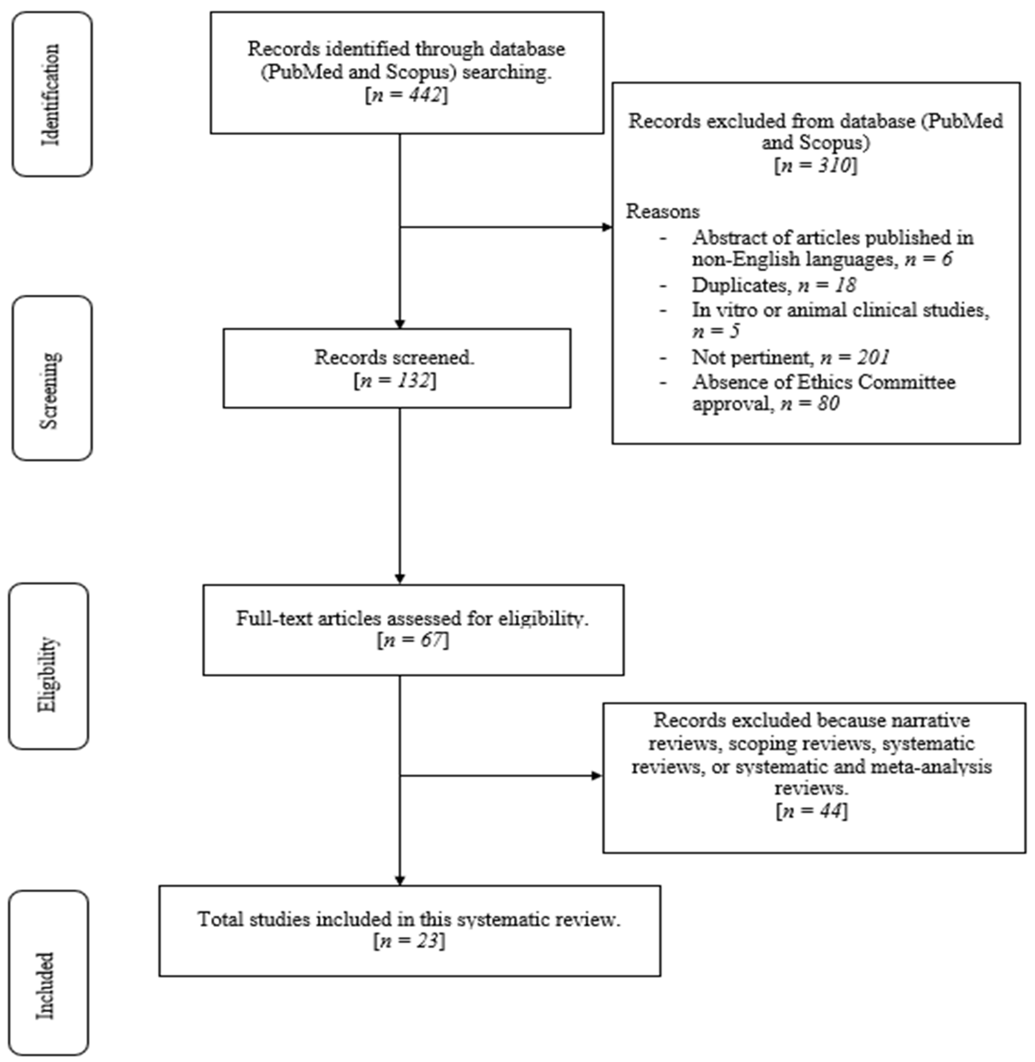
2. Materials and Methods

2.1. Focused Questions

What therapies are currently available for oral mucoceles? What is the management strategy used in oral mucocele therapy? Do surgical therapies have better results than non-surgical approaches regarding oral mucocele management? Are there any recurrences following a surgical approach? Is laser excision effective in oral mucocele treatment?

2.2. Eligibility Criteria

The criteria used to select studies for this review were as follows: (I) study design, which encompassed interventional studies, observational studies, cohort studies, and case series/case reports; (II) study participants, specifically patients diagnosed with an oral mucocele; (III) the interventions of interest, which were surgical and non-surgical methods and laser-assisted protocols; and (IV) the outcome measure, which focused on clinical results for treating oral mucoceles and if a recurrence was reported. Only studies meeting all of these inclusion criteria were included in the examination. Exclusion criteria encompassed: (I) abstracts published in languages other than English; (II) duplicate studies; (III) studies not pertinent to the review’s purpose, including those addressing different supplementary treatments or having full-text content that did not align with their abstracts; (IV) studies conducted on ex vivo or experimental animals; (V) studies lacking Ethics Committee approval; and (VI) narrative reviews, systematic reviews, or systematic and meta-analysis reviews.

2.3. Search Strategy

Following the Joanna Briggs Institute methodology for a scoping review [8], a three-step search process was conducted: (i) an initial limited search on PubMed (MEDLINE) and Scopus; (ii) the selection of key terms from the articles retrieved to formulate the search strategy; and (iii) a search through the reference lists of all included articles to identify additional research [9].
Additionally, the review applied the Population/Concept/Context (PCC) model, which consists of three key elements: population (comprising individuals undergoing surgical and non-surgical procedures), concept (encompassing both surgical and non-surgical treatments for oral mucocele), and context (with no specific cultural or setting restrictions in this review). Abstracts of studies that investigated the impacts of surgical and non-surgical procedures and their clinical outcomes were assessed, including case reports and case series articles, while reviews were excluded from the search. Throughout this scoping review of the literature, adherence to the Preferred Reporting Items for Scoping Reviews (PRISMA-ScR) consensus guidelines was maintained (see Table S1 in the Supplementary Material).

2.4. Research

Medical Subject Heading (MeSH) terms utilized in this study encompassed mucocele, mucoceles, oral mucocele, lip mucocele, tongue mucocele, and oral mucocele therapy. An electronic search was conducted on the PubMed (MEDLINE) and Scopus databases. Articles published between 2010 and 2023 were the target of selection. The data extraction period occurred between May 2023 and June 2023, with the final search conducted on 11 June 2023. Four expert reviewers (F.P., F.D.M., M.G., and G.L.V.) carried out the initial search, with any disagreements or inconsistencies resolved through consensus or consultation with three additional reviewers (F.S., A.S., and M.P.).
All titles and abstracts from the initial search were meticulously assessed, and studies that were not relevant were excluded. Subsequently, all pertinent articles underwent thorough scrutiny, involving an analysis of their full texts, documentation of their findings, and identification of any similar studies that met the predefined inclusion criteria.
The protocol has been registered within the Open Science Framework platform (Registration DOI 10.17605/OSF.IO/Q3NE).
The elaborated strategies applied for each electronic database are exhibited in Table S2 (Supplementary Material).

2.5. Quality Assessment of Included Studies

A methodological quality risk of bias assessment was used in this review, as well as JBI critical appraisal for case reports [9] and JBI critical appraisal for case series [10].

3. Results

The primary search identified 442 articles based on MeSH terms. Following this, 310 articles were removed (6 abstracts of articles published in non-English languages, 18 duplicates, and 201 because they were not pertinent), and 132 articles were screened based on their titles and abstracts. The remaining 67 full-text articles were assessed for eligibility. Additionally, 33 full-text articles were further excluded because they were irrelevant articles. The 30 relevant articles were finally included and analyzed in this review. We included 10 case reports and 13 case series. The flow chart of the review process is described in Figure 1.

Risk of Bias

The JBI critical appraisal tool was applied to assess the risk of bias in the studies included in this review (Table 1 and Table 2), using the judging criteria for risk of bias shown in Tables S2 and S3 (Supplementary Materials) [9,10].

4. Discussion

The management of oral mucoceles involves a spectrum of strategies, both surgical and non-surgical, tailored to the lesion’s specific characteristics and the patient’s requirements. Surgical interventions encompass various techniques, with excision being the most utilized approach. Surgical excision entails the complete removal of the cystic lesion and its associated glandular tissue, minimizing the risk of recurrence. Laser ablation, an increasingly popular choice, provides precise removal with minimal bleeding and reduced scarring. Another surgical method, marsupialization, involves creating a controlled opening to facilitate continuous drainage, thereby reducing the risk of reaccumulation [34,35].
Non-surgical approaches are also considered, particularly for smaller, asymptomatic mucoceles. Cryotherapy employs freezing to eliminate the cystic tissue, while steroid injections help alleviate inflammation and encourage regression [34,35]. However, these methods may have limitations in terms of the lesion’s size and effectiveness. Conservative management, involving observation without active intervention, may be appropriate in specific cases, particularly when the mucocele exhibits signs of spontaneous regression.
The choice between surgical and non-surgical approaches hinges on factors like lesion size, patient discomfort, location, and the potential for recurrence [36,37,38,39]. Surgical methods are more definitive and efficacious for larger, symptomatic mucoceles. Non-surgical techniques, while less invasive, might be preferred for smaller lesions or individuals who wish to avoid surgery. A comprehensive evaluation by a healthcare professional is paramount to determine the optimal approach [39,40]. Collaborative decision-making involving the patient’s preferences, the clinician’s expertise, and the specific characteristics of the mucocele contributes to a successful outcome, minimizing recurrence and ensuring patient satisfaction [41,42].
Surgical or non-surgical management is recommended when the mucocele is causing discomfort, pain, or interference with oral function. Generally, this is a growth-prone lesion, without spontaneous resolution, and can reach a considerable size [43,44,45,46,47,48].

4.1. Laser-Assisted Surgery in Stomatology

Laser-assisted surgery has brought transformative changes to the field of stomatology, offering a precise and minimally invasive approach to treating various oral conditions involving soft tissues. This innovative technique utilizes the energy of focused light to interact with tissues, providing benefits such as reduced bleeding, faster healing, and enhanced patient comfort [48,49,50,51,52,53].
In soft tissue applications, such as gingivectomy and frenectomy, lasers have shown exceptional precision by selectively targeting and removing tissue without causing unnecessary trauma to surrounding healthy areas. This level of precision not only improves surgical outcomes but also reduces postoperative complications, making it a valuable tool in the hands of skilled practitioners [48,49,50,51,52,53].
One of the standout advantages of laser-assisted surgery is its ability to promote hemostasis during procedures. The coagulation effect of lasers significantly reduces bleeding, providing a clear surgical field for the dentist or oral surgeon [48,49,50,51,52,53]. This improved visibility allows for better accuracy in tissue removal and treatment, ultimately leading to a more successful outcome [48,49,50,51,52,53].
Patients undergoing laser-assisted procedures often experience less pain and discomfort compared to traditional surgical methods. The minimally invasive nature of laser surgery reduces nerve stimulation and tissue trauma, resulting in faster recovery times and less reliance on pain management [48,49,50,51,52,53]. Additionally, the reduced need for sutures, due to minimal tissue damage, contributes to a more comfortable and convenient patient experience [48,49,50,51,52,53].
Laser-assisted surgery also offers advantages in terms of sterilization. The high-energy light used in laser procedures possesses inherent sterilizing properties, which can lower the risk of postoperative infections. This is particularly significant in the oral cavity, where infections can lead to serious complications if not managed properly [48,49,50,51,52,53].
Laser surgery plays a vital role in the context of biopsies, ensuring precise and minimally invasive tissue sampling. The utilization of lasers in biopsy procedures presents several benefits, which include enhanced accuracy, reduced patient discomfort, and an increased level of confidence in achieving clear margins [48,51]. During laser surgery, there is a phenomenon known as carbonization, where tissue vaporizes due to the intense heat generated by the laser. While carbonization may raise concerns about the integrity of the tissue sample, contemporary laser systems are designed to minimize this effect [52]. By carefully selecting the appropriate laser parameters, skilled practitioners can prevent excessive carbonization, ensuring that biopsy samples remain viable for precise pathological examination [48,51]. The attainment of clear margins is of paramount importance in biopsy procedures, especially in cases involving suspected malignancies. Laser-assisted surgery offers distinct advantages in ensuring the adequacy of margins [49,52]. The precision of lasers enables healthcare professionals to meticulously remove tissue layers, thereby optimizing the likelihood of achieving complete excision and reducing the necessity for subsequent re-excisions. This is pivotal for accurate staging and treatment planning, particularly in cases involving cancerous tissue [50,51,52]. Diode lasers, despite being the most cost-effective among dental lasers, should not be dismissed as a valuable tool for biopsies. They offer several advantages, including a bloodless surgical field, swift healing, ease of operation, and bactericidal effects that reduce the risk of infection post-treatment. There is evidence to suggest that diode lasers may stimulate clotting factor VII, enhancing hemostasis due to the “hot-tip” effect, causing thermocoagulation [53].
In situations where thermal damage is a concern, irrigation with saline during laser operation can effectively dissipate thermal energy. Studies have shown that a diode laser at 2 W can penetrate tissue effectively up to at least 2 mm, which can be sufficient to seal small lymphatic vessels, reducing postoperative edema. Research demonstrates that biopsies can be collected using diode lasers without altering the microanatomy of the specimen [53]. The choice of wavelength in diode lasers also plays a role, with a 980 nm diode laser cutting slightly faster than an 810 nm diode. This is because the 980 nm diode has greater water absorption, which is the predominant component in most oral tissues, compared to the 810 nm diode, which exhibits less affinity to water. This difference is particularly noticeable when ablating tissues with minimal pigmentation [53]. Neodymium-doped: yttrium aluminum garnet (Nd:YAG) lasers, similar to diode lasers, work well for soft-tissue ablation due to their affinity for pigmented tissues. However, the key difference is the high energy output of Nd:YAG lasers [53]. This high peak power enables the delivery of larger energy bursts in shorter pulses, allowing for deeper tissue penetration and generating a stronger thermal response in deep tissues, often leading to coagulation. In a study involving histological specimens just under 7 mm in size, Nd:YAG lasers exhibited significant thermal effects. The guidelines suggest that using a higher frequency with lower power settings can result in more precise incisions with less tissue damage [53]. Carbon dioxide (CO2) lasers are effective for benign and premalignant intraoral lesion treatment, especially broad-based lesions, due to their collimated beam delivered through a hollow light guide. The wavelength of CO2 lasers creates a thin layer of denatured collagen that acts as a biological wound dressing, reducing pain and sealing sensory nerve endings. While CO2 lasers may result in more scarring compared to a scalpel, their high-water absorption ensures faster and cleaner cutting. Devices with an aerosolized water spray enhance safety during laser use [53]. Erbium lasers, including erbium-doped: yttrium aluminum garnet (Er:YAG) and erbium, chromium-doped: yttrium, scandium, gallium, and garnet (Er,Cr:YSGG), are valuable in dentistry, offering applications in periodontal surgery, cavity preparation, and endodontics. Despite originally being considered hard-tissue devices, erbium lasers now provide predictable hemostasis for soft tissue [53]. Their high water absorption property results in a superficial high-impact effect, which minimizes tissue inflammation and postoperative pain. Histopathological evaluation suggests a thinner thermal damage zone with erbium lasers compared to CO2 lasers. Potassium titanyl phosphate (KTP) lasers are valuable for excisional biopsies. Their green beam is fully absorbed into red tissues, avoiding deep thermal injury, making KTP lasers a favorable option for vascular lesions [53].
Moreover, laser surgery’s ability to provide excellent hemostasis contributes to a clear surgical field, allowing for improved visualization of the margins during the procedure. This is essential for the surgeon to assess the extent of the lesion and ensure that the entire affected area is sampled, reducing the risk of leaving residual abnormal tissue behind [48,49,50,51,52,53].
In a nutshell, laser surgery has made substantial strides in the realm of biopsies, providing a precise and well-regulated approach to obtaining tissue samples. Although carbonization concerns are present, the judicious choice of laser parameters can effectively alleviate this issue. The assurance of achieving clear margins, made possible by the precision and hemostatic abilities of lasers, not only elevates the accuracy of biopsy procedures but also contributes to enhanced patient outcomes [51]. As technology continues its evolution, laser-assisted biopsy techniques are poised to assume a progressively more significant role in diagnostic and treatment protocols [48,49,50,51,52,53].

4.2. Literature Review Results

The literature review considered both case reports and case series, to address the lack of clinical trials conducted on the topic.
Ten case reports [11,12,13,14,15,16,17,18,19,20] and thirteen case series [21,22,23,24,25,26,27,28,29,30,31,32,33] were analyzed, noting the surgical method, the outcome, and the presence of lesion recurrence in the clinical follow-up. Of the studies, 82.61% took into consideration patients under the age of 30, confirming both the prevalence of the lesion in the younger population and the prevalence of the lesion in females. As far as the location of the lesions is concerned, 34.78% of the articles examined reported the oral floor as the location of the finding, 52.17% reported lesions located on the lower lip, 47.83% reported the lingual mucosa as the location, and, finally, there were cases of lesions located in the buccal mucosa, soft palate, and upper labial mucosa, in line with the literature reviews already present on the databases concerning epidemiological data.
The articles included in the review reported different treatment strategies, both regarding surgical and non-surgical procedures. The surgical procedures used were excision by traditional surgery with a scalpel blade [14,17,18], excision by laser-assisted procedures [12,13,20,30], micro-marsupialization [21,23,24,29], and microwave ablation [22]. The non-surgical procedures were cryotherapy [11,16,26], the intralesional application of drugs and medications such as corticosteroids, absolute ethanol, and OK-432 solution [27,28,32,33], sclerotherapy [15,25], acupuncture [19], and the topical application of primrose oil [31]. All the treatments analyzed were aimed at the removal of the lesion;, the surgical treatments resulted in the removal of 100% of the lesion [13,14,17,18,20,21,23,24,29,30]; on the other hand, with regard to the non-surgical treatments, including sclerotherapy with promethazine hydrochloride [15], nitrous oxide cryotherapy [25], dexamethasone [27], and intralesional injection of OK-432 [28], showed a partial resolution of the lesion, or resolution after subsequent sessions. Treatments conducted via acupuncture [19] and the topical application of primrose oil [31] were equally effective in reducing and removing lesions but were proven to be the most time-consuming and challenging, requiring more treatment sessions and extended periods of therapy [19,31]. The outcome also differed for the use or not of sutures: in the surgical methods with traditional cold blade surgery, in all cases it was necessary to suture the wound [14,17,18], while in the cases treated with laser-assisted procedures, the cauterization of the surgical site took place simultaneously with the excision, requiring no further medication [12,13,20,30]. The same result was obtained with cryotherapy which did not require subsequent suturing [11,16,26]. The non-surgical procedures did not require any follow-up medications, other than pain relievers as needed. In terms of recurrence of the lesion, the follow-ups of the cases were very varied; the minimum follow-up was three months up to a maximum of four years. In this period, a recurrence in lesions was noted in patients treated with microwave ablation [22], micro-marsupialization [21,23,24,29], sclerotherapy with promethazine hydrochloride, and intralesional injection of corticosteroids [27,28,32,33].

4.3. Laser-Assisted Procedures

In this review, the use of laser-assisted procedures for the treatment of mucoceles of the oral cavity was investigated. Of the articles reviewed, only four authors used laser-assisted procedures. In Table 3, the laser parameters used in those articles are summarized.
Besbes et al. performed laser-assisted surgery on a patient using a 2 W diode laser on a 0.05 cm lesion of the lower lip. No recurrence was found at the 4-week follow-up [12]. De Falco et al. used a diode laser with a power of 1.5 W and a wavelength of 808 nm on a lesion of the oral floor; the resolution of the lesion was indicated but not the follow-up to investigate any recurrences [13]. In the case of Vitale et al. [20], they always used a diode laser with 3 W power and a wavelength of 810 nm, and there was no recurrence of the lesion at the 4-month follow-up. Romeo et al. [30] presented three cases of laser-assisted excision on three young male patients with lesions with a mean size of 0.5 cm; one case used a Er,Cr:YSGG laser of 2780 nm, one case used a diode laser of 808 nm, and the final case used a KTP 532 nm laser. No recurrences of the lesions were noted in all three patients. In these four articles, it is possible to note the lack of need to suture the surgical wound, as the laser influenced the cauterization of the tissues during surgery and the biostimulation for the induction of healing of the treated site. In all four cases, restitutio ad integrum of the lesion site was reported in 7–10 days with no post-operative discomfort, despite the young ages of some patients. It is also useful to underline the rapidity of the intervention and the need for analgesia only with the use of lidocaine by injection [12,13,20,30]. Table 4 summarizes the results of the research in the literature, highlighting the techniques used for the lesions treated, the clinical outcomes, and whether there were any recurrences found during the follow-up.
According to our focused questions, on which our scoping review is based, we can say that there are different treatment methodologies for the resolution of oral mucoceles, some of which are surgical, such as traditional surgery with a scalpel blade [14,17,18], excision by laser-assisted procedures [12,13,20,30], micro-marsupialization [21,23,24,29], and microwave ablation [22]. The non-surgical procedures were cryotherapy [11,16,26], the intralesional application of drugs and medications such as corticosteroids, absolute ethanol, and OK-432 solution [27,28,32,33], sclerotherapy [15,25], acupuncture [19], and the topical application of primrose oil [31]. Observation and spontaneous healing could be effective in some cases, as smaller mucoceles may resolve on their own without any intervention. This is especially true for mucoceles in children [53]. Observation is a reasonable approach, and patients are advised to avoid aggravating factors like lip biting. It is important to note that, while these therapies are available, not all of them are suitable for every patient or every case of oral mucoceles [53]. The choice of treatment should be made in consultation with a healthcare professional, typically a dentist or oral surgeon, who will consider the specific circumstances and individual patient needs [53]. Additionally, preventing trauma to the oral cavity and maintaining good oral hygiene can help reduce the risk of developing oral mucoceles in the first place [53]. The choice between surgical and non-surgical approaches often depends on factors such as the size and location of the mucocele, the patient’s pain tolerance, the potential for scarring, and the risk of recurrence. Larger or recurrent mucoceles are more likely to benefit from surgical interventions, as these methods offer a more permanent solution. Non-surgical methods are generally preferred for smaller and less bothersome mucoceles [54]. While surgical approaches can be highly effective, non-surgical methods can still provide relief and resolution for many individuals with oral mucoceles, especially in cases of smaller or less symptomatic lesions [54]. The recurrence of oral mucoceles after a surgical approach is possible, but the likelihood of recurrence varies depending on several factors, including the surgical technique used, the skill of the surgeon, and individual patient characteristics [54]. The recurrence rate exhibited notable variations based on both location and age. Notably, oral mucoceles had a significantly higher recurrence rate on the ventral mucosa of the tongue (50.0%) when compared to the labial/buccal mucosa (8.8%). Furthermore, recurrence was notably more frequent among younger patients (aged below 30 years, 16.0%) in contrast to older patients (aged above 30 years, 4.4%). However, there was no significant variance in recurrence rates between surgical techniques employing scalpels and those employing lasers [53,54]. Regular follow-ups with a healthcare provider are recommended to monitor the surgical site and address any concerns promptly. In summary, the risk of recurrence after the surgical treatment of oral mucoceles exists but can be minimized with the proper surgical technique, a skilled surgeon, and attentive post-operative care. The specific risks and outcomes can vary from case to case. As demonstrated by the results of our review, there is no indication in exclusive favor of laser-assisted surgery which is to be considered one of the methods of treatment of the lesion. In fact, to date, there are no clinical trials that justify the superiority of laser-assisted treatment compared to traditional techniques, in terms of limiting the recurrence of the lesion. It would be necessary to conduct randomized clinical trials with adequate follow-ups to evaluate the effectiveness of the technique on long-term outcomes.

5. Conclusions

Oral mucocele surgery is a crucial aspect of oral and maxillofacial surgery, evolving towards minimally invasive methods prioritizing patient comfort and optimal results. Techniques like marsupialization and laser-assisted excision effectively treat mucoceles, with no clear prevalence between them. Laser surgery offers speed, precision, and minimal post-operation discomfort, but randomized trials are needed for conclusive comparisons. Future developments include improved preoperative imaging for precise planning, the integration of robotics and navigation for accuracy, and the exploration of molecular genetics for targeted therapies. Regenerative medicine may hasten healing and reduce recovery times. Collaboration among specialists is essential for refining techniques and advancing personalized treatments. In conclusion, oral mucocele surgery is progressing with minimally invasive approaches, advanced imaging, and multidisciplinary cooperation, promising improved outcomes and patient well-being in the future.

Supplementary Materials

The following supporting information can be downloaded at: https://www.mdpi.com/article/10.3390/app132212327/s1. Table S1: PRISMA-ScR Checklist; Table S2: Search strategies for electronic databases; Table S3: JBI Critical Appraisal checklist for Case Reports; Table S4: JBI Critical Appraisal Checklist for Case Series.

Author Contributions

Conceptualization, F.S.; methodology, F.S.; software, M.P.; validation, M.P., F.P. and F.D.M.; formal analysis, M.P.; investigation, F.P. and G.L.V.; resources, M.G.; data curation, F.D.M.; writing—original draft preparation, F.P.; writing—review and editing, F.S.; visualization, M.P.; supervision, F.S. and A.S.; project administration, F.S. All authors have read and agreed to the published version of the manuscript.

Funding

This research received no external funding.

Institutional Review Board Statement

Not applicable.

Informed Consent Statement

Not applicable.

Data Availability Statement

Upon request to the corresponding author, the data are available for use. The protocol of the review was registered with the Open Science Framework (OSF) with DOI No 10.17605/OSF.IO/Q3NE and registered from osf.io/q3nej.

Conflicts of Interest

The authors declare no conflict of interest.

References

  1. Adachi, P.; Soubhia, A.M.; Horikawa, F.K.; Shinohara, E.H. Mucocele of the glands of Blandin-Nuhn—clinical, pathological, and therapeutical aspects. Oral Maxillofac. Surg. 2011, 15, 11–13. [Google Scholar] [CrossRef] [PubMed]
  2. Agoob Alfergany, M.; Alaijah, F. Overview of the Clinical Benefits Using the Different Diode Laser Wavelengths in Treatment of the Mucocele: Clinical Cases Report Review. Photobiomodulation Photomed. Laser Surg. 2020, 38, 497–506. [Google Scholar] [CrossRef] [PubMed]
  3. Choi, Y.J.; Byun, J.S.; Choi, J.K.; Jung, J.K. Identification of predictive variables for the recurrence of oral mucocele. Med. Oral Patol. Oral Cir. Bucal 2019, 24, e231–e235. [Google Scholar] [CrossRef] [PubMed]
  4. Shapira, M.; Akrish, S. Mucoceles of the oral cavity in neonates and infants—Report of a case and literature review. Pediatr. Dermatol. 2014, 31, e55–e58. [Google Scholar] [CrossRef] [PubMed]
  5. Valerio, R.A.; Mussolino dei Qeiroz, A.; Coutinho Romulado, P.; Brentegani, L.R.; de Paula Silva, F. Mucocele and fibroma: Treatment and Clinical Features for Differential Diagnosis. Braz. Dent. J. 2013, 24, 537–541. [Google Scholar] [CrossRef]
  6. Mínguez-Martinez, I.; Bonet-Coloma, C.; Ata-Ali-Mahmud, J.; Carrillo-García, C.; Peñarrocha-Diago, M.; Peñarrocha-Diago, M. Clinical characteristics, treatment, and evolution of 89 mucoceles in children. J. Oral Maxillofac. Surg. 2010, 68, 2468–2471. [Google Scholar] [CrossRef]
  7. Peters, M.D.J.; Marnie, C.; Tricco, A.C.; Pollock, D.; Munn, Z.; Alexander, L.; McInerney, P.; Godfrey, C.M.; Khalil, H. Updated methodological guidance for the conduct of scoping reviews. JBI Evid. Implement. 2021, 19, 3–10. [Google Scholar] [CrossRef]
  8. Aromataris, E.; Munn, Z. JBI Manual for Evidence Synthesis. 2020. Available online: https://synthesismanual.jbi.global (accessed on 24 April 2023).
  9. Moola, S.; Munn, Z.; Tufanaru, C.; Aromataris, E.; Sears, K.; Sfetcu, R.; Currie, M.; Qureshi, R.; Mattis, P.; Lisy, K.; et al. Chapter 7: Systematic reviews of etiology and risk. Joanna Briggs Inst. Rev. Manual. Joanna Briggs Inst. 2020, 5, 217–269. [Google Scholar]
  10. Munn, Z.; Barker, T.H.; Moola, S.; Tufanaru, C.; Stern, C.; McArthur, A.; Stephenson, M.; Aromataris, E. Methodological quality of case series studies: An introduction to the JBI critical appraisal tool. JBI Evid. Synth. 2020, 18, 2127–2133. [Google Scholar] [CrossRef]
  11. Aulakh, K.K.; Brar, R.S.; Azad, A.; Sharma, S.; Anand, A.; Jyoti, B. Cryotherapy for Treatment of Mouth Mucocele. Niger. J. Surg. 2016, 22, 130–133. [Google Scholar] [CrossRef]
  12. Besbes, A.; Elelmi, Y.; Khanfir, F.; Belgacem, R.; Ghedira, H. Recurrent Oral Mucocele Management with Diode Laser. Case Rep. Dent. 2020, 3, 8855759. [Google Scholar] [CrossRef] [PubMed]
  13. De Falco, D.; Di Venere, D.; Maiorano, E. Diode Laser Excision of Blandin-Nuhn Mucocele. Cureus 2020, 28, e7441. [Google Scholar] [CrossRef]
  14. Essaket, S.; Hakkou, F.; Chbicheb, S. Mucocèle de la muqueuse buccale [Mucocele of the oral mucous membrane]. Pan. Afr. Med. J. 2020, 29, 140. [Google Scholar]
  15. Gaikwad, T.V.; Maini, A.P.; Das, S.; Lokhande, S.; Patil, S.K.; Sarma, A. Nonsurgical Management of Oral Mucocele Occurring on a Rare Site. Contemp. Clin. Dent. 2022, 13, 389–391. [Google Scholar] [CrossRef] [PubMed]
  16. Garg, A.; Tripathi, A.; Chowdhry, S.; Sharma, A.; Biswas, G. Cryosurgery: Painless and fearless management of mucocele in young patient. J. Clin. Diagn. Res. 2014, 8, ZD04–ZD06. [Google Scholar] [CrossRef] [PubMed]
  17. Mouravas, V.; Sfoungaris, D.; Papageorgiou, I.; Kepertis, C.; Spyridakis, I. Mucoceles of the lesser salivary glands in neonates demonstrate a particular clinicopathological pattern and mandate urgent management. J. Stomatol. Oral Maxillofac. Surg. 2018, 119, 238–241. [Google Scholar] [CrossRef]
  18. Nagar, S.R.; Fernandes, G.; Sinha, A.; Rajpari, K.N. Mucocele of the tongue: A case report and review of literature. J. Oral Maxillofac. Pathol. 2021, 25 (Suppl. S1), S37–S41. [Google Scholar] [CrossRef]
  19. Park, S.Y.; Kim, M.H.; Choi, I. Oral mucocele treated with acupuncture: A case report. Acupunct. Med. 2021, 39, 400–402. [Google Scholar] [CrossRef]
  20. Vitale, M.C.; Sfondrini, M.F.; Croci, G.A.; Paulli, M.; Carbone, L.; Gandini, P.; Scribante, A. Diode Laser-Assisted Surgical Therapy for Early Treatment of Oral Mucocele in a Newborn Patient: Case Report and Procedures Checklist. Case Rep. Dent. 2018, 24, 3048429. [Google Scholar] [CrossRef]
  21. Amaral, M.B.; Freitas, I.Z.; Pretel, H.; Abreu, M.H.; Mesquita, R.A. Low level laser effect after micro-marsupialization technique in treating ranulas and mucoceles: A case series report. Lasers Med. Sci. 2012, 27, 1251–1255. [Google Scholar] [CrossRef]
  22. Feng, H.; Wang, S.; Liu, Y.; Liao, X.; Tang, Y.; Liang, X. Microwave Ablation: A Novel Treatment for the Mucoceles of Anterior Lingual Salivary Glands. J. Oral Maxillofac. Surg. 2017, 75, 530–535. [Google Scholar] [CrossRef] [PubMed]
  23. Giraddi, G.B.; Saifi, A.M. Micro-marsupialization versus surgical excision for the treatment of mucoceles. Ann. Maxillofac. Surg. 2016, 6, 204–209. [Google Scholar] [CrossRef] [PubMed]
  24. Graillon, N.; Mage, C.; Le Roux, M.K.; Scemama, U.; Chossegros, C.; Foletti, J.M. Mucoceles of the anterior ventral surface of the tongue and the glands of Blandin-Nuhn: 5 cases. J. Stomatol. Oral Maxillofac. Surg. 2019, 120, 509–512. [Google Scholar] [CrossRef] [PubMed]
  25. Huang, Y.; Yang, C.; Wang, T.; Liu, S.; Chen, A. Preliminary experience with promethazine hydrochloride injection in the sclerotherapy of oral mucocele. Int. J. Oral Maxillofac. Surg. 2021, 50, 516–521. [Google Scholar] [CrossRef]
  26. Farah, C.S.; Koelmeyer, N.; Kaney, A.; Simanovic, B. Nitrous oxide cryotherapy for the management of benign lesions of the oral cavity. J. Oral Pathol. Med. 2019, 48, 611–618. [Google Scholar] [CrossRef]
  27. Mori, K.; Uchino, K.; Komukai, S.; Aijima, R.; Shimohira, D.; Danjo, A.; Yamashita, Y. Clinical evaluation of steroid ointment for the treatment of mucoceles. Oral Surg. Oral Med. Oral Pathol. Oral Radiol. 2021, 132, 307–311. [Google Scholar] [CrossRef]
  28. Ohta, N.; Fukase, S.; Suzuki, Y.; Aoyagi, M. Treatment of salivary mucocele of the lower lip by OK-432. Auris Nasus Larynx 2011, 38, 240–243. [Google Scholar] [CrossRef]
  29. Piazzetta, C.M.; Torres-Pereira, C.; Amenábar, J.M. Micro-marsupialization as an alternative treatment for mucocele in pediatric dentistry. Int. J. Paediatr. Dent. 2012, 22, 318–323. [Google Scholar] [CrossRef]
  30. Romeo, U.; Palaia, G.; Tenore, G.; Del Vecchio, A.; Nammour, S. Excision of oral mucocele by different wavelength lasers. Indian J. Dent. Res. 2013, 24, 211–215. [Google Scholar] [CrossRef]
  31. Sharma, B.B.; Lamey, P.J. Multiple oral mucoceles treated with evening primrose oil: A report of two cases. Br. J. Oral Maxillofac. Surg. 2022, 60, 365–367. [Google Scholar] [CrossRef]
  32. Sinha, R.; Sarkar, S.; Khaitan, T.; Kabiraj, A.; Maji, A. Nonsurgical Management of Oral Mucocele by Intralesional Corticosteroid Therapy. Int. J. Dent. 2016, 2016, 2896748. [Google Scholar] [CrossRef] [PubMed]
  33. Zhang, J.; Wang, C. The Application of Absolute Ethanol in the Treatment of Mucocele of the Glands of Blandin-Nuhn. J. Craniofac. Surg. 2016, 27, e641–e642. [Google Scholar] [CrossRef] [PubMed]
  34. Hong, C.H.L.; Dean, D.R.; Hull, K.; Hu, S.J.; Sim, Y.F.; Nadeau, C.; Gonçalves, S.; Lodi, G.; Hodgson, T.A. World Workshop on Oral Medicine VII: Relative frequency of oral mucosal lesions in children, a scoping review. Oral Dis. 2019, 25 (Suppl. S1), 193–203. [Google Scholar] [CrossRef]
  35. Bowers, E.M.R.; Schaitkin, B. Management of Mucoceles, Sialoceles, and Ranulas. Otolaryngol. Clin. N. Am. 2021, 54, 543–551. [Google Scholar] [CrossRef]
  36. Khandelwal, S.; Patil, S. Oral mucoceles—Review of the literature. Minerva Stomatol. 2012, 61, 91–99. [Google Scholar] [PubMed]
  37. Tandon, A.; Sircar, K.; Chowdhry, A.; Bablani, D. Salivary duct cyst on lower lip: A rare entity and literature review. J. Oral Maxillofac. Pathol. 2014, 18 (Suppl. S1), S151–S156. [Google Scholar] [CrossRef]
  38. Zahid, E.; Bhatti, O.; Zahid, M.A.; Stubbs, M. Overview of common oral lesions. Malays. Fam. Physician 2022, 1, 9–21. [Google Scholar] [CrossRef]
  39. Karthikeyan, M.; Varghese, A.K.; Vasupradha, G.; Dinakaran, J. Mucocele: A diagnostic dilemma!! J. Pharm. Bioallied Sci. 2016, 8 (Suppl. S1), S168–S170. [Google Scholar] [CrossRef]
  40. Nallasivam, K.U.; Sudha, B.R. Oral mucocele: Review of literature and a case report. J. Pharm. Bioallied Sci. 2015, 7 (Suppl. S2), S731–S733. [Google Scholar] [CrossRef]
  41. More, C.B.; Bhavsar, K.; Varma, S.; Tailor, M. Oral mucocele: A clinical and histopathological study. J Oral Maxillofac Pathol. 2014, 18 (Suppl. S1), S72–S77. [Google Scholar] [CrossRef]
  42. Randall, D.A.; Wilson Westmark, N.L.; Neville, B.W. Common Oral Lesions. Am. Fam. Physician 2022, 1, 369–376. [Google Scholar]
  43. Rioboo-Crespo Mdel, R.; Planells-del Pozo, P.; Rioboo-García, R. Epidemiology of the most common oral mucosal diseases in children. Med. Oral Patol. Oral Cir. Bucal 2005, 10, 376–387. [Google Scholar] [PubMed]
  44. Owczarek-Drabińska, J.E.; Nowak, P.; Zimoląg-Dydak, M.; Radwan-Oczko, M. The Prevalence of Oral Mucosa Lesions in Pediatric Patients. Int. J. Environ. Res. Public Health 2022, 8, 11277. [Google Scholar] [CrossRef] [PubMed]
  45. Gonsalves, W.C.; Chi, A.C.; Neville, B.W. Common oral lesions: Part, I. Superficial mucosal lesions. Am. Fam. Physician 2007, 15, 501–507. [Google Scholar]
  46. Çayan, T.; Hasanoğlu Erbaşar, G.N.; Akca, G.; Kahraman, S. Comparative Evaluation of Diode Laser and Scalpel Surgery in the Treatment of Inflammatory Fibrous Hyperplasia: A Split-Mouth Study. Photobiomodul. Photomed. Laser Surg. 2019, 37, 91–98. [Google Scholar] [CrossRef]
  47. Monteiro, L.; Delgado, M.L.; Garcês, F.; Machado, M.; Ferreira, F.; Martins, M.; Salazar, F.; Pacheco, J.J. A histological evaluation of the surgical margins from human oral fibrous-epithelial lesions excised with CO2 laser, Diode laser, Er:YAG laser, Nd:YAG laser, electrosurgical scalpel and cold scalpel. Med. Oral Patol. Oral Cir. Bucal 2019, 1, e271–e280. [Google Scholar] [CrossRef]
  48. Bornstein, M.M.; Suter, V.G.; Stauffer, E.; Buser, D. Der CO2-Laser in der Stomatologie. Teil 1 [The CO2 laser in stomatology. Part 1]. Schweiz. Monatsschr. Zahnmed. 2003, 113, 559–570. [Google Scholar]
  49. Ortega-Concepción, D.; Cano-Durán, J.A.; Peña-Cardelles, J.F.; Paredes-Rodríguez, V.M.; González-Serrano, J.; López-Quiles, J. The application of diode laser in the treatment of oral soft tissues lesions. A literature review. J. Clin. Exp. Dent. 2017, 1, e925–e928. [Google Scholar] [CrossRef]
  50. Tuncer, I.; Ozçakir-Tomruk, C.; Sencift, K.; Cöloğlu, S. Comparison of conventional surgery and CO2 laser on intraoral soft tissue pathologies and evaluation of the collateral thermal damage. Photomed. Laser Surg. 2010, 28, 75–79. [Google Scholar] [CrossRef]
  51. Suter, V.G.; Altermatt, H.J.; Dietrich, T.; Warnakulasuriya, S.; Bornstein, M.M. Pulsed versus continuous wave CO2 laser excisions of 100 oral fibrous hyperplasias: A randomized controlled clinical and histopathological study. Lasers Surg. Med. 2014, 46, 396–404. [Google Scholar] [CrossRef]
  52. Walinski, C.J.; Ou, K.L.; Liang, T.M.; Schafer, D.R. Guidelines for the Biopsy of Oral Mucosal Lesions Using a Laser. Compend. Contin. Educ. Dent. 2020, 41, 313–317. [Google Scholar] [PubMed]
  53. Suter, V.G.A.; Altermatt, H.J.; Bornstein, M.M. A randomized controlled clinical and histopathological trial comparing excisional biopsies of oral fibrous hyperplasias using CO2 and Er:YAG laser. Lasers Med. Sci. 2017, 32, 573–581. [Google Scholar] [CrossRef] [PubMed]
  54. Horvat Aleksijević, L.; Prpić, J.; Muhvić Urek, M.; Pezelj-Ribarić, S.; Ivančić-Jokić, N.; Peršić Bukmir, R.; Aleksijević, M.; Glažar, I. Oral Mucosal Lesions in Childhood. Dent. J. 2022, 9, 214. [Google Scholar] [CrossRef]
Figure 1. Flow chart of the review process.
Applsci 13 12327 g001
Table 1. The risk of bias in case reports is represented by symbols (green for low risk of bias, yellow for high risk of bias). In the first line the questions from the JBI tool checklist and the consequent answers in the subsequent lines are indicated with the letter Q and in increasing numbers.
Author, YearQ1Q2Q3Q4Q5Q6Q7Q8Overall
Appraisal
Aulakh et al.,
2016 [11]
Applsci 13 12327 i001Applsci 13 12327 i001Applsci 13 12327 i001Applsci 13 12327 i001Applsci 13 12327 i001Applsci 13 12327 i002Applsci 13 12327 i002Applsci 13 12327 i001Applsci 13 12327 i001
Besbes et al.,
2020 [12]
Applsci 13 12327 i001Applsci 13 12327 i001Applsci 13 12327 i002Applsci 13 12327 i001Applsci 13 12327 i001Applsci 13 12327 i001Applsci 13 12327 i001Applsci 13 12327 i002Applsci 13 12327 i001
De Falco et al.,
2020 [13]
Applsci 13 12327 i002Applsci 13 12327 i002Applsci 13 12327 i001Applsci 13 12327 i001Applsci 13 12327 i001Applsci 13 12327 i001Applsci 13 12327 i001Applsci 13 12327 i002Applsci 13 12327 i001
Essaket et al.,
2020 [14]
Applsci 13 12327 i001Applsci 13 12327 i001Applsci 13 12327 i001Applsci 13 12327 i002Applsci 13 12327 i001Applsci 13 12327 i001Applsci 13 12327 i001Applsci 13 12327 i002Applsci 13 12327 i001
Gaikwad et al.,
2022 [15]
Applsci 13 12327 i001Applsci 13 12327 i001Applsci 13 12327 i001Applsci 13 12327 i001Applsci 13 12327 i001Applsci 13 12327 i001Applsci 13 12327 i001Applsci 13 12327 i002Applsci 13 12327 i001
Garg et al.,
2014 [16]
Applsci 13 12327 i001Applsci 13 12327 i002Applsci 13 12327 i001Applsci 13 12327 i001Applsci 13 12327 i001Applsci 13 12327 i001Applsci 13 12327 i001Applsci 13 12327 i002Applsci 13 12327 i001
Mouravas et al.,
2018 [17]
Applsci 13 12327 i001Applsci 13 12327 i001Applsci 13 12327 i001Applsci 13 12327 i001Applsci 13 12327 i001Applsci 13 12327 i001Applsci 13 12327 i001Applsci 13 12327 i002Applsci 13 12327 i001
Nagar et al.,
2021 [18]
Applsci 13 12327 i001Applsci 13 12327 i001Applsci 13 12327 i001Applsci 13 12327 i002Applsci 13 12327 i001Applsci 13 12327 i001Applsci 13 12327 i002Applsci 13 12327 i002Applsci 13 12327 i001
Park et al.,
2020 [19]
Applsci 13 12327 i001Applsci 13 12327 i001Applsci 13 12327 i001Applsci 13 12327 i001Applsci 13 12327 i001Applsci 13 12327 i001Applsci 13 12327 i001Applsci 13 12327 i001Applsci 13 12327 i001
Vitale et al.,
2018 [20]
Applsci 13 12327 i001Applsci 13 12327 i001Applsci 13 12327 i001Applsci 13 12327 i001Applsci 13 12327 i001Applsci 13 12327 i001Applsci 13 12327 i001Applsci 13 12327 i001Applsci 13 12327 i001
Table 2. The risk of bias in case series is represented by symbols (green for low risk of bias, yellow for high risk of bias, and blue for uncertain or unavailable data and medium risk of bias). In the first line the questions from the JBI tool checklist and the consequent answers in the subsequent lines are indicated with the letter Q and in increasing numbers.
Author, YearQ1Q2Q3Q4Q5Q6Q7Q8Q9Q10Overall Appraisal
Amaral et al., 2012 [21]Applsci 13 12327 i001Applsci 13 12327 i001Applsci 13 12327 i001Applsci 13 12327 i003Applsci 13 12327 i001Applsci 13 12327 i003Applsci 13 12327 i001Applsci 13 12327 i001Applsci 13 12327 i001Applsci 13 12327 i003Applsci 13 12327 i001
Feng et al., 2017 [22]Applsci 13 12327 i002Applsci 13 12327 i001Applsci 13 12327 i001Applsci 13 12327 i003Applsci 13 12327 i001Applsci 13 12327 i001Applsci 13 12327 i002Applsci 13 12327 i001Applsci 13 12327 i001Applsci 13 12327 i001Applsci 13 12327 i001
Giraddi et al., 2016 [23]Applsci 13 12327 i001Applsci 13 12327 i001Applsci 13 12327 i001Applsci 13 12327 i003Applsci 13 12327 i001Applsci 13 12327 i003Applsci 13 12327 i001Applsci 13 12327 i001Applsci 13 12327 i001Applsci 13 12327 i001Applsci 13 12327 i001
Graillon et al., 2021 [24]Applsci 13 12327 i002Applsci 13 12327 i001Applsci 13 12327 i001Applsci 13 12327 i003Applsci 13 12327 i001Applsci 13 12327 i001Applsci 13 12327 i001Applsci 13 12327 i001Applsci 13 12327 i001Applsci 13 12327 i002Applsci 13 12327 i001
Huang et al., 2021 [25]Applsci 13 12327 i001Applsci 13 12327 i001Applsci 13 12327 i001Applsci 13 12327 i003Applsci 13 12327 i001Applsci 13 12327 i001Applsci 13 12327 i001Applsci 13 12327 i001Applsci 13 12327 i001Applsci 13 12327 i001Applsci 13 12327 i001
Farah et al., 2019 [26]Applsci 13 12327 i001Applsci 13 12327 i001Applsci 13 12327 i001Applsci 13 12327 i003Applsci 13 12327 i001Applsci 13 12327 i001Applsci 13 12327 i001Applsci 13 12327 i001Applsci 13 12327 i001Applsci 13 12327 i001Applsci 13 12327 i001
Mori et al., 2021 [27]Applsci 13 12327 i001Applsci 13 12327 i001Applsci 13 12327 i001Applsci 13 12327 i003Applsci 13 12327 i001Applsci 13 12327 i001Applsci 13 12327 i001Applsci 13 12327 i001Applsci 13 12327 i001Applsci 13 12327 i001Applsci 13 12327 i001
Ohta et al., 2010 [28]Applsci 13 12327 i001Applsci 13 12327 i001Applsci 13 12327 i001Applsci 13 12327 i003Applsci 13 12327 i001Applsci 13 12327 i001Applsci 13 12327 i001Applsci 13 12327 i001Applsci 13 12327 i001Applsci 13 12327 i002Applsci 13 12327 i001
Piazzetta et al., 2012 [29]Applsci 13 12327 i001Applsci 13 12327 i001Applsci 13 12327 i001Applsci 13 12327 i003Applsci 13 12327 i001Applsci 13 12327 i001Applsci 13 12327 i001Applsci 13 12327 i001Applsci 13 12327 i001Applsci 13 12327 i001Applsci 13 12327 i001
Romeo et al., 2013 [30]Applsci 13 12327 i001Applsci 13 12327 i001Applsci 13 12327 i001Applsci 13 12327 i001Applsci 13 12327 i001Applsci 13 12327 i002Applsci 13 12327 i001Applsci 13 12327 i001Applsci 13 12327 i001Applsci 13 12327 i002Applsci 13 12327 i001
Sharma et al., 2022 [31]Applsci 13 12327 i001Applsci 13 12327 i001Applsci 13 12327 i001Applsci 13 12327 i003Applsci 13 12327 i001Applsci 13 12327 i002Applsci 13 12327 i002Applsci 13 12327 i001Applsci 13 12327 i001Applsci 13 12327 i002Applsci 13 12327 i001
Sinha et al., 2016 [32]Applsci 13 12327 i001Applsci 13 12327 i001Applsci 13 12327 i001Applsci 13 12327 i003Applsci 13 12327 i001Applsci 13 12327 i001Applsci 13 12327 i001Applsci 13 12327 i001Applsci 13 12327 i001Applsci 13 12327 i002Applsci 13 12327 i001
Zhang et al., 2016 [33]Applsci 13 12327 i001Applsci 13 12327 i001Applsci 13 12327 i001Applsci 13 12327 i003Applsci 13 12327 i001Applsci 13 12327 i001Applsci 13 12327 i001Applsci 13 12327 i001Applsci 13 12327 i001Applsci 13 12327 i002Applsci 13 12327 i001
Table 3. Laser setting parameters and laser types used in the articles taken into consideration.
Author, YearType of LaserPower SettingWavelength
Besbes et al., 2020 [12]Diode laser2 WNot clear
De Falco et al., 2020 [13]Diode laser1.5 W808 nm
Vitale et al., 2018 [20]Diode laser3 W810 nm
Romeo et al., 2013 [30]Diode laser and Er,Cr:YSGG laserNot clear808 nm for diode laser and 2780 nm for Er,Cr:YSGG laser
Table 4. Results of the literature review, techniques used in the treatment of mucoceles, clinical outcomes, and follow-up.
Author, YearType of ArticleNumber of CasesAgeSexPositionSizeInterventionOutcomeRecurrence
Aulakh et al., 2016 [11]Case report135FFloor of the mouth near 3.60.5 cmCryosurgeryLesion excision and cauterizationNo recurrence, 3-month follow-up
Besbes et al., 2020 [12]Case report110FLower lip0.05 cmDiode laser excision (2 W)Lesion excision and cauterizationNo recurrence, 4-week follow-up
De Falco et al., 2020 [13]Case report128FFloor of the mouth, ventral tongue-Diode laser excision (1.5 W, 800 nm)Lesion excision and cauterizationNot clear
Essaket et al., 2019 [14]Case report143MLower lip2 cm × 1.5 cmScalpel surgeryLesion excision, suture neededNo recurrence, 8-week follow-up
Gaikwad et al., 2022 [15]Case report149MBuccal mucosa4 mmSclerotherapy with sodium tetradecyl sulfate (STS)Lesion removed without surgeryNo recurrence, 6-month follow-up
Garg et al., 2014 [16]Case report110MFloor of the mouth0.5 cmCryosurgeryLesion excision and cauterizationNo recurrence, 6-months follow-up
Nagar et al., 2021 [17]Case report111FLower tongue10 mm × 8 mmScalpel surgeryLesion excision, not clear if suture neededNo recurrence, 1-year follow-up
Mouravas et al., 2018 [18]Case report13 days oldFLateral surface of the tongue0.5 cmScalpel surgery on general anesthesiaLesion excision, suture neededNo recurrence, 4-year follow-up
Park et al., 2021 [19]Case report121MLower lip-Seven sessions of acupuncture, 15 min per session, once or twice a weekLesion removed without surgeryNo recurrence, 15-month follow-up
Vitale et al., 2018 [20]Case report14 monthsFLower lip10 mm × 6 mmDiode laser excision (3 W, 810 nm)Lesion excision and cauterizationNo recurrence, 4-month follow-up
Amaral et al., 2012 [21]Case series11Mean: 17.4 years—range: 5 to 31 years72.7% F—27.3% MFloor of the mouth, lower lip, ventral surface of the tongueMean: 16.7 mmMicro-marsupialization and PBMT with diode laserLaser excision, suture neededNo recurrence, 11-month follow-up
Feng et al., 2017 [22]Case series780–10 y: 53%, 11–20 y: 17%
>21 y: 8%
38.5% M—48% FAnterion lingual mucosa68.2% < 0.5 cm only 7.7% > 1 cmMicrowave ablationLesion removed without surgeryNo recurrence in 88.5% of patients, 11.5% needed a second treatment
Giraddi et al., 2017 [23]Case series20Mean: 19.6 y in group 1—21.9 y in group 250% F—50% MLower lip, floor of the mouth, buccal mucosaMean: 0.98 cm in group 1—1.15 cm in group 2Micro-marsupialization on group 1—scalpel surgery in group 2Lesion excision, suture needed in both groupsMicro-marsupialization showed recurrence in 20% of patients—scalpel surgery showed recurrence in 10% of patients and fibrosis in lower lip in 10% of cases.
Graillon et al., 2021 [24]Case series5Mean: 18.2 y—range: 7 to 39 years90% M—10% FVentral surface of the tongue-Marsupialization and scalpel excisionLesion excision, suture neededNo recurrence, follow-up from 6 to 12 months.
Huang et al., 2021 [25]Case series37Mean: 16 years37.8% M—62.2% FVentral tongue tip, lower lip, floor of the mouthMean: 5 mmSclerotherapy with promethazine hydrochloride (25 mg/mL)Lesion removed without surgeryThirty-three patients showed no recurrence, nine patients needed two sessions of treatment, three patients showed no response to the therapy
Farah et al., 2019 [26]Case series12Mean: 30.9 years50% F—50% MLower lip, floor of the mouthMean: 7.8 mmNitrous oxide cryotherapyLesion removed without surgery and cauterization50% of the patients showed resolution with one application, 67% showed resolution after two visits, 83% showed resolution after three treatment visits, 100% resolution after the fifth treatment.
Mori et al., 2021 [27]Case series91Mean: 13.0 years55% M—45% FLower lip, floor of the mouth, upper labial mucosa, tongue, anterior lingual gland, soft palate, buccal mucosaMean: >6 mmSurgical removal for group 1 and steroid ointment application (dexamethasone 0.1%) for group 2Lesion removedResponse rate of the ointment group was significantly lower than the surgical group.
Ohta et al., 2012 [28]Case series20Mean: 30.8 years16% F—84% MLower lipMean: 7.9 mmInjection of OK-432 solutionLesion removed without surgerySixteen patients showed resolution, four patients showed marked reduction in the lesion size.
Piazzetta et al. 2012 [29]Case Series860–6 years: 15%—7–12 years: 51%—13–18 years: 33%45% M—54% FLower lip, tongue, buccal mucosaMean: 0.6–1 cmMicro-marsupialization vs. scalpel excisionLesion excision, suture neededPatients showed recurrence in 15% of cases treated with micro-marsupialization and in 6% of cases treated with scalpel excision
Romeo et al., 2013 [30]Case series3Mean: 13 years100% MLower lip0.5 cmLaser excision: one case with an Er,Cr:YSGG laser at 2780 nm, one case with a diode laser at 808 nm, one case with a KTP laser at 532 nm.Lesion removed and cauterizationNo recurrence in all cases.
Sharma et al., 2021 [31]Case series2Mean: 50 years100% FSoft palate-Primrose oil application of 500 mg, four times a day for three months.Lesion removed without surgeryNo recurrence in all cases, three-month follow-up.
Sinha et al., 2016 [32]Case series20Range: 10–30 years-Lower lip, buccal mucosa-Intralesional corticosteroid therapy, four injections once a weekLesion removed without surgeryNo recurrence, two cases showed only a reduction in size.
Zhang et al. [33]Case series14Mean: 14.4 years50% F—50% MLingual mucosaMean: 0.9 cm × 0.6 cmIntralesional absolute ethanol (from 0.1 to 0.5 mL according to the lesions’ size) injection.Lesion removed without surgery.No recurrence in 1-year follow-up.
Disclaimer/Publisher’s Note: The statements, opinions and data contained in all publications are solely those of the individual author(s) and contributor(s) and not of MDPI and/or the editor(s). MDPI and/or the editor(s) disclaim responsibility for any injury to people or property resulting from any ideas, methods, instructions or products referred to in the content.

