Article

Fir (Abies alba Mill.) Honeydew Honey Inhibits Growth and Adhesion of Campylobacter jejuni In Vitro

1
Department of Food Science and Technology, Biotechnical Faculty, University of Ljubljana, Jamni karjeva 101, 1000 Ljubljana, Slovenia
2
Department of Microbiology, Faculty of Medicine, University of Rijeka, Braće Branchetta 20, 51000 Rijeka, Croatia
3
Faculty of Health Studies, University of Mostar, Zrinskog Frankopana 34, 88000 Mostar, Bosnia and Herzegovina
4
Division of Toxicology, Institute for Medical Research and Occupational Health, Ksaverska Cesta 2, 10000 Zagreb, Croatia
5
Faculty of Health Studies, University of Rijeka, Viktora Cara Emina 5, 51000 Rijeka, Croatia
*
Author to whom correspondence should be addressed.
Appl. Sci. 2023, 13(23), 12735; https://doi.org/10.3390/app132312735
Submission received: 16 October 2023 / Revised: 22 November 2023 / Accepted: 23 November 2023 / Published: 28 November 2023
(This article belongs to the Special Issue Microorganisms in Foods and Food Processing Environments)

Abstract

:
This study investigated the physicochemical properties of fir (Abies alba Mill.) honeydew honey (FHH) and its biological activity against the leading foodborne bacterium Campylobacter jejuni. Antibacterial activity, bacterial growth and anti-adhesion tests were performed in Mueller Hinton (MH) broth and cow’s milk used as a food model. FHH from the mountainous region of Croatia showed antibacterial activity against C. jejuni in MH broth and milk, with the minimal inhibitory concentration (MIC) values ranging from 12.5 to 50 mg/mL. A transmission electron microscopy (TEM) analysis of C. jejuni cells showed severe cellular damage after exposure to the tested honeydew honey. FHH in concentrations of 3 and 5% (w/v) inhibited growth and prevented adhesion to the polystyrene surface at a concentration of 25 mg/mL or lower of the bacterium C. jejuni in MH broth and milk. The anti-adhesion effect was more pronounced in the milk than in the MH broth. These results suggest that FHH has potent antibacterial activity against C. jejuni due to its particular physicochemical properties.

1. Introduction

Honey is a natural promoter of human health, a fact which has been well known since ancient times, due to its nutritional, antioxidant, anti-inflammatory, and antimicrobial properties [1]. According to the European Council [2] and the Codex Alimentarius [3], honey can have two different botanical origins: blossom honey is produced by bees (Apis mellifera) from nectar contained in the flowers of blossoming plants, while honeydew honey is produced from excretions of plant-sucking insects, mainly from the family Aphididae, on the living parts of plants, mainly from the genera Pinus, Abies, Castanea, and Quercus [4,5,6].
There is an increased commercial interest in honeydew honey because of its lower sweetness (lower content of simple sugars) and more pronounced antioxidant and antibacterial properties compared to blossom honey [4,6]. The broad antimicrobial properties of honeydew honey are the result of its low pH; high osmotic pressure due to the high content of complex sugars; and the presence of bacteriostatic and bactericidal factors such as hydrogen peroxide, bee defensine-1, methylglyoxal, and phenolic compounds (phenolic acids and flavonoids) [1,7].
Among the most appreciated honeydew honeys are those from the genus Abies, known for their high mineral and vitamin content, which gives them a darker colour and consequently better antioxidant properties compared to blossom honey [8]. Fir (Abies alba Mill.) honeydew honey (FHH), produced in the mountainous region of Gorski Kotar in Croatia, is known for its strong antitumour and antibacterial activity, as well as for its high antiradical properties [9,10].
Many pathogens, such as Salmonella, Yersinia, Listeria, Escherichia coli O157, Staphylococcus aureus, and Campylobacter spp., are present in various foods and represent a great risk to public health [11]. According to the 2021 European Food Safety Authority (EFSA) report, the number of cases of campylobacteriosis has been increasing since 2005, and Campylobacters are now the most common food-borne bacterial pathogens, causing 166 million gastrointestinal diseases worldwide [12,13,14]. They are a serious problem in the food industry, since they are able to from biofilms on different surfaces (stainless steel, plastic, glass, rubber, aluminium, etc.), causing economic losses of 2.4 billion € per year [15,16,17]. Campylobacters are most commonly transmitted via contaminated poultry meat, which is a major source of sporadic cases of diarrhoeal disease in humans. Outbreaks are often associated with consumption of contaminated water and raw milk [18]. Raw milk can be easily contaminated with bovine faeces during milking. Direct contamination of milk via mastitis is also possible [19]. The latter should be considered a significant source of Campylobacter spp. [20]. One of the main problems is also the increased incidence and level of multi-antibiotic resistance of Campylobacter isolates [21,22], so it is necessary to search for new antibacterial agents that could be successful in Campylobacter treatment.
Although honeydew honey is known to have stronger antibacterial and antioxidant properties than blossom honey, this specific type of honeydew honey is still less studied and less known to consumers [23]. Therefore, it is necessary to evaluate its physicochemical and antibacterial properties, especially in foods, because it is known that the efficacy of antimicrobial agents depends on the type of food [24,25]. Despite its pronounced health properties, there are no data regarding how this honeydew honey affects bacterial cell structure and whether this type of honey has any antibiofilm properties, including the effect against adhesion, which is necessary for successful biofilm formation. The frequent contamination of milk with Campylobacters, their ability to form biofilm, and the increased incidence and level of multi-antibiotic resistance of Campylobacter isolates motivated us to test FHH as a new antibacterial agent. The aim of this study was therefore to investigate: (i) the physicochemical properties of FHH, (ii) the antibacterial properties of FHH against Campylobacter jejuni in Mueller Hinton (MH) broth and milk, and (iii) the anti-adhesion activity of FHH against C. jejuni in MH broth and milk.

2. Materials and Methods

2.1. Fir (Abies alba Mill.) Honeydew Honey

The sample of FHH was sourced from Gorski d.o.o., Fužine, Croatia. The FHH was collected during the summer of 2018 in Fužine, more specifically in Vrelo, located in the mountainous region of Croatia, Gorski Kotar, with corresponding coordinates 45°19′34″ N, 14°42′16″ W, defined by the Universal Transverse Mercator (UTM) system coordinates. Until the analysis, the FHH was stored at 4 °C in a hermetically closed glass jar.

2.2. Melissopalynological and Physicochemical Analyses

In order to confirm the botanical origin of the FHH, the sample was subjected to melissopalynological assessment [26]. Morphometry of pollen grains and honeydew honey elements (mould hyphae, fungal spores, mycelia, or unicellular algae) was determined using a Hund H500 light microscope (Helmut Hund GmbH, Wetzlar, Germany) equipped with a Dino-Eye AM423U digital camera (Dino-Lite, AnMo Electronics Corp., Hsinchu, Taiwan), coupled to a DinoCapture 2.0 v. 1.4.9 analysis system (Dino-Lite).
In order to additionally confirm the origin of honeydew honey as well as the properties related to the quality of honeydew honey, physicochemical analyses were performed.
An Abeé refractometer (Carl Zeiss, Jena, Germany) was used to determine the water content of the FHH. The refractive index of the FHH was measured at 20 °C, and its water content [%] was obtained from the Chataway table [27].
Electrical conductivity was determined in a solution of 20% (w/v) FHH in a low-conductivity water system at 20 °C using an HI-8733 conductometer (Hanna Instruments, Woonsocket, RI, USA). Ash content was calculated using the electrical conductivity results [28].
To determine the concentrations of glucose, fructose, and sucrose in the FHH, a high-performance liquid chromatograph with a refractive index detector (HPLC-RID) (Knauer, Berlin, Germany) was used [27].

2.3. Determination of the Concentration of Total Phenols and Flavonoids in the Fir Honeydew Honey

The concentration of total phenols and flavonoids in the diluted FHH sample was determined using modified methods developed by Singleton et al. [29] and Kim et al. [30]. First, 2 g of FHH were diluted in 20 mL of the 1:1 mixture of methanol (Honeywell Fluka, Charlotte, NC, USA) and distilled water [pH was calibrated to 2 with concentrated hydrochloric acid (Honeywell Fluka, Charlotte, NC, USA)]. The solution was further filtered through Whatman filter paper (Sigma Aldrich, Darmstadt, Germany).
To determine the concentration of total phenols, gallic acid (Sigma Aldrich, Darmstadt, Germany) was used as a standard to collect data for the calibration curve. Gallic acid (0.01 g) was diluted in 4 mL of the mixture of methanol and distilled water, used for the extraction of FHH. The concentrations of gallic acid for the calibration curve ranged from 0.97 to 125 μg/mL. One hundred μL of diluted FHH sample or gallic acid in different concentrations were added to 2.5 mL of distilled water. As a blank, 100 μL of methanol was used. Furthermore, 100 μL of undiluted 2 M Folin-Ciocalteu (FC) reagent (Sigma Aldrich, Darmstadt, Germany) were added to each mixture. The mixtures were incubated for 6 min at room temperature in the dark. Then, 500 μL of 20% sodium carbonate (Sigma Aldrich, Darmstadt, Germany) were added to each mixture, and the incubation was prolonged by 30 min under the same conditions. After incubation, 100 μL of each sample were transferred to the microtiter plate and the absorbance (760 nm) was measured on a multiplate reader (Varioskan, Thermo Scientific, Waltham, MA, USA). The mean value of three measurements was used and the total phenol content was expressed in mg gallic acid equivalents (mg GAE/100 g honey).
To determine the concentration of total flavonoids, quercetin (Sigma Aldrich, Darmstadt, Germany) was used as a standard to collect data for the calibration curve. Quercetin (0.0005 g) was diluted in 4 mL of the mixture of 2% aluminium chloride (Sigma Aldrich, Darmstadt, Germany) in methanol. The concentrations of quercetin for the calibration curve ranged from 0.24 to 125 μg/mL. Five mL of diluted FHH sample was added to 5 mL of the mixture of 2% aluminium chloride in methanol. As a blank, 5 mL of methanol were used. The mixtures were incubated for 10 min at room temperature in the dark. After incubation, 100 μL of each sample was transferred to the microtiter plate and the absorbance (415 nm) was measured on the multiplate reader. The mean value of three measurements was used, and the total flavonoid content was expressed as mg quercetin equivalents (mg QE/100 g honey).

2.4. Bacterial Strains and Growth Conditions

The C. jejuni NCTC 11168 reference strain and C. jejuni K49/4 strain isolated from poultry meat (Laboratory for Food Microbiology, Biotechnical Faculty, University of Ljubljana) were used [31]. Both strains were stored at −80 °C in a 20% glycerol solution (Kemika, Zagreb, Croatia). Before the experiments, the strains were subcultured on Karmali agar (Biolife, Milan, Italy) in microaerobic atmosphere (85% nitogen, 5% oxygen and 10% carbon dioxide) at 42 °C. Afterwards, the cultures were reinoculated on Mueller Hinton (MH) agar, (BioMérieux, Marcy-l’Étoile, France) and cultivated for another 24 h under the same conditions. After incubation, the bacterial suspension was prepared in MH broth (Oxoid, Basingstoke, UK) using a spectrophotometer (Lambda, Thermo Scientific, Waltham, MA, USA). Bacterial suspension contained 1 × 10⁷ colony forming units (CFU/mL, which corresponds to OD600 = 0.1.

2.5. Food Model

Commercially available pasteurised milk diluted in MH broth was used as the food model (final concentrations in experiments: 1:3). This food model was used for antibacterial testing, evaluation of bacterial growth and anti-adhesion testing (explained in each experiment specifically). FHH was added to the food model at different concentrations. To evaluate the effect on bacterial growth in the food model, 3 and 5% [w/v] solutions of the FHH were used. To determine the anti-adhesion effect of the FHH, a food model supplemented with FHH in concentrations between 3.125 and 100 mg/mL was used. The food model was inoculated with a single bacterial strain to a final concentration of 105 CFU/mL.

2.6. Determination of Antibacterial Activity

The antibacterial activity of FHH in the MH broth and milk was determined via the two-fold microdilution method using resazurin staining assay [32,33]. This assay, used for determining the viability of bacterial cells, is based on the reduction of blue, non-fluorescent and non-toxic dye, resazurin, to pink fluorescent resorufin. Sterile 96-well microtiter plates (Nunc 266 120 polystyrene plates; Nunc, Roskilde, Denmark) were used to prepare series of two-fold dilutions of FHH in the MH broth. As described above, a volume of 100 μL of each sample prepared in the concentration from 1.5625 to 100 mg/mL was mixed with the same volume of the bacterial suspension. The positive control was bacterial suspension without FHH. Negative controls were sterile MH broth or milk. After incubation for 24 h at 42 °C in microaerobic atmosphere, 10 μL of resazurin was added to each well of the microtiter plate and further incubated for 2 h at 42 °C in microaerobic atmosphere. The minimal inhibitory concentration (MIC) was determined as the lowest concentration at which no colour change was detected.

2.7. Transmission Electron Microscopy (TEM)

The morphologies of the bacteria exposed to the FHH were determined using TEM. Briefly, C. jejuni NCTC 11168 was grown on Karmali agar (Biolife, Milan, Italy) in microaerobic atmosphere at 42 °C. The bacterial suspension was made in Ringer solution up to the final concentration of 108 CFU/mL and then exposed to the FHH in 1× MIC (12.5 mg/mL) and 2× MIC (25 mg/mL) concentration for 24 h at 42 °C. Ten μL of bacterial suspension were placed for 2 min on Formvar-coated copper grids (Agar Scientific Ltd., Stansted Mountfitchet, UK). The excess liquid was removed with filter paper. The bacteria on the grids were contrasted for 1 min with 1% phosphotungstic acid (PTA; Sigma-Aldrich, St. Louis, MI, USA). The grids were dried in the air for a few minutes. As a control, the bacteria were incubated in Ringer solution without FHH. The bacterial morphology was analysed on a JEM-2100F TE microscope (Jeol, Tokyo, Japan).

2.8. Bacterial Growth Inhibition in MH Broth or Milk

To evaluate the effect on bacterial growth in the MH broth or milk at different time points (0, 2, 4, 6, 8, and 24 h), 3 and 5% [w/v] solutions of the FHH were used. The initial stock solution in MH broth contained FHH at a concentration of 1 g/mL. Five mL of MH broth or milk, with or without addition of FHH, were inoculated with bacterial suspension prepared in MH broth at a final concentration of 105 CFU/mL. The cultures were incubated at 42 °C in microaerobic atmosphere for 24 h. After incubation, ten-fold serial dilutions were made and plated on MH agar plates. The plates were incubated for 24 h at 42 °C in a microaerobic atmosphere. After incubation, the number of CFU/mL was determined. The antibacterial efficacy of the FHH was determined as a percentage of growth inhibition, as described by Klančnik et al. [25]. The percentages of bacterial growth inhibition (I) at the indicated times were calculated as follows:
I = C T / C × 100 ,
where C is the cell concentration under control treatment and T is the cell concentration under FHH treatment.

2.9. Anti-Adhesion Assay

To determine the anti-adhesion effect of the FHH, polystyrene microtiter plates (Nunc 266 120 polystyrene plates; Nunc, Roskilde, Denmark) were used. Fifty μL of MH broth or milk supplemented with FHH at concentrations between 3.125 and 100 mg/mL were added to the wells. Subsequently, 50 μL of bacterial suspension prepared in MH broth containing 105 CFU/mL were transferred to the wells of the polystyrene microtiter plate. The polystyrene microtiter plates were then incubated for 24 h at 42 °C in a microaerobic atmosphere. After incubation, the plates were aspirated (Integra, Zizers, Switzerland) and washed three times with phosphate buffered saline (PBS) (Oxoid, Basingstoke, UK) to remove non-attached bacterial cells. One hundred µL of PBS were added to each well of a microtiter plate and then sonicated in an ultrasonic bath (HAAKE, Vreden, Germany) to release the attached cells. Ten-fold serial dilutions of bacterial suspensions were made and plated on MH agar plates. The plates were incubated at 42 °C under microaerobic atmosphere for 24 h. After incubation, the number of CFU/mL was determined.

2.10. Statistical Analysis

All experiments were performed in three or more independent procedures. The data were statistically analysed using Origin 2018 (OriginLab, Northampton, MA, USA). To determine statistical significance, Student’s t-test was used for two independent means. The results were statistically significant at p < 0.05 in the 95% confidence interval.

3. Results and Discussion

In our preliminary study, antibacterial activity against C. jejuni NCTC 11168 was found for several different honey samples. These honeys were from different sources (chestnut, buckwheat, acacia, linden, fir) and regions (Osrednjoslovenska, Koroška, Pomurska, Koriška, Gorenjska, Savinjska, Podravska in Slovenia and Gorski Kotar in Croatia). Most of them exhibited antibacterial activity against C. jejuni NCTC 11168 with MIC values 31.25–250 mg/mL. Among them, one stood out–FHH from Gorski Kotar in Croatia, with a MIC value of 12.5 mg/mL against C. jejuni NCTC 11168. As FHH from Gorski Kotar showed the strongest antibacterial activity, we decided to determine its physicochemical properties as well as its biological activity, including the effect on bacterial growth, adhesion and cell structure.

3.1. Fir Honeydew Honey Sample

As many studies have shown that the biological activity of honey depends on its botanical and geographical origin, as well as on the weather conditions during its production, it is always necessary to characterise the honey in question [34]. Microscopic analyses of pollen grain and honeydew honey elements of tested samples confirmed that the tested honeydew honey sample was FHH [26].
The electrical conductivity of the tested FHH was 1.23 mS/cm; the ash and water content were 0.62% and 17.9%, respectively, which is comparable to the results of different Croatian honeydew honey samples tested by Primorac et al. [35]. Honeydew honeys are generally characterised by higher values of electrical conductivity, ash and water content compared to blossom honeys [8,36,37]. The concentrations of sugars were: glucose 24.46 g/100 g, fructose 30.34 g/100 g, and both fructose and glucose 54.80 g/100 g, which complies with EU Directive 110/2001 [2]. Sucrose was not present. These data are comparable to the concentrations of glucose and fructose found in fir honey produced in France and Spain [38,39].
The total content of phenolic compounds in the analysed FHH was 220 mg GAE/100 g of honey, and the total content of flavonoids was 2.7 mg QE/100 g of honey, which met the criteria for honeydew honeys [40,41]. Our data showed that FHH collected in the mountainous region of Gorski Kotar in Croatia contained higher amounts of total polyphenols (~50%) than the FHH collected in Transylvania in Romania; Slovenia; Poland; and Italy [42,43,44,45]. Also, the amount of flavonoids in FHH from Gorski Kotar, Croatia, was higher (~45%) than in the FHH from Transylvania [42]. Polyphenols and flavonoids in honey are very important for biological activity, including antitumour, antibacterial and antioxidant properties [46,47].

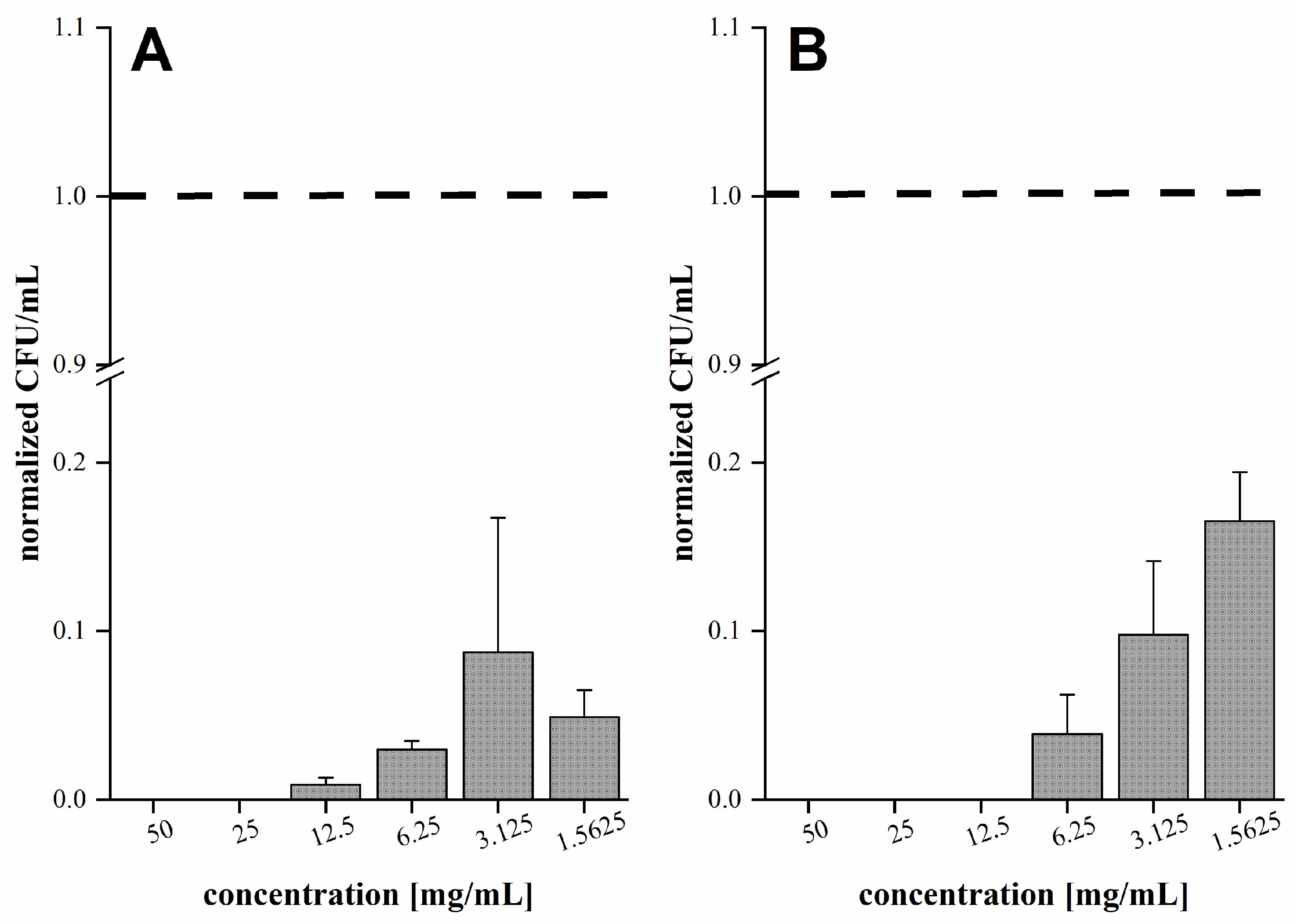
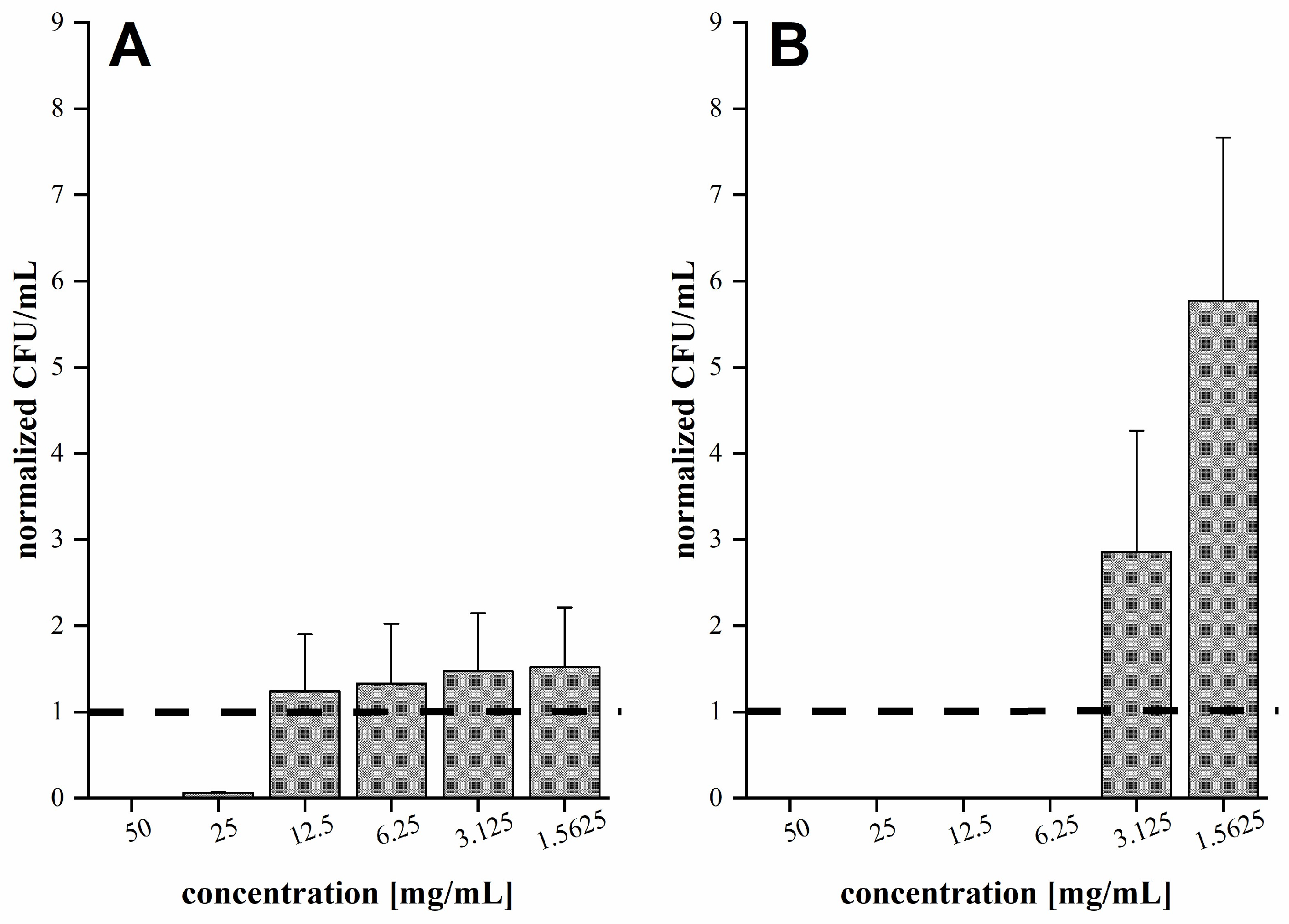
3.2. Antibacterial Activity of Fir Honeydew Honey

Both of the tested strains of C. jejuni were sensitive to FHH in MH broth and milk, with MICs ranging from 12.5 to 50 mg/mL (Table 1). C. jejuni NCTC 11168 was more sensitive to FHH in MH broth and milk compared to C. jejuni K49/4. There is a limited number of studies that have demonstrated the antibacterial effect of FHH, especially in milk. It has been shown that different honeys are effective against different bacteria, but results cannot be directly compared to other studies because there are differences in samples, concentrations, and methods [48,49]. Broznić et al. [10] showed that FHHs from Gorski Kotar, Croatia, had an antibacterial effect against Staphylococcus spp. and different strains of Acinetobacter bumanii in the MH broth. The antibacterial effect of different samples of FHH determined by Broznić et al. [10] was comparable to the effect of the FHH used in this study. Also, Bucekova et al. [50] showed that honeydew honey samples collected in several regions of Slovakia had an antibacterial effect on bacteria Staphylococcus aureus and Pseudomonas aeruginosa. FHH also has an antibacterial effect against C. jejuni in milk. As can be seen in Table 1, the MIC was higher in milk than in MH broth, which may be due to the high fat content in milk that influences the activity of the sample studied [51].

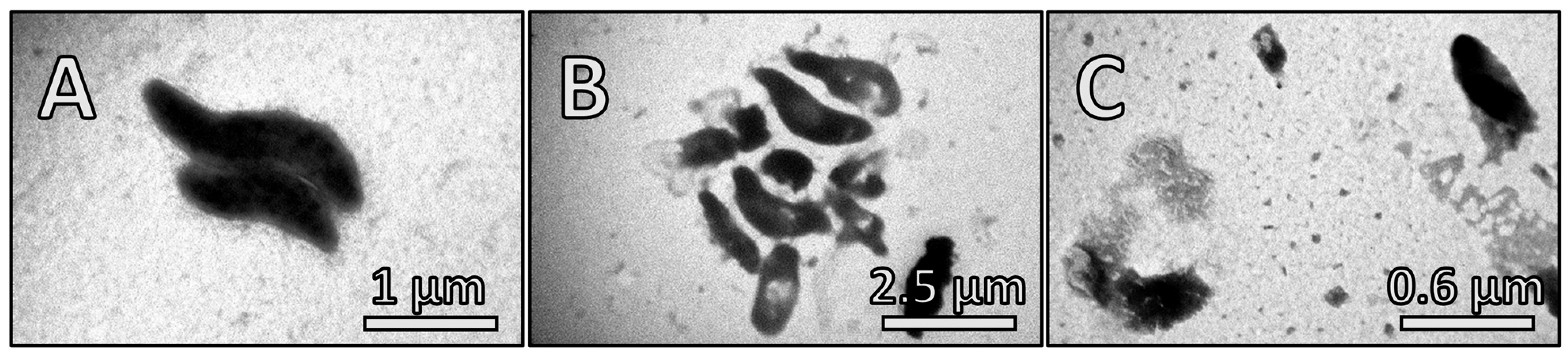
3.3. The Effect of Fir Honeydew Honey on Bacterial Growth and Cell Structure

In order to determine the effect of FHH on bacterial growth, solutions containing 5% [w/v] and 3% [w/v] of FHH in MH broth and milk were prepared. As expected, a higher concentration of FHH had a stronger effect on the growth of C. jejuni NCTC 11168 and C. jejuni K49/4 in the MH broth and milk. FHH at a concentration of 5% [w/v] completely inhibited the growth of C. jejuni NCTC 11168 in MH broth and milk at each time point (Table 2 and Table 3). At the lower concentration (3% [w/v]), a statistically significant effect was not observed after 24 h of incubation in MH broth (Table 2). After 24 h incubation in milk, to which FHH (3% [w/v]) was added, the growth of C. jejuni NCTC 11168 was reduced by 74.4 ± 8.3% (Table 3). There was a significant effect of FHH on the growth of C. jejuni K49/4 in MH broth and milk at the concentration of 5% [w/v], but the lower concentration (3% [w/v]) showed no inhibitory effect on the growth of C. jejuni K49/4 in MH broth and milk (Table 3).
C. jejuni NCTC 11168 was more sensitive compared to C. jejuni K49/4. C. jejuni K49/4 is a strain isolated from heat-treated poultry meat, which could be the reason why this strain has a better stress response than C. jejuni NCTC 11168 [31]. Many antimicrobial substances in honeydew honey, including hydrogen peroxide and various polyphenols, as well as their interactions [50,51,52,53,54], can affect the growth of C. jejuni.
The effect of FHH in milk was as good as in MH broth (Table 3). Milk contains various bioactive peptides [55], which can improve the antibacterial effect of honey. Milk also contains smaller peptides that can diffuse rapidly into the cell membrane and cause cell leakage. Immunological peptides in milk can also reduce the growth of bacteria [56].
Inhibition of C. jejuni growth after the addition of FHH was the consequence of stress, which includes osmotic stress and stress caused by the antimicrobial agents to which bacteria were exposed [52]. This was confirmed using TEM (Figure 1), which showed that FHH induced morphological changes in bacterial cell shape similar to those found by Shen and Chou [53] on Campylobacter cells after exposure to stress conditions. The changes caused by FHH are shown in Figure 1B,C. A concentration of 1× MIC (12.5 mg/mL) of FHH affected the shape of the bacterial cells. Cells were more clustered and small buds were formed on the cell wall. The morphology of the cells was changed from a curly to an oval form. A disorganized cytoplasm was also observable. The 2× MIC (25 mg/mL) concentration of FHH caused the complete decay of the bacterial cell wall, which led to cell leakage (Figure 1C).

3.4. Anti-Adhesion Effect of Fir Honeydew Honey

FHH at concentrations from 1.5625 to 50 mg/mL in MH broth and milk significantly reduced the adhesion of C. jejuni NCTC 11168 on the polystyrene microtiter plate after 24 h of incubation. As shown in Figure 2, the reduction was stronger when the concentration of FHH in the MH broth was higher, with complete reduction at 25 and 50 mg/mL. The same was observed in milk, where concentrations of 12.5, 25 and 50 mg/mL completely reduced the adhesion of C. jejuni NCTC 11168 on the polystyrene surface (p < 0.05).
FHH had a lower effect on the adhesion of C. jejuni K49/4 on a polystyrene microtiter plate after 24 h incubation in MH broth compared to the effect on C. jejuni NCTC 11168. Only concentrations of 25 and 50 mg/mL significantly reduced the adhesion of C. jejuni K49/4 in MH broth (Figure 3). FHH had a similar effect on the adhesion of C. jejuni K49/4 on the polystyrene surface of the microtiter plate after 24 h incubation in milk compared to the effect on C. jejuni NCTC 11168, where concentrations of 6.25, 12.5, 25, and 50 mg/mL completely reduced the adhesion of C. jejuni K49/4 (p < 0.05).
Our results showed that the adhesion on the polystyrene surface of both strains was lower in milk compared to MH broth. Even lower concentrations of FHH reduced the adhesion of C. jejuni on the polystyrene surface in milk. The reason for the higher anti-adhesion effect in milk can include milk proteins, which can form a conditioning layer and prevent bacteria from adhering to the polystyrene surface. Also, the ionic composition of the suspending medium can influence the attachment of the cells to the abiotic surface [57]. To overcome the limitations of this research, further studies will include more bacterial strains that will be tested on several food models.

4. Conclusions

Physicochemical properties confirmed that the FHH used in this study belongs to the honeydew honey group. Its biological activity was also demonstrated via in vitro assays, which confirmed that FHH has a strong antibacterial and anti-adhesion potential against C. jejuni in MH broth and milk. These results point to the potential usage of FHH in low concentrations as food preservatives that can reduce the potential contamination of food products.

Author Contributions

Conceptualization, I.G. and D.V.; methodology, D.R., A.P. and, S.S.M.; investigation, D.R., A.P. and S.S.M.; resources, I.G.; writing—original draft preparation, D.R., I.G., D.V. and I.B.K.; writing—review and editing, I.G., D.V. and I.B.K.; supervision, I.G. and D.V.; project administration, I.G. All authors have read and agreed to the published version of the manuscript.

Funding

This research was funded by a grant from the University of Rijeka (Grant no. uniribiomed-18-171).

Institutional Review Board Statement

Not applicable.

Informed Consent Statement

Not applicable.

Data Availability Statement

The data presented in this study are available in the article.

Acknowledgments

This study was performed using the facilities and equipment funded within the European Regional Development Fund project KK.01.1.1.02.0007 “Research and Education Centre of Environmental Health and Radiation Protection—Reconstruction and Expansion of the Institute for Medical Research and Occupational Health”.

Conflicts of Interest

The authors declare no conflict of interest.

References

  1. Fernández-Estellé, M.; Hernández-González, V.; Saurina, J.; Núñez, O.; Sentellas, S. Characterization and classification of Spanish honeydew and blossom honeys based on their antioxidant capacity. Antioxidants 2023, 12, 495. [Google Scholar] [CrossRef] [PubMed]
  2. 581. Off. J. Eur. Communities. 2002. Available online: https://eur-lex.europa.eu/LexUriServ/LexUriServ.do?uri=OJ:L:2002:010:0047:0052:EN:PDF (accessed on 10 September 2023).
  3. FAO; WHO. Codex Alimentarius Commission—Procedural Manual, 27th ed.; FAO: Rome, Italy; WHO: Rome, Italy, 2019; ISBN 978-92-5-131099-1. [Google Scholar]
  4. Pita-Calvo, C.; Vázquez, M. Differences between honeydew and blossom honeys: A review. Trends Food Sci. Technol. 2017, 59, 79–87. [Google Scholar] [CrossRef]
  5. Iglesias, M.T.; De Lorenzo, C.; Polo, M.D.C.; Martín-Álvarez, P.J.; Pueyo, E. Usefulness of amino acid composition to discriminate between honeydew and floral honeys. Application to honeys from a small geographic area. J. Agric. Food Chem. 2004, 52, 84–89. [Google Scholar] [CrossRef] [PubMed]
  6. Pita-Calvo, C.; Vázquez, M. Honeydew honeys: A review on the characterization and authentication of botanical and geographical origins. J. Agric. Food Chem. 2018, 66, 2523–2537. [Google Scholar] [CrossRef] [PubMed]
  7. Kunat-Budzynska, M.; Rysiak, A.; Wiater, A.; Graz, M.; Andrejko, M.; Budzynski, M.; Brys, M.S.; Sudzinski, M.; Tomczyk, M.; Gancarz, M.; et al. Chemical composition and antimicrobial activity of new honey varietals. Int. J. Environ. Res. Public Health 2023, 20, 2458. [Google Scholar] [CrossRef] [PubMed]
  8. Vela, L.; De Lorenzo, C.; Pérez, R.A. Antioxidant capacity of Spanish honeys and its correlation with polyphenol content and other physicochemical properties. J. Sci. Food Agric. 2007, 87, 1069–1075. [Google Scholar] [CrossRef]
  9. Lušić, D.; Koprivnjak, O.; Ćurić, D.; Sabatini, A.G.; Conte, L.S. Volatile profile of Croatian lime tree (Tilia sp.), fir honeydew (Abies alba) and sage (Salvia officinalis) honey. Food Technol. Biotechnol. 2007, 45, 156–165. [Google Scholar]
  10. Broznić, D.; Ratkaj, I.; Malenica, M.; Pavelić, S.; Zurga, P.; Bubalo, D.; Gobin, I. Evaluation of the antioxidant capacity, antimicrobial and antiproliferative potential of fir (Abies alba Mill.) honeydew honey collected from Gorski Kotar (Croatia). Food Technol. Biotechnol. 2018, 56, 533–545. [Google Scholar] [CrossRef]
  11. Artursson, K.; Schelin, J.; Thisted Lambertz, S.; Hansson, I.; Olsson Engvall, E. Foodborne pathogens in unpasteurized milk in Sweden. Int. J. Food Microbiol. 2018, 284, 120–127. [Google Scholar] [CrossRef]
  12. European Food Safety Authority; European Centre for Disease Prevention and Control. The European Union One Health 2021 Zoonoses Report. EFSA J. 2022, 20, e07666. [Google Scholar] [CrossRef]
  13. García-Sánchez, L.; Melero, B.; Rovira, J. Campylobacter in the Food Chain. Adv. Food Nutr. Res. 2018, 86, 215–252. [Google Scholar] [PubMed]
  14. Oh, E.; Andrews, K.J.; Jeon, B. Enhanced biofilm formation by ferrous and ferric iron through oxidative stress in Campylobacter jejuni. Front. Microbiol. 2018, 9, 1204. [Google Scholar] [CrossRef] [PubMed]
  15. Gölz, G.; Kittler, S.; Malakauskas, M.; Alter, T. Survival of Campylobacter in the food chain and the environment. Curr. Clin. Microbiol. Rep. 2018, 5, 126–134. [Google Scholar] [CrossRef]
  16. Johnson, T.J.; Shank, J.M.; Johnson, J.G. Current and potential treatments for reducing Campylobacter colonization in animal hosts and disease in humans. Front. Microbiol. 2017, 8, 487. [Google Scholar] [CrossRef] [PubMed]
  17. Teh, A.H.T.; Lee, S.M.; Dykes, G.A. Does Campylobacter jejuni form biofilms in food-related environments? Appl. Environ. Microbiol. 2014, 80, 5154–5160. [Google Scholar] [CrossRef] [PubMed]
  18. Burnham, P.M.; Hendrixson, D.R. Campylobacter jejuni: Collective components promoting a successful enteric lifestyle. Nat. Rev. Microbiol. 2018, 16, 551–565. [Google Scholar] [CrossRef] [PubMed]
  19. Modi, S.; Brahmbhatt, M.N.; Chatur, Y.A.; Nayak, J.B. Prevalence of Campylobacter species in milk and milk products, their virulence gene profile and antibiogram. Vet. World 2015, 8, 1–8. [Google Scholar] [CrossRef]
  20. Del Collo, L.P.; Karns, J.S.; Biswas, D.; Lombard, J.E.; Haley, B.J.; Kristensen, R.C.; Kopral, C.A.; Fossler, C.P.; Van Kessel, J.A.S. Prevalence, antimicrobial resistance, and molecular characterization of Campylobacter spp. in bulk tank milk and milk filters from US dairies. J. Dairy Sci. 2017, 100, 3470–3479. [Google Scholar] [CrossRef]
  21. Kurin, M.; Berce, I.; Zorman, T.; Smole Možina, S. The prevalence of multiple antibiotic resistance in Campylobacter spp. from retail poultry meat. Food Technol. Biotechnol. 2005, 43, 157–163. [Google Scholar]
  22. Smole Možina, S.; Kurinčič, M.; Klančnik, A.; Mavri, A. Campylobacter and its multi-resistance in the food chain. Trends Food Sci. Technol. 2011, 22, 91–98. [Google Scholar] [CrossRef]
  23. Vasić, V.; Gašić, U.; Stanković, D.; Lušić, D.; Vukić-Lušić, D.; Milojković-Opsenica, D.; Tešić, Ž.; Trifković, J. Towards better quality criteria of European honeydew honey: Phenolic profile and antioxidant capacity. Food Chem. 2019, 274, 629–641. [Google Scholar] [CrossRef] [PubMed]
  24. Gutierrez, J.; Barry-Ryan, C.; Bourke, P. Antimicrobial activity of plant essential oils using food model media: Efficacy, synergistic potential and interactions with food components. Food Microbiol. 2009, 26, 142–150. [Google Scholar] [CrossRef] [PubMed]
  25. Klančnik, A.; Piskernik, S.; Bucar, F.; Vučković, D.; Smole Možina, S.; Jeršek, B. Reduction of microbiological risk in minced meat by a combination of natural antimicrobials: Effects of antimicrobials on a bacterial cocktail. J. Sci. Food Agric. 2014, 94, 2758–2765. [Google Scholar] [CrossRef] [PubMed]
  26. Louveaux, J.; Maurizio, A.; Vorwohl, G. Methods of melissopalynology. Bee World 1978, 59, 139–157. [Google Scholar] [CrossRef]
  27. Bogdanov, S. Harmonised methods of the International Honey Commission. Int. Honey Comm. 2002, 1–62. [Google Scholar]
  28. Bogdanov, S.; Lüllmann, C.; Martin, P.; Von Der Ohe, W.; Russmann, H.; Vorwohl, G.; Oddo, L.P.; Sabatini, A.-G.; Marcazzan, G.L.; Piro, R.; et al. Honey quality and international regulatory standards: Review by the International Honey Commission. Bee World 1999, 80, 61–69. [Google Scholar] [CrossRef]
  29. Singleton, V.L.; Orthofer, R.; Lamuela-Raventós, R.M. Analysis of Total Phenols and Other Oxidation Substrates and Antioxidants by Means of Folin-Ciocalteu Reagent. Methods Enzymol. 1999, 299, 152–178. [Google Scholar]
  30. Kim, D.-O.; Jeong, S.W.; Lee, C.Y. Antioxidant capacity of phenolic phytochemicals from various cultivars of plums. Food Chem. 2003, 81, 321–326. [Google Scholar] [CrossRef]
  31. Zorman, T.; Smole Možina, S. Classical and molecular identification of thermotolerant Campylobacters from poultry meat. Food Technol. Biotechnol. 2002, 40, 177–184. [Google Scholar]
  32. Klančnik, A.; Guzej, B.; Kolar, M.H.; Abramovič, H.; Smole Možina, S. In vitro antimicrobial and antioxidant activity of commercial rosemary extract formulations. J. Food Prot. 2009, 72, 1744–1752. [Google Scholar] [CrossRef]
  33. Kovač, J.; Šimunović, K.; Wu, Z.; Klančnik, A.; Bucar, F.; Zhang, Q.; Smole Možina, S. Antibiotic resistance modulation and modes of action of (-)-α-pinene in Campylobacter jejuni. PLoS ONE 2015, 10, e0122871. [Google Scholar] [CrossRef] [PubMed]
  34. Warui, M.W.; Hansted, L.; Gikungu, M.; Mburu, J.; Kironchi, G.; Bosselmann, A.S. Characterization of Kenyan honeys based on their physicochemical properties, botanical and geographical origin. Int. J. Food Sci. 2019, 2019, 2932509. [Google Scholar] [CrossRef] [PubMed]
  35. Primorac, L.; Angelkov, B.; Mandić, M.; Čačić Kenjerić, D.; Nedeljko, M.; Flanjak, I.; Pirički, A.; Arapcheska, M. Comparison of the Croatian and Macedonian honeydew honey. J. Cent. Eur. Agric. 2009, 10, 263–270. [Google Scholar]
  36. Diez, M.J.; Andres, C.; Terrab, A. Physicochemical parameters and pollen analysis of Moroccan honeydew honeys. Int. J. Food Sci. Technol. 2004, 39, 167–176. [Google Scholar] [CrossRef]
  37. Golob, T.; Plestenjak, A. Quality of Slovene honey. Food Technol. Biotehnol. 1999, 37, 195–201. [Google Scholar]
  38. Delafuente, E.; Sanz, M.; Martinezcastro, I.; Sanz, J.; Ruizmatute, A. Volatile and carbohydrate composition of rare unifloral honeys from Spain. Food Chem. 2007, 105, 84–93. [Google Scholar] [CrossRef]
  39. Devillers, J.; Morlot, M.; Pham-Delègue, M.H.; Doré, J.C. Classification of monofloral honeys based on their quality control data. Food Chem. 2004, 86, 305–312. [Google Scholar] [CrossRef]
  40. Al, M.L.; Daniel, D.; Moise, A.; Bobis, O.; Laslo, L.; Bogdanov, S. Physico-chemical and bioactive properties of different floral origin honeys from Romania. Food Chem. 2009, 112, 863–867. [Google Scholar] [CrossRef]
  41. Meda, A.; Lamien, C.E.; Romito, M.; Millogo, J.; Nacoulma, O.G. Determination of the total phenolic, flavonoid and proline contents in Burkina Fasan honey, as well as their radical scavenging activity. Food Chem. 2005, 91, 571–577. [Google Scholar] [CrossRef]
  42. Bobis, O.; Marghitas, L.; Krisztina, R.; Niculae, M.; Dezmirean, D. Honeydew honey: Correlations between chemical composition, antioxidant capacity and antibacterial effect. Lucr. Ştiinţ. Zooteh. Biotehnol. Timis. 2008, 41, 1–9. [Google Scholar]
  43. Kuś, P.M.; Jerković, I.; Marijanović, Z.; Tuberoso, C.I.G. Screening of Polish fir honeydew honey using GC/MS, HPLC-DAD, and physical-chemical parameters: Benzene derivatives and terpenes as chemical markers. Chem. Biodivers. 2017, 14, e1700179. [Google Scholar] [CrossRef] [PubMed]
  44. Bertoncelj, J.; Dobersek, U.; Jamnik, M.; Golob, T. Evaluation of the phenolic content, antioxidant activity and colour of Slovenian honey. Food Chem. 2007, 105, 822–828. [Google Scholar] [CrossRef]
  45. Castiglioni, S.; Stefano, M.; Astolfi, P.; Carloni, P. Chemometric approach to the analysis of antioxidant properties and colour of typical Italian monofloral honeys. Int. J. Food Sci. Technol. 2017, 52, 1138–1146. [Google Scholar] [CrossRef]
  46. Moniruzzaman, M.; Yung An, C.; Rao, P.V.; Hawlader, M.N.I.; Azlan, S.A.B.M.; Sulaiman, S.A.; Gan, S.H. Identification of phenolic acids and flavonoids in monofloral honey from Bangladesh by high performance liquid chromatography: Determination of antioxidant capacity. BioMed Res. Int. 2014, 2014, 737490. [Google Scholar] [CrossRef] [PubMed]
  47. Socha, R.; Juszczak, L.; Pietrzyk, S.; Gałkowska, D.; Fortuna, T.; Witczak, T. Phenolic profile and antioxidant properties of Polish honeys: Antioxidant properties of honeys. Int. J. Food Sci. Technol. 2011, 46, 528–534. [Google Scholar] [CrossRef]
  48. Al-Waili, N.S.; Salom, K.; Butler, G.; Al Ghamdi, A.A. Honey and microbial infections: A review supporting the use of honey for microbial control. J. Med. Food 2011, 14, 1079–1096. [Google Scholar] [CrossRef]
  49. Kuś, P.M.; Szweda, P.; Jerković, I.; Tuberoso, C.I.G. Activity of Polish unifloral honeys against pathogenic bacteria and its correlation with colour, phenolic content, antioxidant capacity and other parameters. Lett. Appl. Microbiol. 2016, 62, 269–276. [Google Scholar] [CrossRef]
  50. Bucekova, M.; Buriova, M.; Pekarik, L.; Majtan, V.; Majtan, J. Phytochemicals-Mediated production of hydrogen peroxide is crucial for high antibacterial activity of honeydew honey. Sci. Rep. 2018, 8, 9061. [Google Scholar] [CrossRef]
  51. Ananda Baskaran, S.; Kazmer, G.W.; Hinckley, L.; Andrew, S.M.; Venkitanarayanan, K. Antibacterial effect of plant-derived antimicrobials on major vacterial mastitis pathogens in vitro. J. Dairy Sci. 2009, 92, 1423–1429. [Google Scholar] [CrossRef]
  52. Matzen, R.D.; Zinck Leth-Espensen, J.; Jansson, T.; Nielsen, D.S.; Lund, M.N.; Matzen, S. The antibacterial effect in vitro of honey derived from various Danish flora. Dermatol. Res. Pract. 2018, 2018, 7021713. [Google Scholar] [CrossRef]
  53. Shen, J.-P.; Chou, C.-F. Morphological plasticity of bacteria-open questions. Biomicrofluidics 2016, 10, 031501. [Google Scholar] [CrossRef] [PubMed]
  54. Bucekova, M.; Jardekova, L.; Juricova, V.; Bugarova, V.; Di Marco, G.; Gismondi, A.; Leonardi, D.; Farkasovska, J.; Godocikova, J.; Laho, M.; et al. Antibacterial activity of different blossom honeys: New findings. Molecules 2019, 24, 1573. [Google Scholar] [CrossRef] [PubMed]
  55. Mohanty, D.; Jena, R.; Choudhury, P.; Pattnaik, R.; Mohapatra, S.; Saini, M. Milk derived antimicrobial bioactive peptides: A Review. Int. J. Food Prop. 2016, 19, 837–846. [Google Scholar] [CrossRef]
  56. Khan, M.U.; Pirzadeh, M.; Förster, C.Y.; Shityakov, S.; Shariati, M.A. Role of milk-derived antibacterial peptides in modern food biotechnology: Their synthesis, applications and future perspectives. Biomolecules 2018, 8, 110. [Google Scholar] [CrossRef]
  57. Barnes, L.-M.; Lo, M.F.; Adams, M.R.; Chamberlain, A.H.L. Effect of milk proteins on adhesion of bacteria to stainless steel surfaces. Appl. Environ. Microbiol. 1999, 65, 4543–4548. [Google Scholar] [CrossRef]
Figure 1. Transmission electron microscopy images of Campylobacter jejuni NCTC 11168 of samples not treated (A) and treated with fir honeydew honey at concentrations of 12.5 mg/mL [1× minimal inhibitory concentration (MIC)] (B) and 25 mg/mL (2× MIC) (C). Samples were scanned after 24 h of incubation at 42 °C in microaerobic atmosphere in Ringer solution, where fir honeydew honey was not (A) or was added (B,C).
Applsci 13 12735 g001
Figure 2. Normalised adhesion of Campylobacter jejuni NCTC 11168 to a polystyrene surface after 24 h incubation in Mueller Hinton (MH) broth (A) and milk (B) under microaerobic atmosphere at 42 °C. Cells were exposed to fir honeydew honey at a concentration from 1.5625 to 50 mg/mL The adhered cells in the microtiter plates were suspended in phosphate buffered saline (PBS) by ultrasonic bath and their concentration was determined via the plate counting method. The CFU is given per mL of PBS. Normalised mean values (control = 1) + standard deviation are given.
Applsci 13 12735 g002
Figure 3. Normalised adhesion of Campylobacter jejuni K49/4 to polystyrene surface after 24 h incubation in Mueller Hinton (MH) broth (A) and milk (B) under microaerobic atmosphere at 42 °C. Cells were exposed to fir honeydew honey at a concentration from 1.5625 to 50 mg/mL The adhered cells in the microtiter plates were suspended in phosphate buffered saline (PBS) via ultrasonic bath and their concentration was determined via the plate counting method. The CFU is given per mL of PBS. Normalised mean values (control = 1) + standard deviation are given.
Applsci 13 12735 g003
Table 1. Minimal inhibitory concentration (MIC) of fir honeydew honey aginst bacteria Campylobacter jejuni NCTC 11168 and C. jejuni K49/4 determined in Mueller Hinton (MH) broth and milk.
MIC (mg/mL)
Bacterial StrainMH BrothMilk
C. jejuni NCTC 1116812.525
C. jejuni K49/42550
Table 2. Inhibitory effects of fir honeydew honey at concentrations 5% [w/v] and 3% [w/v] in Mueller Hinton (MH) broth on Campylobacter jejuni NCTC 11168 and C. jejuni K49/4 growth, determined at different time points, at 42 °C.
Fir Honeydew Honey
in MH Broth
Time (h)Growth Inhibition ± SD [%]
C. jejuni NCTC 11168C. jejuni K49/4
5% [w/v]2100 ± 0.057.4 ± 7.0
4100 ± 0.099.9 ± 0.0
6100 ± 0.099.9 ± 0.0
8100 ± 0.099.9 ± 0.0
24100 ± 0.099.9 ± 0.0
3% [w/v]299.1 ± 0.05.3 ± 3.3
498.9 ± 0.630.5 ± 27.8
699.4 ± 0.119.4 ± 12.3
899.8 ± 0.013.2 ± 10.2
2428.6 ± 12.115.5 ± 3.6
Table 3. Inhibitory effects of fir honeydew honey at concentrations 5% [w/v] and 3% [w/v] in milk on Campylobacter jejuni NCTC 11168 and C. jejuni K49/4 growth, determined at different time points at 42 °C.
Fir Honeydew Honey
in Milk
Time (h)Growth Inhibition ± SD [%]
C. jejuni NCTC 11168C. jejuni K49/4
5% [w/v]2100 ± 0.084.3 ± 6.6
4100 ± 0.098.1 ± 0.4
6100 ± 0.0100 ± 0.0
8100 ± 0.0100 ± 0.0
24100 ± 0.099.9 ± 0.0
3% [w/v]299.3 ± 0.035.5 ± 11.1
499.2 ± 0.135.8 ± 12.3
695.7 ± 1.8ND *
898.3 ± 0.2ND *
2474.4 ± 8.316.3 ± 21.6
* ND–not determined.
Disclaimer/Publisher’s Note: The statements, opinions and data contained in all publications are solely those of the individual author(s) and contributor(s) and not of MDPI and/or the editor(s). MDPI and/or the editor(s) disclaim responsibility for any injury to people or property resulting from any ideas, methods, instructions or products referred to in the content.

