Article

The Effect of a Static Magnetic Field on microRNA in Relation to the Regulation of the Nrf2 Signaling Pathway in a Fibroblast Cell Line That Had Been Treated with Fluoride Ions

by
Magdalena Kimsa-Dudek
*,
Agata Krawczyk
and
Agnieszka Synowiec-Wojtarowicz
Department of Nutrigenomics and Bromatology, Faculty of Pharmaceutical Sciences in Sosnowiec, Medical University of Silesia, Katowice, Jednosci 8, 41-200 Sosnowiec, Poland
*
Author to whom correspondence should be addressed.
Appl. Sci. 2023, 13(3), 1470; https://doi.org/10.3390/app13031470
Submission received: 3 December 2022 / Revised: 18 January 2023 / Accepted: 19 January 2023 / Published: 22 January 2023

Abstract

:
Many physical and chemical factors can influence the Nrf2 signaling pathway, and its deregulation has been associated with various disease states. Nrf2 and Keap1 molecules are key elements that regulate the response under cell stress conditions. Hence, the aim of the research was to assess the impact of a static magnetic field that was generated by permanent magnets on the transcriptional activity of the NFE2L2 and KEAP1 genes in a human dermal fibroblast cell line that had been cultured with fluoride ions and in an environment without them. Additionally, our research aimed to answer the question of whether the regulatory RNA molecules—miRNAs—are involved in these effects. Experiments were performed on an NHDF cell line that had been treated with fluoride ions, along with simultaneous exposure to a moderate-intensity static magnetic field. The RT-qPCR method was used to determine the mRNA level of the NFE2L2 and KEAP1 genes and the miRNA level of miR-28-3p, miR-29b-1-5p, miR-141-3p and miR-365a-3p. Our results indicate that fluoride ions influence the Nrf2 signaling pathway and that miRNAs are involved in regulating it. Moreover, the static magnetic field had a beneficial effect on the cells with fluoride-induced oxidative stress due to stimulating the antioxidant defense.

1. Introduction

A microRNA molecule (miRNA) is a type of RNA that controls the expression of mRNA [1]. Recently, miRNAs have become an area of interest for many researchers. The mechanism of action of miRNAs has been extensively described [2,3]. Most of the attention has been paid to the interaction of miRNAs with mRNA, which affects the level of gene expression. This is important because the level of miRNA expression affects the overall cell homeostasis [2]. MiRNAs have become popular because of their wide range of properties and applications. They affect many biological processes, including proliferation, differentiation and apoptosis. Their usefulness as biomarkers of many diseases, including carcinogenesis and diseases that are associated with oxidative stress, has also been mentioned [4,5].
One of the defense models used to maintain cell homeostasis against oxidative stress is the NRF2/KEAP1 axis (NF-E2-related factor 2/Kelch-like ECH-associated protein 1) [6]. Moi et al. [7] were the first to describe the transcription factor Nrf2. At that time, attention was paid to its role in controlling cellular oxidative stress. It has been shown that it is the main regulator of gene transcription, the products of which protect cells against the harmful effects of oxidative stress, including enzymes that catalyze glutathione metabolism, ROS (reactive oxygen species)-detoxifying enzymes (glutathione peroxidase, glutathione S-transferases) and the enzymes that are involved in drug detoxification: phase I (aldo-keto reductases, carbonyl reductases, aldehyde dehydrogenase 1, NAD(P)H quinone oxidoreductase 1 and various cytochrome P450s), phase II (glutathione S-transferases, UDP-glucuronosyltransferases and heme oxygenase-1) and phase III (drug efflux transporters). Moreover, Nrf2 regulates the expression of the genes that participate in reducing oxidized protein thiols (thioredoxin, thioredoxin reductase 1, sulfiredoxin). The induction of the Nrf2 signaling cascade has been found to counteract the effects of the disruption of cellular redox homeostasis [8].
The generation of oxidative stress is influenced by many factors, including stress, environmental pollution [9], smoking [10] and overexposure to solar radiation [11]. In addition, factors that increase the production of reactive oxygen species (ROS) also include fluoride compounds, which people are often exposed to during their everyday lives. Despite the documented positive effects of fluoride on the body (including maintaining the proper structure of the skeleton), it should be borne in mind that exposure to fluoride and its compounds in incorrect doses may have negative effects and cause many unfavorable changes in the body [12,13]. An inadequate intake of fluoride may adversely affect the functioning of many organs, including the kidneys and liver, by, e.g., contributing to an increased risk of developing renal failure or necrotic changes in the liver. Fluoride hyperglycemia has also been reported. The neurotoxicity of fluoride compounds consists of the appearance of neurodegenerative changes in the brain structures and the reduction in the receptors in the brain. In addition, fluoride can lead to changes in the functioning of the reproductive system as well as changes in the mineral status of the body. The harmful effects of this compound depend on the duration of the exposure. The dose and its ability to accumulate in the tissues are also not insignificant [14,15].
These changes can result from the role of the fluoride compounds in modifying the molecular pathways, including the NRF2/KEAP1 pathway in cells. Histological changes in the myocardium after NaF (sodium fluoride) had been administered, which was the result of an excessive accumulation of reactive oxygen species and was related to the above-mentioned molecular pathway, have also been demonstrated [16]. Moreover, in an animal model, the negative effect of fluoride on lung cells as a result of oxidative stress has also been confirmed. Simultaneously, the same study confirmed the protective effect of epigallocatechin gallate (EGCG) on the lungs by inhibiting the oxidative stress that was caused by the activation of Nrf2 [17]. In turn, Mukhopadhyay et al. [18] showed that the exposure of adult zebrafish (Danio rerio) to NaF resulted in an increased production of ROS and the up-regulation of NFE2L2 (NFE2-Like BZIP Transcription Factor 2) gene expression. Published data on animal models have also demonstrated that miRNAs can constitute new biomarkers of fluoride toxicity due to the considerable alterations in the expression of many of them after fluoride exposure [19]. In addition, their usefulness as diagnostic markers and therapeutic targets in fluoride-induced liver disorders has also been proved [20].
The effect of a static magnetic field (SMF) on the life processes of organisms has become quite an interesting topic in recent years. Because of their complex mechanism of action, the effects of an SMF on the body are not yet fully understood. It is quite an interesting topic because of the widespread human exposure to SMFs in the environment, including at work [21,22]. Previous studies have shown that an SMF can restore redox homeostasis in cells that had been treated with chemical agents [23]. It is known that one consequence of the physical impact of an SMF on cells is a change in the concentrations of the calcium, sodium and potassium ions inside them, which could have a modulating effect on many important signaling pathways. There are currently no studies that have shown a relationship between exposure to a static magnetic field and miRNA expression. However, potential associations between electromagnetic field (EMF) exposure and miRNAs have been demonstrated in spermatocyte-derived mouse GC-2 cells as well as in peripheral human blood cells [24].
Taking into account the widespread exposure of the human body to the effects of both fluoride compounds and SMFs, the impact of fluoride on the human body in terms of molecular changes in the signaling pathways that are related to oxidative stress as well as their modification under the influence of an SMF becomes puzzling. We believe that the conducted study could have a significant impact on expanding the knowledge in the field on the changes in the molecular signaling pathways that are related to the oxidative process under the influence of external factors. On the other hand, the inclusion of miRNA molecules in the analyses could also be considered innovative.

2. Materials and Methods

The research was conducted using the NHDF cell line (Normal Human Dermal Fibroblasts, Clonetics, CC-2511, San Diego, CA, USA), which was purchased from Lonza (Basel, Switzerland). The choice of this cell line was justified due to the influence of fluoride on both soft tissues and various biological processes, including type I collagen synthesis, proliferation and morphology of fibroblasts.

2.1. Cell Culture Conditions

The cells were cultured in a fibroblast growth basal medium (FBMTM, Lonza, Basel, Switzerland), which was enriched by adding 10% fetal bovine serum (FBS), gentamicin, insulin and basic human fibroblast growth factor (FGM™ SingleQuots™; Lonza, Basel, Switzerland). The cells were grown in monolayers in 25 cm2 culture flasks (Sarstedt, Nümbrecht, Germany).
NHDF cells that were between passages four and eight were used to evaluate the influence of fluoride and an SMF. Their number and viability were assessed using a Countess TM Automated Cell Counter (Invitrogen, Carlsbad, CA, USA) and 0.4% trypan blue.
For the research, the cells were seeded into culture flasks at a density of 1 × 10⁶ cells and were then incubated for 24 h. Next, the cells for the experiment were divided into four groups: (1) a control group (C), which was represented by cells that had not been treated with NaF (sodium fluoride) and had not been exposed to an SMF; (2) a group that had been treated sodium fluoride (F), which was represented by cells that had only been treated with NaF at a concentration of 0.3 mmol/l (Sigma-Aldrich, St Louis, MO, USA); (3) a group that had been exposed to an SMF (SMF), which was represented by cells that had only been exposed to an SMF with magnetic flux density of 0.65 T; and (4) group that had been exposed to an F + SMF, which was represented by cells that had been exposed to a combination of NaF and an SMF. After 24 h of being cultured with the tested factors, the cells were washed with phosphate-buffered saline (PBS, Lonza, Basel, Switzerland) and the pellets were collected for further analysis.
An SMF was generated in a special magnetic chamber that was designed for in vitro research and consisted of a ferromagnetic yoke and a permanent magnet. For chambers that were fully fit to the cell flask, the uniformity of the static magnetic field induction was maintained on the entire surface of the culture [25,26]. For the unexposed cultures, the control chambers in which steel (0.0 T) was used instead of a permanent magnet were used. The concentration of fluoride ions and magnetic induction used were decided based on recent studies that showed that they had no significant effect on cell viability [23,27]. In addition, a concentration of NaF at which the magnesium and calcium fluorides did not precipitate, which would have affected the functioning of the cells, was chosen. Moreover, the selected concentration of NaF (0.3 mmol/l) was higher than is usually observed in healthy adult serum [28], which could be clinically significant. The method for exposing the cell cultures to both factors has been previously described [23].

2.2. Molecular Analyses

The RNA was extracted using an miRNeasy Mini Kit (Qiagen, Valencia, CA) according to the manufacturer’s protocols. The quality and quantity of the RNA extracts were evaluated using agarose gel electrophoresis and a MaestroNano MN-913 (MaestroGen Inc., Las Vegas, NV, USA) spectrophotometer.
The RNA was used as a template for the RT-qPCR assay to assess the NFE2L2 and KEAP1 gene expression. The reaction was performed using SYBR Green I chemistry (SensiFAST SYBR No-ROX One-Step, Bioline, London, UK) and a LightCycler® 480 Instrument II (Roche Life Science, Basel, Switzerland). Commercially available oligonucleotide-specific primers were used (Sigma-Aldrich, St. Louis, MO, USA) (Table 1). The reaction parameters were as follows: reverse transcription, 45 °C—10 min; activation, 95 °C—2 min; 45 cycles: denaturation, 95 °C—5 s; annealing, 60 °C—10 s; and extension, 72 °C—5 s. Finally, a melting curve analysis was performed. Each PCR was repeated three times with three replicates for each sample.
To evaluate the expression of the selected miRNAs, a reverse transcription reaction was conducted using an miRCURY LNA RT Kit (Qiagen, Valencia, CA, USA). The cDNA samples that were obtained were used in a qPCR assay. The miRNA level of miR-28-3p, miR-29b-1-5p, miR-141-3p and miR-365a-3p was evaluated using a miRCURY LNA SYBR® Green PCR Kit, LightCycler® 480 Instrument II (Roche Life Science, Basel, Switzerland) and the appropriate miRCURY LNA miRNA PCR assays (Qiagen, Valencia, CA, USA). The following thermal profile was applied: activation, 95 °C—2 min; 45 cycles: denaturation, 95 °C—10 s; annealing/extension, 56 °C—60 s. Finally, a melting curve analysis was performed. All of the qPCR reactions were conducted in triplicate for each cDNA sample.
The tested miRNAs that regulate the expression of NFE2L2 mRNA (miR-28-3p, miR-29b-1-5p and miR-365a-3p) and KEAP1 mRNA (miR-141-3p) were selected in silico using the miRTar database (http://mirtar.mbc.nctu.edu.tw/human/ accessed on 18 January 2023) and also based on a previous report [29,30].
The relative expression levels of the mRNAs and miRNAs were calculated based on the 2−ΔCt relative quantification method after normalization with the reference controls: for mRNA—β-actin and for miRNA—RNU5G [31]. Small nuclear RNA, such as RNU5G, is constitutively and relatively stably expressed across different human tissues and often used to evaluate the miRNA expression. RNU5G also showed a low variation among our samples.

2.3. Statistical Analyses

Statistica 13.3 software (StatSoft, Tulsa, OK, USA) was used to perform the statistical analyses. The results with p < 0.05 were considered to be statistically significant. One-way ANOVA and Tukey post hoc tests were used in the statistical analyses due to the normal distribution of the data. Moreover, Pearson’s correlation tests were performed to verify the relationships between expression profiles of the selected miRNAs and mRNAs.

3. Results

3.1. The mRNA Expression Level of NFE2L2 and KEAP1 in the NHDF Cells after Treatment with Fluoride Ions and an SMF

Our results reveal that both factors may influence the expression of the NFE2L2 gene. It was observed that fluoride reduced the mRNA level of this gene (Tukey post hoc test, p = 0.073, a trend toward statistical significance) compared to the control cells, while the effect of an SMF was associated with a trend of increased expression compared to the cells that had only been treated with fluoride (Figure 1A, Table 2). In the case of the KEAP1 gene, there were no statistically significant differences between the control and the fluoride-treated cells. On the other hand, exposure to an SMF induced a significant reduction in the level of KEAP1 mRNA compared to the untreated cells (Tukey post hoc test, p = 0.003) and the cells that had only been treated with fluoride (Tukey post hoc test, p = 0.008) (Figure 1B, Table 2).

3.2. The miRNA Expression Level in the NHDF Cells after Treatment with Fluoride Ions and an SMF

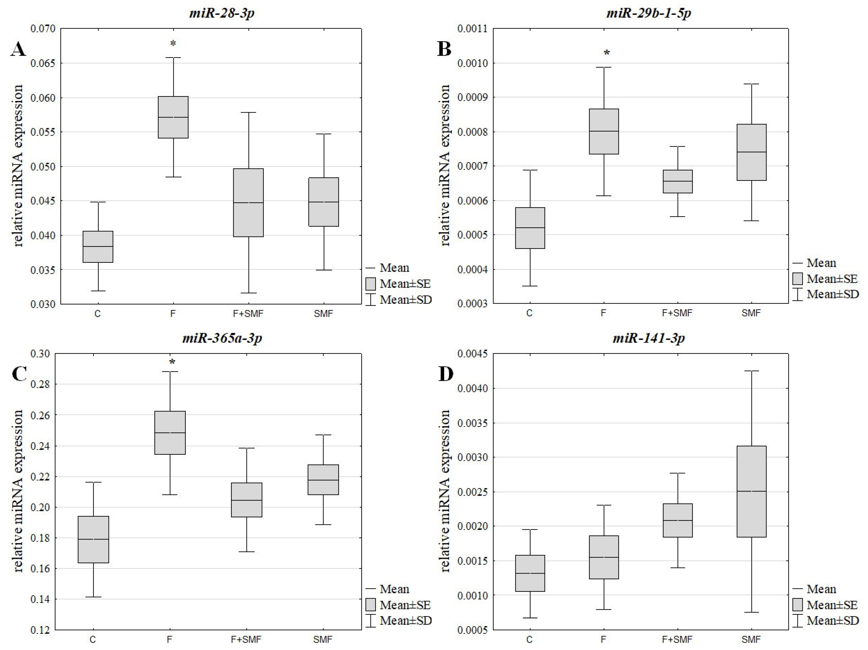
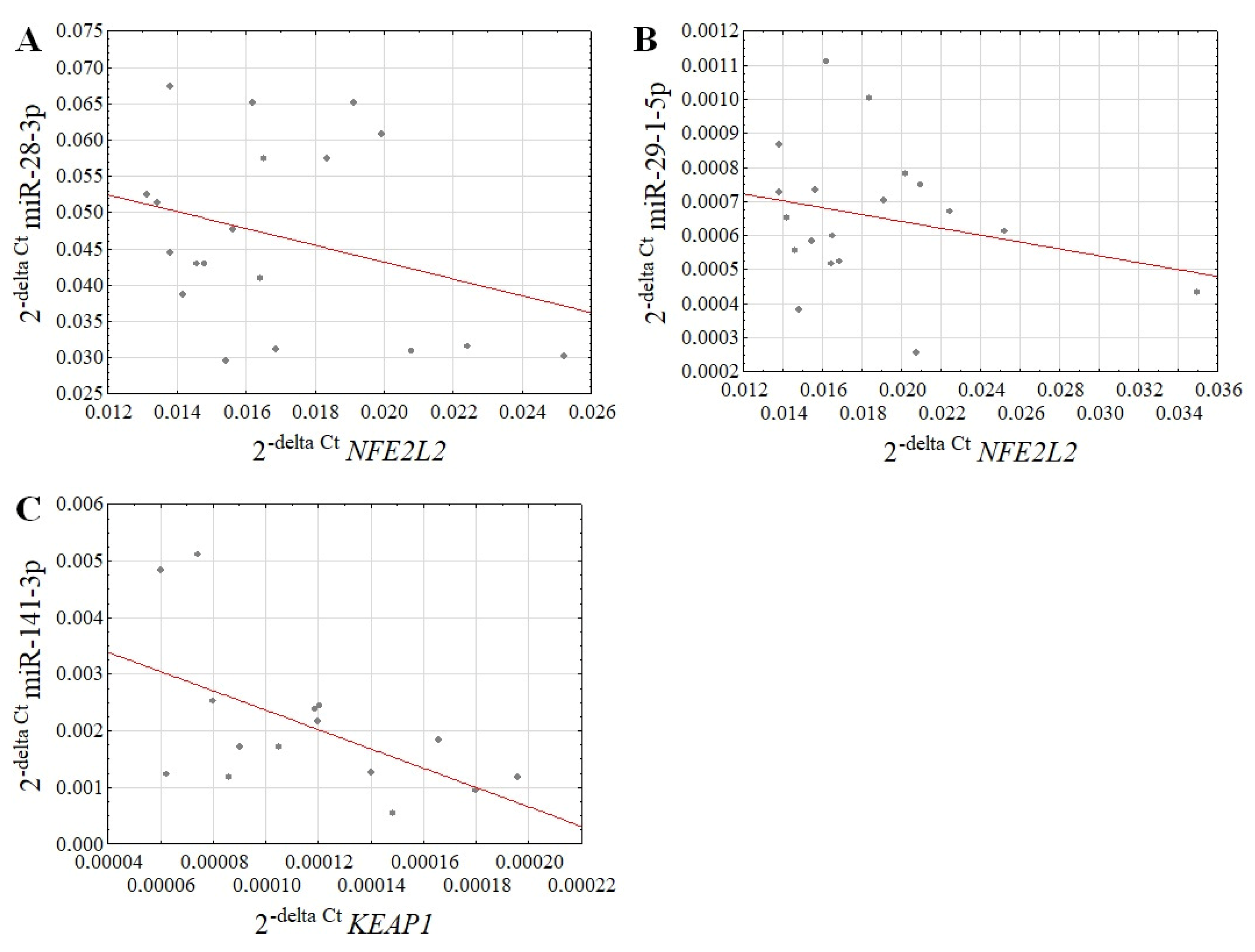
Analysis of our results showed that the miRNAs that regulate the expression of the NFE2L2 gene, i.e., miR-28-3p, miR-29b-1-5p and miR-365a-3p, were overexpressed in the NHDF cells that had been treated with fluoride compared to the control (Tukey post hoc test, p = 0.003, p = 0.010 and p = 0.005, respectively), while the action of an SMF caused their silencing compared to the F--treated cells (miR-28-3p: Tukey post hoc test, p = 0.086 for F + SMF vs. F and p = 0.076 for SMF vs. F; miR-365a-3p: Tukey post hoc test, p = 0.070 for F + SMF vs. F; a trend toward statistical significance) (Figure 2A–C, Table 3). There was also a negative correlation between the NFE2L2 gene expression and the selected miRNAs (miR-28-3p: r = −0.30, p = 0.050 and miR-29b-1-5p: r = −0.25, p = 0.062), which confirms their roles in regulating the NFE2L2 gene expression (Figure 3A,B). However, no such relationship was observed for miR-365a-3p (r = 0.043).
In the case of miR-141-3p regulating the expression of the KEAP1 gene, no statistically significant differences in its level were observed between the studied groups of cells (Figure 2D, Table 3). However, it can be seen that an SMF may increase its expression level. Moreover, a statistically significant negative correlation between the level of miR-141-3p and the level of KEAP1 mRNA was observed (r = −0.55, p = 0.032), which suggests its regulatory role in the expression of this gene (Figure 3C).

4. Discussion

As is already well known, oxidative stress is a factor that negatively affects the functioning of the body, thus causing a number of adverse effects. The Nrf2 signaling pathway is significantly related to oxidative stress as its activation is caused, inter alia, by reactive oxygen species. Under physiological conditions, the transcription factor Nrf2 is bound to the Keap 1 (kelch-like ECH = associated protein 1) protein in the cytoplasm. In the presence of ROS, this complex is degraded, which leads to the translocation of the Nrf2 factor to the cell nucleus, and consequently, causes its attachment to the ARE (antioxidant response element) sequence and the transcription of the genes whose protein products have cytoprotective activity. It has been proven that its activation provides protection against oxidative stress [32]. There are many reports in the latest scientific literature about the involvement of Nrf2 in many disease entities [33,34]. Nrf2 could play an important role in the prevention of various diseases, as well as during the therapy that is used to treat them. First of all, this applies to cancer as well as other human diseases, including cardiovascular diseases and diabetes as well as neurodegenerative diseases, e.g., Alzheimer’s disease, or other disorders of the internal organs. The function of the Keap1-Nrf2 system in monitoring cellular redox imbalance, and thus in the aging process, has also been widely described. Other more complex regulatory networks that are associated with this signaling pathway have been identified as well [35,36]. Thang et al. [37] proved that Nrf2 protects the cells of the nucleus pulposus against oxidative stress. These reports are identical to the conclusions that were drawn in the course of the presented study in terms of fluoride-induced redox homeostasis disorders and the role of the Nrf2 factor.
This and our previous studies [38,39] have shown that fluoride disrupts redox homeostasis and disrupts the ability of cells to protect themselves against oxidative stress by reducing the NFE2L2 expression. In the NHDF cells that had been treated with NaF, there was an increase in malondialdehyde (MDA) concentration, which is an indicator of lipid peroxidation as well as an increase in the intracellular ROS. At the same time, the presence of fluoride reduced the activity of the antioxidant enzymes superoxide dismutase (SOD) and glutathione peroxidase (GPx) [39]. In turn, the exposure of cells to a static magnetic field restored the redox balance and activated the cells, allowing them to defend themselves against stress, which was manifested by the up-regulation of the NFE2L2 expression and also increased activity of the antioxidant enzymes (superoxide dismutase—SOD and glutathione peroxidase—GPx) compared to the cells that had only been treated with fluoride [39]. Superoxide dismutase, catalase and glutathione peroxidase are key enzymes that constitute the first line of antioxidant defense, and their role is to prevent the formation of hydroxyl radicals. Furthermore, the combined exposure of fibroblasts to fluoride and an SMF resulted in a decrease in the production of intracellular ROS and a decrease in the MDA concentration, as was shown in our previous report [39]. Moreover, the RT-qPCR analysis showed statistically significant changes in the expression of the genes such as SOD1, SOD2 and GSR (glutathione reductase) that belong to the Nrf2 downstream genes. The mRNA level of these genes was lower in the NaF-treated fibroblasts than in the control cells. However, a significant increase in their expression was revealed in the cells that had been co-exposed to fluoride and an SMF with a 0.65 T flux density [23,38]. A similar trend was followed by Liu et al. [40]. They observed that the level of intracellular ROS increased dramatically after treatment with NaF. It was then observed that glycine had ameliorated this phenomenon [40]. Additionally, in a study that was conducted in an animal model (mice), the effect of NaF on an increase in ROS generation was also observed [41]. In turn, Zhou et al. [42] noted that exposure to fluoride in a concentration range of 0.25–2.00 mM caused an increase in the Nrf2 expression in mice due to endoplasmic reticulum stress, but that a higher concentration of fluoride (4.00 mM) led to its significant down-regulation.
Moreover, our findings demonstrate that an SMF may reduce the expression of the KEAP1 gene, which may also have an influence on the activation of the Nrf2 factor. This is quite a controversial issue, and scientific reports on this topic conflict. Alipour et al. [43] showed that an SMF increased the ROS level in mesenchymal stem cells. This is in contrast to our observations. However, the same authors showed an increase in the expression of Nrf2 and the antioxidant enzymes, which indicates an improvement in the antioxidant system. This situation could arise from the activation of the defense system of cells in response to increased ROS production [43]. However, Feng et al. [44] proved that in fibroblast cells (NIH3T3), an SMF caused a decrease in ROS level and thus reduced the oxidative stress.
In our report, the proposed mechanism for regulating the expression of the above-mentioned genes under the influence of both chemical and physical factors is the use of miRNA molecules. These small regulatory non-coding RNAs regulate various cellular signaling pathways, which helps to maintain the proper functioning of cells. However, changes in the miRNA expression profile can lead to a dysregulation of the cellular signaling pathways and the subsequent development of various diseases. On the other hand, it provides the opportunity to use them as potential diagnostic and therapeutic biomarkers. Many studies have shown the crucial role of miRNAs in regulating the Nrf2 signaling pathway as well. As was indicated in this report, selected miRNAs are involved in regulating NFE2L2 and KEAP1 activity in fibroblasts in the presence of fluoride and a static magnetic field of a moderate intensity. A negative correlation between the expression of the NFE2L2 gene and miR-28-3p or miR-29b-1-5p confirms their roles in regulating this gene. This may suggest the usefulness of the tested miRNAs that regulate NFE2L2 as potential biomarkers of the toxic effects of fluoride. A similar relationship was observed for KEAP1 and miR-141-3p. Li et al. [45] also demonstrated a dependence between KEAP1 and its regulatory miRNA. MiR-141-3p overexpression has been shown to attenuate the effects of oxidative stress in patients’ liver cells [44]. Moreover, De Blasio et al. [46] showed that the activation of Nrf2 caused breast cancer cell growth. At the same time, an overexpression of miR-29b-1-5p caused the opposite effect, including increased ROS production. To summarize, miR-29b-1-5p is down-regulated by Nrf2 [45]. Interestingly, the team of Yang et al. [47] also found a correlation between the Nrf2 mRNA level and miR-28 expression in breast epithelial cells, but through a Keap1-independent mechanism. The up-regulation of Nrf2 mRNA via the inhibition of miR-28 was also demonstrated by Dongoran and Wu [48] in Hep2G cells after diterpene quinone–cryptotanshinone treatment. However, in our study, there was no correlation between Nrf2 and miR-365a-3p expression in the cells that had been co-treated with both factors. The role of the miR-365a-3p/Nrf2 axis was also investigated by Gai et al. [49] in non-small-cell lung cancer. In turn, they showed that the metallothionein 1D pseudogene could induce ferroptosis that was mediated by the miR-365a-3p/Nrf2 signaling pathway. These reports suggest that the miRNAs that regulate the Nrf2 pathway could be susceptible to changes in expression under the influence of natural bioactive compounds, physical factors or other agents.
In conclusion, fluoride influences the Nrf2 signaling pathway and miRNAs are also involved in regulating it. Moreover, an SMF has beneficial effects on cells that are suffering from fluoride-induced oxidative stress via the stimulation of antioxidant defense. However, the main limitation of our research is the lack of investigation at a protein level. Therefore, additional studies are necessary to confirm the effect of fluoride ions and a static magnetic field on the Nrf2 signaling pathway. Further research is also necessary in relation to the possible use of a static magnetic field in the therapy of diseases in which oxidative stress plays a role in the pathogenesis. Our research is an attempt to explain the molecular mechanism of action of an SMF as well, which is not yet fully understood.

Author Contributions

Conceptualization, M.K.-D.; methodology, M.K.-D. and A.K.; investigation, M.K.-D., A.K. and A.S.-W.; writing—original draft preparation, M.K.-D. and A.K.; writing—review and editing, M.K.-D. and A.S.-W. All authors have read and agreed to the published version of the manuscript.

Funding

This research was funded by grant PCN-1-024/N/1/I from the Medical University of Silesia, Katowice, Poland.

Institutional Review Board Statement

Not applicable.

Informed Consent Statement

Not applicable.

Data Availability Statement

Not applicable.

Conflicts of Interest

The authors declare no conflict of interest.

References

  1. Correia de Sousa, M.; Gjorgjieva, M.; Dolicka, D.; Sobolewski, C.; Foti, M. Deciphering miRNAs’ Action through miRNA Editing. Int. J. Mol. Sci. 2019, 20, 6249. [Google Scholar] [CrossRef] [PubMed] [Green Version]
  2. Hill, M.; Tran, N. miRNA interplay: Mechanisms and consequences in cancer. Dis. Model. Mech. 2021, 14, dmm047662. [Google Scholar] [CrossRef] [PubMed]
  3. Ali Syeda, Z.; Langden, S.S.S.; Munkhzul, C.; Lee, M.; Song, S.J. Regulatory Mechanism of MicroRNA Expression in Cancer. Int. J. Mol. Sci. 2020, 21, 1723. [Google Scholar] [CrossRef] [PubMed] [Green Version]
  4. Kura, B.; Szeiffova Bacova, B.; Kalocayova, B.; Sykora, M.; Slezak, J. Oxidative Stress-Responsive MicroRNAs in Heart Injury. Int. J. Mol. Sci. 2020, 21, 358. [Google Scholar] [CrossRef] [Green Version]
  5. Wang, X.; Zhou, Y.; Gao, Q.; Ping, D.; Wang, Y.; Wu, W.; Lin, X.; Fang, Y.; Zhang, J.; Shao, A. The Role of Exosomal microRNAs and Oxidative Stress in Neurodegenerative Diseases. Oxid. Med. Cell. Longev. 2020, 2020, 3232869. [Google Scholar] [CrossRef]
  6. Bellezza, I.; Giambanco, I.; Minelli, A.; Donato, R. Nrf2-Keap1 signaling in oxidative and reductive stress. Biochim. Biophys. Acta Mol. Cell Res. 2018, 1865, 721–733. [Google Scholar] [CrossRef]
  7. Moi, P.; Chan, K.; Asunis, I.; Cao, A.; Kan, Y.W. Isolation of NF-E2-related factor 2 (Nrf2), a NF-E2-like basic leucine zipper transcriptional activator that binds to the tandem NF-E2/AP1 repeat of the beta-globin locus control region. Proc. Natl. Acad. Sci. USA 1994, 91, 9926–9930. [Google Scholar] [CrossRef] [Green Version]
  8. He, F.; Antonucci, L.; Karin, M. NRF2 as a regulator of cell metabolism and inflammation in cancer. Carcinogenesis 2020, 41, 405–416. [Google Scholar] [CrossRef]
  9. Saenen, N.D.; Martens, D.S.; Neven, K.Y.; Alfano, R.; Bové, H.; Janssen, B.G.; Roels, H.A.; Plusquin, M.; Vrijens, K.; Nawrot, T.S. Air pollution-induced placental alterations: An interplay of oxidative stress, epigenetics, and the aging phenotype? Clin. Epigenetics 2019, 11, 124. [Google Scholar] [CrossRef] [PubMed]
  10. Ahmed, N.J.; Husen, A.Z.; Khoshnaw, N.; Getta, H.A.; Hussein, Z.S.; Yassin, A.K.; Jalal, S.D.; Mohammed, R.N.; Alwan, A.F. The Effects of Smoking on IgE, Oxidative Stress and Haemoglobin Concentration. Asian Pac. J. Cancer Prev. 2020, 21, 1069–1072. [Google Scholar] [CrossRef]
  11. Albrecht, S.; Jung, S.; Müller, R.; Lademann, J.; Zuberbier, T.; Zastrow, L.; Reble, C.; Beckers, I.; Meinke, M.C. Skin type differences in solar-simulated radiation-induced oxidative stress. Br. J. Dermatol. 2019, 180, 597–603. [Google Scholar] [CrossRef]
  12. Srivastava, S.; Flora, S.J.S. Fluoride in Drinking Water and Skeletal Fluorosis: A Review of the Global Impact. Curr. Environ. Health Rep. 2020, 7, 140–146. [Google Scholar] [CrossRef] [PubMed]
  13. Rezaee, T.; Bouxsein, M.L.; Karim, L. Increasing fluoride content deteriorates rat bone mechanical properties. Bone 2020, 136, 115369. [Google Scholar] [CrossRef]
  14. Pereira, H.A.B.D.S.; Dionizio, A.S.; Araujo, T.T.; Fernandes, M.D.S.; Iano, F.G.; Buzalaf, M.A.R. Proposed mechanism for understanding the dose- and time-dependency of the effects of fluoride in the liver. Toxicol. Appl. Pharmacol. 2018, 358, 68–75. [Google Scholar] [CrossRef] [PubMed]
  15. Ma, Y.; Yao, Y.; Zhong, N.; Angwa, L.M.; Pei, J. The dose-time effects of fluoride on the expression and DNA methylation level of the promoter region of BMP-2 and BMP-7 in rats. Environ. Toxicol. Pharmacol. 2020, 75, 103331. [Google Scholar] [CrossRef]
  16. Labib, H.; Badr, A.M.; Abdelgwad, M.; Abd El-Galil, T.I. Keap1/Nrf2 pathway in sodium fluoride-induced cardiac toxicity and the prophylactic role of vitamin C versus platelet-rich plasma. Folia Morphol. 2022, 81, 663–678. [Google Scholar] [CrossRef] [PubMed]
  17. Shanmugam, T.; Selvaraj, M.; Poomalai, S. Epigallocatechin gallate potentially abrogates fluoride induced lung oxidative stress, inflammation via Nrf2/Keap1 signaling pathway in rats: An in-vivo and in-silico study. Int. Immunopharmacol. 2016, 39, 128–139, Erratum in Int. Immunopharmacol. 2020, 80, 106203. [Google Scholar] [CrossRef] [PubMed]
  18. Mukhopadhyay, D.; Srivastava, R.; Chattopadhyay, A. Sodium fluoride generates ROS and alters transcription of genes for xenobiotic metabolizing enzymes in adult zebrafish (Danio rerio) liver: Expression pattern of Nrf2/Keap1 (INrf2). Toxicol. Mech. Methods 2015, 25, 364–373. [Google Scholar] [CrossRef] [PubMed]
  19. Chen, J.; Luo, Y.; Cao, J.; Xie, L. Fluoride exposure changed the expression of microRNAs in gills of male zebrafish (Danio rerio). Aquat. Toxicol. 2021, 233, 105789. [Google Scholar] [CrossRef] [PubMed]
  20. Zhao, Y.; Yu, Y.; Ommati, M.M.; Xu, J.; Wang, J.; Zhang, J.; Sun, Z.; Niu, R.; Wang, J. Multiomics Analysis Revealed the Molecular Mechanism of miRNAs in Fluoride-Induced Hepatic Glucose and Lipid Metabolism Disorders. J. Agric. Food Chem. 2022, 70, 14284–14295. [Google Scholar] [CrossRef] [PubMed]
  21. Izzo, L.; Tunesi, M.; Boeri, L.; Laganà, M.; Giordano, C.; Raimondi, M.T. Influence of the static magnetic field on cell response in a miniaturized optically accessible bioreactor for 3D cell culture. Biomed. Microdevices 2019, 21, 29. [Google Scholar] [CrossRef] [Green Version]
  22. Chekhun, V.F.; Demash, D.V.; Nalieskina, L.A. Evaluation of biological effects and possible mechanisms of action of static magnetic field. Fiziol. Zh. 2012, 58, 85–94. [Google Scholar] [CrossRef]
  23. Kimsa-Dudek, M.; Synowiec-Wojtarowicz, A.; Derewniuk, M.; Gawron, S.; Paul-Samojedny, M.; Kruszniewska-Rajs, C.; Pawłowska-Góral, K. Impact of fluoride and a static magnetic field on the gene expression that is associated with the antioxidant defense system of human fibroblasts. Chem. Biol. Interact. 2018, 287, 13–19. [Google Scholar] [CrossRef] [PubMed]
  24. Lamkowski, A.; Kreitlow, M.; Radunz, J.; Willenbockel, M.; Stiemer, M.; Fichte, L.O.; Rädel, C.F.; Majewski, M.; Ostheim, P.; Port, M.; et al. Analyzing the impact of 900 MHz EMF short-term exposure to the expression of 667 miRNAs in human peripheral blood cells. Sci. Rep. 2021, 11, 4444. [Google Scholar] [CrossRef] [PubMed]
  25. Gawron, S.; Glinka, M.; Wolnik, T. Magnetyczna komora badawcza dedykowana do hodowli komórek. Zesz. Probl. Masz. Elektr. 2012, 4, 11–16. [Google Scholar]
  26. Glinka, M.; Gawron, S.; Sieroń, A.; Pawłowska-Góral, K.; Cieślar, G.; Sieroń-Stołtny, K. Test chambers for cell culture in static magnetic field. J. Magn. Mater. 2013, 331, 208–215. [Google Scholar] [CrossRef]
  27. Dini, L.; Abbro, L. Bioeffects of moderate-intensity static magnetic fields on cell cultures. Micron 2005, 36, 195–217. [Google Scholar] [CrossRef]
  28. Allison, S.J.; Docherty, P.D.; Pons, D.; Chase, J.G. Serum fluoride levels in ambulance staff after commencement of methoxyflurane administration compared to meta-analysis results for the general public. Int. J. Occup. Med. Environ. Health 2021, 34, 767–777. [Google Scholar] [CrossRef]
  29. Hsu, J.B.; Chiu, C.M.; Hsu, S.D.; Huang, W.Y.; Chien, C.H.; Lee, T.Y.; Huang, H.D. miRTar: An integrated system for identifying miRNA-target interactions in human. BMC. Bioinformatics 2011, 12, 300. [Google Scholar] [CrossRef] [PubMed] [Green Version]
  30. Ayers, D.; Baron, B.; Hunter, T. miRNA Influences in NRF2 Pathway Interactions within Cancer Models. J. Nucleic Acids 2015, 2015, 143636. [Google Scholar] [CrossRef] [Green Version]
  31. Schmittgen, T.D.; Livak, K.J. Analyzing real-time PCR data by the comparative C(T) method. Nat. Protoc. 2008, 3, 1101–1108. [Google Scholar] [CrossRef] [PubMed]
  32. Ray, P.D.; Huang, B.W.; Tsuji, Y. Reactive oxygen species (ROS) homeostasis and redox regulation in cellular signaling. Cell Signal. 2012, 24, 981–990. [Google Scholar] [CrossRef] [PubMed] [Green Version]
  33. Zhan, X.; Li, J.; Zhou, T. Targeting Nrf2-Mediated Oxidative Stress Response Signaling Pathways as New Therapeutic Strategy for Pituitary Adenomas. Front. Pharmacol. 2021, 12, 565748. [Google Scholar] [CrossRef]
  34. Li, L.; Cheng, S.Q.; Guo, W.; Cai, Z.Y.; Sun, Y.Q.; Huang, X.X.; Yang, J.; Ji, J.; Chen, Y.Y.; Dong, Y.F.; et al. Oridonin prevents oxidative stress-induced endothelial injury via promoting Nrf-2 pathway in ischaemic stroke. J. Cell. Mol. Med. 2021, 25, 9753–9766. [Google Scholar] [CrossRef] [PubMed]
  35. Chen, Q.M.; Maltagliati, A.J. Nrf2 at the heart of oxidative stress and cardiac protection. Physiol. Genom. 2018, 50, 77–97. [Google Scholar] [CrossRef] [PubMed] [Green Version]
  36. Yu, C.; Xiao, J.H. The Keap1-Nrf2 System: A Mediator between Oxidative Stress and Aging. Oxid. Med. Cell. Longev. 2021, 2021, 6635460. [Google Scholar] [CrossRef]
  37. Tang, Z.; Hu, B.; Zang, F.; Wang, J.; Zhang, X.; Chen, H. Nrf2 drives oxidative stress-induced autophagy in nucleus pulposus cells via a Keap1/Nrf2/p62 feedback loop to protect intervertebral disc from degeneration. Cell Death Dis. 2019, 10, 510. [Google Scholar] [CrossRef] [PubMed] [Green Version]
  38. Kimsa-Dudek, M.; Krawczyk, A.; Synowiec-Wojtarowicz, A. The Protective Effect of Static Magnetic Fields with Different Magnetic Inductions against Fluoride Toxicity Is Related to the NRF2 Signaling Pathway. Appl. Sci. 2020, 10, 6509. [Google Scholar] [CrossRef]
  39. Kimsa-Dudek, M.; Synowiec-Wojtarowicz, A.; Derewniuk, M.; Paul-Samojedny, M.; Pawłowska-Góral, K. The effect of simultaneous exposure of human fibroblasts to fluoride and moderate intensity static magnetic fields. Int. J. Radiat. Biol. 2019, 95, 1581–1587. [Google Scholar] [CrossRef]
  40. Liu, Y.; Sun, B.; Zhang, S.; Li, J.; Qi, J.; Bai, C.; Zhang, J.; Liang, S. Glycine alleviates fluoride-induced oxidative stress, apoptosis and senescence in a porcine testicular Sertoli cell line. Reprod. Domest. Anim. 2021, 56, 884–896. [Google Scholar] [CrossRef]
  41. Lu, Y.; Luo, Q.; Cui, H.; Deng, H.; Kuang, P.; Liu, H.; Fang, J.; Zuo, Z.; Deng, J.; Li, Y.; et al. Sodium fluoride causes oxidative stress and apoptosis in the mouse liver. Aging 2017, 9, 1623–1639. [Google Scholar] [CrossRef] [PubMed]
  42. Zhou, X.; Chen, Z.; Zhong, W.; Yu, R.; He, L. Effect of fluoride on PERK-Nrf2 signaling pathway in mouse ameloblasts. Hum. Exp. Toxicol. 2019, 38, 833–845. [Google Scholar] [CrossRef] [PubMed]
  43. Alipour, M.; Hajipour-Verdom, B.; Javan, M.; Abdolmaleki, P. Static and Electromagnetic Fields Differently Affect Proliferation and Cell Death through Acid Enhancement of ROS Generation in Mesenchymal Stem Cells. Radiat. Res. 2022, 198, 384–395. [Google Scholar] [CrossRef] [PubMed]
  44. Feng, C.; Yu, B.; Song, C.; Wang, J.; Zhang, L.; Ji, X.; Wang, Y.; Fang, Y.; Liao, Z.; Wei, M.; et al. Static Magnetic Fields Reduce Oxidative Stress to Improve Wound Healing and Alleviate Diabetic Complications. Cells 2022, 11, 443. [Google Scholar] [CrossRef]
  45. Li, T.; Chen, Q.; Dai, J.; Huang, Z.; Luo, Y.; Mou, T.; Pu, J.; Yang, H.; Wei, X.; Wu, Z. MicroRNA-141-3p attenuates oxidative stress-induced hepatic ischemia reperfusion injury via Keap1/Nrf2 pathway. Mol. Biol. Rep. 2022, 49, 7575–7585. [Google Scholar] [CrossRef]
  46. De Blasio, A.; di Fiore, R.; Pratelli, G.; Drago-Ferrante, R.; Saliba, C.; Baldacchino, S.; Grech, G.; Scerri, C.; Vento, R.; Tesoriere, G. A loop involving NRF2, miR-29b-1-5p and AKT, regulates cell fate of MDA-MB-231 triple-negative breast cancer cells. J. Cell. Physiol. 2020, 235, 629–637. [Google Scholar] [CrossRef] [PubMed]
  47. Yang, M.; Yao, Y.; Eades, G.; Zhang, Y.; Zhou, Q. MiR-28 regulates Nrf2 expression through a Keap1-independent mechanism. Breast Cancer Res. Treat. 2011, 129, 983–991. [Google Scholar] [CrossRef] [Green Version]
  48. Dongoran, R.A.; Wu, T.Y. Cryptotanshinone activate Nrf2 expression through microRNA regulations. Cancer Res. 2017, 77 (Suppl. 13), 5269. [Google Scholar] [CrossRef]
  49. Gai, C.; Liu, C.; Wu, X.; Yu, M.; Zheng, J.; Zhang, W.; Lv, S.; Li, W. MT1DP loaded by folate-modified liposomes sensitizes erastin-induced ferroptosis via regulating miR-365a-3p/NRF2 axis in non-small cell lung cancer cells. Cell Death Dis. 2020, 11, 751. [Google Scholar] [CrossRef] [PubMed]
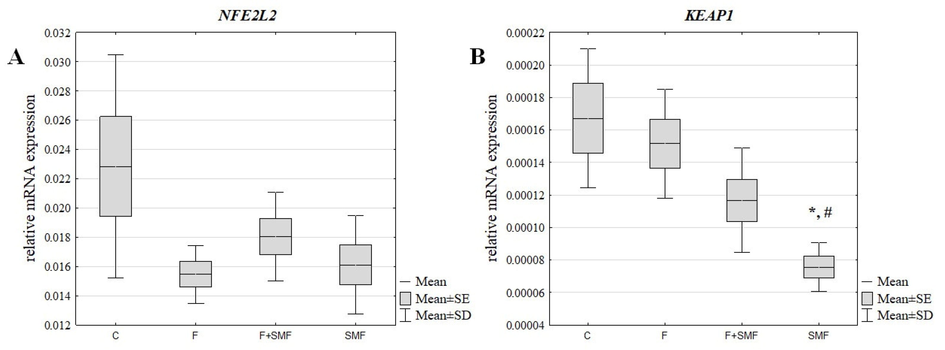
Figure 1. Relative mRNA expression of the NFE2L2 (A) and KEAP1 (B) genes in cells that had been treated with fluoride and cells that had been exposed to a static magnetic field. The results are presented as the mean ± SD; C—control cells; F—cells that had only been treated with fluoride; SMF—cells that had only been exposed to a static magnetic field; F + SMF—cells that had simultaneously been exposed to fluoride and a static magnetic field; * p < 0.05 vs. C, # p < 0.05 vs. F.
Applsci 13 01470 g001
Figure 2. Relative miRNA expression of miR-28-3p (A), miR-29b-1-5p (B), miR-365a-3p (C) and miR-141-3p (D) in the cells that had been treated with fluoride and the cells that had been exposed to a static magnetic field. The results are presented as the mean ± SD; C—control cells; F—cells that had been treated with fluoride; SMF—cells that had been exposed to a static magnetic field; F + SMF—cells that had simultaneously been exposed to fluoride and a static magnetic field; * p < 0.05 vs. C.
Applsci 13 01470 g002
Figure 3. Correlation between the selected mRNA and miRNA: miR-28-3p vs. NFE2L2 (A), mir-29-1-5p vs. NFE2L2 (B) and miR-14-1-3p vs. KEAP1 (C).
Applsci 13 01470 g003
Table 1. Characteristics of the primers that were used in the study.
Gene NameOligo NameSequence (5′-3′)
NFE2-like bZIP transcription factor 2 (NFE2L2)FH1_NFE2L2Forward: CGTTTGTAGATGACAATGAGG
BH1_NFE2L2Reverse: AGAAGTTTCAGGTGACTGAG
Kelch-like ECH-associated protein 1 (KEAP1)FH1_KEAP1Forward: CGTTTGTAGATGACAATGAGG
BH1_KEAP1Reverse: CTCCAAGGACGTAGATTCTC
β-actin (ACTB)FH1_ACTBForward: GACGACATGGAGAAAATCTG
BH1_ACTBReverse: ATGATCTGGGTCATCTTCTC
Table 2. Comparison of the gene expression levels (fold change) among the studied cell groups.
GroupsmRNA Level
NFE2L2KEAP1
FC vs. CFC vs. FFC vs. CFC vs. F
F−1.48-−1.10-
F + SMF−1.271.17−1.43−1.30
SMF−1.421.04−2.21−2.00
FC—fold change; C—control cells; F—cells that had been treated with fluoride ions; F + SMF—cells that had been treated with fluoride ions and simultaneously exposed to a static magnetic field; SMF—cells that had only been exposed to a static magnetic field.
Table 3. Comparison of the miRNA expression levels (fold change) among studied cell groups.
GroupsmiRNA Level
miR-28-3pmiR-29b-1-5pmiR-365a-3pmiR-141-3p
FC vs. CFC vs. FFC vs. CFC vs. FFC vs. CFC vs. FFC vs. CFC vs. F
F1.49-1.54-1.39-1.18-
F + SMF1.17−1.281.26−1.221.14−1.211.581.34
SMF1.17−1.271.42−1.081.22−1.141.901.62
FC—fold change; C—control cells; F—cells that had been treated with fluoride ions; F + SMF—cells that had been treated with fluoride ions and simultaneously exposed to a static magnetic field; SMF—cells that had been exposed to a static magnetic field.
Disclaimer/Publisher’s Note: The statements, opinions and data contained in all publications are solely those of the individual author(s) and contributor(s) and not of MDPI and/or the editor(s). MDPI and/or the editor(s) disclaim responsibility for any injury to people or property resulting from any ideas, methods, instructions or products referred to in the content.