

Unilateral Condylar Hyperplasia in Surgeons’ Perspective—A Narrative Review

 ,
, 
 ,
,  , ,
, , 
Abstract
1. Introduction
2. Mandibular Asymmetries Differentiation
3. Classifications
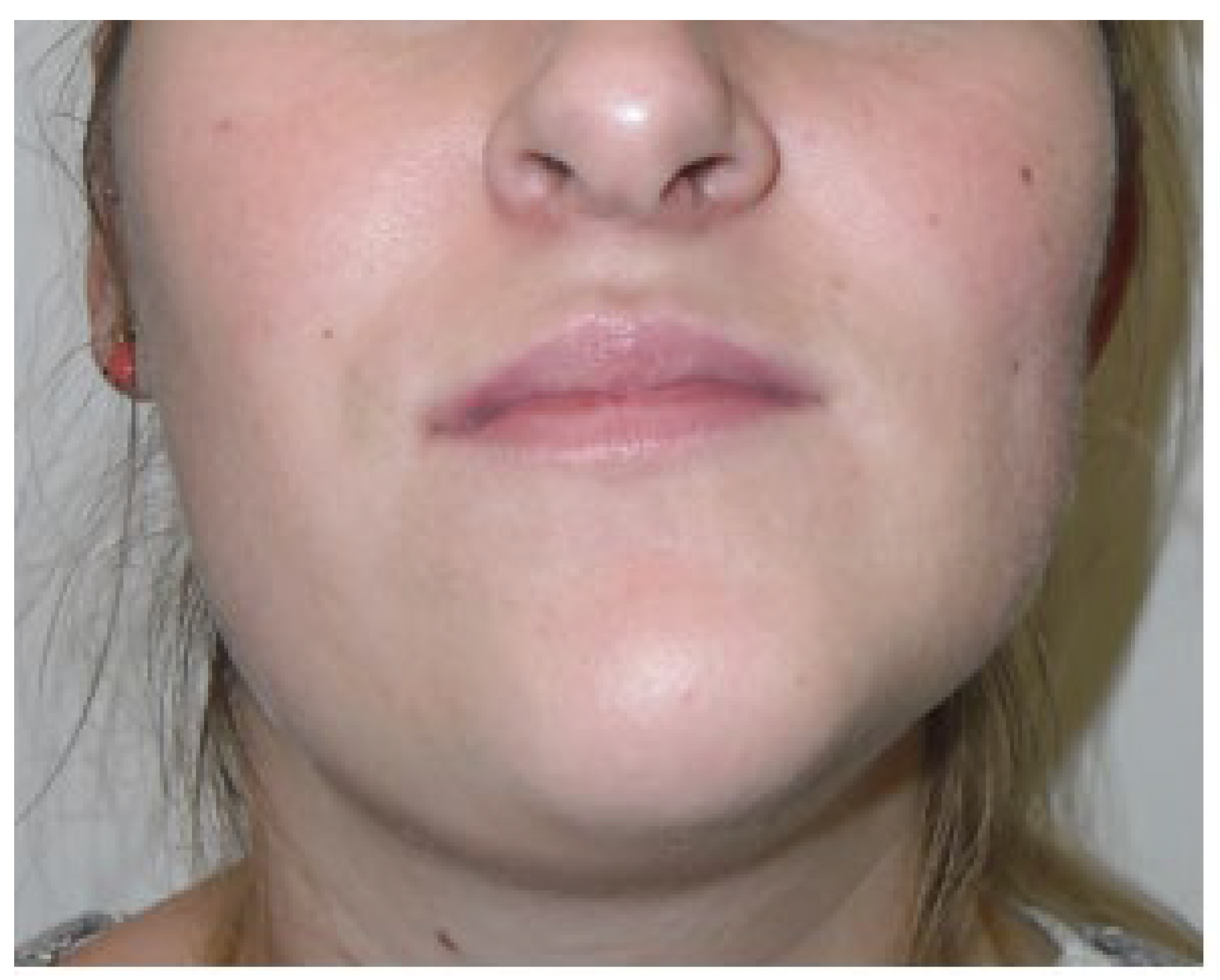
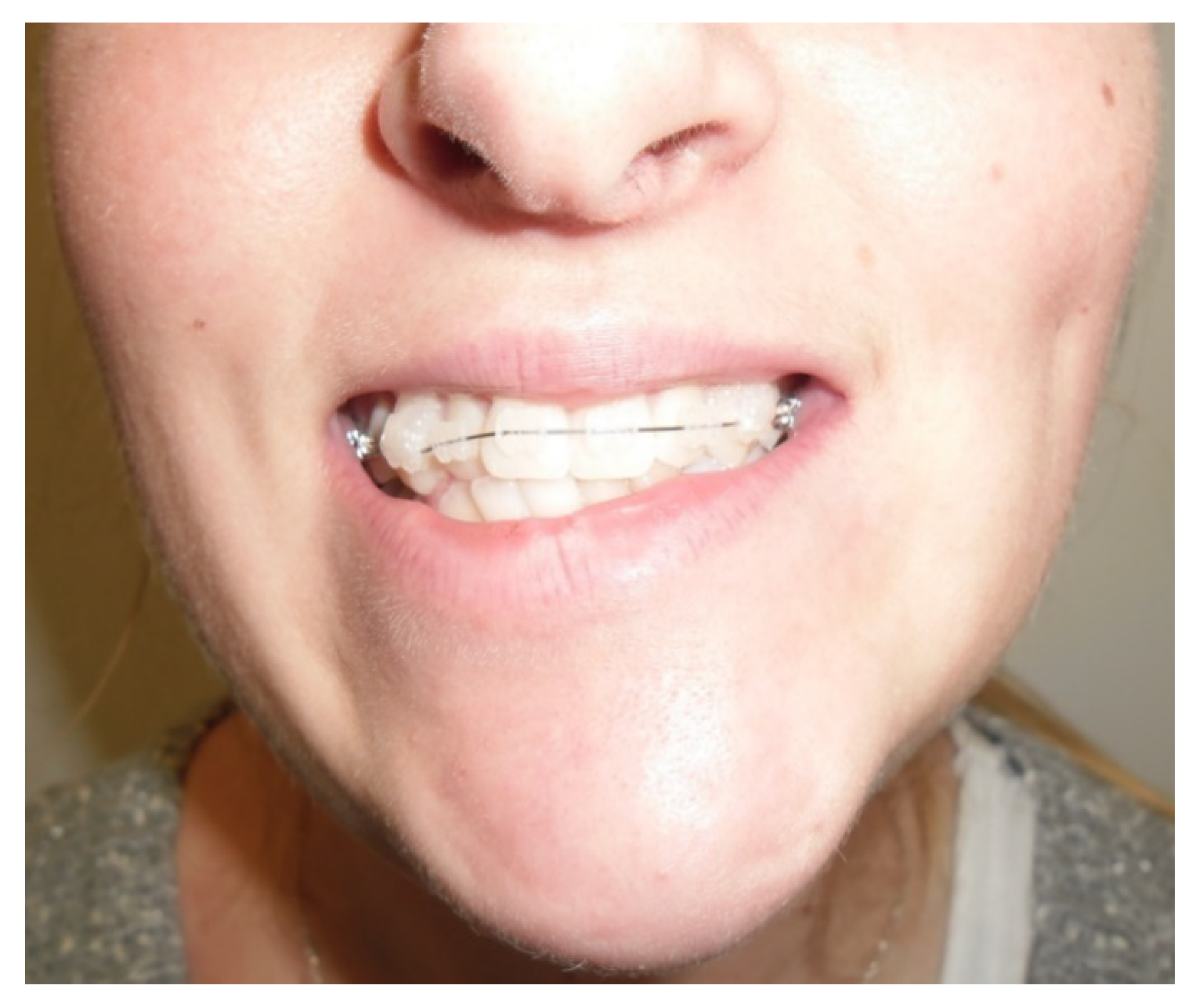
4. Signs and Symptoms
5. Imaging Tests for Diagnosis
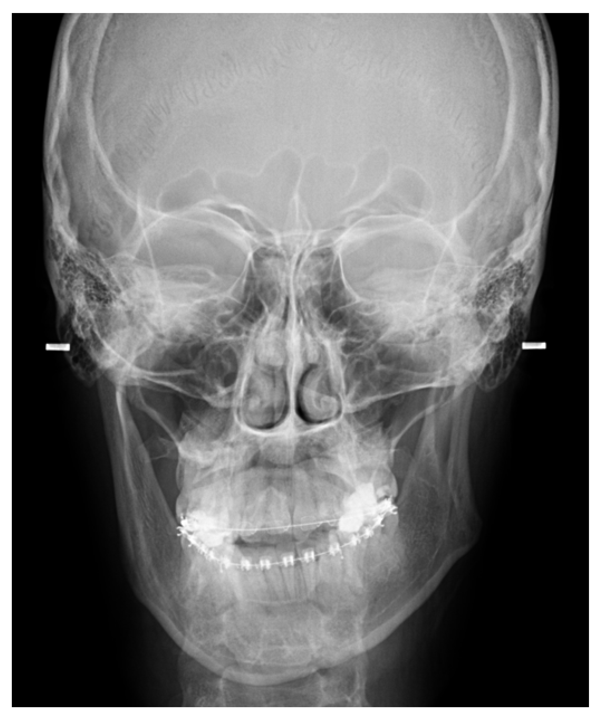
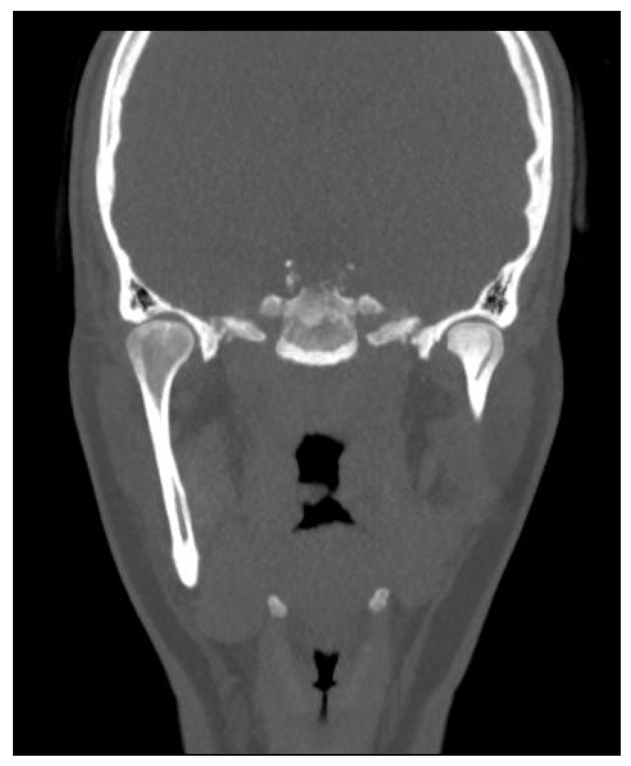
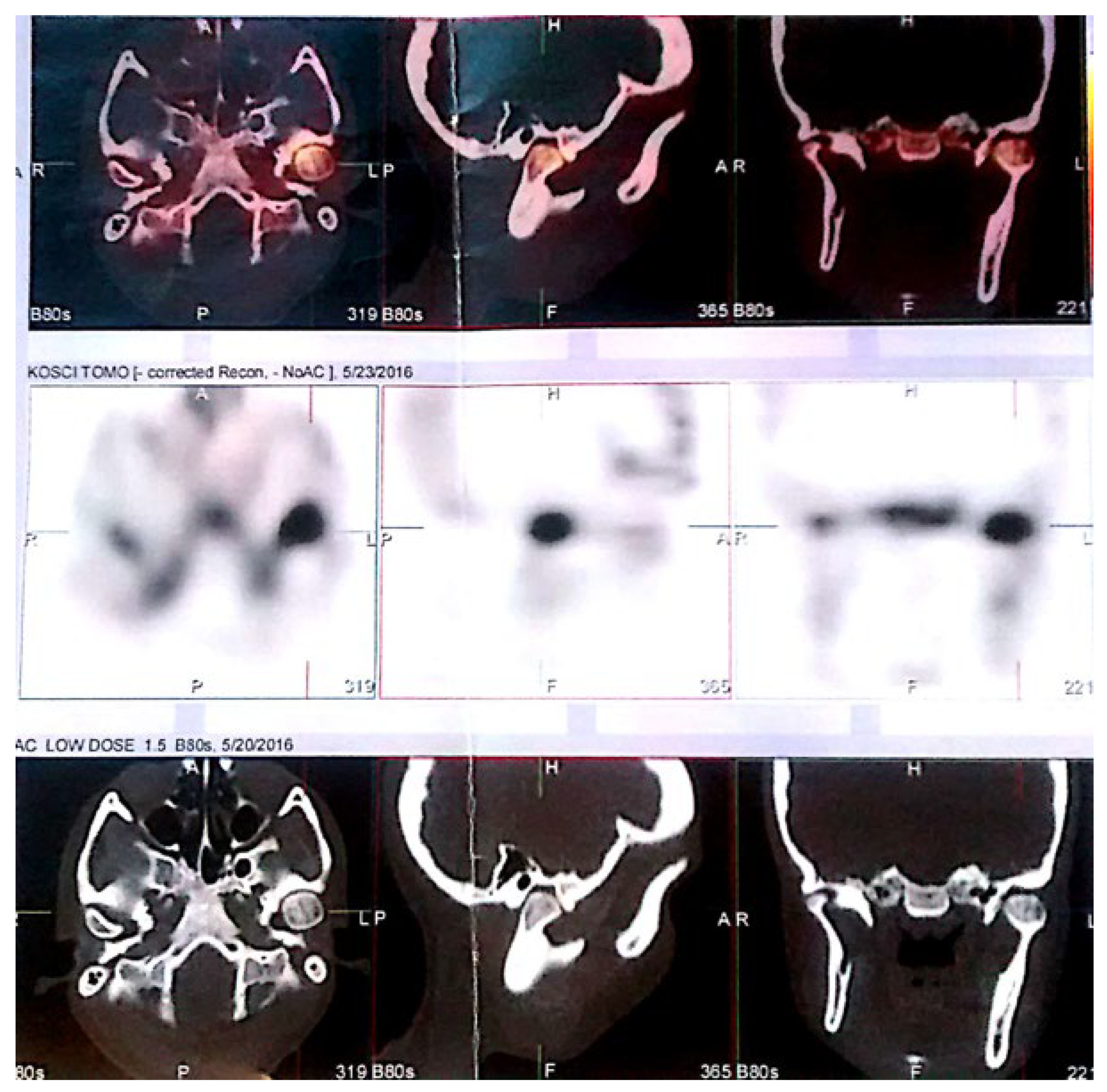
5.1. Panoramic Radiographs and Computed Tomography
5.2. Scintigraphy and Growth Activity
6. Dental Treatment
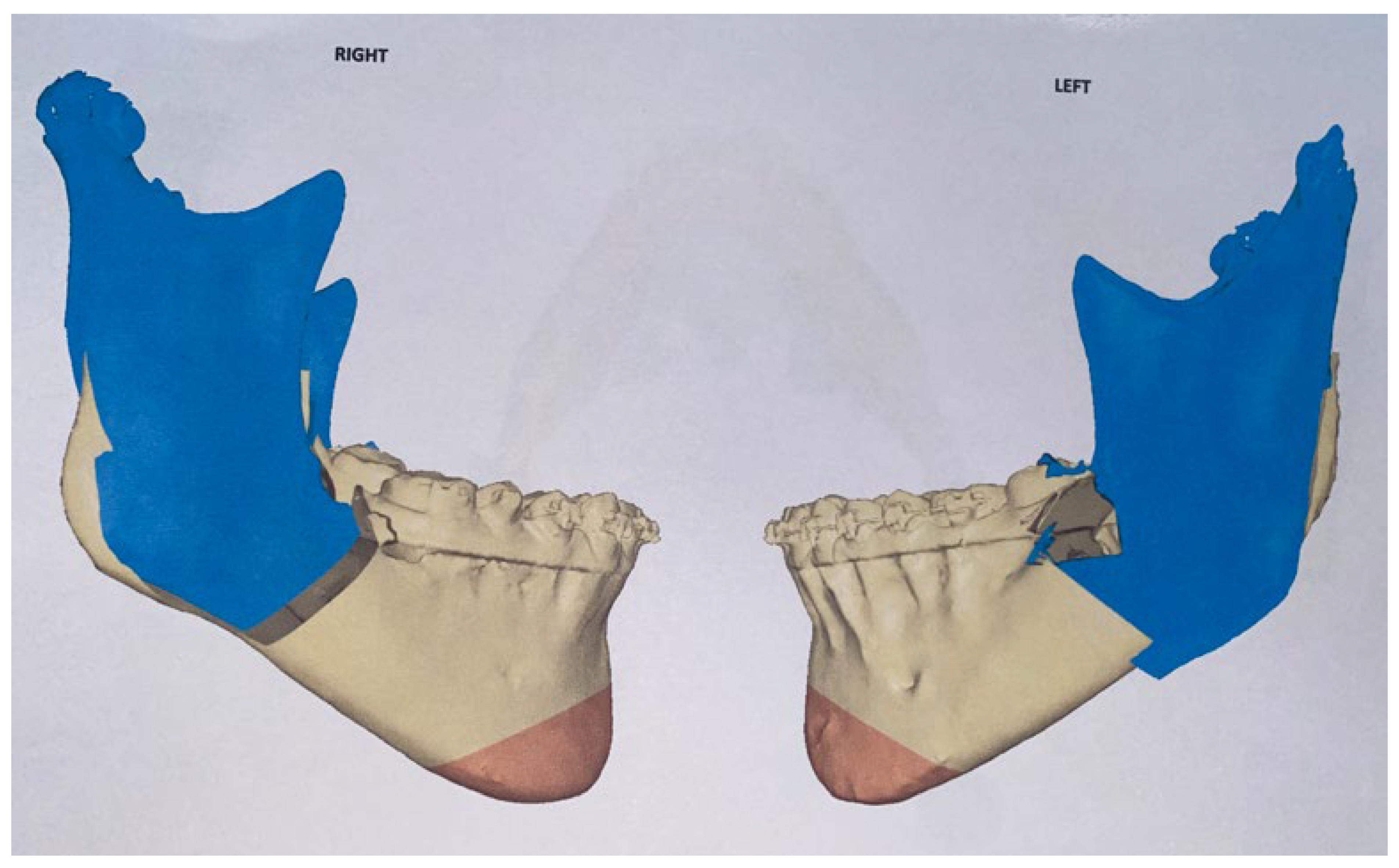
7. Treatment from Surgeons’ Perspective
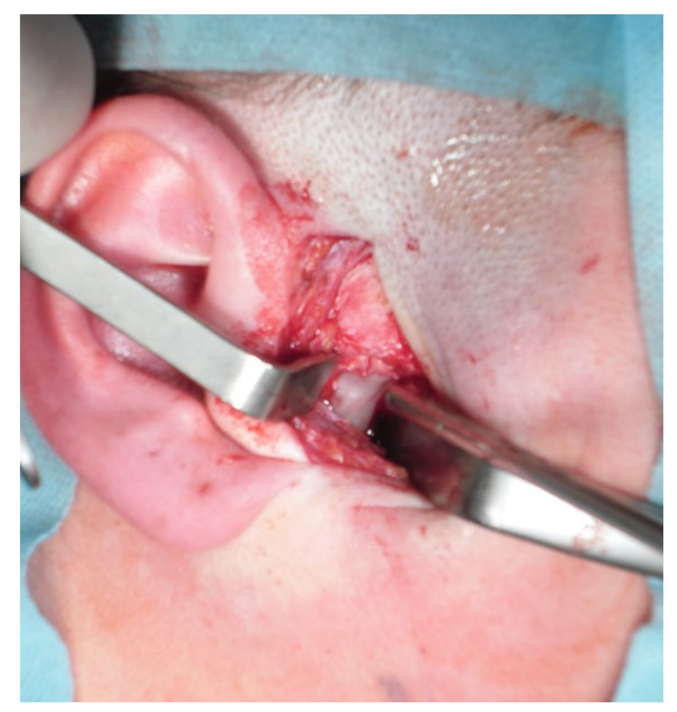
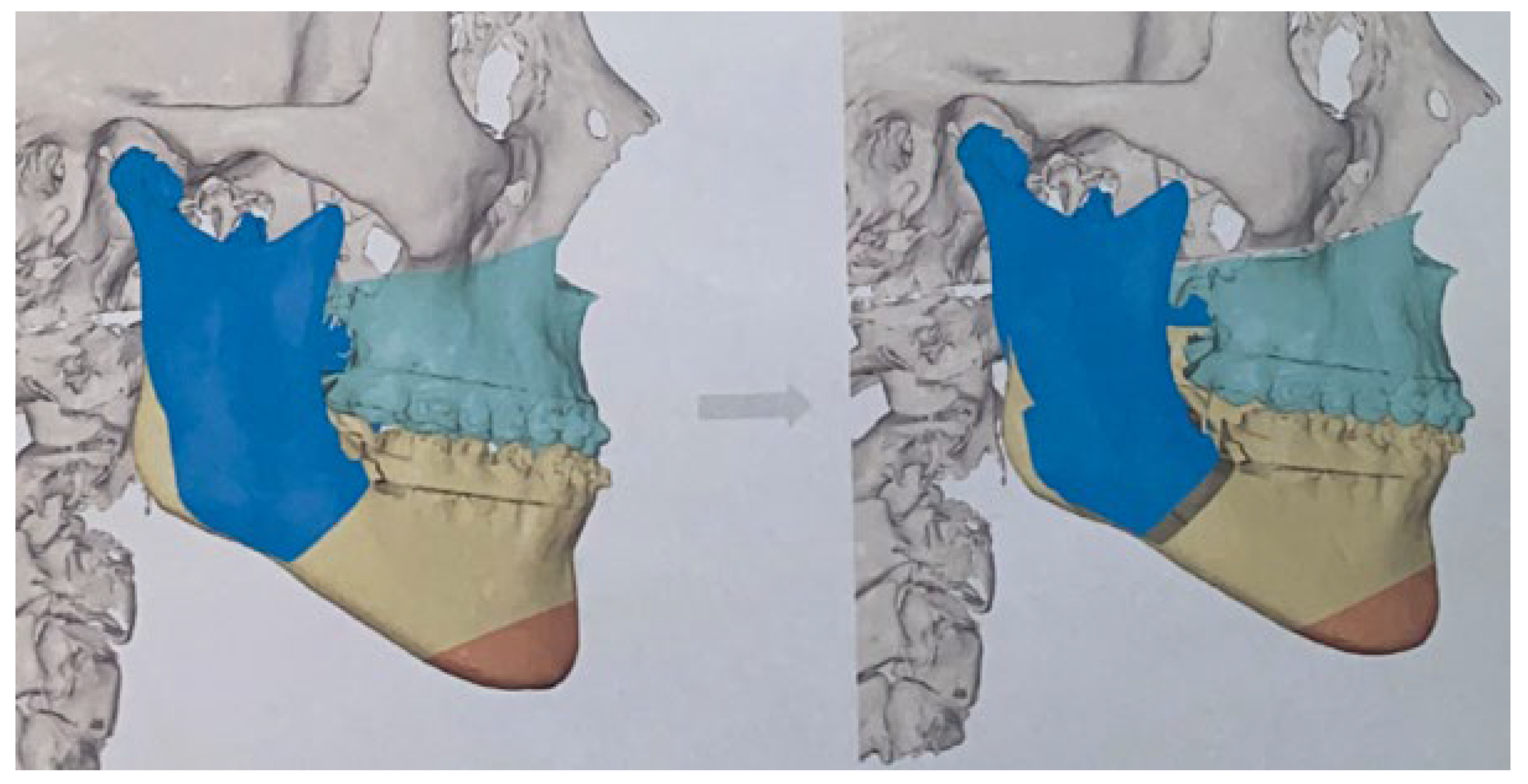
7.1. Condylectomy
- (1)
- High condylectomy, Lippold et al.: excision of 4–5 mm of bone from the superior condyle pole is enough when this amount of bone is cut vertically from the lateral to medial pole [80,81,82]. High condylectomy as a sole operative treatment might be sufficient in some cases of UCH. The use of orthodontic treatment is suitable for some small or mild cases of overgrowth. The problem remains crucial when there are differences in the ramus vertical height and maxillary asymmetry with their own growth on the affected side of UCH [83,84,85].
- (2)
- Low condylectomy: dedicated to removing a larger volume of MCH with just the far inferior part left alone. On the other hand, Wolford et al.’s study on 37 UCH-2 (osteochondroma of mandibular condylectomy associated with mandibular deformity) patients used a low condylectomy approach in all patients with recontouring of the condylar neck to form a new condyle, repositioning of the articular disc over the condylar stump, and repositioning of the contralateral disc if any symptoms of displacement or malposition might occur, and any indicated orthognathic surgical procedures [6,75,86]. Further, follow-ups concluded that low condylectomy and orthognathic surgery improved not only TMJ function and proper joint movements but reduced any pain or other related symptoms [29,87,88,89].
- (3)
- A total condylectomy procedure (TCP) is rarely used in UCH and is mostly dedicated to condyle tumors, such as osteochondromas (OC). Domingues et al., after total condylectomy for condyle repair, used a costochondral graft and preserved the articular disc with good outcomes. Some authors advise leaving the resected condyle in the joint without reconstruction or suturing with pterygoid muscles only or using other modifications [13,90,91]. The neocondyle condyle might be unstable for quite some time, and patient education on chewing, biting, diet, and other factors is necessary.
- (4)
- Proportional condylectomy is a special technique that compares the head and ramus’s vertical and horizontal diameters to ensure a more balanced result after the procedure, and therefore this amount of condyle is excised. Mouallem et al.’s retrospective study on 73 patients with UCH was divided into vertical (61.6%) and transversal (38.4%) forms of UCH according to Delaire’s classification [92]. Using proportional condylectomy in the authors’ approach, followed by indicated orthognathic techniques, maxilla–mandibular elastic therapy, and rehabilitation, is a good, accurate, and reliable option for treating UCH, regardless of the activity status of the pathology [93].
- (5)
- Condylar head reshaving and modeling with bone drills, chisels, and piezosurgery (the most superior part of the MCH) [94].
- (6)
- Transoral approach. This might include techniques with mandibular coronoid process osteotomy to gain more visibility toward the condyle, as reported by Choung et al. [95]. A similar approach to the intraoral approach was described by Wang et al., which might be combined with coronoid process resection and an IVRO procedure–intraoral vertical ramus osteotomy [96,97]. Approaches with computer-guided/3D navigation techniques can also be useful. The intraoral approach for a condylectomy is challenging and can be used in various technique alternations. Some authors combine it with a coronoidectomy, while others combine it with temporal coronoid process osteosynthesis to improve access to the MC. The presented method by Deng et al. described new insights into accurate and safe intraoral approaches [98]. The endoscopic-assisted transoral approach is a similar approach and technique used for intraoral condyle fracture osteosynthesis after fractures and can also be used with a great deal of success to improve intraoral condylectomy [99,100,101].
- (7)
- Three-dimensional-guided and similar planning guides: Cascone et al. introduced the usage of 3D mandible and skull cast models based on CBCT to establish the best methods of surgical planning on 3D printed models [102]. The herein-mentioned planning on two cases indicates that proportional condylectomy might be used as the sole procedure for UCH treatment when detailed measurements on 3D models, either virtual or printed, are used to improve the surgical outcomes [103,104].
- (8)
- Conservative condylectomy is another type of condylectomy that includes a conservative approach. Kim et al.’s study on five patients concludes that a conservative condylectomy without any additional orthognathic surgery should include the presence of the vertical height of the condylar process [105]. Additional intermaxillary fixation and elastics are necessary to maintain a stable outcome. Additional orthodontic treatment is also reported to be a valuable and accurate method if condylectomy is the sole procedure [86,106,107].
- (9)
- Other perspectives include head shaving, remodeling, and reshaping into desired forms, but these are rarely known or used. On the other hand, the condylar head after condylectomy might be influenced by many remodeling forces, both from inside and outside the joint. A recent article by Rojare et al. evaluated condyles in CT and expressed that condyle shape and size after a condylectomy might vary greatly [108]. Secondly, the authors reported that all operated condyles developed new cortical bone, and some type of visible glenoid fossa thickening was also present. The scope of the removed volume of the condyle is mostly case- and growth-related [109].
7.2. Further Surgical Steps
- (1)
- Mandibular ramus osteotomy on the affected condyle’s side (unilateral osteotomy) suggested by Motamedi, 1996 [114]: this is a simple and accurate method; however, the rotation of the unaffected condyle might become troublesome;
- (2)
- Condylectomy followed by a surgery-first approach after a month (SFA) suggested by Lopez et al., 2017 [115]: when patients benefit from achieving a balanced facial profile faster without any orthodontic approach, which is mostly scheduled after the surgery in a short period of time;
- (3)
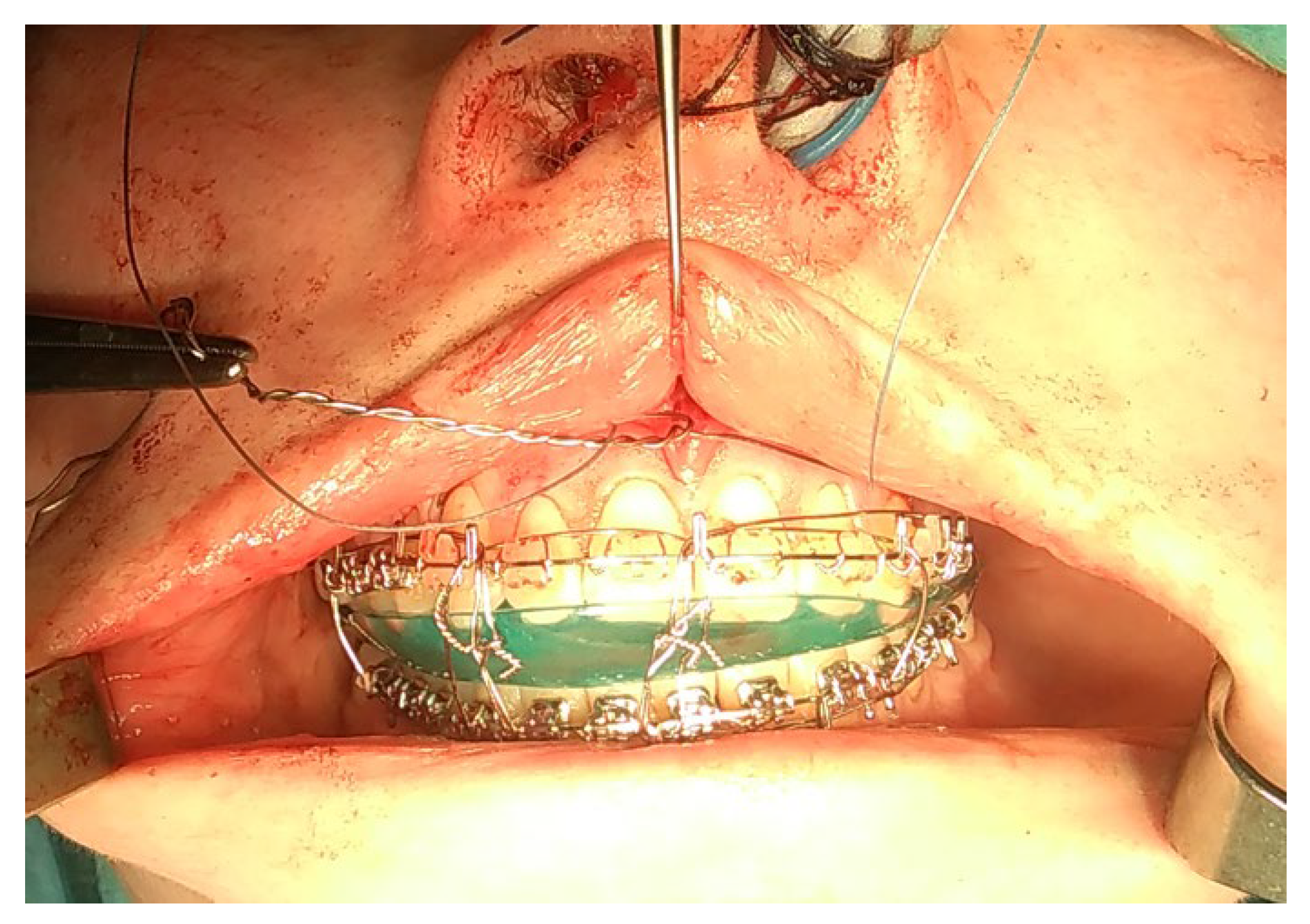
- Orthognathic surgery: BSSO or BSSO with Lefort I on severely asymmetric maxillary and mandibular bones with a prognathic profile and open bite [116];
- (4)
- BSSO with Ferguson modification [117]: for achieving a more balanced chin and lower mandibular border symmetry;
- (5)
- Only BSSO or Lefort I, depending on the scope of asymmetry and profile changes after condylectomy [84];
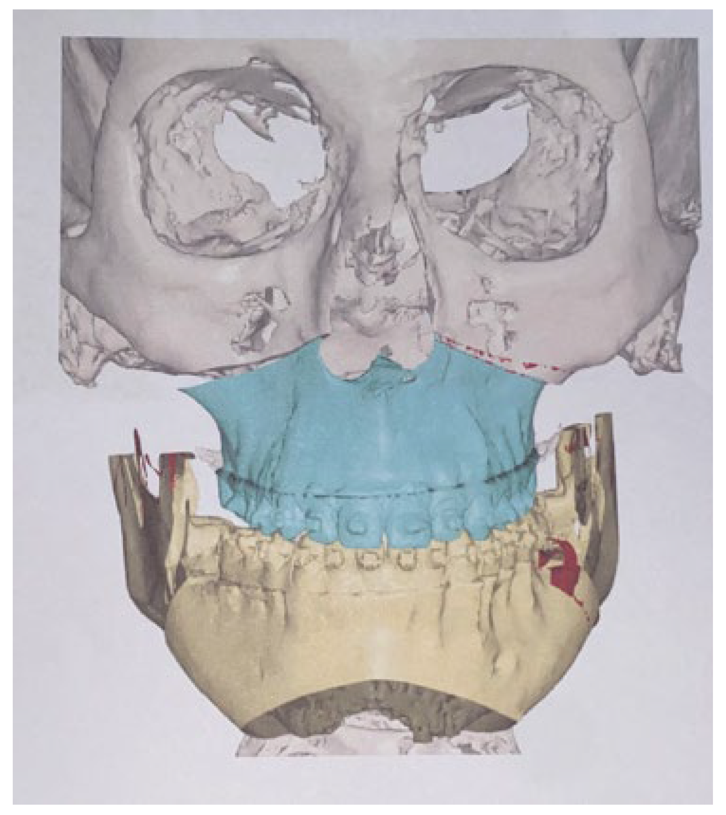
- (6)
- A condylectomy with BSSO and Lefort I simultaneously was described by Wolford in 2002; however, some authors report operated TMJ joint instability, which might be related to the degree of excised bone and poor condyle stabilization with at least lateral pterygoid muscle reattachment or other, perhaps even related with IMF intramaxillary fixation devices to stabilize the occlusion [75] (Figure 9);
- (7)
- Surgical camouflage such as marginectomy, chin shift procedure, bone osteomodeling, or basic orthognathic surgery in cases of UCH growth cessation/growth end [118];
- (8)
- (9)
- (10)
- A modification of a wing osteotomy, suggested by Wenghoefer et al., describing a chin-wing osteotomy to restore the balance of the inferior border of the mandible [121];
- (11)

8. Clinical Outcome
9. Conclusions
Author Contributions
Funding
Institutional Review Board Statement
Informed Consent Statement
Data Availability Statement
Conflicts of Interest
References
- Obwegeser, H.L.; Makek, M.S. Hemimandibular Hyperplasia—Hemimandibular Elongation. J. Maxillofac. Surg. 1986, 14, 183–208. [Google Scholar] [CrossRef]
- Rodrigues, D.B.; Castro, V. Condylar Hyperplasia of the Temporomandibular Joint: Types, Treatment, and Surgical Implications. Oral Maxillofac. Surg. Clin. N. Am. 2015, 27, 155–167. [Google Scholar] [CrossRef]
- Chia, M.S.; Naini, F.B.; Gill, D.S. The Aetiology, Diagnosis and Management of Mandibular Asymmetry. Orthod. Updat. 2008, 1, 44–52. [Google Scholar] [CrossRef]
- Madrid, J.R.P.; Montealegre, G.; Gomez, V. A New Classification Based on the Kaban’s Modification for Surgical Management of Craniofacial Microsomia. Craniomaxillofac. Trauma. Reconstr. 2010, 3, 1–7. [Google Scholar] [CrossRef]
- Wink, J.D.; Goldstein, J.A.; Paliga, J.T.; Taylor, J.A.; Bartlett, S.P. The Mandibular Deformity in Hemifacial Microsomia: A Reassessment of the Pruzansky and Kaban Classification. Plast. Reconstr. Surg. 2014, 133, 174e–181e. [Google Scholar] [CrossRef]
- Wolford, L.M.; Movahed, R.; Perez, D.E. A Classification System for Conditions Causing Condylar Hyperplasia. J. Oral Maxillofac. Surg. 2014, 72, 567–595. [Google Scholar] [CrossRef]
- Bishara, S.E.; Burkey, P.S.; Kharouf, J.G. Dental and Facial Asymmetries: A Review. Angle Orthod. 1994, 64, 89–98. [Google Scholar] [CrossRef]
- Reyneke, J.P. Orthognathic Essentials of Surgery: Second Edition; Quintessence Books: Chandler Drive, IL, USA, 2010. [Google Scholar]
- Iyer, J.; Hariharan, A.; Cao, U.M.N.; Tran, S.D. Acquired Facial, Maxillofacial, and Oral Asymmetries—A Review Highlighting Diagnosis and Management. Symmetry 2021, 13, 1661. [Google Scholar] [CrossRef]
- Andrade, N.N.; Mathai, P.; Aggarwal, N. Facial Asymmetry. In Oral and Maxillofacial Surgery for the Clinician; Bonanthaya, K., Panneerselvam, E., Manuel, S., Kumar, V.V., Rai, A., Eds.; Springer Nature: Singapore, 2021; pp. 1549–1576. ISBN 9789811513466. [Google Scholar]
- Almeida, L.E.; Zacharias, J.; Pierce, S. Condylar Hyperplasia: An Updated Review of the Literature. Korean J. Orthod. 2015, 45, 333–340. [Google Scholar] [CrossRef]
- Raijmakers, P.G.; Karssemakers, L.H.E.; Tuinzing, D.B. Female Predominance and Effect of Gender on Unilateral Condylar Hyperplasia: A Review and Meta-Analysis. J. Oral Maxillofac. Surg. 2012, 70, e72–e76. [Google Scholar] [CrossRef]
- Nelke, K.H.; Pawlak, W.; Morawska-Kochman, M.; Łuczak, K. Ten Years of Observations and Demographics of Hemimandibular Hyperplasia and Elongation. J. Craniomaxillofac. Surg. 2018, 46, 979–986. [Google Scholar] [CrossRef]
- Mahajan, M. Unilateral Condylar Hyperplasia—A Genetic Link? Case Reports. Natl. J. Maxillofac. Surg. 2017, 8, 58–63. [Google Scholar] [CrossRef]
- Yang, J.; Lignelli, J.L.; Ruprecht, A. Mirror Image Condylar Hyperplasia in Two Siblings. Oral Surg. Oral Med. Oral Pathol. Oral Radiol. Endod. 2004, 97, 281–285. [Google Scholar] [CrossRef]
- Portelli, M.; Gatto, E.; Matarese, G.; Militi, A.; Catalfamo, L.; Gherlone, E.; Lucchese, A. Unilateral Condylar Hyperplasia: Diagnosis, Clinical Aspects and Operative Treatment. A Case Report. Eur. J. Paediatr. Dent. 2015, 16, 99–102. [Google Scholar]
- Mehrotra, D.; Dhasmana, S.; Kamboj, M.; Gambhir, G. Condylar Hyperplasia and Facial Asymmetry: Report of Five Cases. J. Maxillofac. Oral Surg. 2011, 10, 50–56. [Google Scholar] [CrossRef]
- Singh, V.; Verma, A.; Attresh, G.; Batra, J. Ortho-Surgical Management of Condylar Hyperplasia: Rare Case Reports. Natl. J. Maxillofac. Surg. 2014, 5, 54–59. [Google Scholar] [CrossRef]
- Ma, X.; Wang, H.; Zhang, X. [Orthognathic surgery in the treatment of condylar osteochondroma]. Hua Xi Kou Qiang Yi Xue Za Zhi 2014, 32, 150–152. [Google Scholar] [CrossRef]
- Fisch, A.W.; Espinosa, C.I.; Quezada, S.R. Facial Asymmetry Secondary to Mandibular Condylar Hyperplasia. A Case Report. Rev. Odontol. Mex. 2011, 15, 251–256. [Google Scholar]
- Brionne, C.; Cadre, B.; Laroche, Y.; Lhotellier, J.; Maze, M.; Raffre, A.; Sorel, O. The Diagnosis of Mandibular Assymmetries. J. Dentofac. Anom. Orthod. 2013, 16. [Google Scholar] [CrossRef]
- Alyamani, A.; Abuzinada, S. Management of Patients with Condylar Hyperplasia: A Diverse Experience with 18 Patients. Ann. Maxillofac. Surg. 2012, 2, 17–23. [Google Scholar] [CrossRef]
- Arora, K.S.; Bansal, R.; Mohapatra, S.; Pareek, S. Review and Classification Update: Unilateral Condylar Hyperplasia. BMJ Case Rep. 2019, 12, e227569. [Google Scholar] [CrossRef]
- Nitzan, D.W.; Katsnelson, A.; Bermanis, I.; Brin, I.; Casap, N. The Clinical Characteristics of Condylar Hyperplasia: Experience with 61 Patients. J. Oral Maxillofac. Surg. 2008, 66, 312–318. [Google Scholar] [CrossRef]
- Khawaja, S.N.; Crow, H.; Gonzalez, Y. Goldenhar Syndrome and Pain-Related Temporomandibular Disorders. A Case Report. N. Y. State Dent. J. 2016, 82, 21–24. [Google Scholar]
- GN, S.; Sharma, M.L.; JK, D.R.; Goel, S.; Srivastava, S. Facial Asymmetry in Young Adults with Condylar Hyperplasia-Unusual Changes in the Facial Bones. J. Clin. Diagn. Res. 2015, 9, ZD21–ZD23. [Google Scholar] [CrossRef] [PubMed]
- Kawamoto, H.K.; Kim, S.S.; Jarrahy, R.; Bradley, J.P. Differential Diagnosis of the Idiopathic Laterally Deviated Mandible. Plast. Reconstr. Surg. 2009, 124, 1599–1609. [Google Scholar] [CrossRef] [PubMed]
- De Stefano, A.A.; Di Chicco, A.; Alessandra, I.; Emanuela, S.; Guercio-Mónaco, E.; Galluccio, G. Unilateral Condylar Hyperplasia: A Thee-Dimensional CBCT Morphometric and Volumetric Evaluation of Mandibular Condyle by Open-Source Softwares. Int. J. Morphol. 2021, 39. [Google Scholar] [CrossRef]
- Fariña, R.; Bravo, R.; Villanueva, R.; Valladares, S.; Hinojosa, A.; Martinez, B. Measuring the Condylar Unit in Condylar Hyperplasia: From the Sigmoid Notch or from the Mandibular Lingula? Int. J. Oral Maxillofac. Surg. 2017, 46, 857–860. [Google Scholar] [CrossRef]
- Olate, S.; Cantín, M.; Alister, J.P.; Uribe, F.; Navarro, P.; Olate, G.; de Moraes, M. Relationship Between Condylar Size and Transverse Facial Asymmetry in Subject with Condylar Hyperplasia. Int. J. Morphol. 2013, 31, 937–941. [Google Scholar] [CrossRef]
- Saridin, C.P.; Raijmakers, P.; Becking, A.G. Quantitative Analysis of Planar Bone Scintigraphy in Patients with Unilateral Condylar Hyperplasia. Oral Surg. Oral Med. Oral Pathol. Oral Radiol. Endodontol. 2007, 104, 259–263. [Google Scholar] [CrossRef]
- Olate, S.; Netto, H.D.; Rodriguez-Chessa, J.; Alister, J.P.; de Albergaria-Barbosa, J.; de Moraes, M. Mandible Condylar Hyperplasia: A Review of Diagnosis and Treatment Protocol. Int. J. Clin. Exp. Med. 2013, 6, 727. [Google Scholar]
- Di Luzio, C.; Caputo, M.; Favale, M.L.; Bellisario, A.; Squillace, F. Spect Bone Scintigraphy in the Diagnosis of Unilateral Condylar Hyperplasia: A Systematic Review. 2017. Available online: https://www.webmedcentral.com/article_view/5377 (accessed on 12 November 2022).
- Saridin, C.P.; Raijmakers, P.G.; Tuinzing, D.B.; Becking, A.G. Bone Scintigraphy as a Diagnostic Method in Unilateral Hyperactivity of the Mandibular Condyles: A Review and Meta-Analysis of the Literature. Int. J. Oral Maxillofac. Surg. 2011, 40, 11–17. [Google Scholar] [CrossRef] [PubMed]
- Liu, P.; Shi, J. Growth Trends Analysis of Unilateral Condylar Hyperplasia Followed up with Planar Scintigraphy. Medicine 2021, 100, e28226. [Google Scholar] [CrossRef] [PubMed]
- Higginson, J.A.; Bartram, A.C.; Banks, R.J.; Keith, D.J.W. Condylar Hyperplasia: Current Thinking. Br. J. Oral Maxillofac. Surg. 2018, 56, 655–662. [Google Scholar] [CrossRef]
- Kambylafkas, P.; Murdock, E.; Gilda, E.; Tallents, R.H.; Kyrkanides, S. Validity of Panoramic Radiographs for Measuring Mandibular Asymmetry. Angle Orthod. 2006, 76, 388–393. [Google Scholar] [CrossRef] [PubMed]
- Van Elslande, D.C.; Russett, S.J.; Major, P.W.; Flores-Mir, C. Mandibular Asymmetry Diagnosis with Panoramic Imaging. Am. J. Orthod. Dentofac. Orthop. 2008, 134, 183–192. [Google Scholar] [CrossRef]
- Nelke, K.; Łuczak, K.; Janeczek, M.; Pasicka, E.; Barnaś, S.; Dobrzyński, M. What Features on Routine Panoramic Radiographs Could Help Orthodontists to Estimate the Occurrence of Condylar Hyperplasia from Other Mandibular Asymmetries—Retrospective Analysis Study. Symmetry 2022, 14, 1287. [Google Scholar] [CrossRef]
- Takahashi-Ichikawa, N.; Susami, T.; Nagahama, K.; Ohkubo, K.; Okayasu, M.; Uchino, N.; Uwatoko, K.; Saijo, H.; Mori, Y.; Takato, T. Evaluation of Mandibular Hypoplasia in Patients with Hemifacial Microsomia: A Comparison between Panoramic Radiography and Three-Dimensional Computed Tomography. Cleft Palate Craniofac. J. 2013, 50, 381–387. [Google Scholar] [CrossRef]
- Nolte, J.W.; Karssemakers, L.H.E.; Grootendorst, D.C.; Tuinzing, D.B.; Becking, A.G. Panoramic Imaging Is Not Suitable for Quantitative Evaluation, Classification, and Follow up in Unilateral Condylar Hyperplasia. Br. J. Oral Maxillofac. Surg. 2015, 53, 446–450. [Google Scholar] [CrossRef]
- Nolte, J.W.; Verhoeven, T.J.; Schreurs, R.; Bergé, S.J.; Karssemakers, L.H.E.; Becking, A.G.; Maal, T.J.J. 3-Dimensional CBCT Analysis of Mandibular Asymmetry in Unilateral Condylar Hyperplasia. J. Craniomaxillofac. Surg. 2016, 44, 1970–1976. [Google Scholar] [CrossRef]
- Sun, R.; Sun, L.; Sun, Z.; Li, G.; Zhao, Y.; Ma, X.; Sun, C. A Three-Dimensional Study of Hemimandibular Hyperplasia, Hemimandibular Elongation, Solitary Condylar Hyperplasia, Simple Mandibular Asymmetry and Condylar Osteoma or Osteochondroma. J. Craniomaxillofac. Surg. 2019, 47, 1665–1675. [Google Scholar] [CrossRef]
- Cavagnetto, D.; Abate, A.; Caprioglio, A.; Cressoni, P.; Maspero, C. Three-Dimensional Volumetric Evaluation of the Different Mandibular Segments Using CBCT in Patients Affected by Juvenile Idiopathic Arthritis: A Cross-Sectional Study. Prog. Orthod. 2021, 22, 32. [Google Scholar] [CrossRef] [PubMed]
- Hatamleh, M.M.; Yeung, E.; Osher, J.; Huppa, C. Novel Treatment Planning of Hemimandibular Hyperplasia by the Use of Three-Dimensional Computer-Aided-Design and Computer-Aided-Manufacturing Technologies. J. Craniofac. Surg. 2017, 28, 764–767. [Google Scholar] [CrossRef] [PubMed]
- Rizwana, A.; Mohan, N.; Kumar, P.T.R.; Karthik, R.; Gokulraj, S. Panoramic Radiograph and CBCT in Unilateral Mandibular Condylar Hyperplasia—A Case Report. Indian J. Radiol. Imaging 2021, 31, 1070–1074. [Google Scholar] [CrossRef] [PubMed]
- Yilmaz, E.; Kansu, Ö.; Özgen, B.; Akçiçek, G.; Kansu, H. Radiographic Manifestations of the Temporomandibular Joint in a Case of Proteus Syndrome. Dentomaxillofac. Radiol. 2013, 42, 58444855. [Google Scholar] [CrossRef]
- Hodder, S.C.; Rees, J.I.; Oliver, T.B.; Facey, P.E.; Sugar, A.W. SPECT Bone Scintigraphy in the Diagnosis and Management of Mandibular Condylar Hyperplasia. Br. J. Oral Maxillofac. Surg. 2000, 38, 87–93. [Google Scholar] [CrossRef]
- Yang, Z.; Reed, T.; Longino, B.H. Bone Scintigraphy SPECT/CT Evaluation of Mandibular Condylar Hyperplasia. J. Nucl. Med. Technol. 2016, 44, 49–51. [Google Scholar] [CrossRef]
- Buitrago, D.F.L.; Botero, J.R. Asymmetry of Glenoid Fossa as Differential Diagnosis for Hemimandibular Elongation. Rev. Mex. De Ortod. 2017, 5, 221–230. [Google Scholar]
- López, B.D.F.; Corral, S.C.M. Comparison of Planar Bone Scintigraphy and Single Photon Emission Computed Tomography for Diagnosis of Active Condylar Hyperplasia. J. Craniomaxillofac. Surg. 2016, 44, 70–74. [Google Scholar] [CrossRef]
- Wen, B.; Shen, Y.; Wang, C.-Y. Clinical Value of 99Tcm-MDP SPECT Bone Scintigraphy in the Diagnosis of Unilateral Condylar Hyperplasia. Sci. World J. 2014, 2014, 1–6. [Google Scholar]
- DeLone, D.R.; Brown, W.D.; Gentry, L.R. Proteus Syndrome: Craniofacial and Cerebral MRI. Neuroradiology 1999, 41, 840–843. [Google Scholar] [CrossRef]
- Pripatnanont, P.; Vittayakittipong, P.; Markmanee, U.; Thongmak, S.; Yipintsoi, T. The Use of SPECT to Evaluate Growth Cessation of the Mandible in Unilateral Condylar Hyperplasia. Int. J. Oral Maxillofac. Surg. 2005, 34, 364–368. [Google Scholar] [CrossRef] [PubMed]
- Saridin, C.P.; Raijmakers, P.G.; Slootweg, P.J.; Tuinzing, D.B.; Becking, A.G.; van der Waal, I. Unilateral Condylar Hyperactivity: A Histopathologic Analysis of 47 Patients. J. Oral Maxillofac. Surg. 2010, 68, 47–53. [Google Scholar] [CrossRef] [PubMed]
- Saridin, C.P.; Raijmakers, P.; Al Shamma, S.; Tuinzing, D.B.; Becking, A.G. Comparison of Different Analytical Methods Used for Analyzing SPECT Scans of Patients with Unilateral Condylar Hyperactivity. Int. J. Oral Maxillofac. Surg. 2009, 38, 942–946. [Google Scholar] [CrossRef] [PubMed]
- Warhekar, A.M.; Wanjari, P.V.; Phulambrikar, T. Unilateral Trifid Mandibular Condyle: A Case Report. Cranio 2011, 29, 80–84. [Google Scholar] [CrossRef]
- Katti, G.; Najmuddin, M.; Fatima, S.; Unnithan, J. Bifid Mandibular Condyle. BMJ Case Rep. 2012, 2012, bcr-2012-007051. [Google Scholar] [CrossRef]
- John, L.; Yin, Y.Y.; David, Y.W.C.; Kwong, C.L. Planar Scintigraphy in Assessment of Mandibular Asymmetry: Unilateral Condylar Hyperplasia vs. Asymmetric Mandibular Hyperplasia. 2012. Available online: https://www.scirp.org/journal/paperinformation.aspx?paperid=26244 (accessed on 12 November 2022).
- Goulart, D.R.; Muñoz, P.; Cantín López, M.G.; de Moraes, M.; Olate, S. Comparative Evaluation of Condylar Volume between Patients with Unilateral Condylar Hyperplasia and Class III Dentofacial Deformity. J. Oral Maxillofac. Surg. 2017, 75, 180–188. [Google Scholar] [CrossRef]
- Sedano-Balbin, G.; Romero-Tapia, P.; Pérez-Vargas, F.; Alvitez-Temoche, D.; Mayta-Tovalino, F. Three-Dimensional Correlation between Condylar Size and Mandibular Asymmetry with Type 1B Condylar Hyperplasia: A Quasi-Experimental Study from PERU. J. Int. Oral Health 2020, 12, 323. [Google Scholar] [CrossRef]
- Nah, K.-S. Condylar Bony Changes in Patients with Temporomandibular Disorders: A CBCT Study. Imaging Sci. Dent. 2012, 42, 249–253. [Google Scholar] [CrossRef]
- Angiero, F.; Farronato, G.; Benedicenti, S.; Vinci, R.; Farronato, D.; Magistro, S.; Stefani, M. Mandibular Condylar Hyperplasia: Clinical, Histopathological, and Treatment Considerations. Cranio 2009, 27, 24–32. [Google Scholar] [CrossRef]
- Luther, F.; Morris, D.O.; Hart, C. Orthodontic Preparation for Orthognathic Surgery: How Long Does It Take and Why? A Retrospective Study. Br. J. Oral Maxillofac. Surg. 2003, 41, 401–406. [Google Scholar] [CrossRef]
- El Mozen, L.A.; Meng, Q.-G.; Li, Y.-J.; Long, X.; Chen, G.-X. Condylar and Occlusal Changes after High Condylectomy and Orthodontic Treatment for Condylar Hyperplasia. J. Huazhong Univ. Sci. Technol. Med. Sci. 2015, 35, 265–270. [Google Scholar] [CrossRef] [PubMed]
- Goulart, D.R.; Muñoz, P.; Olate, S.; de Moraes, M.; Fariña, R. No Differences in Morphological Characteristics between Hyperplastic Condyle and Class III Condyle. Int. J. Oral Maxillofac. Surg. 2015, 44, 1281–1286. [Google Scholar] [CrossRef] [PubMed]
- Li, Y.; Zheng, Y.; Cai, H.; Meng, Q.; Fang, W.; Ke, J.; Long, X.; Chen, G. Standard Orthodontic Treatment after Condylectomy for Patients with Active Unilateral Condylar Hyperplasia. Am. J. Orthod. Dentofac. Orthop. 2022, 161, 404–415.e1. [Google Scholar] [CrossRef] [PubMed]
- Shivhare, P.; Lata, S.; Balaji, P.; Gupta, A. Non-Syndromic Bilateral Condylar Aplasia: A Rare Case. Indian J. Dent. Res. 2015, 26, 210–213. [Google Scholar] [CrossRef] [PubMed]
- Canger, E.M.; Celenk, P. Aplasia of the Mandibular Condyle Associated with Some Orthopaedic Abnormalities. Dentomaxillofac. Radiol. 2012, 41, 259–263. [Google Scholar] [CrossRef] [PubMed]
- Kaneyama, K.; Segami, N.; Hatta, T. Congenital Deformities and Developmental Abnormalities of the Mandibular Condyle in the Temporomandibular Joint. Congenit. Anom. 2008, 48, 118–125. [Google Scholar] [CrossRef]
- Bertolini, F.; Bianchi, B.; De Riu, G.; Di Blasio, A.; Sesenna, E. Hemimandibular hyperplasia treated by early high condylectomy: A case report. Int. J. Adult Orthod. Orthognath. Surg. 2001, 16, 227–234. [Google Scholar]
- Tun Oo, L.; Miyamoto, J.J.; Takada, J.-I.; Moriyama, K. Correlation between the Position of the Glenoid Fossa and Condylar Translational Movement in Skeletal Class III Mandibular Asymmetry Patients. Eur. J. Orthod. 2022, 44, 294–302. [Google Scholar] [CrossRef]
- Damstra, J.; Fourie, Z.; Ren, Y. Evaluation and comparison of postero-anterior cephalograms and cone-beam computed tomography images for the detection of mandibular asymmetry. Eur. J. Orthod. 2013, 35, 45–50. [Google Scholar] [CrossRef]
- Shivhare, P.; Shankarnarayan, L.; Kumar, M.; Sowbhagya, M.B. Condylar Aplasia and Hypoplasia: A Rare Case. Case Rep. Dent. 2013, 2013, 745602. [Google Scholar] [CrossRef]
- Wolford, L.M.; Mehra, P.; Reiche-Fischel, O.; Morales-Ryan, C.A.; García-Morales, P. Efficacy of High Condylectomy for Management of Condylar Hyperplasia. Am. J. Orthod. Dentofac. Orthop. 2002, 121, 136–150, discussion 150–151. [Google Scholar] [CrossRef] [PubMed]
- Da Costa Araújo, F.A.; de Santana Santos, T.; de Oliveira e Silva, E.D.; Filho, J.R.L. One-Stage Treatment of Hemimandibular Hyperplasia. J. Craniofac. Surg. 2012, 23, e635. [Google Scholar] [CrossRef] [PubMed]
- Xavier, S.P.; Santos, T.D.S.; Silva, E.R.; Faria, A.C.; de Mello Filho, F.V. Two-Stage Treatment of Facial Asymmetry Caused by Unilateral Condylar Hyperplasia. Braz. Dent. J. 2014, 25, 257–260. [Google Scholar] [CrossRef] [PubMed]
- Diniz, J.A.; dos Santos Siqueira, A.; Torres, L.H.S.; Faro, T.F.; Rodrigues, E.D.R.; Laureano Filho, J.R. Virtual Surgical Planning and One-Stage Treatment of Active Hemimandibular Hyperplasia. J. Craniofac. Surg. 2019, 30, e679–e681. [Google Scholar] [CrossRef] [PubMed]
- Ferri, J.; Raoul, G.; Potier, J.; Nicot, R. Temporomandibular Joint (TMJ): Condyle Hyperplasia and Condylectomy. Rev. De Stomatol. De Chir. Maxillo-Faciale Et De Chir. Orale 2016, 117, 259–265. [Google Scholar]
- Lippold, C.; Kruse-Losler, B.; Danesh, G.; Joos, U.; Meyer, U. Treatment of Hemimandibular Hyperplasia: The Biological Basis of Condylectomy. Br. J. Oral Maxillofac. Surg. 2007, 45, 353–360. [Google Scholar] [CrossRef]
- Pereira-Santos, D.; De Melo, W.M.; Souza, F.Á.; De Moura, W.L.; de Paulo Cravinhos, J.C. High Condylectomy Procedure: A Valuable Resource for Surgical Management of the Mandibular Condylar Hyperplasia. J. Craniofac. Surg. 2013, 24, 1451–1453. [Google Scholar] [CrossRef]
- Ghawsi, S.; Aagaard, E.; Thygesen, T.H. High Condylectomy for the Treatment of Mandibular Condylar Hyperplasia: A Systematic Review of the Literature. Int. J. Oral Maxillofac. Surg. 2016, 45, 60–71. [Google Scholar] [CrossRef]
- Muñoz, M.F.; Monje, F.; Goizueta, C.; Rodríguez-Campo, F. Active Condylar Hyperplasia Treated by High Condylectomy: Report of Case. J. Oral Maxillofac. Surg. 1999, 57, 1455–1459. [Google Scholar] [CrossRef]
- Maniskas, S.A.; Ly, C.L.; Pourtaheri, N.; Parsaei, Y.; Steinbacher, D.M. Concurrent High Condylectomy and Orthognathic Surgery for Treatment of Patients With Unilateral Condylar Hyperplasia. J. Craniofac. Surg. 2020, 31, 2217–2221. [Google Scholar] [CrossRef]
- Wu, C.; Meng, Q.; Deng, M.; Cai, H.; Ke, J.; Long, X. Cone–Beam Computed Tomographic Analysis of Maxillary and Mandibular Changes after High Condylectomy Combined with Orthodontic Treatment for Active Unilateral Condylar Hyperplasia. Br. J. Oral Maxillofac. Surg. 2018, 56, 692–697. [Google Scholar] [CrossRef]
- Wolford, L.M.; Movahed, R.; Dhameja, A.; Allen, W.R. Low Condylectomy and Orthognathic Surgery to Treat Mandibular Condylar Osteochondroma: A Retrospective Review of 37 Cases. J. Oral Maxillofac. Surg. 2014, 72, 1704–1728. [Google Scholar] [CrossRef] [PubMed]
- Fariña, R.A.; Becar, M.; Plaza, C.; Espinoza, I.; Franco, M.E. Correlation between Single Photon Emission Computed Tomography, AgNOR Count, and Histomorphologic Features in Patients with Active Mandibular Condylar Hyperplasia. J. Oral Maxillofac. Surg. 2011, 69, 356–361. [Google Scholar] [CrossRef] [PubMed]
- Fariña, R.; Pintor, F.; Pérez, J.; Pantoja, R.; Berner, D. Low Condylectomy as the Sole Treatment for Active Condylar Hyperplasia: Facial, Occlusal and Skeletal Changes. An Observational Study. Int. J. Oral Maxillofac. Surg. 2015, 44, 217–225. [Google Scholar] [CrossRef] [PubMed]
- Alsayegh, H.A.; Alsubaie, Z.A.; Alwayil, A.R.; Alqadhi, M.A.; Alawadh, A.M. Unilateral Condylar Hyperplasia With Active Bony Overgrowth: A Case Report. Cureus 2021, 13, e5840. [Google Scholar] [CrossRef] [PubMed]
- Dominguez, M.; Castillo, J.L.D.; Guerra, M.M.; Sanchez, R.; La Plata, M.M.D. Condylar Osteochondroma Treated with Total Condylectomy and Preservation of the Articular Disc: A Case Report. Craniomaxillofac. Trauma Reconstr. 2015, 8, 136–140. [Google Scholar] [CrossRef]
- De Melo, W.M.; Pereira-Santos, D.; Sonoda, C.K.; De Moura, W.L.; de Paulo-Cravinhos, J.C. Conservative Condylectomy for Management of Osteochondroma of the Mandibular Condyle. J. Craniofac. Surg. 2013, 24, e209–e211. [Google Scholar] [CrossRef]
- Mouallem, G.; Vernex-Boukerma, Z.; Longis, J.; Perrin, J.-P.; Delaire, J.; Mercier, J.-M.; Corre, P. Efficacy of Proportional Condylectomy in a Treatment Protocol for Unilateral Condylar Hyperplasia: A Review of 73 Cases. J. Cranio-Maxillofac. Surg. 2017, 45, 1083–1093. [Google Scholar] [CrossRef]
- Niño-Sandoval, T.C.; Maia, F.P.A.; Vasconcelos, B.C.E. Efficacy of Proportional versus High Condylectomy in Active Condylar Hyperplasia—A Systematic Review. J. Craniomaxillofac. Surg. 2019, 47, 1222–1232. [Google Scholar] [CrossRef]
- Dattani, A.; Heggie, A. Hemifacial Hyperplasia: A Case Series and Review of the Literature. Int. J. Oral Maxillofac. Surg. 2021, 50, 341–348. [Google Scholar] [CrossRef]
- Choung, P.H.; Nam, I.W. An Intraoral Approach to Treatment of Condylar Hyperplasia or High Condylar Process Fractures Using the Intraoral Vertico-Sagittal Ramus Osteotomy. J. Oral Maxillofac. Surg. 1998, 56, 563–570, discussion 571. [Google Scholar] [CrossRef] [PubMed]
- Wang, X.; Li, Z.; Yi, B.; Liang, C.; Li, Y.; Wang, X. [Preliminary study of condylectomy via intraoral approach]. Zhonghua Kou Qiang Yi Xue Za Zhi 2012, 47, 305–309. [Google Scholar] [CrossRef] [PubMed]
- Wang, X.; Li, Z.; Yi, B.; Liang, C.; Tian, K.; Wang, X. [Clinical application of condylectomy via intraoral approach under computer assisted surgical navigation]. Zhonghua Kou Qiang Yi Xue Za Zhi 2013, 48, 350–354. [Google Scholar] [CrossRef] [PubMed]
- Deng, M.; Long, X.; Cheng, A.H.A.; Cheng, Y.; Cai, H. Modified Trans-Oral Approach for Mandibular Condylectomy. Int. J. Oral Maxillofac. Surg. 2009, 38, 374–377. [Google Scholar] [CrossRef] [PubMed]
- Li, B.; Sun, H.; Zhang, L.; Wang, X. Simple Way of Facilitating Intraoral Condylectomy and Securing the Excised Condyle: Technical Note. Br. J. Oral Maxillofac. Surg. 2013, 51, e305–e306. [Google Scholar] [CrossRef] [PubMed]
- You, H.-J.; Moon, K.-C.; Yoon, E.-S.; Lee, B.-I.; Park, S.-H. Clinical and Radiological Outcomes of Transoral Endoscope-Assisted Treatment of Mandibular Condylar Fractures. Int. J. Oral Maxillofac. Surg. 2016, 45, 284–291. [Google Scholar] [CrossRef]
- Schmelzeisen, R.; Cienfuegos-Monroy, R.; Schön, R.; Chen, C.-T.; Cunningham Jr, L.; Goldhahn, S. Patient Benefit from Endoscopically Assisted Fixation of Condylar Neck Fractures—A Randomized Controlled Trial. J. Oral Maxillofac. Surg. 2009, 67, 147–158. [Google Scholar] [CrossRef]
- Cascone, P.; Cicero, B.; Ramieri, V.; Germano, F.; Vellone, V. Treatment of Unilateral Condylar Hyperplasia with Proportional Condylectomy and Orthodontic Aligners. J. Clin. Orthod. JCO 2020, 54, 611–619. [Google Scholar]
- Abotaleb, B.; Bi, R.; Telha, W.; Zhao, W.; Li, Y.; Zhu, S. Treatment Measures of Hemimandibular Hyperplasia and Associated Facial Deformities. J. Cranio-Maxillofac. Surg. 2021, 49, 126–134. [Google Scholar] [CrossRef]
- Qi, L.; Cao, N.; Ge, W.; Jiang, T.; Fan, L.; Zhang, L. A New Method for Individual Condylar Osteotomy and Repositioning Guides Used in Patients with Severe Deformity Secondary to Condylar Osteochondroma. Orphanet J. Rare Dis. 2021, 16, 1–11. [Google Scholar] [CrossRef]
- Kim, D.S.; Kim, J.-Y.; Jeong, C.-W.; Park, K.-H.; Huh, J.-K. Conservative Condylectomy Alone for the Correction of Mandibular Asymmetry Caused by Osteochondroma of the Mandibular Condyle: A Report of Five Cases. J. Korean Assoc. Oral Maxillofac. Surg. 2015, 41, 259. [Google Scholar] [CrossRef] [PubMed]
- Wolford, L.M.; Mehra, P.; Franco, P. Use of Conservative Condylectomy for Treatment of Osteochondroma of the Mandibular Condyle. J. Oral Maxillofac. Surg. 2002, 60, 262–268. [Google Scholar] [CrossRef] [PubMed]
- Santos, G.S.; Gomes, J.B.; de Sousa Maia, S.; Bermejo, P.R.; Shinohara, E.H.; Sonoda, C.K.; de Melo, W.M. Using Conservative Condylectomy for Management of a Large Osteochondroma of the Mandibular Condyle with 6-Year Follow-Up. J. Craniofac. Surg. 2014, 25, e102–e104. [Google Scholar] [CrossRef] [PubMed]
- Rojare, C.; Wojcik, T.; Coussens, C.; Ferri, J.; Pertuzon, B.; Raoul, G. Condylar Hyperplasia: Qualitative and Quantitative Study of Temporomandibular Joints Remodeling after Condylectomy. L’Orthodontie Française 2014, 85, 189–200. [Google Scholar] [CrossRef]
- Gc, R.; Muralidoss, H.; Ramaiah, S. Conservative Management of Unilateral Condylar Hyperplasia. Oral Maxillofac. Surg. 2012, 16, 201–205. [Google Scholar] [CrossRef]
- Nolte, J.W.; Schreurs, R.; Karssemakers, L.H.E.; Tuinzing, D.B.; Becking, A.G. Demographic Features in Unilateral Condylar Hyperplasia: An Overview of 309 Asymmetric Cases and Presentation of an Algorithm. J. Cranio-Maxillofac. Surg. 2018, 46, 1484–1492. [Google Scholar] [CrossRef]
- Açikgöz, A. Bilateral Bifid Mandibular Condyle: A Case Report. J. Oral Rehabil. 2006, 33, 784–787. [Google Scholar] [CrossRef]
- Neelakandan, R.S.; Bhargava, D. Bifid hyperplastic mandibular condyle. J. Maxillofac. Oral Surg. 2013, 12, 466–471. [Google Scholar] [CrossRef]
- Butt, F.M.A.; Guthua, S.W.; Nganga, P.; Edalia, M.; Dimba, E.A.O. One-Stage Treatment of Acquired Facial Deformity Caused by Severe Unilateral Condylar Hyperplasia. J. Craniofac. Surg. 2011, 22, 1966–1968. [Google Scholar] [CrossRef]
- Motamedi, M.H. Treatment of Condylar Hyperplasia of the Mandible Using Unilateral Ramus Osteotomies. J. Oral Maxillofac. Surg. 1996, 54, 1161–1169, discussion 1169–1170. [Google Scholar] [CrossRef]
- López, D.F.; Aristizábal, J.F.; Martínez-Smit, R. Condylectomy and “Surgery First” Approach: An Expedited Treatment for Condylar Hyperplasia in a Patient with Facial Asymmetry. Dent. Press J. Orthod. 2017, 22, 86–96. [Google Scholar] [CrossRef] [PubMed]
- Gallagher, A.L.; Ruellas, A.C.D.O.; Benavides, E.; Soki, F.N.; Aronovich, S.; Magraw, C.B.L.; Turvey, T.; Cevidanes, L. Mandibular Condylar Remodeling Characteristics after Simultaneous Condylectomy and Orthognathic Surgery. Am. J. Orthod. Dentofac. Orthop. 2021, 160, 705–717. [Google Scholar] [CrossRef] [PubMed]
- Ferguson, J.W. Definitive Surgical Correction of the Deformity Resulting from Hemimandibular Hyperplasia. J. Craniomaxillofac. Surg. 2005, 33, 150–157. [Google Scholar] [CrossRef] [PubMed]
- Nelke, K.; Łuczak, K.; Janeczek, M.; Pasicka, E.; Morawska-Kochman, M.; Guziński, M.; Dobrzyński, M. Methods of Definitive Correction of Mandibular Deformity in Hemimandibular Hyperplasia Based on Radiological, Anatomical, and Topographical Measurements—Proposition of Author’s Own Protocol. Int. J. Env. Res. Public Health 2022, 19, 10005. [Google Scholar] [CrossRef]
- Cascone, P.; D’Alessandro, F.; Gallo, E.; Cicero, G.; Vellone, V. The Role of Three-Dimensional Printing Technology as an Additional Tool in Unilateral Condylar Hyperplasia Surgical Planning. J. Craniofac. Surg. 2020, 31, e735–e738. [Google Scholar] [CrossRef]
- Ha, S.-W.; Choi, J.-Y.; Baek, S.-H. Correction of Unilateral Condylar Hyperplasia and Posterior Open Bite with Proportional Condylectomy and Fixed Orthodontic Treatment. Angle Orthod. 2020, 90, 144–158. [Google Scholar] [CrossRef]
- Wenghoefer, M.; Peters, A.; Reich, R.H. Modified Triaca-Style Wing Osteotomy to Correct Facial Asymmetry in a Patient With Horizontal Growth–Type Condylar Hyperplasia. J. Craniofac. Surg. 2013, 24, 1769–1771. [Google Scholar] [CrossRef]
- Nagasaka, H.; Sugawara, J.; Kawamura, H.; Nanda, R. “ Surgery First” Skeletal Class III Correction Using the Skeletal Anchorage System. J. Clin. Orthod. JCO 2009, 43, 97–105. [Google Scholar]
- Janakiraman, N.; Feinberg, M.; Vishwanath, M.; Jayaratne, Y.S.N.; Steinbacher, D.M.; Nanda, R.; Uribe, F. Integration of 3-Dimensional Surgical and Orthodontic Technologies with Orthognathic “Surgery-First” Approach in the Management of Unilateral Condylar Hyperplasia. Am. J. Orthod. Dentofac. Orthop. 2015, 148, 1054–1066. [Google Scholar] [CrossRef]
- Peng, S.P.; Ko, W.-C.E.; Lin, C.-H.; Ho, C.-Y. Hemimandibular Hyperplasia Treated by Orthognathic Surgery and Condylectomy in a Young Woman. Taiwan. J. Orthod. 2021, 33, 5. [Google Scholar] [CrossRef]
- Vernucci, R.A.; Mazzoli, V.; Galluccio, G.; Silvestri, A.; Barbato, E. Unilateral hemimandibular hyperactivity: Clinical features of a population of 128 patients. J. Craniomaxillofac. Surg. 2018, 46, 1105–1110. [Google Scholar] [CrossRef] [PubMed]
- Verhelst, P.-J.; Shaheen, E.; de Faria Vasconcelos, K.; Van der Cruyssen, F.; Shujaat, S.; Coudyzer, W.; Salmon, B.; Swennen, G.; Politis, C.; Jacobs, R. Validation of a 3D CBCT-Based Protocol for the Follow-up of Mandibular Condyle Remodeling. Dentomaxillofac. Radiol. 2020, 49, 20190364. [Google Scholar] [CrossRef] [PubMed]
- Nelke, K.H.; Morawska-Kochman, M.; Nienartowicz, J.; Grzelak, J.; Staszak, K.; Fraczek, M.; Luczak, K.; Guzinski, M. Anatomical and Clinical Implications in Neocondyle Stability After a Condylectomy. J. Craniofac. Surg. 2020, 31, 241–250. [Google Scholar] [CrossRef] [PubMed]


| 1. | Growth Activity | SPECT Monitoring | Condylectomy | Maxillary Tilting | Presence of Overgrowth | |
|---|---|---|---|---|---|---|
| No growth present | Are orthodontics sufficient? | Surgical camo | ||||
| A | Not present | One study | NO | ORT or surgical camo | Genioplasty - Wing-osteotomy - Marginectomy - Bone drilling and chiseling - Corrective ostectomies - Bone remodeling procedures - Facial contouring - Facial implants | |
| Some forms of growth: | YES | NO | ||||
| B | Active growth: rapidly progressive in time | SPECT: at least two studies | YES | ± BIMAX BSSO/Lefort I ± ORT ± ORT camo ± Surg camo | ± BSSO ± ORT ± ORT camo ± Surg camo | |
| C | Active growth: slowly progressive in time | SPECT: at least two studies | YES | ± BIMAX BSSO/Lefort I ± ORT ± ORT camo ± Surg camo | ± BSSO ± ORT ± ORT camo ± Surg camo | |
| D | Growth self-limiting in time | Each one per year till absence of growth | NO | ± Surg camo BSSO/Lefort I ± ORT ± ORT camo | ± BSSO ± ORT ± ORT camo ± Surg camo | |
| E | Growth cessation | One study | NO | ± Lefort I ± BSSO ± ORT ± ORT camo | ± ORT ± ORT camo ± Surg camo | |
Disclaimer/Publisher’s Note: The statements, opinions and data contained in all publications are solely those of the individual author(s) and contributor(s) and not of MDPI and/or the editor(s). MDPI and/or the editor(s) disclaim responsibility for any injury to people or property resulting from any ideas, methods, instructions or products referred to in the content. |
© 2023 by the authors. Licensee MDPI, Basel, Switzerland. This article is an open access article distributed under the terms and conditions of the Creative Commons Attribution (CC BY) license (https://creativecommons.org/licenses/by/4.0/).
Share and Cite
Nelke, K.; Łuczak, K.; Pawlak, W.; Janeczek, M.; Pasicka, E.; Morawska-Kochman, M.; Błaszczyk, B.; Błaszczyk, T.; Dobrzyński, M. Unilateral Condylar Hyperplasia in Surgeons’ Perspective—A Narrative Review. Appl. Sci. 2023, 13, 1839. https://doi.org/10.3390/app13031839
Nelke K, Łuczak K, Pawlak W, Janeczek M, Pasicka E, Morawska-Kochman M, Błaszczyk B, Błaszczyk T, Dobrzyński M. Unilateral Condylar Hyperplasia in Surgeons’ Perspective—A Narrative Review. Applied Sciences. 2023; 13(3):1839. https://doi.org/10.3390/app13031839
Chicago/Turabian StyleNelke, Kamil, Klaudiusz Łuczak, Wojciech Pawlak, Maciej Janeczek, Edyta Pasicka, Monika Morawska-Kochman, Bartłomiej Błaszczyk, Tomasz Błaszczyk, and Maciej Dobrzyński. 2023. "Unilateral Condylar Hyperplasia in Surgeons’ Perspective—A Narrative Review" Applied Sciences 13, no. 3: 1839. https://doi.org/10.3390/app13031839
APA StyleNelke, K., Łuczak, K., Pawlak, W., Janeczek, M., Pasicka, E., Morawska-Kochman, M., Błaszczyk, B., Błaszczyk, T., & Dobrzyński, M. (2023). Unilateral Condylar Hyperplasia in Surgeons’ Perspective—A Narrative Review. Applied Sciences, 13(3), 1839. https://doi.org/10.3390/app13031839