Communication

Lichens as a Natural Source of Compounds Active on Microorganisms of Human Health Interest

Department of Life Sciences, University of Modena and Reggio Emilia, Via Giuseppe Campi 103/287, 41125 Modena, Italy
*
Author to whom correspondence should be addressed.
Appl. Sci. 2023, 13(3), 1976; https://doi.org/10.3390/app13031976
Submission received: 11 January 2023 / Revised: 30 January 2023 / Accepted: 31 January 2023 / Published: 3 February 2023
(This article belongs to the Special Issue New Trends in Biosciences III)

Abstract

:

Featured Application

Natural compound extracts from lichens were able to decrease and/or inhibit the growth of microorganisms of interest in human health, thus providing a good alternative alone or in combination with drugs for therapy.

Abstract

The antimicrobial properties of two lichen extracts (LC1 and LC2 solutions extracted in acetone and cyclohexane, respectively) were investigated against both Gram-positive and Gram-negative microorganisms through the agar well diffusion assay. Results displayed that both samples were similarly effective against all the indicator strains. The antimicrobial activity was maintained up to 30 days against Candida albicans ATCC 10231 with an inhibition zone of 38 mm and 37 mm for the LC1 and LC2 solutions extracted, respectively. In order to separate the single chemical components and to associate them with the biological activity, the two extracts were subjected to an activity-guided fractionation followed by a liquid chromatography mass spectrometry (LC–MS) Ion Trap 6310A for the chemical characterization. Chromatogram analysis of each sample that maintained an antimicrobial activity revealed the presence of a significant peak, at a retention time (tR) of 10.8 min, corresponding to a scabrosin derivative that could likely be associated with the antimicrobial activity. Results obtained in the present investigation, especially against the opportunistic pathogen C. albicans, are encouraging and could represent a preliminary step to a future solution toward a microorganism responsible for fungal infections, mainly occurring in immunocompromised patients and recently caused by drug-resistant strains.

1. Introduction

In recent years, a strong interest has been observed in achieving the characterization of biologically active compounds from natural sources especially for their potential therapeutic use in infectious diseases, including fungal infections that can have important consequences in immunocompromised patients [1]. Some sources of biodiversity that may be largely explored are lichens [2]. Lichens can grow on a wide variety of substrates, and they are adapted to survive at any latitude and longitude, in various extreme and adverse environments. Lichens are a well-known symbiotic association of two organisms functioning as a single entity, derived from the close partnership between fungi and algae and/or cyanobacteria [3,4]. This association allows the lichen to obtain simple sugars, which the algae and/or cyanobacteria transfer to the fungus through photosynthesis, thereby becoming able to biosynthesize complex and specific metabolites [5]. The production of a wide range of primary (intracellular) and secondary (extracellular) compounds gives the lichen much of its intrinsic resistance [6]. Furthermore, lichens produce a wide variety of secondary metabolites that belong to low molecular weight aliphatic and aromatic chemicals that accumulate mainly in the outer cortex or medullary layer [7,8]. Various compounds have been identified, such as monoaromatics, depsides, depsidones, pulvinates, dibenzofurans, anthraquinones, and xanthones [9,10]. Lichens are still poorly understood as a source of biological compounds, and their potential needs to be fully explored and utilized as they could represent a promising source of bioactive natural products capable of sustaining human life. Some of these substances, similarly to other plants or plant derivatives, exhibit various biological activities to potentially use them in the pharmaceutical field [11,12,13,14]. Antimicrobial activity is frequently found in lichens that produce compounds such as atranorin, salazinic acid, lecanoric acid, and usnic acid, with both antibacterial and antifungal properties against microorganisms of interest in human health [15]. The antibacterial activity of four ethanolic extracts of lichens was reported toward Gram-negative and Gram-positive pathogens [16,17], and lichen active compounds were found to be also effective against ARB (antibiotic-resistant bacteria) strains, such as Enterococcus faecalis and Staphylococcus aureus [18,19,20,21]. Among these lichen active compounds, usnic acid is the most studied for its strong biological activities (e.g., antibacterial, antiviral, and antioxidant properties). The antiviral activity of usnic acid is due to the inhibition of transcription, just as the antibacterial activity is given by the inhibition of DNA and RNA synthesis in sensitive bacteria cells. Thus, the antimicrobial activity is expressed mainly by the interference with RNA synthesis and secondarily by the impairment of bacterial DNA replication, especially for Gram-positive bacteria [22]. Considering the growing interest in the antibiotic resistance phenomenon, the reduced sensitivity of pathogens to different antimicrobials, and the lack of new antimicrobial drugs, the research and identification of alternative natural products active alone [23,24,25] or in combination with synthetic drugs [26,27,28] might be a significant step in the discovery of new antimicrobial compounds. The field of medicine was revolutionized by the discovery of antibiotics, but the increase in their production and their excessive and improper use have caused a constantly increasing selective pressure on bacterial populations, with consequent widespread genes responsible for antibiotic resistance. The massive use of antibiotics is a problem not only in the field of medicine, but also in agriculture, animal husbandry, and aquaculture, and it is increasingly linked to the presence of resistant bacteria in these fields. Most of the antibiotics consumed are excreted unchanged and are then introduced into the environment either directly or through waste streams [29]. The effects of these wrong behaviors on microbial communities are wide-ranging, as resistance to antibiotics disseminates rapidly not only in the clinic but also across different ecosystems around the world. For these reasons, many Gram-positive and Gram-negative antimicrobial-resistant (AMR) pathogens represent a great concern not only in the nosocomial field, but also in the community. The use of antimicrobials, for the treatment and control of infections in humans and for the promotion of growth in the livestock field, can cause the selection of both Gram-positive and Gram-negative resistant strains [30].
In recent years, many nosocomial and community-acquired pathogens have developed high-level resistance to antimicrobial drugs. Of particular importance are vancomycin-resistant enterococci (VRE) [31], methicillin-resistant Staphylococcus aureus (MRSA) [32], third- and fourth-generation cephalosporin-resistant Enterobacteriaceae (ESBL) [33], and multidrug-resistant Pseudomonas aeruginosa (MDRPA), which severely limit available antimicrobial alternatives [34].
With regard to antifungal activity, there are several studies on the activity of lichen derivatives alone [35,36] or in combination with antifungal agents [37,38], particularly toward Candida albicans, a most common human opportunistic pathogen. Candida albicans is often associated with severe fungal infections treated with a limited number of antifungal agents, especially azole compounds. The massive use of these antifungal agents has led to the expansion of drug resistance in the treatment of this pathogen, a problem of increasing importance [39]. The increase in bacterial and fungal resistance to existing drugs, associated with the difficulty of finding new ones, is leading researchers to seek alternative agents to prevent the evolution of drug resistance.
This preliminary study was designed to explore the antimicrobial activity of extracts obtained from the lichen Physconia grisea collected from waste made of branches in the Bologna area (Emilia-Romagna, Italy) against different human pathogens.

2. Materials and Methods

2.1. Human Pathogens Strains and Culture Conditions

Both classified ATCC (American Type Culture Collection) microorganisms (Staphylococcus aureus ATCC 6538, Enterococcus faecalis ATCC 29212, Bacillus subtilis ATCC 6633, Escherichia coli ATCC 8739, Pseudomonas aeruginosa ATCC 27853, and Candida albicans ATCC 10231) and bacteria isolated in our laboratory (labeled with the acronym MM—Modena Microbiology) (Burkholderia cepacia MM 08, Klebsiella pneumoniae MM 01) were used as microbial indicators in this study. All strains from −80 °C glycerol stocks were revitalized and grown in both Tryptic Soy Broth (TSB, Oxoid S.p.A, Milan, Italy), Tryptic Soy Agar (TSA, Oxoid S.p.A, Milan, Italy), Sabouraud Dextrose Broth (SDB, Oxoid S.p.A, Milan, Italy), and Sabouraud Dextrose Agar (SDA, Oxoid S.p.A, Milan, Italy) for 18–24 or 48 h (for Candida albicans ATCC 10231) at 37 °C or 30 °C, depending on the target microorganism’s optimal growth condition.

2.2. Extraction of Lichen Samples

The investigated lichen was collected in the cold season (March) in the Bologna area (Emilia-Romagna, Italy) from waste made of branches. After harvesting, the lichens were air-dried at room temperature for 48 h, and then placed in a desiccator for 2 days at 20 °C. Then, the samples were placed in a sterile Petri dish whose bottom contained a filter paper moistened with distilled water and left at room temperature until use. At the moment of employment, 5 g of finely cut thalli were put into a Soxhlet extractor and underwent consecutive cycles of extraction, using cyclohexane and acetone [40]. Then, 300 mL of solvent was submitted to the extraction procedure, and two different extraction procedures were carried out, one for cyclohexane and the other one for acetone. The extracts were filtered and then concentrated under reduced pressure in a rotary evaporator. The obtained extracts were stored at room temperature until they were used to test their antimicrobial activity.
The yield of the extraction process was calculated as follows:
(g final extracted compound/g initial compound) × 100.
Yields of 1.0% (w/w) for solution LC1 and 2.4% (w/w) for solution LC2 were obtained.

2.3. Screening of the Antimicrobial Activity of Lichen Extracts

The sensitivity of microorganisms to lichen extracts was analyzed by the agar well diffusion assay [41]. In detail, the dried extracts obtained from cyclohexane and acetone solvents were named LC1 and LC2, respectively, dissolved in 5 mL of dimethyl sulfoxide (DMSO, Sigma Aldrich St. Louis, MO, USA). Then, 100 μL of each solution in the concentration of 1000 µg/mL was tested on wells of about 6 mm in diameter hollowed out in TSA plates. All plates were placed under a biosafety cabinet to allow a correct diffusion of the solution. On each plate, 6 mL of soft agar containing 106 CFU/mL of each indicator strain was inoculated, and plates were incubated for 24 h at 37 °C or 30 °C. A clear zone of inhibition around wells quantified the antimicrobial activity. The assay was repeated at exact time intervals (7, 15, and 30 days) to determine the stability of the antibacterial activity over time of lichen extracts stored at room temperature. All experiments were performed in triplicate, and a DMSO solution (diluted 1:10 in sterile distilled water) was used as a negative control.

2.4. Fractionation of Lichen Extracts

Since both LC1 and LC2 showed some antimicrobial activity (see below), in order to identify the components linked with this biological characteristic, they were subjected to an activity-guided fractionation on a silica gel column (10 mm × 50 cm). The column was developed with petroleum ether, ethyl acetate, and methanol in a ratio of 6:4:0.5. The fractions were collected, diluted in a DMSO solution (1:10 in sterile distilled water), and tested through the agar well diffusion assay. All the solvents (cyclohexane, acetone, and DMSO) were used as a negative control.

2.5. Liquid Chromatography Mass Spectrometry Analysis

The two lichen extracts and each fraction obtained from the fractionation procedure were analyzed using a liquid chromatography mass spectrometry (LC–MS) Ion Trap 6310A (Agilent Technologies, Santa Clara, CA, USA) to determine their chemical composition. For each sample, 100 µL was diluted in 900 µL of sterile distilled water and 100 µL of acetonitrile. Compounds in the extracts were partitioned on an Agilent 1200 series HPLC (Agilent Technologies, Inc., Santa Clara, CA, USA) consisting of a vacuum degasser, an autosampler, and a binary pump equipped with a RP C18 analytical column (3 mm ID × 210 mm, 5 µm particle size, Agilent Zorbax SB). Acidified water (0.1% formic acid v/v) and acidified acetonitrile (0.1% formic acid v/v) were employed as the mobile phases A and B, respectively. The mobile phase was designed as follows: column flow, 0.300 mL/min; stop time, 20.00 min; post time, 10.00 min; timetable, 0 min at 40% B, 10 min at 100% B, 12 min at 100% B, and 15 min at 40% B; Injection volume, 10.0 µL.
For LC–MS/MS analysis, the Agilent 1200 LC was coupled to an Agilent 6310A Ion Trap (Agilent Technologies Inc., Palo Alto, CA, USA) equipped with an electrospray interface in negative mode, where the ion trap examined at 50–800 m/z ranges at 1300 u/s during partitioning and detection. The maximum accumulation time for the ion trap was set at 10 ms, the target count was set at 8000, and the compound stability was set at 100%. The optimal values of the ESI-MS conditions were as follows: capillary voltage, 3.5 kV; drying gas temperature, 320 °C; drying gas flow, 10.0 L/min; nebulizing gas pressure, 32.0 psi.
Each analysis was preceded by a thin-layer chromatography (TLC) analysis. The TLC test was conducted on precoated Si60 F254 Silica plates (Merck-Sigma Aldrich, Darmstadt, Germany). Typically, 3 µL solutions were spread on the plate and dried. The plate was placed in a saturated chamber with a blend of cyclohexane and ethyl acetate in a ratio of 7:3. Spots were observed under an ultraviolet lamp.

2.6. Statistical Analyses

All values are the arithmetic mean of three determinations ± SD (standard deviation). The statistical significance was determined by t-test and ANOVA test using statistical program GraphPad Prism 9.2.0. (San Diego, CA, USA). The p-values were declared significant at ≤0.05.

3. Results

3.1. Extraction of Lichen Samples

The crude extracts obtained at room temperature by the Soxhlet apparatus in cyclohexane and acetone appeared as pale-yellow oil. The extraction protocol led to extraction yields of 1.0% and 2.4% for the two solvents, respectively.

3.2. Screening of the Antimicrobial Activity of Lichen Extracts

The antimicrobial activity of LC1 and LC2 lichen extracts, tested at the concentration of 1000 µg/mL by the agar well diffusion assay against relevant human pathogens, is shown in Table 1. Both extracts showed the same antimicrobial activity 1 day after the extraction procedure, albeit with some differences among the indicator strains according to the diameter of the inhibition zone. No activity was detected for the DMSO solution used as a control.
According to the results, 24 h after the extraction procedure, B. cepacia MM 08 was the least sensitive microorganism to lichen extracts, as shown by the small diameter of the inhibition zone (6 mm), followed by S. aureus ATCC 6538 (8 mm), K. pneumoniae MM 01 (12 mm), and B. subtilis ATCC 6633 (16 mm). More relevant inhibition halos were observed for the other indicator strains (E. faecalis ATCC, E. coli ATCC 8739, P. aeruginosa ATCC 902, and C. albicans ATCC 10231) compared to the previous ones. The highest sensitivity was detected against Candida albicans ATCC 10231, recording a diameter of 40 mm. Analyzing the long-term antimicrobial effects (7, 15, and 30 days after the extraction procedure), both lichen extracts were active for no longer than 1 day against the majority of indicator strains. The two extracts maintained their activity up to 7 days (Figure 1) against the three Gram-positive pathogens E. faecalis ATCC 29212, S. aureus ATCC 6538, and B. subtilis ATCC 6633, whereas the activity was retained up to 30 days toward C. albicans ATCC 10231.

3.3. Fractionation of Lichen Extracts

From the chromatographic separation of LC1 and LC2, 19 and 18 fractions were obtained, respectively. Among the 19 samples of LC1, TLC analysis showed organic compounds only in fractions 5–17 (Figure 2); on the contrary, LC2 fractions did not exhibit a clear composition. Nevertheless, both LC1 and LC2 fractions were analyzed for antimicrobial activity through the agar well diffusion assay as previously described. Results obtained for LC1 fractions showed a good antimicrobial activity in samples 8–16 toward all indicator strains, although with a reduced diameter of the inhibition zone, compared to the total LC1 extract, probably due to the fractionation of the active compound into the various samples. On the contrary, in all LC2 fractions, no antimicrobial activity was detected; therefore, the chemical compound associated with the antimicrobial activity was probably diluted during the procedure for the separation into fractions.

3.4. Liquid Chromatography/Mass Spectrometry Analysis

The most relevant and interesting peaks of LC1 and LC2 extracts were obtained at retention times (tR) of 6.3 min (Figure 3), 10.6 min (Figure 4), and 11.5 min (Figure 5), corresponding to a wide diversity of secondary metabolites. Acetone exhibited higher a extraction yield than hexane as it is a more polar solvent that allowed the extraction of polar components, which could explain the higher yield (Table 2).
Chromatograms of samples 8–16 obtained from LC1 fractionation revealed the presence of a significant peak at tR 10.8 min (Figure 6). Liquid chromatography/mass spectrometry (LC–MS) analysis, in accordance with the GNPS database [42], showed that this compound was traceable to a scabrosin (MW = 532.6) (Figure 7) derivative that was absent in the remaining fractions of LC1, as well as in all the LC2 samples. These results suggest that the antimicrobial activity can most be likely associated with the scabrosin derivative that generates the peak at 10.8 min.

4. Discussion

The research and the use of new drugs from natural sources have been accelerated in recent years. Researchers are increasingly focusing on the discovery of new compounds, which could be used for the treatment of various infectious diseases, especially those caused by antibiotic-resistant microorganisms. A few chemical compounds have been associated with the antimicrobial activity of lichens. Usnic acid is the most examined among lichen compounds, and several studies have been developed to investigate its antimicrobial properties [43,44]. Results obtained in our study demonstrate the presence in the investigated lichen of bioactive compounds with a wide spectrum of activity against yeasts and bacteria, which could be associated with a scabrosin derivative on the basis of the chemical characterization. The scabrosin esters belong to the epipolythiodioxopiperazine (ETP) family of fungal metabolites, and they are the first ETP compounds isolated from lichens [45]. ETP compounds display an extensive range of biological activity, including antibacterial and antifungal properties [46]. ETP scabrosin ester inhibits mitochondrial function, which results in the release of calcium and magnesium, leading to cell death by apoptosis [47].
Our results suggest that natural compounds extracted from lichens can be effective against pathogenic microorganisms, thus providing an interesting alternative to the use of traditional antimicrobials. In detail, the two lichen extracts were able to inhibit the growth of bacteria of interest in human health, showing the same activity, especially against Gram-positive bacteria. This can be explained by the fact that LC–MS analysis revealed the two extracts to contain the same components. Evidently, the two solvents were both effective in extracting the bioactive metabolites. The most interesting and relevant result was obtained against Candida albicans, which was susceptible to the extracts up to 30 experimental days. The Candida genus is responsible for many fungal infections, and the species C. albicans and Candida glabrata are involved in the 65–75% of infections in immunocompromised patients [48,49]. In addition, considering the emergence of drug-resistant C. albicans strains, especially in clinical settings, due to the excessive use of fluconazole and to the restricted number of new antifungal drugs, the development of new effective anti-Candida approaches is necessary to target fungal diseases. Many studies have demonstrated the antimicrobial activity of lichen extracts against Candida species, used alone or in combination with other drugs [17,50,51]. Some studies on the anti-Candida activity of usnic acid showed how this compound can alter the pro-oxidant-antioxidant balance, which causes cell death due to irreversible tissue damage [52]. Furthermore, usnic acid was able to reduce Candida biofilm by decreasing the amount of sugars present in the exopolysaccharide (EPS) layer [53].
With regard to the amount of extracted active metabolites, as for other plant derived natural compounds, the different environmental factors (light, temperature, precipitation rates, and humidity) are particularly important in determining the growth differences of lichens and the consequent production of secondary metabolites [54]. However, the basis of the biosynthesis of active lichen compounds and their functions related to climatic factors is poorly understood [55]. Some studies have shown that the concentrations of secondary metabolites such as atranorin and salazinic acid differ according to altitude [56], while usnic acid concentrations are affected by seasonal climatic changes [57]. This variability in the production and concentration of active lichen compounds, determined by place of growth, microclimatic factors, and seasonality, may partially represent a limitation of research in this field. The encouraging results on the biological activities that emerged in the present investigation, however, represent a starting point for further in-depth studies of the most represented active metabolites. In conclusion, the preliminary results obtained in the present investigation are of interest and encouraging, especially the prolonged antimicrobial activity shown against the opportunistic pathogen Candida albicans. The use of metabolites derived from lichens might represent a significant alternative or as a synergistic adjuvant in clinical field. Additional experiments are necessary to isolate and characterize the lichen compound associated with the anti-Candida activity, in order to test it alone or in combination with other natural or synthetic drugs, and to validate its use as a natural antimicrobial product.

Author Contributions

Conceptualization, P.M.; methodology, R.I., C.C., I.A. and L.F.; validation, R.I. and L.F.; investigation, R.I., C.C., I.A. and L.F.; resources, P.M. and C.S.; data curation, R.I. and C.C.; writing original draft preparation, C.C. and P.M.; writing—review and editing, P.M., R.I., L.F. and C.S.; visualization, R.I. and C.C.; supervision, P.M., L.F. and C.S.; project administration, P.M. and C.S. All authors have read and agreed to the published version of the manuscript.

Funding

This research received no external funding.

Institutional Review Board Statement

Not applicable.

Informed Consent Statement

Not applicable.

Data Availability Statement

Not applicable.

Conflicts of Interest

The authors declare no conflict of interest.

References

  1. Akhtar, N.; Ayoubi, R.; Kour, V.; Gautam, U.; Amin-ul Mannan, M. Natural products for fungal diseases management and prevention. J. Nat. Prod. 2022, 12, 60–69. [Google Scholar] [CrossRef]
  2. Adenubi, O.T.; Famuyide, I.M.; McGaw, L.J.; Eloff, J.N. Lichens: An update on their ethnopharmacological uses and potential as sources of drug leads. J. Ethnopharmacol. 2022, 298, 115657. [Google Scholar] [CrossRef] [PubMed]
  3. Hawksworth, D.; Grube, M. Lichens redefined as complex ecosystems. New Phytol. 2020, 227, 1281–1283. [Google Scholar] [CrossRef]
  4. Elkhateeb, W.A.; Daba, G.M.; Sheir, D.; Nguyen, T.; Hapuarachchi, K.K.; Thomas, P.W. Mysterious world of lichens: Highlights on their history, applications, and pharmaceutical potentials. Nat. Prod. J. 2021, 3, 275–287. [Google Scholar] [CrossRef]
  5. Girardot, M.; Millot, M.; Hamion, G.; Billard, J.L.; Juin, C.; Ntoutoume, G.M.A.N.; Sol, V.; Mambu, L.; Imbert, C. Lichen polyphenolic compounds for the eradication of Candida albicans biofilms. Front. Cell. Infect. Microbiol. 2021, 11, 698883. [Google Scholar] [CrossRef]
  6. Sargsyan, R.; Gasparyan, A.; Tadevosyan, G.; Panosyan, H. Antimicrobial and antioxidant potentials of non-cytotoxic extracts of corticolous lichens sampled in Armenia. AMB Express 2021, 11, 110. [Google Scholar] [CrossRef]
  7. Brunauer, G.; Hager, A.; Grube, M.; Türk, R.; Stocker-Wörgötter, E. Alterations in secondary metabolism of aposymbiotically grown mycobionts of Xanthoria elegans and cultured resynthesis stages. Plant Physiol. Biochem. 2007, 45, 146–151. [Google Scholar] [CrossRef]
  8. Elix, J.; Stocker-Wörgötter, E. Biochemistry and secondary metabolites. Lichen Biol. 1996, 1, 154–180. [Google Scholar]
  9. Gandhi, A.D.; Umamahesh, K.; Sathiyaraj, S.; Suriyakala, G.; Velmurugan, R.; Al Farraj, D.A.; Gawwad, M.R.A.; Murugan, K.; Babujanarthanam, R.; Saranya, R. Isolation of bioactive compounds from lichen Parmelia sulcata and evaluation of antimicrobial property. J. Infect. Public Health. 2022, 15, 491–497. [Google Scholar] [CrossRef]
  10. Calcott, M.J.; Ackerley, D.F.; Knight, A.; Keyzers, R.A.; Owen, J.G. Secondary metabolism in the lichen symbiosis. Chem. Soc. Rev. 2018, 47, 1730–1760. [Google Scholar] [CrossRef]
  11. Shukla, V.; Joshi, G.P.; Rawat, M.S.M. Lichens as a potential natural source of bioactive compounds: A review. Phytochem. Rev. 2010, 9, 303–314. [Google Scholar] [CrossRef]
  12. Molnár, K.; Farkas, E. Current results on biological activities of lichen secondary metabolites: A review. Z. Naturforsch. C. J. Biosci. 2010, 65, 157–173. [Google Scholar] [CrossRef]
  13. Jayanthi, S.; Priya, P.; Monica Devi, D.; Benila Smily, J.M. Lichens: Origin, types, secondary metabolites and applications. J. Acad. Indus. Res. 2012, 1, 45–49. [Google Scholar]
  14. Zambare, V.P.; Christopher, L.P. Biopharmaceutical potential of lichens. Pharm. Biol. 2012, 50, 778–798. [Google Scholar] [CrossRef]
  15. Maurya, I.K.; Singh, S.; Tewari, R.; Tripathi, M.; Upadhyay, S.; Joshi, Y. Antimicrobial activity of Bulbothrix setschwanensis (Zahlbr.) Hale lichen by cell wall disruption of Staphylococcus aureus and Cryptococcus neoformans. Microb. Pathog. 2018, 115, 12–18. [Google Scholar] [CrossRef]
  16. Shrestha, G.; St. Clair, L.L. Lichens: A promising source of antibiotic and anticancer drugs. Phytochem. Rev. 2013, 12, 229–244. [Google Scholar] [CrossRef]
  17. Plaza, C.M.; Salazar, C.P.; Plaza, R.E.; Vizcaya, M.; Rodriguez-Castillo, G.; Medina-Ramirez, G. In vitro analysis of antibacterial and antifungal potential of lichen species of Everniastrum cf vexans, Parmotrema blanquetianum, Parmotrema reticulatum and Peltigera laciniata. MOJ. Drug Des. Develop. Ther. 2018, 2, 125–134. [Google Scholar] [CrossRef]
  18. Boustie, J.; Grube, M. Lichens, a promising source of bioactive secondary metabolites. Plant. Genet. Resour. Charact. 2005, 3, 273–287. [Google Scholar] [CrossRef]
  19. Kokubun, T.; Shiu, W.; Gibbons, S. Inhibitory activities of lichen-derived compounds against methicillin- and multidrug-resistant Stahylococcus aureus. Planta Med. 2007, 73, 176–179. [Google Scholar] [CrossRef]
  20. Resende, D.I.S.P.; Pereira-Terra, P.; Inácio, Â.S.; Costa, P.M.D.; Pinto, E.; Sousa, E.; Pinto, M.M.M. Lichen Xanthones as models for new antifungal agents. Molecules. 2018, 23, 2617. [Google Scholar] [CrossRef]
  21. Bate, P.N.N.; Orock, A.E.; Nyongbela, K.D.; Babiaka, S.B.; Kukwah, A.; Ngemenya, M.N. In vitro activity against multi-drug resistant bacteria and cytotoxicity of lichens collected from Mount Cameroon. J. King Saud Univ. Sci. 2020, 32, 614–619. [Google Scholar] [CrossRef]
  22. Maciąg-Dorszyńska, M.; Węgrzyn, G.; Guzow-Krzemińska, B. Antibacterial activity of lichen secondary metabolite usnic acid is primarily caused by inhibition of RNA and DNA synthesis. FEMS Microbiol. Lett. 2014, 353, 57–62. [Google Scholar] [CrossRef] [PubMed]
  23. Nourbakhsh, F.; Lotfalizadeh, M.; Badpeyma, M.; Shakeri, A.; Soheili, V. From plants to antimicrobials: Natural products against bacterial membranes. Phytother. Res. 2022, 36, 33–52. [Google Scholar] [CrossRef] [PubMed]
  24. Dwibedi, V.; Rath, S.K. New insight into and updates on antimicrobial agents. In Emerging Modalities in Migration Antimicrobial Resistance; Akthar, N., Singh, K.S., Goyal, D., Eds.; Springer: Cham, Switzerland, 2022; pp. 179–217. [Google Scholar]
  25. Khameneh, B.; Iranshahy, M.; Soheil, V.; Bazzaz, B.S.F. Review on plant antimicrobials: A mechanistic viewpoint. Antimicrob. Resist. Infect. Control 2019, 8, 118. [Google Scholar] [CrossRef]
  26. Hemaiswarya, S.; Kruthiventi, A.K.; Doble, M. Synergism between natural products and antibiotics against infectious diseases. Phytomedicine 2008, 15, 639–652. [Google Scholar] [CrossRef]
  27. Chanda, S.; Rakholiya, K. Combination therapy: Synergism between natural plant extracts and antibiotics against infectious diseases. In Science against Microbial Pathogens: Communicating Current Research and Technological Advances; FORMATEX: Badajoz, Spain, 2011; Volume 1, pp. 520–529. [Google Scholar]
  28. Cheesman, M.J.; Ilanko, A.; Blonk, B.; Cock, I.E. Developing new antimicrobial therapies: Are synergistic combination of plants extracts/compounds with conventional antibiotics the solution? Pharmacogn. Rev. 2017, 11, 57–72. [Google Scholar] [CrossRef]
  29. Sarmah, A.K.; Meyer, M.T.; Boxall, A.B. A global perspective on the use, sales, exposure pathways, occurrence, fate and effects of veterinary antibiotics (VAs) in the environment. Chemosphere 2006, 65, 725–759. [Google Scholar] [CrossRef]
  30. Witte, W. Selective pressure by antibiotic use in livestock. Int. J. Antimicrob. Agents 2000, 16, 19–24. [Google Scholar] [CrossRef]
  31. Franz, C.M.; Holzapfel, W.H.; Stiles, M.E. Enterococci at the crossroads of food safety? In. J. Food Microbiol. 1999, 47, 1–24. [Google Scholar] [CrossRef]
  32. Denu, R.A.; Patel, D.; Becker, B.J.; Shiffler, T.; Kleinschmidt, P. MRSA septicemia with septic arthritis and prostatic, intraretinal, periapical, and lung abscesses. WMJ 2020, 119, 62–65. [Google Scholar]
  33. Li, X.Z.; Mehrotra, M.; Ghimire, S.; Adewoye, L. Beta-Lactam resistance and beta-lactamases in bacteria of animal origin. Vet. Microbiol. 2007, 121, 197–214. [Google Scholar] [CrossRef]
  34. Seiffert, S.N.; Hilty, M.; Perreten, V.; Endimiani, A. Extended-spectrum cephalosporin-resistant Gram-negative organisms in livestock: An emerging problem for human health? Drug Resist. Updat. 2013, 16, 22–45. [Google Scholar] [CrossRef]
  35. Schmeda-Hirschmann, G.; Tapia, A.; Lima, B.; Pertino, M.; Sortino, M.; Zacchino, S.; Arias, A.R.; Feresin, G.E. A new antifungal and antiprotozoal depside from the Andean lichen Protousnea poeppigii. Phytother. Res. 2008, 22, 349–355. [Google Scholar] [CrossRef]
  36. Popovici, V.; Bucur, L.; Calcan, S.I.; Cucolea, E.I.; Costache, T.; Rambu, D.; Schröder, V.; Gîrd, C.E.; Gherghel, D.; Vochita, G.; et al. Elemental analysis and in vitro evaluation of antibacterial and antifungal activities of Usnea barbata (L.) Weber ex F.H. Wigg from Călimani Mountains, Romania. Plants 2021, 11, 32. [Google Scholar] [CrossRef]
  37. Sun, L.; Sun, S.; Cheng, A.; Wu, X.; Zhang, Y.; Lou, H. In vitro activities of retigeric acid B alone and in combination with azole antifungal agents against Candida albicans. Antimicrob. Agents Chemother. 2009, 53, 1586–1591. [Google Scholar] [CrossRef]
  38. Li, Y.; Chang, W.; Zhang, M.; Li, X.; Jiao, Y.; Lou, H. Synergistic and drug-resistant reversing effects of diorcinol D combined with fluconazole against Candida albicans. FEMS Yeast Res. 2015, 15, fov001. [Google Scholar] [CrossRef]
  39. Zida, A.; Bamba, S.; Yacouba, A.; Ouedraogo-Traore, R.; Guiguemdé, R.T. Anti-Candida albicans natural products, sources of new antifungal drugs: A review. J. Mycol. Med. 2017, 27, 1–19. [Google Scholar] [CrossRef]
  40. Soxhlet, F. Die gewichtsanalytische bestimmung des milchfettes. Dinglers Polytech. J. 1879, 232, 461–465. [Google Scholar]
  41. Rogers, A.M.; Montville, T.J. Improved agar diffusion assay for nisin quantification. Food Biotechnol. 1991, 5, 161–168. [Google Scholar] [CrossRef]
  42. Olivier-Jimenez, D.; Chollet-Krugler, M.; Rondeau, D.; Beniddir, M.A.; Ferron, S.; Delhaye, T.; Allard, P.M.; Wolfender, J.L.; Sipman, H.J.M.; Lücking, R.; et al. A database of high-resolution MS/MS spectra for lichen metabolites. Sci. Data 2019, 6, 294. [Google Scholar] [CrossRef]
  43. Cocchietto, M.; Skert, N.; Nimis, P.L.; Sava, G. A review on usnic acid, an interesting natural compound. Naturwissenschaften 2002, 89, 137–146. [Google Scholar] [CrossRef]
  44. Ingólfsdóttir, K. Usnic acid. Phytochem 2002, 61, 729–736. [Google Scholar] [CrossRef]
  45. Moerman, K.L.; Chai, C.L.L.; Waringb, P. Evidence that the lichen-derived scabrosin esters target mitochondrial ATP-synthase in P388D1 cells. Toxicol. Appl. Pharmacol. 2003, 190, 232–240. [Google Scholar] [CrossRef]
  46. Waring, P.; Beaver, J. Gliotoxin and related epipolythiodioxopiperazines. Gen. Pharmacol. 1996, 27, 1311–1316. [Google Scholar] [CrossRef]
  47. Gardiner, D.M.; Waring, P.; Howlett, B.J. The epipolythiodioxopiperazine (ETP) class of fungal toxins: Distribution, mode of action, functions and biosynthesis. Microbiology 2005, 151, 1021–1032. [Google Scholar] [CrossRef]
  48. Pfaller, M.A.; Diekema, D.J. Epidemiology of invasive candidiasis: A persistent public health problem. Clin. Microbiol. Rev. 2007, 20, 133–163. [Google Scholar] [CrossRef]
  49. Kriengkauykiat, J.; Ito, J.I.; Dadwal, S.S. Epidemiology and treatment approaches in management of invasive fungal infections. Clin. Epidemiol. 2011, 3, 175–191. [Google Scholar] [CrossRef]
  50. Güvenç, A.; Akkol, E.K.; Süntar, I.; Keleş, H.; Yildiz, S.; Çaliş, I. Biological activities of Pseudevernia furfuracea (L.) Zopf extracts and isolation of the active compounds. J. Ethnopharmacol. 2012, 144, 726–734. [Google Scholar] [CrossRef]
  51. Türkan, M.F.; Aslan, A.; Yapici, A.N.; Meriçli Yapici, B.; Bilgi, S.T. Assessment of antimicrobial activity of natural leathers treated with Pseudevernia furfuracea (L.) Zopf extracts. Text. Appar. 2013, 23, 176–180. [Google Scholar]
  52. Peralta, M.A.; da Silva, M.A.; Ortega, M.G.; Cabrera, J.L.; Paraje, M.G. Usnic acid activity on oxidative and nitrosative stress of azole-resistant Candida albicans biofilm. Planta Med. 2017, 83, 326–333. [Google Scholar] [CrossRef]
  53. Nithyanand, P.; Beema Shafreen, R.M.; Muthamil, S.; Karutha Pandian, S. Usnic acid inhibits biofilm formation and virulent morphological traits of Candida albicans. Microbiol. Res. 2015, 179, 20–28. [Google Scholar] [CrossRef]
  54. Armstrong, R.A. The influence of environmental factors on the growth of lichens in the field. In Recent Advances in Lichenology; Upreti, D.K., Shukla, V., Divakar, P.K., Bajpai, R., Eds.; Springer: New Delhi, India, 2015; pp. 1–18. [Google Scholar] [CrossRef]
  55. Singh, G.; Calchera, A.; Schulz, M.; Drechsler, M.; Bode, H.B.; Schmitt, I.; Dal Grande, F. Climate-specific biosynthetic gene clusters in populations of a lichen-forming fungus. Environ. Microbiol. 2021, 23, 4260–4275. [Google Scholar] [CrossRef] [PubMed]
  56. Shukla, V.; Patel, D.K.; Bajpai, R.; Semwal, M.; Upreti, D.K. Ecological implication of variation in the secondary metabolites in Parmelioid lichens with respect to altitude. Environ. Sci. Pollut. Res. Int. 2016, 23, 1391–1397. [Google Scholar] [CrossRef]
  57. Bjerke, J.W.; Elvebakk, A.; Domínguez, E.; Dahlback, A. Seasonal trends in usnic acid concentrations of Arctic, alpine and Patagonian populations of the lichen Flavocetraria nivalis. Phytochemistry 2005, 66, 337–344. [Google Scholar] [CrossRef]
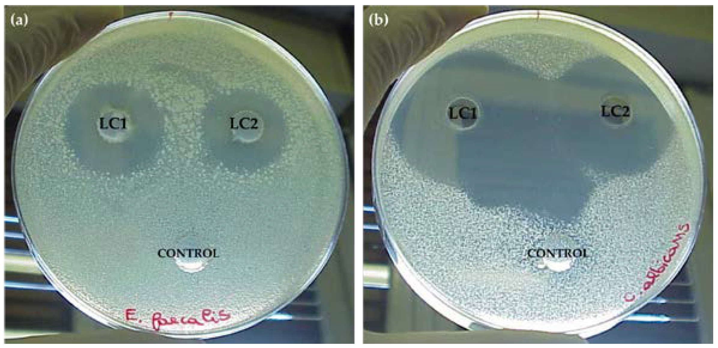
Figure 1. Antimicrobial activity of LC1 and LC2 lichen extracts against (a) Enterococcus faecalis ATCC 29212 and (b) Candida albicans ATCC 10231 7 and 30 days, respectively, after the extraction procedure.
Applsci 13 01976 g001
Figure 2. Results of thin-layer chromatography (TLC) analysis of LC1 solution’s fractions.
Applsci 13 01976 g002
Figure 3. Peak at retention time of 6.3 min for solution LC1 (black) and solution LC2 (red).
Applsci 13 01976 g003
Figure 4. Peak at retention time of 10.6 min for solution LC1 (blue) and solution LC2 (green).
Applsci 13 01976 g004
Figure 5. Peak at retention time of 11.5 min for solution LC1 (green) and solution LC2 (gray).
Applsci 13 01976 g005
Figure 6. Example of a LC1 fraction HPLC chromatogram (fraction 8) showing the antimicrobial activity against Candida albicans ATCC 10231. The peak at a retention time (tR) of 10.8 min corresponds to a scabrosin derivative.
Applsci 13 01976 g006
Figure 7. Example of the mass spectrometry (MS) spectrum related to the peak at 10.8 min.
Applsci 13 01976 g007
Table 1. Antimicrobial activity of cyclohexane (LC1) and acetone (LC2) lichen extracts against human pathogens using the agar well diffusion assay. Data are expressed in mm (diameter of inhibition zone) detected 1, 7, 15, and 30 days (d) after extraction procedure. Values are the arithmetic mean (n = 3) ± SD (standard deviation). Values marked with letters were statistically different according to t-test and ANOVA; p < 0.05 (a), p < 0.01 (b), p < 0.001 (c), and p < 0.0001 (d).
LC1LC2
1 d7 d15 d30 d1 d7 d15 d30 d
Staphylococcus aureus ATCC 65388 ± 24 ± 0.5 a0 b0 b8 ± 13 ± 1.5 b0 b0 b
Enterococcus faecalis ATCC 2921224 ± 224 ± 10 d0 d24 ± 0.522 ± 10 d0 d
Bacillus subtilis ATCC 663316 ± 215 ± 10 c0 c16 ± 0.514 ± 10 c0 c
Escherichia coli ATCC 873924 ± 0.50 d0 d0 d24 ± 1.50 d0 d0 d
Pseudomonas aeruginosa ATCC 2785322 ± 20 d0 d0 d22 ± 10 d0 d0 d
Burkholderia cepacia MM 086 ± 10 d0 d0 d6 ± 20 d0 d0 d
Klebsiella pneumoniae MM 0112 ± 0.50 d0 d0 d12 ± 2.50 d0 d0 d
Candida albicans ATCC 1023140 ± 240 ± 339 ± 0.538 ± 340 ± 140 ± 137 ± 237 ± 1
Table 2. Extraction yields and main metabolites in LC1 and LC2 lichen extracts.
Lichen ExtractsSolvent% YieldMain Matabolites
LC1Cyclohexane1.0variolatic acid scabrosin derivates
LC2Acetone2.4variolatic acid scabrosin derivates
Disclaimer/Publisher’s Note: The statements, opinions and data contained in all publications are solely those of the individual author(s) and contributor(s) and not of MDPI and/or the editor(s). MDPI and/or the editor(s) disclaim responsibility for any injury to people or property resulting from any ideas, methods, instructions or products referred to in the content.