Article

Accuracy of Guided Endodontics in Posterior Teeth

1
Department of Periodontology, Endodontology and Cariology, University Center for Dental Medicine Basel UZB, University of Basel, Mattenstrasse 40, 4058 Basel, Switzerland
2
Department of Oral Surgery, University Center for Dental Medicine Basel UZB, University of Basel, Mattenstrasse 40, 4058 Basel, Switzerland
3
Center for Dental Imaging, University Center for Dental Medicine Basel UZB, University of Basel, Mattenstrasse 40, 4058 Basel, Switzerland
*
Author to whom correspondence should be addressed.
These authors contributed equally to this work.
Appl. Sci. 2023, 13(4), 2321; https://doi.org/10.3390/app13042321
Submission received: 13 January 2023 / Revised: 3 February 2023 / Accepted: 9 February 2023 / Published: 10 February 2023
(This article belongs to the Special Issue Innovative Techniques in Endodontics and Restorative Dentistry)

Abstract

:
The purpose of this ex vivo study was to determine the accuracy of template-based guided endodontics for access cavity preparation and root canal detection in posterior teeth. First, three maxillary and four mandibular models were constructed using 67 premolars and molars, with a total number of 135 main root canals. Cone beam computed tomography (CBCT) and three-dimensional 3D surface scans of each model were performed and matched in order to plan access cavity preparation and design templates virtually. Template-guided access cavity preparation was then performed for each tooth, followed by postoperative CBCT scanning. Deviations between planned and prepared access cavities were measured after superimposition of the pre- and postoperative CBCT scans, and they were analyzed using descriptive and multivariate statistics. All root canals (135/135) were detected utilizing guided endodontics. The mean angle deviation was 1.4 degrees, and the mean deviations at the tip and base of the bur were 0.24–0.31 mm and 0.26–0.29 mm, respectively. This study demonstrated that guided endodontics is an accurate and predictable method for endodontic access cavity preparation in posterior teeth.

1. Introduction

The aim of any root canal treatment is to cure or prevent apical periodontitis [1]. This is mainly achieved by disinfecting the root canal system [2,3].
In teeth with severe pulp canal calcification (PCC), endodontic access cavity preparation and identification of the root canal orifice can be very difficult, time-consuming and prone to a higher risk of iatrogenic damage (e.g., tooth structure loss and root perforation) [4]. An increased loss of enamel and dentin may result in an increased risk of fractures, ultimately worsening the prognosis of the tooth [4,5]. Guided endodontics (GE) is an innovative method that uses 3D imaging (cone beam computed tomography, CBCT) and surface scans for virtual preoperative planning of minimally invasive access cavity preparation. A bur is guided through a template sleeve system to the planned position, similarly to the technique of guided implantology [6]. Guided endodontics enables the operator to locate the root canal orifice even in teeth with severe PCC [7,8], and is less invasive than conventional endodontic techniques [9].
Since CBCT was introduced into dentistry, it has been widely used for diagnostics and treatment planning [10]. It is extremely important to adhere to the ALARA principle (as low as reasonably achievable) in order to keep the radiation exposure for patients as low as possible [11]. A CBCT in endodontics may be considered when the conventional radiograph is insufficient for diagnosis, and the additional information from the CBCT is likely to facilitate diagnosis and further treatment. The current guidelines explicitly state that implementation of a CBCT may be justified for the planning of GE in teeth with PCC [12].
Guided endodontics is already being implemented in the clinical setting. However, it is mainly used in anterior teeth [8,13], and only a few publications have provided details regarding the accuracy of GE in posterior teeth [14,15]. Therefore, the aim of this study was to determine the general accuracy of template-guided endodontics for access cavity preparation in posterior teeth, and to evaluate variables that might affect the accuracy of access cavity preparation, such as the type of tooth (molar vs. premolar), location of the posterior teeth (maxilla vs. mandible) and the number of root canals per tooth.

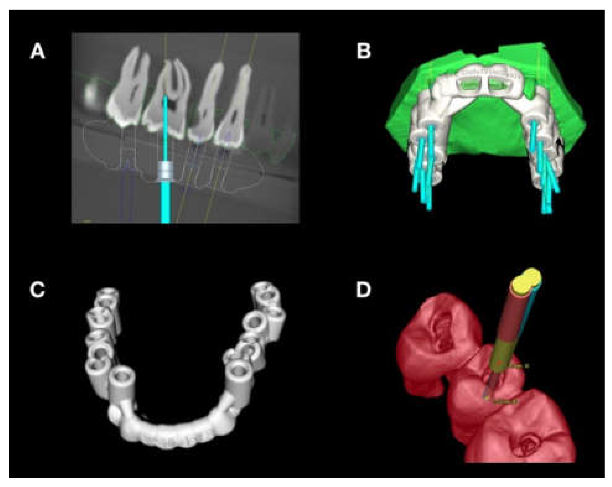
2. Materials and Methods

Sixty-seven premolars and molars extracted for reasons unrelated to this study were used to construct three maxillary and four mandibular models. Only premolars and molars with complete root formation and without resorptions or restorations were included. Teeth with profound caries extending to the pulp were excluded. The extracted teeth were placed in their anatomically correct arch positions and fixed onto a permanent methyl methacrylate copolymer base. Afterwards, optical surface scans of the models were made using an intraoral scanner (3Shape TRIOS 3; Institut Straumann AG, Basel, Switzerland) and saved in surface tessellation language (STL) format. Additionally, CBCT scans of each model were made with a voxel size of 0.16 mm, 90 kV, 6 mA and an 8 × 8 cm field of view (3D Accuitomo 170; Morita Manufacturing Corp., Kyoto, Japan) and saved in digital imaging and communications in medicine (DICOM) format. The STL and DICOM data sets were then imported into 3D planning software (coDiagnostiX; Dental Wings Inc, Montreal, Canada) and virtually matched based on the available matching tool without further corrections. For each main root canal of the 67 posterior teeth, a guided access cavity was planned in the 3D planning software by virtually positioning a true-to-dimension bur to allow straight-line access to the root canal orifice, followed by the construction of templates (Figure 1).
A total of 11 templates were virtually designed with computer-aided design/computer-aided manufacturing (CAD/CAM) and manufactured from polymethyl methacrylate discs (98.5 mm; Yamahachi Dental, Gamagori, Japan) in a 3D milling machine (Motion 2; Amann Girrbach AG, Koblach, Austria) [16]. The fabricated templates were checked for proper fit on the models and adjusted as needed. Afterwards, the tip of the guided endodontics bur was stained with caries marker solution (Karies Marker, VOCO GmbH, Cuxhaven, Germany). The template, with the sleeve inserted, was placed on the model in the end position and the stained bur was inserted in the sleeve, leaving a mark indicating the occlusal position of the access cavity. The enamel at the access cavity site was then removed using a diamond bur in a contra-angle handpiece without using the template. With dentin exposed, the endodontic bur (ATEC Endoseal, steco-system-technik GmbH & Co KG, Hamburg, Germany) was set to a speed of 10,000 rpm and guided through the sleeve of the template using slow and short up-and-down movements while regularly clearing debris from the bur and access cavity. Guided access cavity preparation was completed once the bur reached the defined bur stop. Endodontic hand files were then utilized to confirm the root canal location (Figure 2). After access cavity preparation for each tooth, the bur was inspected for deformation or damage, and was replaced after completing all teeth on a model or beforehand if any damage was observed.
After all access cavities were prepared, postoperative CBCT scans of each model were created using the same CBCT machine settings as those used preoperatively. Using the planning software, the pre- and postoperative CBCT data were matched in the 3D planning software. The treatment evaluation tool integrated into the software was used to determine the accuracy of the prepared vs. virtually planned access cavities. Because the direction of each access cavity could be derived from the postoperative CBCT data, virtual bur placement was possible for each prepared access cavity. The software then automatically calculated the angular and spatial deviation in different planes at the tip and base of the bur. Descriptive analysis was performed for all access cavities (Table 1). The results for posterior tooth location (maxilla vs. mandible), type (molar vs. premolar) and number of main root canals per tooth (one, two or three) were calculated as mean values with 95% confidence intervals. In addition, a multivariate analysis of variance (MANOVA), followed by Bonferroni post hoc tests for the variable “number of main root canals”, were performed using SPSS V. 28.0.1 (IBM Corp, Armonk, NY, USA) in order to assess the influence of different variables with regard to the accuracy of the prepared access cavities in different and angular spatial dimensions. The level of significance was set at α = 0.05.

3. Results

All of the 135 root canals (100%) were detected after GE access cavity preparation. No root perforations were observed. The overall mean angular deviation between the planned and prepared access cavities was 1.39 degrees (details are shown in Table 1). The distribution of angular deviation for all models is presented in Figure 3.
The mean angular and spatial deviations in the different dimensions, in addition to their 95% confidence intervals for maxillary vs. mandibular teeth and for molars vs. premolars, are shown in Figure 4. Regarding angular deviation, the MANOVA revealed that access cavity preparation was significantly less accurate in maxillary teeth compared to mandibular teeth (1.70° vs. 1.04°; p = 0.03). The results are presented in Table 2.
MANOVA revealed that the number of root canals per tooth had a significant effect on the angular and spatial deviation at the tip of the bur in the bucco-oral direction (p < 0.05). The results for accuracy with regard to mean deviation, according to the number of main roots canals per tooth, are shown in Figure 5. Teeth with one canal had significantly higher mean angle deviation than those with two canals (2.08 vs. 0.96 degrees; p = 0.006). The deviation in bucco-oral dimension at the tip of the bur was also significantly greater in teeth with one canal compared to those with two canals (0.42 vs. 0.27 mm; p = 0.04). Further details are shown in Table 3 and Table 4.

4. Discussion

This ex vivo proof-of-principle study demonstrates that GE allows for precise and predictable access cavity preparation in posterior teeth. Other ex vivo studies showing that GE is a viable, fast and accurate method for the preparation of endodontic access cavities in anterior teeth already exist [7,8,16], and there are clinical case reports showing the feasibility of guided endodontic treatment of the upper second and third molars [14] as well as the first lower molar [17].
The evidence indicates that GE is operator-independent and allows less experienced operators to reliably access calcified root canals. The overall accuracy (mean angular deviation for all access cavities) measured in this study (1.39 degrees) is comparable to that reported in previous studies [7,18].
One interesting new finding of this study is that the number of main root canals per tooth had a significant effect on angular deviation and spatial deviation at the tip of the bur in the bucco-lingual dimension. Access cavity preparation was less accurate in teeth with one main root canal than in multi-canal teeth. This could be explained by the fact that the teeth used in this study did not have severe calcification, and that the teeth with one root canal had a larger, possibly oval root canal lumen. Since there was a certain looseness of fit between the utilized burs and sleeves, the bur might have been able to center itself in smaller and rounder root canals once the root canal orifice was reached. This would explain the smaller deviation in teeth with two or three main root canals per tooth.
However, operator-related sources of error (e.g., manual positioning during certain steps) may have led to inaccuracies in access cavity preparation as well. Semi-automatic registration of CBCT and surface scan data by the planning software could be another source of inaccuracy, as there is some evidence suggesting that full arch surface scans may be subject to local deviation [16]. Other sources of inaccuracy described in previous studies also apply to the present study (e.g., CAD/CAM processing of templates or loose fit between the bur and sleeve) [19].
There are limiting factors to consider before performing GE in the posterior region [7]. A limited mouth-opening ability and, therefore, limited space for the GE template may complicate the application of GE in posterior regions. However, it should be noted that the templates used in clinical practice can be designed with a lower height than the ones used in this study.
For GE, the root canal orifice must be located in a position that can be reached by a straight and rigid bur [7]. Therefore, root curvature is considered a contraindication if the root canal orifice is located far apically. In contrast to anterior teeth, molars often have roots with greater curvature. Fortunately, curvatures most commonly occur in the apical third of molar roots, resulting in a generally promising prognosis for guided endodontics treatment in molars, since root canal calcifications rarely occur up to the apical third of the root canal of these teeth [15].
Even though complex canal morphologies can be a limiting factor for the application of GE, on the other hand, GE may be advantageous in teeth with root variations. GE could be aid in the identification of a calcified radix entomolaris or a middle mesial canal.
The planning effort has surely increased with the application of GE; however, the effective treatment time for the patient is probably reduced, as the tedious search for PCC is omitted. Therefore, GE may possibly improve the patient’s comfort, even though additional treatment steps such as intraoral scanning are necessary. In addition, numerous case reports and studies regarding GE have already been published, focusing mainly on anterior teeth [20].
CBCT has become an important tool for preoperative planning in dentistry. In endodontics, CBCT aids in identifying resorptions, calcifications, root canal morphologies and apical pathologies [21]. However, CBCT results in higher radiation doses than conventional radiography techniques. Therefore, the indication for CBCT must be carefully evaluated. According to a joint statement of the American Association of Endodontists and the American Academy of Oral and Maxillofacial Radiology, the use of limited field-of-view (FOV) is indicated to help identify and locate calcified canals and depict complex canal morphology [22].
A limitation of this ex vivo study is related to the fact that none of the treated teeth were severely calcified. Severe calcification could have negative implications for virtual access cavity planning and may result in lower overall accuracy. However, it is difficult to find extracted teeth that are severely calcified, yet caries- and restoration-free, at the same time. In recent years, 3D-printed teeth have also been used in the field of endodontic teaching and research. The major advantage of these teeth over extracted teeth is the possibility to digitally create root canal calcification as needed. However, studies have also shown that various types of 3D-printed teeth cannot fully reproduce the optical and haptic properties of human dentin [23].
In GE, additional time is required to perform CBCT and intraoral scanning, to complete the virtual planning and to fabricate the guide template. Further studies are needed to address aspects such as the differences between conventional freehand and GE access cavity preparation in posterior teeth with regard to time, operator experience, possible additional costs and the amount of tooth substance loss that is associated with the techniques.

5. Conclusions

Guided endodontics is an accurate and predictable technique for access cavity preparation and root canal detection in posterior teeth. All root canals were successfully detected due to a low deviation in angle (1.4°) and a low mean deviation at the tip of the bur (0.24–0.31 mm). GE was even more accurate in teeth with multiple root canals than in teeth with only one root canal.

Author Contributions

Conceptualization, T.C. and R.W.; methodology, T.C.; software, S.K. and W.L.; formal analysis, W.L.; investigation, B.H.; data curation, B.H. and W.L.; writing—original draft preparation, B.H.; writing—review and editing, B.H., W.L., T.C., E.M., R.W., D.D.-B. and S.K.; visualization, B.H. and W.L.; supervision, T.C.; funding acquisition. All authors have read and agreed to the published version of the manuscript.

Funding

This research received no external funding.

Institutional Review Board Statement

Ethical approval was obtained from the local Research Ethics Committee (EKNZ UBE-15/111).

Informed Consent Statement

Not applicable.

Data Availability Statement

Not applicable.

Acknowledgments

This article was supported by the following dentists, who provided access to the necessary infrastructure: Sabine Teubner, Eckart Teubner, and Stefanie Hirt.

Conflicts of Interest

The authors declare that they have no conflict of interest.

References

  1. European Society of Endodontology. Quality guidelines for endodontic treatment: Consensus report of the European Society of Endodontology. Int. Endod. J. 2006, 39, 921–930. [Google Scholar] [CrossRef] [PubMed]
  2. Schilder, H. Cleaning and shaping the root canal. Dent. Clin. N. Am. 1974, 18, 269–296. [Google Scholar] [CrossRef] [PubMed]
  3. Kakehashi, S.; Stanley, R.; Fitzgerald, R.J. The effects of surgical exposures of dental pulps in germ-free and conventional laboratory rats. Oral. Surg. Oral. Med. Oral. Pathol. 1965, 20, 340–349. [Google Scholar] [CrossRef] [PubMed]
  4. Cvek M, G.L.; Lundberg, M. Failures and healing in endodontically treated non-vital anterior teeth with posttraumatically reduced pulpal lumen. Acta Odontol. Scand. 1982, 40, 223–228. [Google Scholar] [CrossRef] [PubMed]
  5. Lang, H.; Korkmanz, Y.; Schneider, K.; Raab, W.H. Impact of Endodontic Treatments on the Rigidity of the Root. J. Dent. Res. 2006, 85, 364–368. [Google Scholar] [CrossRef] [PubMed]
  6. Kühl, S.; Payer, M.; Zitzmann, N.U.; Lambrecht, J.T.; Filippi, A. Technical accuracy of printed surgical templates for guided implant surgery with the coDiagnostiX software. Clin. Implant Dent. Relat. Res. 2015, 17, 177–182. [Google Scholar] [CrossRef]
  7. Connert, T.; Zehnder, M.S.; Weiger, R.; Kühl, S.; Krastl, G. Microguided Endodontics: Accuracy of a Miniaturized Technique for Apically Extended Access Cavity Preparation in Anterior Teeth. Int. Endod. J. 2017, 43, 787–790. [Google Scholar] [CrossRef] [PubMed]
  8. Krastl, G.; Zehnder, M.S.; Connert, T.; Weiger, R.; Kuhl, S. Guided Endodontics: A novel treatment approach for teeth with pulp canal calcification and apical pathology. Dent. Traumatol. 2015, 32, 240–246. [Google Scholar] [CrossRef]
  9. Connert, T.; Krug, R.; Eggmann, F.; Emsermann, I.; ElAyouti, A.; Weiger, R.; Kuhl, S.; Krastl, G. Guided Endodontics versus Conventional Access Cavity Preparation: A Comparative Study on Substance Loss Using 3-dimensional-printed Teeth. J. Endod. 2019, 45, 327–331. [Google Scholar] [CrossRef]
  10. Mozzo, P.; Procacci, C.; Tacconi, A.; Martini, P.T.; Andreis, I.A. A new volumetric CT machine for dental imaging based on the cone-beam technique: Preliminary results. Eur. Radiol. 1998, 8, 1558–1564. [Google Scholar] [CrossRef]
  11. Farman, A.G. ALARA still applies. Oral. Surg. Oral. Med. Oral. Pathol. Oral. Radiol. Endod. 2005, 4, 395–397. [Google Scholar] [CrossRef] [PubMed]
  12. Patel, S.; Brown, J.; Semper, M.; Abella, F.; Mannocci, F. European Society of Endodontology position statement: Use of cone beam computed tomography in Endodontics. Int. Endod. J. 2019, 52, 1675–1678. [Google Scholar] [CrossRef] [PubMed]
  13. van der Meer, W.J.; Vissink, A.; Ng, Y.L.; Gulabivala, K. 3D Computer aided treatment planning in endodontics. J. Dent. 2016, 45, 67–72. [Google Scholar] [CrossRef] [PubMed]
  14. Lara-Mendes, S.T.O.; Barbosa, C.F.M.; Santa-Rosa, C.C.; Machado, V.C. Guided Endodontic Access in Maxillary Molars Using Cone-beam Computed Tomography and Computer-aided Design/Computer-aided Manufacturing System: A Case Report. J. Endod. 2018, 44, 875–879. [Google Scholar] [CrossRef]
  15. Lee, J.K.; Ha, B.H.; Choi, J.H.; Heo, S.M.; Perinpanayagam, H. Quantitative three-dimensional analysis of root canal curvature in maxillary first molars using micro-computed tomography. J. Endod. 2006, 32, 941–945. [Google Scholar] [CrossRef] [PubMed]
  16. Leontiev, W.; Bieri, O.; Madorin, P.; Dagassan-Berndt, D.; Kuhl, S.; Krastl, G.; Krug, R.; Weiger, R.; Connert, T. Suitability of Magnetic Resonance Imaging for Guided Endodontics: Proof of Principle. J. Endod. 2021, 47, 954–960. [Google Scholar] [CrossRef]
  17. Shi, X.; Zhao, S.; Wang, W.; Jiang, Q.; Yang, X. Novel navigation technique for the endodontic treatment of a molar with pulp canal calcification and apical pathology. Aust. Endod. J. 2018, 44, 66–70. [Google Scholar] [CrossRef]
  18. Zehnder, M.S.; Connert, T.; Weiger, R.; Krastl, G.; Kühl, S. Guided endodontics: Accuracy of a novel method for guided access cavity preparation and root canal location. Int. Endod. J. 2016, 49, 966–972. [Google Scholar] [CrossRef]
  19. Krug, R.; Reich, S.; Connert, T.; Kess, S.; Soliman, S.; Reymus, M.; Krastl, G. Guided endodontics: A comparative in vitro study on the accuracy and effort of two different planning workflows. Int. J. Comput. Dent. 2020, 23, 119–128. [Google Scholar]
  20. Connert, T.; Weiger, R.; Krastl, G. Present status and future directions—Guided endodontics. Int. Endod. J. 2022, 10, 995–1002. [Google Scholar] [CrossRef] [PubMed]
  21. Patel, S.; Durack, C.; Abella, F.; Shemesh, H.; Roig, M.; Lemberg, K. Cone beam computed tomography in Endodontics—A review. Int. Endod. J. 2015, 48, 3–15. [Google Scholar] [CrossRef] [PubMed]
  22. Endodontics, A.A.O. AAE and AAOMR Joint Position Statement—Use of Cone Beam Computed Tomography in Endodontics. Available online: https://www.aae.org/specialty/wp-content/uploads/sites/2/2017/06/conebeamstatement.pdf (accessed on 12 December 2022).
  23. Reymus, M.; Fabritius, R.; Keßler, A.; Hickel, R.; Edelhoff, D.; Stawarczyk, B. Fracture load of 3D-printed fixed dental prostheses compared with milled and conventionally fabricated ones: The impact of resin material, build direction, post-curing, and artificial aging-an in vitro study. Clin. Oral. Investig. 2020, 24, 701–710. [Google Scholar] [CrossRef] [PubMed]
Figure 1. Digital workflow: (A) virtually planned straight-line access cavity, (B) multiple access cavities planned for posterior teeth in a maxillary model, (C) virtually designed template, (D) 3D rendered view of accuracy evaluation revealing the deviation between planned and performed access cavity preparation.
Applsci 13 02321 g001
Figure 2. Access cavity preparation: (A) CAD/CAM-fabricated template placed on a model, (B) sleeve inserted for static navigation of the bur, (C) access cavity preparations in maxillary premolars and molar, (D) locating three main root canals with endodontic hand files.
Applsci 13 02321 g002
Figure 3. Angle deviation between the planned and prepared access cavities on the three maxillary and four mandibular models.
Applsci 13 02321 g003
Figure 4. Mean angle deviation (°) and mean deviation at the base and the tip of the bur in the mesio-distal and bucco-oral dimensions (mm) by tooth location (maxilla vs. mandible) and type (premolar (pm) vs. molar (m)).
Applsci 13 02321 g004
Figure 5. Mean angle deviation (°) and mean deviation at the tip of the bur in the bucco-oral dimension (mm) for teeth with one, two or three main root canals, respectively.
Applsci 13 02321 g005
Table 1. Deviation between planned and prepared access cavities regarding angle deviation and mesio-distal and bucco-oral deviation at the tip and base of the bur.
Deviation Angle
[Degrees]
Mesio-Distal, at Base of the Bur [mm]Bucco-Oral, at Base of the Bur [mm]Mesio-Distal, at Tip of the Bur [mm]Bucco-Oral, at Tip
of the Bur [mm]
Mean1.390.260.290.240.31
Median1.100.200.210.180.21
Minimum 0.00.00.00.00.0
Maximum7.81.191.961.611.36
Table 2. Mean angle deviation (°) and deviation in the mesio-distal and bucco-oral direction (mm) at the base and tip of the bur between maxillary vs. mandibular and premolar vs. molar teeth.
Deviation TypeMean
MaxillaMandiblep-ValuePremolarMolarp-Value
Angle [°]1.701.040.031.521.340.89
Mesiodistal, Base [mm]0.310.270.990.290.290.11
Buccolingual, Base [mm]0.290.230.780.220.280.77
Mesiodistal, Apex [mm]0.340.280.460.350.300.14
Buccolingual, Apex [mm]0.260.210.970.160.270.77
Table 3. Mean difference in angle deviation between planned and prepared access cavities according to the number of main root canals per tooth (CPT).
Angle Deviation [°]
Group 1Group 2Mean Difference: Group 1 vs. Group 2 [°]p-Value
1 CPT2 CPT1.120.006
3 CPT0.550.36
2 CPT 1 CPT−1.120.006
3 CPT−0.560.13
3 CPT1 CPT−0.550.36
2 CPT 0.560.13
Table 4. Mean difference in bucco-oral deviation at the tip of the bur between planned and prepared access cavities according to the number of main root canals per tooth (CPT).
Bucco-Oral Deviation at Tip of the Bur [mm]Mean Difference: Group 1 vs. Group 2 [mm]p-Value
Group 1Group 2
1 CPT2 CPT0.160.04
3 CPT0.120.21
2 CPT 1 CPT−0.160.04
3 CPT−0.041.00
3 CPT1 CPT−0.120.21
2 CPT 0.041.00
Disclaimer/Publisher’s Note: The statements, opinions and data contained in all publications are solely those of the individual author(s) and contributor(s) and not of MDPI and/or the editor(s). MDPI and/or the editor(s) disclaim responsibility for any injury to people or property resulting from any ideas, methods, instructions or products referred to in the content.

