Article

Somatotropin Penetration Testing from Formulations Applied Topically to the Skin

Department of Pharmaceutical Technology, Faculty of Pharmaceutical Sciences, Medical University of Silesia, Kasztanowa 3, 41-200 Sosnowiec, Poland
*
Author to whom correspondence should be addressed.
Appl. Sci. 2023, 13(4), 2588; https://doi.org/10.3390/app13042588
Submission received: 14 December 2022 / Revised: 8 February 2023 / Accepted: 15 February 2023 / Published: 17 February 2023
(This article belongs to the Special Issue Advances in Biomaterials and Drug Technology)

Abstract

:
Growth hormone (somatotropin—STH) deficiency therapy requires daily injections of recombinant human growth hormone. The FDA approved treatment with STH with one dose per week for the first time in 2021. However, injectable drug application is accompanied by numerous inconveniences. Therefore, an attempt was made to formulate a less invasive STH formulation for topical application to the skin. A substrate was prepared based on a polymer, methylcellulose (MC), into which STH was introduced at a concentration of 1 mg/g. Simultaneously, formulations were made with STH, to which albumin (ALB) was added at different concentrations: 0.1%, 0.2% and 0.5%. A test of the degree of STH permeation was carried out, as well as the effect of ALB on STH permeation parameters. Selected rheological properties of the formulations obtained were investigated. A test of STH permeation in simulated in vivo conditions through porcine skin indicated a relatively good bioavailability of over 80% and confirmed the effectiveness of MC as a carrier for growth hormone. ALB prolonged the STH penetration rate and increased the penetration degree of STH to 93%. The hydrogels obtained were found to be typical shear-thinning, thixotropic fluids.

1. Introduction

Somatotropin (STH), known as growth hormone (GH), is a polypeptide produced by the somatotropic cells of the anterior lobe of the pituitary gland [1]. The hormone is secreted pulsatilely in response to the action of somatoliberin and somatostatin secreted from hypothalamic neurons [2]. STH consists of 191 amino acid residues with a molecular weight (Mw) of 22 kDa [1,3]. The cycle of GH release is pulsatile, with the largest diurnal increase in GH secretion occurring shortly after the onset of sleep, and smaller pulses occurring at intervals throughout the day, several hours after eating a meal [4,5]. The total daily GH production of approximately 0.5 mg is highest in childhood and adolescence, while it decreases with age [5]. GH and insulin-like growth factor (IGF-1) levels in the aged decrease by 30–60%. Its pulsatile secretion, which is the most important factor regulating IGF-1, results in a rapid time half-life of growth hormone, ranging from 17 to 45 min [2,5,6]. The production of biosynthetic GH allows for the treatment of people with its deficiency [7], in low-growth conditions in which GH deficiency is not found, such as Turner syndrome, Prader-Willi syndrome, Noonan syndrome, idiopathic low-growth syndrome and chronic renal failure. STH has also been used in diseases that cause a loss of muscle tissue, cachexia due to HIV, cystic fibrosis, inflammatory bowel disease and neurodegenerative disorders [8]. Registered preparations of STH come in the form of a solution and a powder and solvent for preparing a solution for injection.
Human serum albumin (ALB) physiologically maintains the effective osmotic pressure of colloids in plasma, influences the integrity of the vascular endothelium, has antioxidant and anti-inflammatory effects, maintains acid-base equilibrium, and participates in the transport, distribution and metabolism of various endogenous and exogenous substances. ALB can have a stabilizing effect on certain proteins [9].
ALB is composed of 585 amino acids [10]. It is a simple polypeptide with a Mw of 66.5 kDa and has no prosthetic groups [9].
Currently, almost all available protein preparations (hormone treatment, enzymes, growth factor, coagulation factor, interferon) are administered at large doses at frequent intervals, which can result in dose fluctuations and patient neglect. At present, technologies make it possible to extend the half-life of proteins, which can prevent rapid degradation in vivo by fusing with immunoglobulins or serum proteins (ALB) and conjugating with natural or synthetic polymers [11].
Siemiradzka et al. [12] demonstrated the effect of the concentration of ALB on the timing and extent of corticotropin (ACTH) permeation from hydrogels. ALB caused a delay in penetration that was dependent on the ratio of ALB to ACTH. Apparently, this effect was seen at a 1:1 ratio of ALB and ACTH (1.5% ALB and 1.5% ACTH) [12].
Drugs used on the skin enter through the upper epidermis layer—the stratum corneum, which varies in thickness from 10 to 80 µm (stratum corneum, SC)—or one of the appendages (hair follicles). Passage leads over subsequent layers, dermis and subcutaneous tissue, creating the potential for systemic distribution of the drug [13]. There are many therapeutics administered to the skin that exhibit both superficial and systemic effects [14,15]. Transport through the stratum corneum occurs generally by passive diffusion.
Permeation depends on the affinity of the compound to the lipid environment [13]. The epidermis, which comprises most of the skin, provides a barrier to both hydrophilic and lipophilic substances [16]. The permeation of active substances through the skin depends on their physicochemical properties—small, oval-shaped molecules with high concentrations in the formulation penetrate most easily [17].
Therapeutic substance diffusion is improved via the use of carriers or sorption promoters. Factors including enzymatic activity, blood supply and hydration decrease the passage of drugs, and the site of the skin to which the preparation is applied varies in SC thickness and lubrication. Drugs penetrate the mucous membranes most easily; less easily through the back (0.29 mg/cm2/h) and trunk (0.34 mg/cm2/h); and with most difficulty through the palms (1.14 mg/cm2/h) and the soles of the feet [17,18]. Adequate moisturization of the skin, as a consequence of which corneocytes in the SC swell, makes for the easier diffusion of active substances [19].
An important group of gels used in pharmaceutics is hydrogels, usually composed of hydrophilic polymers, which under certain conditions and at a certain polymer concentration undergo gelation—that is, the binding of a solvent (a mixture of water and glycerol or propylene glycol) to a polymer network (cellulose derivatives, carbomers, poloxamers, starch) [20]. Hydrogels can be applied to skin, mucous membranes, wounds, and can also be inserted into body cavities. Hydrogels have many advantages, including that they do not leave an oily film on the skin, can be simply removed from skin with water, and are mucoadhesive. Their structure and compatibility with the aqueous environment make them very attractive, biocompatible drug carriers [21]. Thanks to their flexibility, porosity and high water content, hydrogels can be used as vehicles for numerous drug substances, including peptides and proteins (ACTH); the pore size can be kept under control by adjusting the cross-linking of the polymer. The water interface of hydrogels is able to stabilize cellular and compound drugs, including peptides, proteins, oligonucleotides and DNA [22,23].
In their trial, Lamb et al. described the approval of a long-acting STH in pediatrics [24]. Daily therapy with frequent non-adherence and fluctuations in dose can affect the following treatment outcomes. A long-acting growth hormone analogue was developed that allowed for less frequent administration [24,25]. In 2021, the FDA approved treatment by the once-weekly administration of the STH preparation lonapegsomatropin-tcgd, SKYTROFA® (Ascendis Pharma, Denmark), for pediatric patients. SKYTROFA® is a long-acting pro-drug of human growth hormone. Using E. Coli recombinant DNA technology, STH was produced and conjugated to methoxypolyethylene glycol (4 × 10 DamPEG) using a patented TransCon linker [26]. After the subcutaneous administration of lonapegsomatropin at a dose of 0.24 mg/kg/week in pediatrics, the mean observed half-life was 30.7 h [27]. The side-effect profile was comparable to STH administered daily, and indicators such as fasting glucose and hemoglobin in those receiving lonapegsomatropin were mostly within normal limits [28].
Due to the numerous inconveniences that accompany injectable drug application, this study attempts to formulate a much less invasive skin formulation containing the peptide substance STH. For skin formulations, it is important to ensure convenient application and effective therapeutic concentration of the active substance. The purpose of this project was to a make a formulation that would allow diffusion of the active substance from the substrate and have appropriate rheological and physicochemical properties. To this end, we planned to trace the permeation of the active substance from the obtained formulation through a natural membrane, such as pig skin. To compare the permeation process, the process was also carried out using an artificial membrane—a cellulose membrane. Since STH has a short half-life, efforts have been made to prepare hydrogel formulations with the addition of the transport protein ALB, which could prolong the permeation time, and thus, increase the therapeutic efficacy of STH.

2. Materials and Methods

2.1. Materials

The study material was: STH—Genotropin® 12 GoQuick (12 mg, 36 IU), Pfizer, New York, USA, series: GF1722, ALB—Human Albumin CSL Behring 200 g/L (200 g/L total protein, including at least 96% human ALB, sodium caprylate, sodium N-acetyl-D, L-tryptophan, sodium chloride, sodium hydroxide, water for injection), CSL Behring GmbH, Marburg, Germany.
The following were used as auxiliary substances: MC (Fluka Chemie, Buchs, Switzerland); glycerol 85% (Galfarm, Krakow, Poland); sodium chloride (Avantor Performance Material S.A., Gliwice, Poland); water for injection (Galfarm, Krakow, Poland); disodium hydrogen phosphate (Avantor Performance Material S.A., Gliwice, Poland); potassium dihydrogen phosphate (Avantor Performance Material S.A., Gliwice, Poland). The substances used were of the PD class and met the requirements of standards for pharmaceutical raw materials.

2.2. Hydrogel Preparation

To prepare the hydrogel substrates, the liquid components—water and glycerol—were weighed and heated, not exceeding 80 °C. Then, while stirring vigorously (250 RPM) the water–glycerol solution was placed on a Fisherbrand™ Isotemp™ magnetic stirrer, and methylcellulose (MC) was added in portions and stirred. MC-based hydrogel was brought to ambient temperature. STH from Genotropin 12 was dissolved in a small amount of water and introduced into the prepared hydrogel base. Next, hydrogels were formulated with STH and the addition of ALB. ALB was introduced into the hydrogels with STH as an aqueous solution in appropriate amounts in the ratio of STH and ALB: 1:1, 1:2 and 1:5, and the formulations were mixed thoroughly on a magnetic stirrer (100 RPM). The amount of water for the formulation of all hydrogels containing both STH and STH with ALB was reduced by the amount for STH dissolution and the amount of added ALB solution, respectively. The hydrogels thus made were stored at 2–8 °C. The composition of the prepared formulations is shown in Table 1.

2.3. Measurement Method of STH

The content of STH in the acceptor fluid (phosphate-buffered sodium chloride (PBS) at pH = 7.4) was determined using a validated spectrophotometric method. Absorbance measurements were performed using a Cecil CE 3021 UV-VIS spectrophotometer (Cecil Instruments Limited, Cambridge, England) at a wavelength of λ = 276 nm. The photometric accuracy of the spectrophotometer was ±0.005 A. Based on the results obtained, the calibration curves of the dependence of absorbance on substance concentration were plotted. The linearity of the method was determined by 3-fold measuring the absorbance of STH solutions in 10 concentrations from 0.01 to 1.0 mg/mL, and it was described by the linear regression equation y = ax + b. The fit of the equation to the experimental data was described by the correlation coefficient (R2). Each sample taken was analyzed six times. The spectrophotometric method used was specific for the determination of the corresponding protein–peptide substance.

2.4. STH Permeation Study

2.4.1. Franz’s Vertical Diffusion Cells

Franz’s diffusion cells (Xenometrix, Allschwil, Switzerland) were used for the permeation study. The permeation study is described in detail in the paper by Siemiradzka, during the study of ALB permeation through the skin [29].
STH content was calculated from a curve with the equation y = 0.8953x + 0.0032; R2 = 0.999. The amount of STH permeated was measured spectrophotometrically at λ = 276 nm, using the PBS solution as a reference. The average of six repeats was calculated.

2.4.2. Membranes for Permeation Studies

Preliminary studies of the permeation process of STH were carried out using the artificial membrane Spectra/Por®DialysisMembrane, Biotech CE, MWCO: 100 kDa, Spectrum Laboratories Inc. The membrane was immersed in injection water for 30 min before testing.
The model membrane for the skin permeation study of STH was porcine skin, which was obtained from a local farmer. Skin sections were trimmed to a diameter of 2.5 ± 0.5 cm2. Each sample was spread over the skin surface. The preparation of porcine skin is described in Siemiradzka et al. [29].

2.4.3. Determination of Permeation Parameters of STH

The permeation of STH via the skin was traced by plotting the dependence of the released dose of STH C (%) in the time t = 6 h in preliminary studies using a cellulose membrane, and at 24 h using porcine skin.
Based on the results, the rate constant for the STH permeation process and the half-life of t50% were calculated.
The AUC (area under the curve) of the change in drug concentration over time is one of the parameters characterizing bioavailability. It shows the total amount of drug absorbed into the body. The triangle and trapezoid method was used to calculate the area under the curve—concentration of the substance: time, between 0 and 6 h—AUC(0–6 h) in preliminary studies using cellulose membrane, and between 0 and 24 h—AUC(0–24 h) using porcine skin. The degree of relative availability was calculated for the STH-only formulation, for which the STH availability was assumed to be 100%.
The penetration kinetics of STH matched the Higuchi model, the Korsmayer–Peppas model and the first-order model [30,31].
The results of the STH permeation study through the cellulose membrane and pig skin and the results of the viscosity measurements were statistically analyzed using Microsoft Excel and Statistica 13. A model-independent mathematical was used to compare the dissolution profiles of the samples and the reference product referring to the difference factor (f1) and similarity factor (f2) [32].

2.5. Rheological Properties

The viscosity of the hydrogel preparations was measured using an RM 200 Touch rotational rheometer, which was equipped with a CP1 Plus thermostat. Tests were carried out in a plate-to-plate arrangement using a 2445 MS CP measuring system. The rheometer with all the equipment and software is from Lamy Rheology Instruments (Champagne au Mont d’Or, France). Before measurement, the samples were placed in a CLW 53 STD hothouse, POL-EKO Aparatura sp.j., Wodzisław Śląski, Poland, at 32.0 ± 0.5 °C. After 30 min, the sample was placed on the lower plate of the Peltier effect thermostatic system. Viscosity measurements were carried out at 32.0 ± 0.5 °C at two shear rates of 15 s−1 and 30 s−1. Pre-shear was carried out at a shear rate of 5 s−1 for 2 min after sample application. All measurements were repeated 6 times (n = 6).
The flow curve was performed in the shear rate range D = 5–50 s−1 for 30 s. This was followed by a step-by-step test at a shear rate of D = 5–110 s−1. The number of measuring points taken at equal intervals—every 10 s—was 12 for each curve at increasing and decreasing shear rates.

2.6. Sensory Evaluation

A sensory evaluation of the prepared formulations was carried out [33].
Uniformity and smoothness were assessed on a plate by spreading approximately 0.5 cm3 of product in a circular motion, assessing the presence of lumps and air bubbles. Consistency determines density, and consistency was examined by freely dipping a finger at an angle of 45–60º into approximately 20 cm3 of the product placed in a beaker and quickly removing it. Note the resistance of the cream when dipping the finger and the contact of the finger with the hydrogel when pulling it out. The pillow effect is a parameter that determines the amount of product felt between the fingers when rubbing them against each other. The more product felt between the fingers, the stronger the pillow effect. An amount of 0.5 cm3 of the product was placed between the thumb and index finger; by rubbing the fingers against each other, the palpable amount of hydrogel was determined. Adhesion determines the ability to scoop the product onto the fingertip. Good adhesion characterizes a preparation that is easily picked up, does not run off and forms a permanent characteristic cone on the fingertip (does not spill). From the vessel, in which approximately 20 cm3 of hydrogel was placed, the product was freely picked up with a fingertip. Stickiness is a parameter determining the ability of the product to leave a sticky, adhesive layer on the skin. It should be spread on the skin surface with the fingers, or the product should be left spread between the fingers and checked with the other hand after a while to see if it is sticky. Greasiness and greasing evaluate the ability of the product to leave an oily film on the skin immediately after application (greasiness) and after 30 min (greasing).
The formulations were subjected to preliminary observations in terms of color, odor and appearance. The analysis for color was carried out in daylight on glass plates placed on a white matte background.

2.7. Stability Test

The stability of the hydrogels was examined by the biotechnology/biological products stability test under the conditions specified by ICH, Q5C: 25 ± 1 °C and 5 ± 3 °C. The samples were stored for 4 weeks [34]. The hydrogels were controlled by observing sensory parameters, color, drug content, pH, viscosity and shear stress. The pH was measured using an In Lab Expert Pro-ISM electrode, part number 30014096, Mettler Toledo AG, Greifensee, Switzerland. The amount of STH was determined by spectrophotometry, using a CECIL apparatus (UV-VIS Cecil CE 3021 Instruments Limited, Cambridge, UK) at λ = 276 nm. For rheological tests, a Lamy RM 200 Touch rotational rheometer (Lamy Rheology Instruments, Champagne au Mont d’Or, France) was used.

3. Results

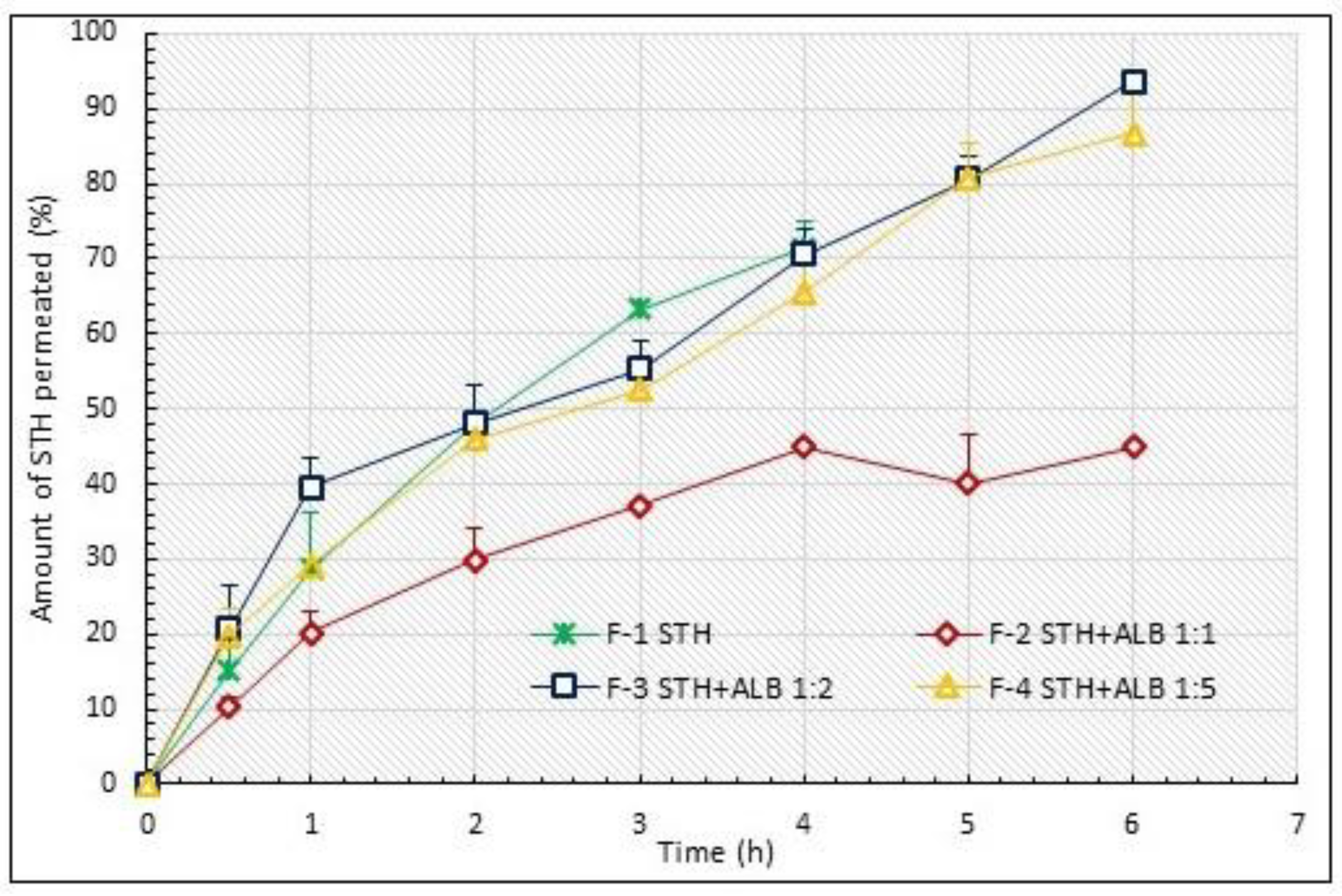
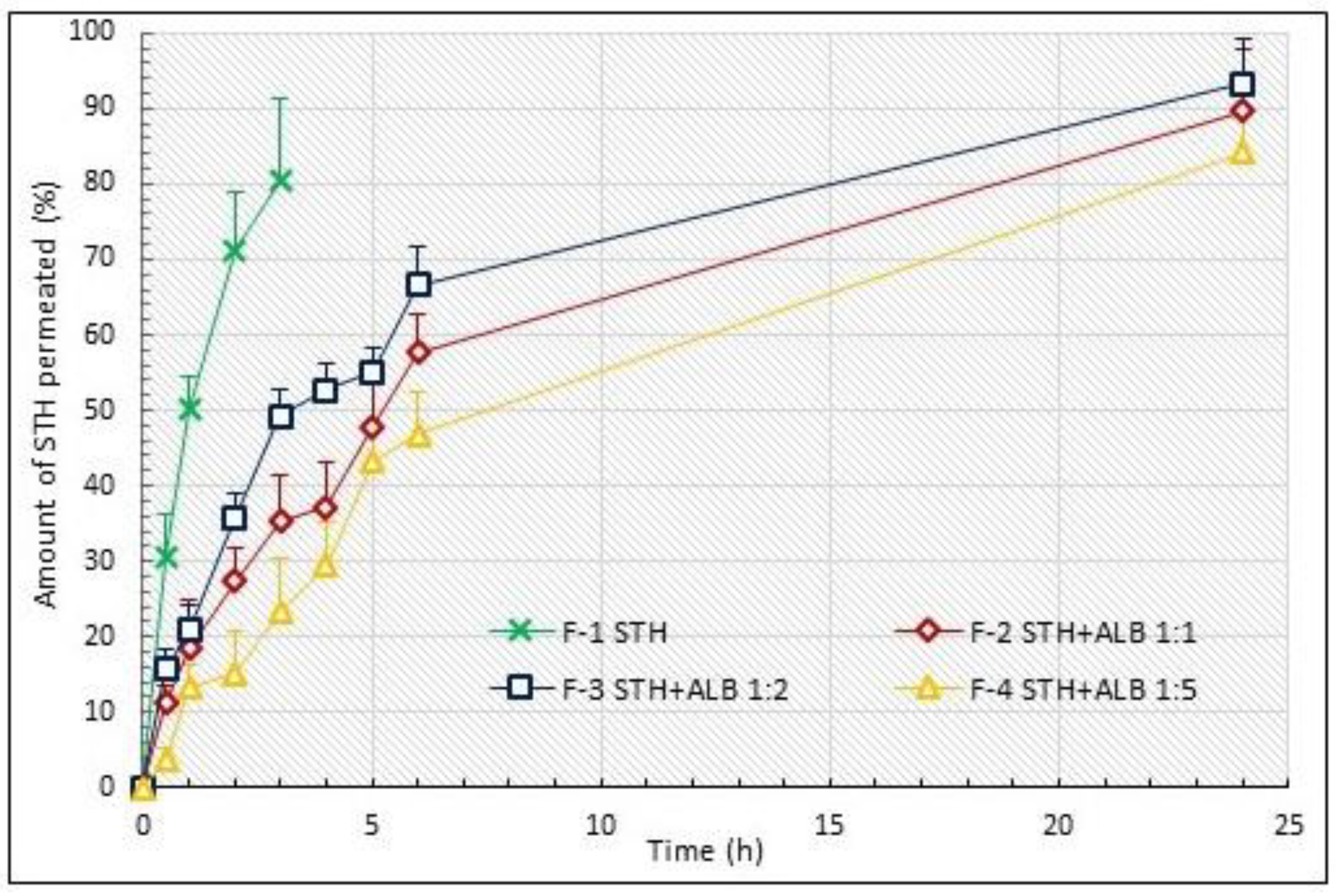
3.1. STH Permeation Study

Initially, the permeation process of STH was examined using Spectra/Por® Dialysis Membrane, a cellulose membrane with a pore size of 100 kDa. The study was conducted for 6 h. ALB-free STH was found to penetrate at t = 4 h. From all ALB-added formulations, STH permeated throughout the time that the permeation test was conducted. No decrease in the amount of STH in the acceptor fluid was noted after this time. The permeation process of STH from the prepared MC-based hydrogels through the artificial membrane, Spectra/Por® Dialysis Membrane with a pore size of 100 kDa, is shown in Figure 1.
The process of the penetration of STH via porcine skin was tested for 24 h. The test conditions using a natural membrane were intended to resemble simulated in vivo conditions. The permeation process of STH via porcine skin from prepared hydrogels is shown in Figure 2.

3.2. Permeation Parameters of STH

The permeation profiles of STH with added ALB at three ratios: 1:1, 1:2 and 1:5 (F-2–F-4), were compared with the permeation from the STH formulation without ALB, and the ALB formulations alone were compared with each other. The degree of STH permeation, AUC values and relative availability are shown in Table 2.
In a study of the permeation process of STH through a cellulose membrane, the most STH permeated from the F-3 formulation containing STH in a 1:2 ratio to ALB (93.59 ± 0.99%), which was 23.5% more than from the formulation containing only STH (71.44 ± 3.43%). The least STH permeated the formulation containing STH in a 1:1 ratio to ALB (44.96 ± 0.1%), which was 37% less than the formulation containing STH without ALB. The largest area under the permeation curve, AUC(0–6 h), was characterized by formulation F-3, containing STH and ALB in a 1:2 ratio, and the smallest by formulation F-1, containing STH without ALB, indicating a significant effect of ALB on STH availability.
The examination of the permeation process of STH through porcine skin confirmed that ALB significantly prolongs the permeation process of STH to at least 24 h, whereas from the formulation containing STH alone (F-1), the entire amount of STH permeated in 3 h. A prolongation of the STH permeation time was evident when ALB was added to the formulations at all ratios. In the porcine skin permeation study, the highest amount of STH also permeated from formulation F-3, with an STH to ALB ratio of 1:2 (93.22 ± 6.83%), and this was approximately 14% higher than from the formulation without added ALB (F-1)—80.37 ± 10.78%, from which STH permeated in the lowest amount. Slightly less STH than from the F-3 formulation permeated the other ALB formulations: from F-2, with a STH-ALB ratio of 1:1, approximately 4% less STH permeated (89.57 ± 7.83%), and from the F-4 formulation, with a STH-ALB ratio of 1:5, nearly 10% less STH permeated (84.11 ± 5.38). The largest area AUC(0–24 h) was characterized by the F-3 formulation, and the smallest by the F-1 formulation, which does not contain ALB, confirming the effect of ALB in increasing STH availability (Table 2).

3.3. Permeation Kinetics

To define the kinetics of the permeation process, a kinetic model was then fitted based on the amount of STH permeated (at 6 h for cellulose membrane and 24 h for porcine skin) (Table 3).
The STH permeation process was assumed to follow first-order kinetics. To calculate the rate constant of the STH permeation process, a formula based on the first-order kinetics model was used (R2 = 0.9768 for the cellulose membrane and R2 = 0.9812 for the skin). In a test of STH permeation through the cellulose membrane, STH permeated most rapidly from formulation F-3, containing STH and ALB in a 1:2 ratio. ALB increased the permeation rate with respect to the formulation without ALB by 1.4-fold. For the F-4 formulation, it only slightly increased the permeation rate (1.1-fold). In contrast, ALB added in a 1:1 ratio to STH reduced the permeation rate by three-fold.
In the STH skin permeation test, STH permeated most rapidly from the formulation without ALB (F-1), while the addition of ALB reduced the permeation rate in all formulations: most significantly in formulation F-4 by approximately 8-fold, in F-2 by 5.5-fold and in formulation F-3 by 4.5-fold. ALB increased the half-life of STH permeation. When tested over 24 h, the longest t50% = 9.10 ± 0.83 (h) was observed for the formulation with the highest ALB content (0.5%), while the shortest half-life was observed for the STH-containing hydrogel without ALB, which was t50% = 1.17 ± 0.16 (h).

3.4. Evaluation of the Similarity of the STH Permeation Profiles

The release profiles of the tested formulations were compared (Table 4). No similarity was found between the compared profiles. The release profiles were assumed to be similar when F-1 was in the range of 0–15 and F-2 was in the range of 50–100 [35].

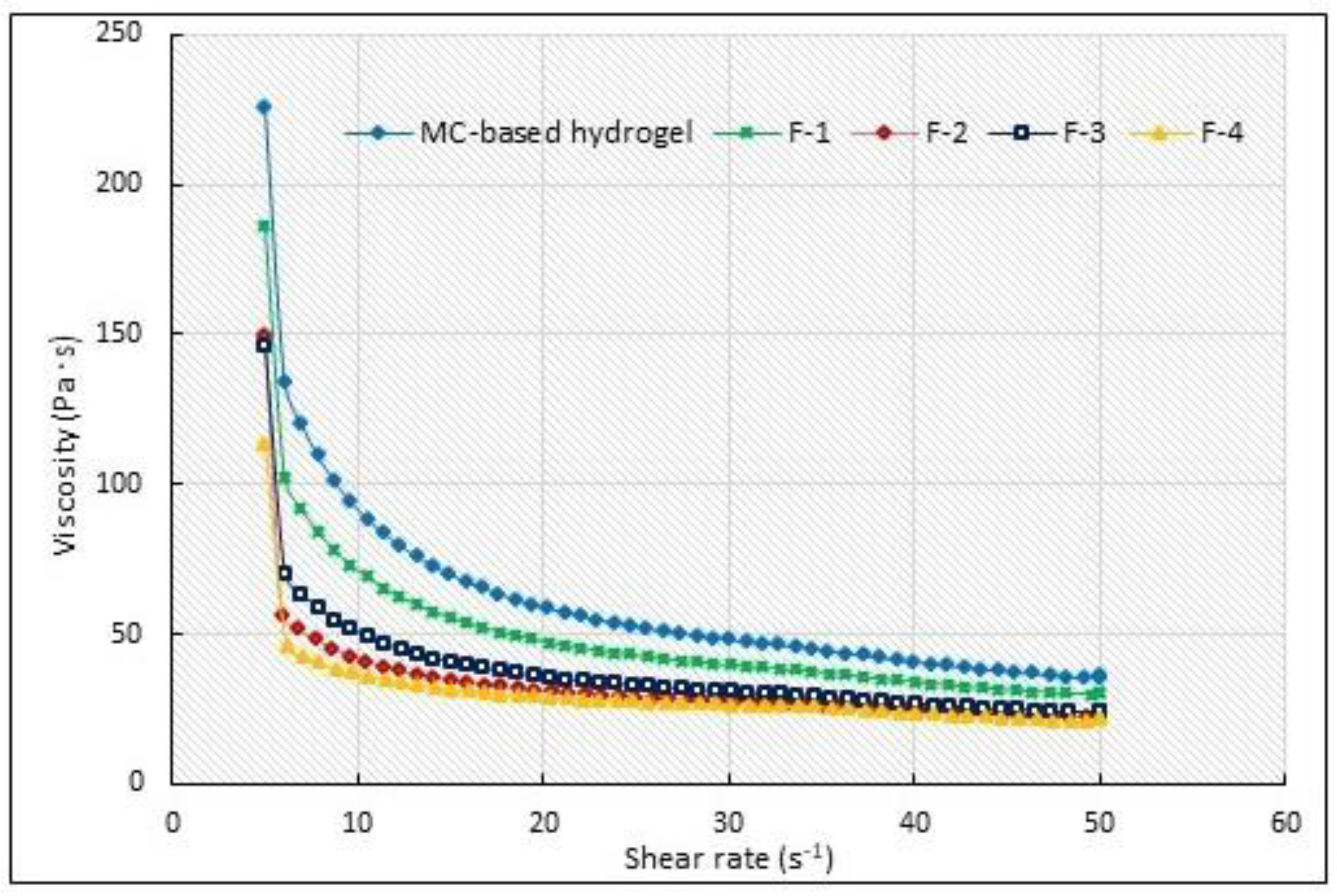
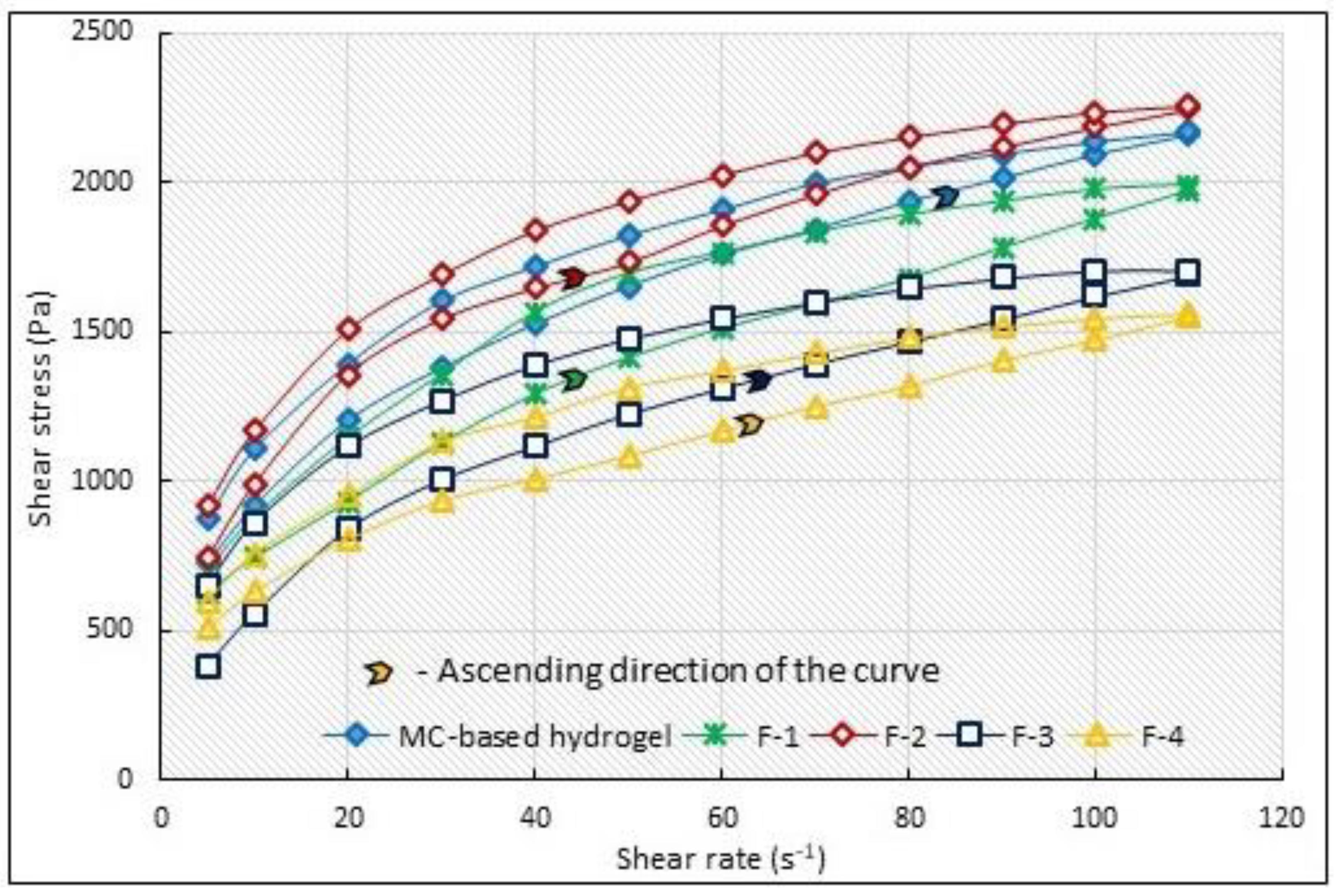
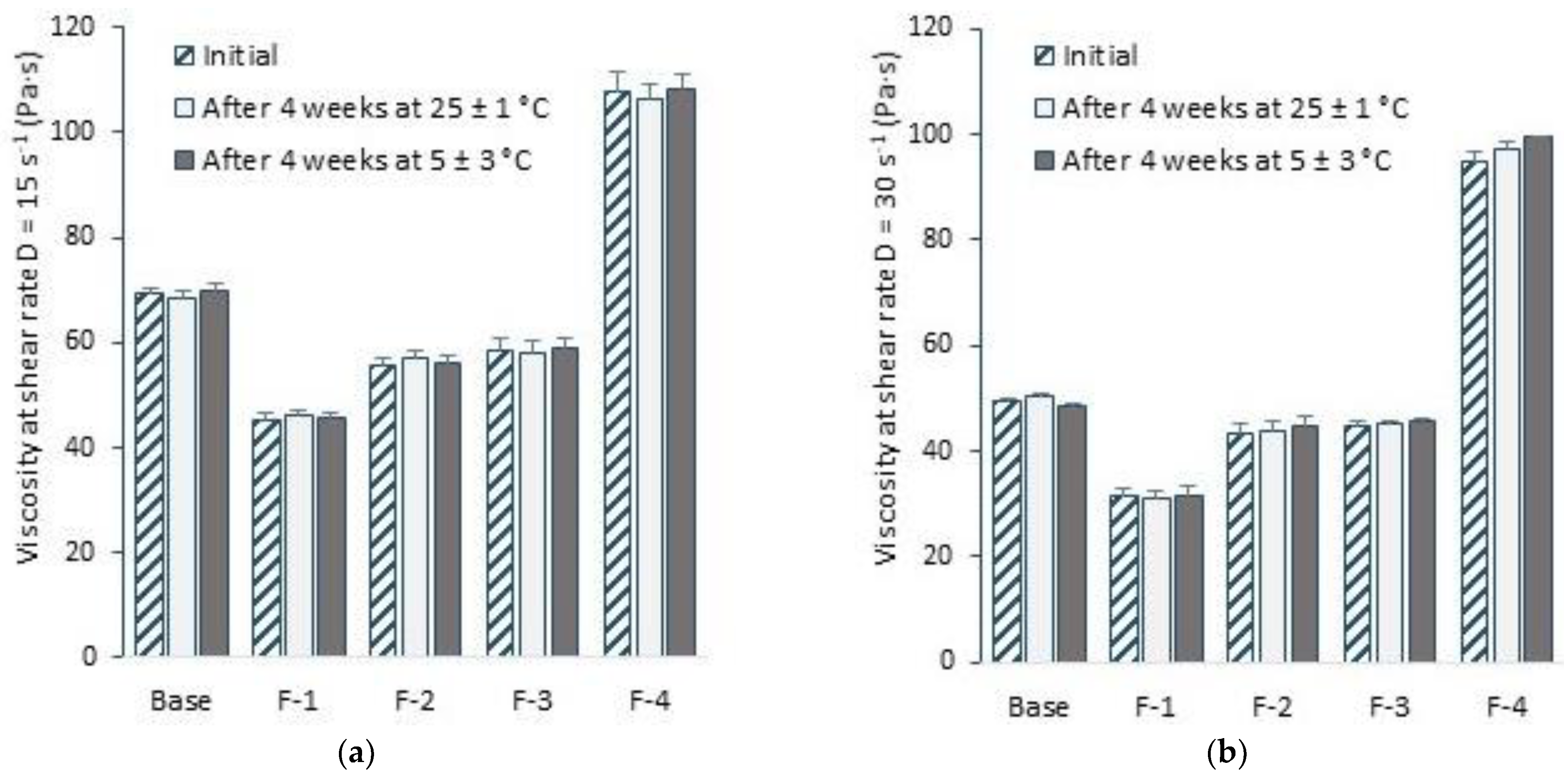
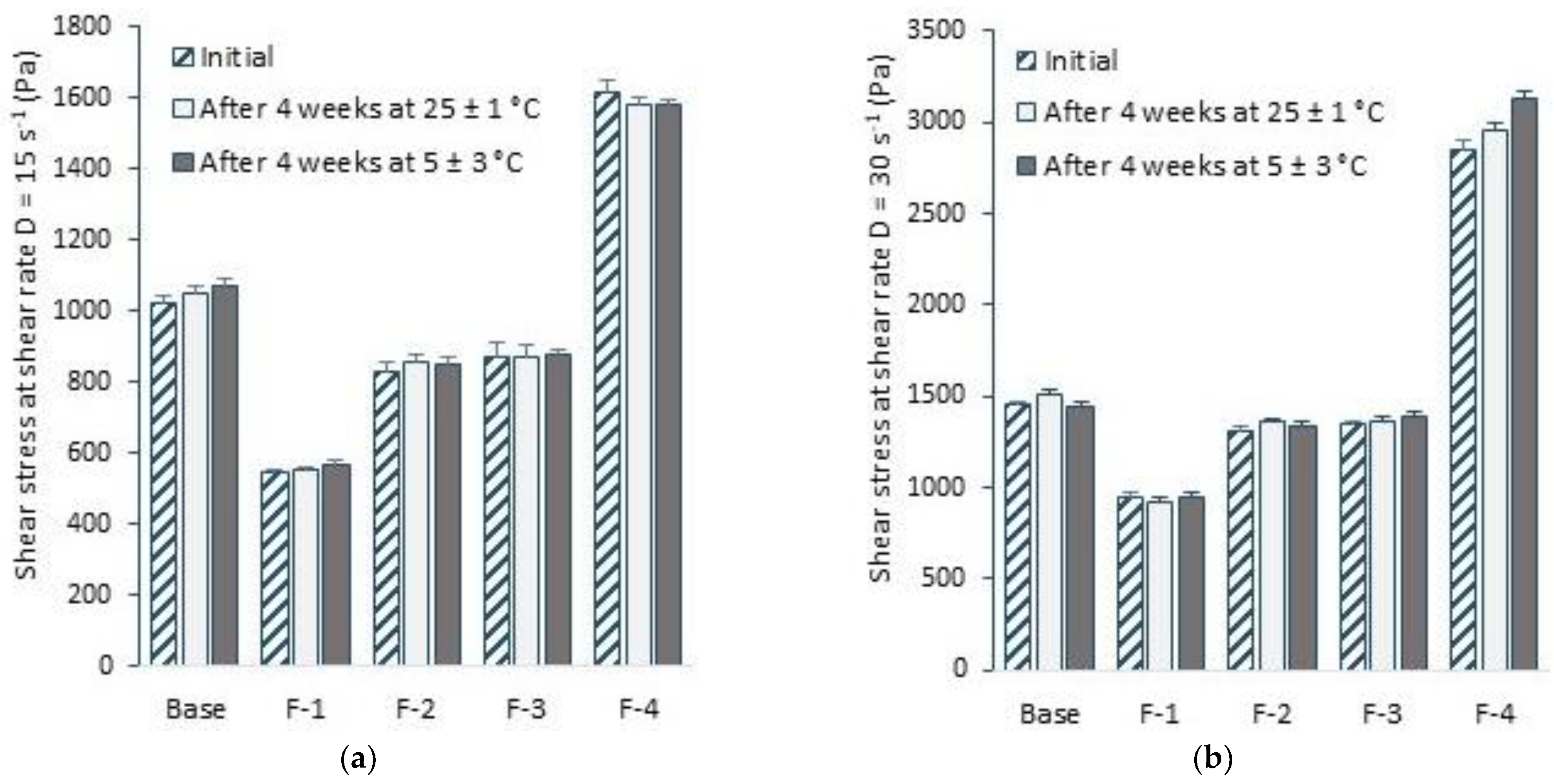
3.5. Selected Rheological Properties

Table 5 shows the results of a constant shear rate viscosity test carried out for two shear rates: D = 15 s−1 and D = 30 s−1.
STH caused a decrease in viscosity compared to the MC-based hydrogel alone at both shear rates. ALB, on the other hand, increased the viscosity of hydrogels with STH to an extent that depended on the amount of ALB added. The higher the amount of ALB, the higher the viscosity of the formulations, although the formulations with a concentration of ALB of 0.1% and 0.2% still had a lower viscosity than the unloaded hydrogel. ALB at a concentration of 0.5% significantly increased the viscosity of the F-4 formulation, which correlates with a significant increase in permeation time. The lower the viscosity, the faster the STH permeated from the hydrogel formulations through the porcine skin (lowest viscosity in formulation F-1: 45.41 ± 1.19, at D = 15 s−1 and 31.25 ± 1.62 at D = 30 s−1, and highest in F-4: 107.70 ± 3.47 at D = 15 s−1 and 95.00 ± 1.74 at D = 30 s−1). Despite the differences in the viscosity of the protein-loaded formulations, they showed better spreading on the skin compared to the unloaded substrate, as observed when applying the formulation samples to the skin surface in the STH skin permeation test. The addition of ALB has a beneficial effect on the form of the hydrogel for application to the skin. The hydrogel will not flow, allowing for a longer contact time with the skin.
Based on the flow curve (shear stress vs. shear rate relationship) in the shear rate range D = 5–50 s−1, viscosity curves were determined (Figure 3). As can be seen from Figure 3, with increasing the shear rate, the viscosity of the obtained hydrogels decreases, the system being shear-thinning.
The classic method for assessing thixotropic properties is to perform a hysteresis loop test. A step-by-step flow curve in the form of a hysteresis loop using the step-by-step test is presented in Figure 4. This technique involves measuring the shear stress of the sample under increasing shear rate conditions, until a maximum value is reached, and then at a decreasing shear rate. If the ‘up’ curve does not coincide with the ‘down’ curve, this is an indication of rheological instability. When the ‘up’ curve lies under the ‘down’ curve, this indicates rheopexy. Comparison of the melt curve, plotted at an increasing shear rate, with the curve at a decreasing shear rate, allows the extent of destruction or internal structure of the system to be determined.
The hysteresis areas of the examined formulations at T = 32 °C were as follows: MC-hydrogel base 7445.11 Pa/s, F-1 19288.89 Pa/s, F-2 12598.38 Pa/s, F-3 19310.05 Pa/s and F-4 14945.83 Pa/s. The smallest area characterized the MC-based substrate, the largest—2.6 times the area of the substrate—characterized the F-3 formulation with ALB added at an STH-ALB ratio of 1:2, and F-1, containing STH without ALB added. The hysteresis area is a measure of the breakdown of the internal hydrogel structure. It is suggested that a larger hysteresis loop area increases the penetration of active substances through the stratum corneum. During topical application, formulations deform and become more fluid, which promotes the diffusion of active substances [36,37].

3.6. Sensory Evaluation

All tested formulations subjected to initial observations showed similarity in color, odor and appearance, compared to the control formulation, regardless of storage conditions. Sensory evaluation parameters are summarized in Table 6.

3.7. Stability Test

All examined formulations maintained their initial pH (Table 7), and viscosity and shear stress values under varying storage conditions (Figure 5 and Figure 6). All prepared formulations were found to be stable. The API content was within the acceptable limit of 90% of the initial value.
The pH values of all the obtained formulations are acidic. The addition of the protein substances STH and ALB results in a statistically significant increase in pH: from 4.58 ± 0.043 (pH of the substrate alone) to 5.52 ± 0.090 for the hydrogel containing STH 1 mg/g, and to 5.91 ± 0.026 for the hydrogel with STH and the highest ALB content (0.5%). These values remain within the physiological pH of the skin, and their reaction remains acidic, so they can be safely applied to the skin without the risk of dryness and hypersensitivity.

4. Discussion

The transdermal delivery of therapeutic substances has many advantages over conventional paths of administration. However, the penetration of biological molecules through the skin layers is limited by their dimensions and characteristics. The enhancers of the transdermal delivery of therapeutic peptides and proteins are the subject of ongoing testing [38,39]. While overcoming the living layers of the skin, peptide drugs can undergo enzymatic degradation [40,41,42]. However, as a consequence of quick uptake into the circulatory system after reaching the highly vascularized papillary skin zone, the metabolism of peptides/proteins most likely occurs while passing through the epidermal layer of the skin [43]. However, that route of administration appears favorable due to the prevention of enzymatic degradation in the digestive tract and liver. There are penetration enhancement techniques that can also affect enzyme activity, as described for ultrasound, which can contribute to the deactivation of certain skin enzymes [44,45]. Encapsulation may provide some protection against skin enzymes, although the exact moment at which the drug leaves its “protective capsule” as it passes through the tissue has yet to be defined [38,39].
There are many reports that ALB binds to the drug molecule and has a protective effect on it. Binding to plasma proteins prevents substances from being oxidized, decreases their toxicity and elongates their half-life, and drugs which are tightly associated with plasma proteins tend to have a low first-pass effect [46].
The biologically active glucagon-like peptide-1, GLP-1 (7–37), has a half-life of 1.5 to 2 min. Liraglutide is an ALB-bond form of GLP-1 and is modified with myristic acid. Unlike GLP-1, liraglutide is stable to metabolic degradation and has a plasma half-life of 11–15 h after subcutaneous administration, allowing it to be administered once daily [47]. When administered transdermally, ALB nanoparticles could, however, bypass the stratum corneum barrier for delivery through the hair follicle. Furthermore, the nanoparticles could be kept in the hair follicles for a sufficiently long period. This would allow systemic absorption and topical activity, and possibly provide a prolonged release effect [15,48].
Hydrogels using cellulose derivatives as hormone carriers (testosterone, proges-terone, insulin, estradiol) are known to be used [49]. MC, sodium carboxymethylcellulose (CMC) and hydroxypropylmethylcellulose (HPMC) have been used to obtain hydrogels. The efficacy of using HPMC as a testosterone carrier was reported by Heo et al. [50] In a study on designing a hydrogel matrix for insulin, Ostróżka-Cieślik et al. [51] studied the usefulness of Carbopol Ultrez 10, Carbopol Ultrez 30, MC and glycerol hydrogel. They observed that the largest amount of insulin was released from the MC-based gel—75%. The hormone was released sequentially for up to nine hours. The MC hydrogel showed very favorable properties, enabling its spreadability on the skin [50]. Siemiradzka et al. confirmed the effectiveness of MC in an ALB permeation test. The largest amount of ALB permeated via porcine skin from MC-based hydrogel—35.6 percent within 24 h. The use of MC with chitosan as a matrix for ALB reduced the amount of permeated ALB from the hydrogel by 1.7-fold; sodium alginate reduced it by 2.5-fold, while HPMC caused 3-fold less ALB to permeate [29].
Şenyiğit et al. used chitosan and bovine serum ALB to prepare nanogels containing mupirocin (MPR). Both the ALB and the mucilaginous ability of chitosan could prolong the duration of Col/haNPs presence at the destination, favoring a longer drug release. However, to improve the chitosan gelation (adhesion) properties, Carbopol 940 had to be added at concentration 2% and 3%. The addition of MPR to the nanogel formulation resulted in a reduction in viscosity and, inversely, viscosity values increased together with Carbopol 940 concentration [52].
In this study, the prolonged permeation time of STH up to 24 h in the present test confirms the effect of ALB on the length of this process. STH penetrated from the MC hydrogel in 3 h through porcine skin from the formulation without ALB addition. The addition of ALB, added in each amount used in the formulations produced, prolonged the permeation process. At the same time, ALB at a concentration of 0.2% (STH-ALB 1:1) increased the amount of STH permeated by 14%.
When testing the process of STH permeation through the cellulose membrane, a strong kinetics match was observed for all three models used. In the test of STH permeation through porcine skin, a slightly better fit was recorded for the first-order model, while the Higuchi and Korsmayer–Peppas models showed a very similar fit. This effect may result from the complexity of the hydrogel system—degradation mechanism, pore size, and the feature of drug substance. These models are dedicated to specific systems, have different assumptions and differ from each other.
Due to the slightly higher values of the regression coefficients and the results obtained in previous tests of peptide substances, the permeation rate was determined based on the first-order model. Drug release and subsequent skin permeation, which followed first-order kinetics, is described by drug concentrations obtained from an in vitro dissolution test plotted against time—ln % of remaining drug to time. This relationship can refer to drug forms containing water-soluble substances encapsulated in a porous matrix, such as MC-based hydrogel. The average regression coefficients’ R2 was 0.9812.
In a test of STH permeation through the skin, the regression coefficients for the Higuchi and Korsmayer–Peppas models were lower than those of the first-order model.
The kinetics of the process of release and permeation of therapeutic substances vary greatly. This is influenced by many factors responsible for the form of the drug: the physicochemical nature of the excipients and therapeutic substances, as well as the drug form itself and its properties. The hydrogel used for the preparation of formulations with STH and ALB is hydrophilic in nature, and the therapeutic substances are water-soluble peptides, which can affect the process of release—especially their viscosity, swelling ability, as well as the ability of ALB to transport other substances and prolong their t50%. Due to the heterogeneous process of the degradation of polymeric matrices, characterized by different pore sizes, a differential fit of the kinetic models of the process of therapeutic substances’ release and permeation is observed [53,54,55]. Unambiguous interpretation of the course of these processes is not entirely possible. The kinetics of active ingredient permeation from hydrogels is an extremely difficult issue, and there is no clear interpretation.
The effect of ACTH amount on its penetration degree from topical formulations has been reported. It was found that the higher ACTH concentration, the lower the degree of its penetration through the skin [22].
Another study by Siemiradzka et al. examined the effect of another protein, ALB, on the permeation of ACTH from topical hydrogels for skin application. They observed that both the concentration of ALB and the amount of ALB in relation to ACTH were significant. The 2% of ALB did not increase the amount of ACTH released over 24 h. A greater effect was observed at lower concentrations, i.e., 1.5% of ALB and 1.5% of ACTH (1:1). The degree of ACTH penetration was approximately 2.7 times higher (40.76 ± 3.51%). ALB also increased the rate of ACTH permeation by approximately 1.4-fold (p < 0.05) [12].
The results of the permeation process through the artificial and natural membrane may indicate the influence of the membrane on the transport of active substances. In the present study, STH penetrated the porcine skin to a greater extent. From the formulation containing STH without ALB, 1.1 times more STH permeated through the porcine skin, from the formulation containing STH and ALB in a ratio of 1:1 to 2 times more, while with increasing the amount of ALB in formulation F-3 the amount of STH was very similar, and in formulation F-4 the amount of STH permeated decreased slightly. The size of the pores in the membrane changes the permeation process, and the skin has more restrictions. Sometimes, however, natural skin can be more permeable to molecules of therapeutic substances, which is mostly determined by the characteristics of the molecule (size, shape, pH, hydrophilic–lipophilic nature, concentration). In a test by Siemiradzka et al., a 2.3-fold increase in permeation through a natural membrane compared to an artificial cellulose membrane was observed for a hydrogel with ACTH without ALB addition, and for a hydrogel with ALB addition in a 1:1 ratio [12].
Examination of the selected physicochemical properties of the produced hydrogel formulations allowed an assessment of their suitability in terms of application of the formulations to the skin. Time-dependent viscosity variation is a desirable characteristic in pharmaceutical forms due to the requirement for elasticity in drug supply. In contrast, the rebuilding of the structure and the accompanying increase in viscosity can prevent sedimentation of the preparation particles, and the increasing viscosity over time also prevents the preparation from running off the skin’s surface. At high shear stresses, partial or complete destruction of the hydrogel structure occurs very fast. Much more prolonged, and thus more significant because of the durability of the topical formulation, is the rebuilding of the structure of the viscoplastic system. The area bounded by the curves forming the hysteresis loop is considered to be a measure of the thixotropic/rheopexy characteristics of the system. STH at a concentration of 0.1% has been shown to decrease the viscosity of an MC-based hydrogel. In doing so, easier spreading of the formulation was noted. The addition of ALB at concentrations of 0.1% and 0.2% slightly increased the viscosity, but the value was still lower than that of the unloaded substrate. ALB in the amount of 0.5 mg/g significantly enhanced the viscosity value, which translated into the lowest permeation rate. The relatively narrow space between the ascending and descending curves indicates satisfactory recovery of the formulation, which is conducive to increasing adhesion to the skin, thus providing a better treatment outcome.
Glycerol hydrogel was similarly affected by ACTH incorporated into the hydrogel matrix, which increased viscosity with increasing ACTH concentration, whereas the addition of ALB at a concentration of 1% significantly decreased viscosity, and at a concentration of 0.01% virtually produced no change in viscosity compared to the unloaded substrate [12].
A test of ALB permeation depending on the substrate and concentration used found that ALB at a concentration of 2% did not change the viscosity of sodium alginate, increased the viscosity of HPMC, decreased the viscosity of MC and slightly decreased that of MC with chitosan (CH), while it did not change the viscosity at lower concentrations: 1% and 1.5% of the latter hydrogel (CH-MC) [29].
The effect of protein concentration on viscosity is not always predictable. It depends largely on the nature and properties of the polymer matrix, but also on other substances, such as sugars and polyols, which are added to fix the tertiary structure of the protein [56,57]. Developments in the field of protein therapeutics require counteracting the difficulties accompanying the processing of protein solutions, which are aggregation and viscosity. Proteins delivered to the body require high concentrations, which promotes the increase in viscosity of these solutions. The viscosity of protein solutions is due to interactions between protein molecules: electrostatic repulsion, exclusion volume effect by steric repulsion or attractive interactions. Electrostatic repulsion reduces the mobility of the molecule, which results in the increased viscosity of the solution. In practice, this can be prevented by introducing small-molecule substances, such as arginine hydrochloride (ArgHCl) [56], so that the preparation is easily applied and fulfills its therapeutic function.
Sensory evaluation of the adhesion, elasticity and consistency of the obtained hydrogels confirmed the favorable effect of STH and ALB in concentrations of 0.1% and 0.2% on the mentioned parameters. The examined formulations showed stability during storage under specified temperature conditions for a period of 4 weeks. The ingredients that were used to prepare the hydrogels do not exhibit irritant effects, so they should not disturb the lipid mantle of the skin, nor will they damage the skin. They offer the possibility of application to hairy and hairless skin. They can be used with concomitant exudative lesions. The pH values of the formulations obtained remained within the physiological pH of the skin, and their reaction remained acidic, so they can be safely applied to the skin without the risk of dryness and hypersensitivity. The use of MC and glycerol to prepare the base for the semi-solid drug formulation turned out to be advantageous for a variety of reasons: simple and inexpensive preparation technology, high yield, high biocompatibility, low sensitizing potential, and an aesthetic and modern form of application. The high-water content and hydration effect on the skin promote the increased transport of active ingredients through the skin. They also show high stability of the physical form.
All hydrogels were formulated on the basis of hydrophilic polymer MC at a concentration 6%. After the addition of protein substances, STH, STH and ALB showed favorable properties based on the evaluation of such parameters as uniformity, consistency, pillow effect, adhesion, stickiness, greasiness and greasing, compared to a clean base. The formulations were characterized by greater spreadability, softer consistency and lower viscosity. Only the F-4 formulation, containing STH 0.1% and ALB 0.5%, showed similar viscosity to the substrate itself; however, the consistency remained soft, and the formulation was very easily spreadable. A small pillow effect, no greasy feeling left behind, adequate stickiness and adhesion make such formulations readily used by patients. However, sensory parameters also affect the stability of hydrogels and the therapeutic substance. Uniformity allows for uniform distribution of the active substance and the expected therapeutic effect. The evaluation of physical properties provided important, useful information for predicting the sensory qualities of topical products and can lead to the design of products that meet patient expectations and increase acceptability. The work of Savary et al. presented an objective method for characterizing the texture properties of topical products applied to the skin. Firmness, viscosity, spreadability and amount of residue were considered, as these characteristics play a key role in the design of topical products. Sensory tests showed a wide variety of texture properties of the selected topical preparations, and the testing of rheological and mechanical properties showed perceptual differences between the samples. Firmness and stickiness were related to the mechanical response and flow behavior of the systems during deformation. Spreadability depended on the presence or not of a hydrophilic polymer. Gels, unlike emulsions, showed less noticeable residual films after application in terms of quantity and tactile friction properties [58].
Storage period and stability are important issues in evaluating the suitability of topical drug delivery systems. In the case of hydrophilic gels, contact with the temperature of the body surface, the pH of the skin, as well as changes under atmospheric conditions, are important in determining the stability of the introduced drug substances and the safety and efficacy of the drug product. Such tests are important in selecting the appropriate formulation and packaging, as well as proper storage conditions or shelf life and use.
When hydrogels are applied to the skin, only the active ingredient penetrates the skin. The temperature at the skin surface is approximately 32 °C, and the prepared formulations with STH were stored at 5 ± 3 °C and 25 ± 1 °C for 28 days and were stable. Ostróżka et al. prepared insulin-containing hydrogels based on MC for skin application and demonstrated the stability of these formulations under the same storage conditions (time and temperature) [51,59].
In a test conducted by Siddig et al., the visual appearance of nine formulations prepared at three concentrations (1%, 5% and 10%) containing MC, Carbomer B and white petrolatum was evaluated. The preparations appeared transparent and uniform in consistency and showed no noticeable change in transparency, color or odor. A microscopic evaluation of the formulations for particulate matter showed no significant particles visible under the microscope. The slides met the physical stability requirement and the requirement for the absence of particulate matter and granularity, which is desirable for topical preparations [59].
Siddig et al. tested the stability of Piperacillin formulations at 1% and 10% concentrations, which were prepared using MC as a polymer matrix with concentrations ranging from 0.5 to 5%. Hydrogels with MC were stored in stability chambers for 12 weeks at three temperatures: 25 °C—ambient temperature, 37 °C body temperature and possibly an extreme ambient temperature of 45 °C. The effect of skin pH at different storage and use temperatures on drug stability was tested at pH 5 and 7. MC hydrogels, unlike complex CMC, are not sensitive to pH changes due to the absence of carboxylate groups. MC-based hydrogels were selected for physical and chemical degradation tests as formulations with better physicochemical properties and better drug permeation and release characteristics. The authors found that changes in pH from 7 to 5 for MC-based hydrogels (all concentrations) do not affect the viscosity of this base, and the change in temperature does not affect the stability of this hydrogel substrate. In the case of gels with 10% piperacillin, there was no statistically significant difference in drug stability at these temperatures for this formulation. The authors concluded that increased drug loading in MC gels confirmed the significant stability of the drug substance [59].

5. Conclusions

In this study, the permeation of STH from semi-solid hydrogels for skin application across an artificial cellulose membrane was traced in vitro. Following this, a permeation test was conducted under simulated in vivo conditions using porcine skin. It was shown that STH loaded into the polymeric network of hydrogels intended for the skin can act systemically in hormone supplementation. The availability of this hormone was more than 80%. The addition of ALB as a transport protein for STH resulted in an elongation of the penetration time through pig skin to at least 24 h. ALB, depending on its concentration, could increase the availability of STH by up to 93%. At the same time, it caused an increase in the half-life of permeation.

Author Contributions

Conceptualization, W.S., B.D. and A.F.; methodology, W.S. and A.F.; software, W.S. and A.F.; validation, W.S., A.F. and L.B.; formal analysis, W.S.; investigation, W.S. and A.F.; resources, B.D., L.B. and W.S.; data curation, W.S., A.F. and L.B.; writing—original draft preparation, W.S. and A.F.; writing—review and editing, W.S. and A.F.; visualization, W.S.; supervision, W.S.; project administration, B.D., L.B. and W.S.; funding acquisition, B.D., L.B. and W.S. All authors have read and agreed to the published version of the manuscript.

Funding

This research was funded by the Medical University of Silesia in Katowice, grant number PCN-1-056/K/2/F.

Institutional Review Board Statement

Not applicable.

Informed Consent Statement

Not applicable.

Data Availability Statement

Not applicable.

Conflicts of Interest

The authors declare no conflict of interest.

References

  1. Kato, Y.; Murakami, Y.; Sohmiya, M.; Nishiki, M. Regulation of human growth hormone secretion and its disorders. Intern. Med. 2002, 41, 7–13. [Google Scholar] [CrossRef] [PubMed] [Green Version]
  2. Ashpole, N.M.; Sanders, J.E.; Hodges, E.L.; Yan, H.; Sonntag, W.E. Growth hormone, insulin-like growth factor-1 and the aging brain. Exp. Gerontol. 2015, 68, 76–81. [Google Scholar] [CrossRef] [Green Version]
  3. Korcz-Iżykowska, M.; Majewska, K.; Kędzia, A. Effects of growth hormone and recombinant human growth hormone on skin homeostasis. Endokrynol. Ped. 2018, 4, 245–250. [Google Scholar]
  4. Strous, G.J.; Almeida, A.D.S.; Putters, J.; Schantl, J.; Sedek, M.; Slotman, J.A.; Nespital, T.; Hassink, G.C.; Mol, J.A. Growth hormone receptor regulation in cancer and chronic diseases. Front. Endocrinol. 2020, 11, 597573. [Google Scholar] [CrossRef] [PubMed]
  5. Siebert, D.M.; Rao, A.L. The use and abuse of human growth hormone in sports. Sports Health 2018, 10, 419–426. [Google Scholar] [CrossRef]
  6. Romer, T. Growth hormone. Production and secretion. In Treatment with Hormones and Hormone Derivatives, 2nd ed.; Pawlikowski, M., Ed.; Medical Publishing House: Warsaw, Poland, 1996; p. 46. [Google Scholar]
  7. Boguszewski, M.C.S.; Cardoso-Demartini, A.A.; Boguszewski, C.L.; Chemaitilly, W.; Higham, C.E.; Johannsson, G.; Yuen, K.C.J. Safety of growth hormone (GH) treatment in GH deficient children and adults treated for cancer and non-malignant intracranial tumors-a review of research and clinical practice. Pituitary 2021, 24, 810–827. [Google Scholar] [CrossRef]
  8. Harvey, S.; Martinez-Moreno, C.G. Growth hormone: Therapeutic possibilities—An overview. Int. J. Mol. Sci. 2018, 19, 2015. [Google Scholar] [CrossRef] [Green Version]
  9. Raoufinia, R.; Mota, A.; Keyhanvar, N.; Safari, F.; Shamekhi, S.; Abdolalizadeh, J. Overview of albumin and its purification methods. Adv Pharm Bull. 2016, 6, 495–507. [Google Scholar] [CrossRef] [Green Version]
  10. Steglich, M.; Lombide, R.; López, I.; Portela, M.; Fló, M.; Marín, M.; Alvarez, B.; Turell, L. Expression, purification and initial characterization of human serum albumin domain I and its cysteine 34. PLoS ONE 2020, 15, e0240580. [Google Scholar] [CrossRef]
  11. Zaman, R.; Islam, R.A.; Ibnat, N.; Othman, I.; Zaini, A.; Lee, C.Y.; Chowdhury, E.H. Current strategies in extending half-lives of therapeutic proteins. J. Control. Release 2019, 301, 176–189. [Google Scholar] [CrossRef]
  12. Siemiradzka, W.; Dolińska, B.; Ryszka, F. Modelling and control of corticotropin permeation from hydrogels across a natural membrane in the presence of albumin. Processes 2021, 9, 1674. [Google Scholar] [CrossRef]
  13. Jepps, O.G.; Dancik, Y.; Anissimov, Y.G.; Roberts, M.S. Modeling the human skin barier—towards a better understanding of dermal absorption. Adv. Drug Deliv. Rev. 2013, 65, 152–168. [Google Scholar] [CrossRef] [PubMed]
  14. Kontermann, R.E. Half-life extended biotherapeutics. Expert Opin. Biol. Ther. 2016, 16, 903–915. [Google Scholar] [CrossRef] [PubMed]
  15. Lademann, J.; Richter, H.; Teichmann, A.; Otberg, N.; Blume-Peytavi, U.; Luengo, J.; Sterry, W. Nanoparticles–an efficient carrier for drug delivery into the hair follicles. Eur. J. Pharm. Biopharm. 2007, 66, 159–164. [Google Scholar] [CrossRef]
  16. Baroni, A.; Buommino, E.; De Gregorio, V.; Ruocco, E.; Ruocco, V.; Wolf, R. Structure and function of the epidermis related to barrier properties. Clin. Dermatol. 2012, 30, 257–262. [Google Scholar] [CrossRef]
  17. Marwah, H.; Garg, T.; Goyal, A.K.; Rath, G. Permeation enhancer strategies in transdermal drug delivery. Drug Deliv. 2016, 23, 564–578. [Google Scholar] [CrossRef]
  18. N’Da, D.D. Prodrug strategies for enhancing the percutaneous absorption of drugs. Molecules 2014, 19, 20780–20807. [Google Scholar] [CrossRef] [Green Version]
  19. Bäsler, K.; Bergmann, S.; Heisig, M.; Naegel, A.; Zorn-Kruppa, M.; Brandner, J.M. The role of tight junctions in skin barrier function and dermal absorption. J. Control. Release 2016, 242, 105–118. [Google Scholar] [CrossRef]
  20. Maqbool, A. Semisolid dosage forms manufacturing: Tools, critical process parameters, strategies, optimization, and recent advances. Indo. Am. J. Pharm. Sci. 2017, 7, 882–893. [Google Scholar]
  21. Narayanaswamy, R.; Torchilin, V.P. Hydrogels and their applications in targeted drug delivery. Molecules 2019, 24, 603. [Google Scholar] [CrossRef] [Green Version]
  22. Siemiradzka, W.; Dolińska, B.; Ryszka, F. Influence of concentration on release and permeation process of model peptide substance-corticotropin-from Semisolid Formulations. Molecules 2020, 25, 2767. [Google Scholar] [CrossRef] [PubMed]
  23. Chai, Q.; Jiao, Y.; Yu, X. Hydrogels for biomedical applications: Their characteristics and the mechanisms behind them. Gels 2017, 3, 6. [Google Scholar] [CrossRef] [PubMed] [Green Version]
  24. Lamb, Y.N. Lonapegsomatropin: Pediatric First Approval. Pediatr. Drugs 2022, 24, 83–90. [Google Scholar] [CrossRef] [PubMed]
  25. Yuen, K.C.J.; Miller, B.S.; Boguszewski, C.L.; Hoffman, A.R. Usefulness and potential pitfalls of long-acting growth hormone analogs. Front. Endocrinol. 2021, 12, 637209. [Google Scholar] [CrossRef]
  26. Sravanthi, S.; Kumari, M.M.; Sharma, J.V.C. A critique view on SKYTROFA®. IJRESM 2021, 4, 188–191. [Google Scholar]
  27. USA Food and Drug Administration. SKYTROFA® (Lonapegsomatropin-Tcgd): Highlights of Prescribing Information. 2021. Available online: https://www.accessdata.fda.gov/ (accessed on 12 October 2021).
  28. Maniatis, A.; Nadgir, U.; Hofman, P.; Saenger, P.; Chertok, E.; Aghajanova, E. Lonapegsomatropin (TransCon hGH) in children with growth hormone deficiency: Efficacy and safety of up to 2 years of treatment [poster]. In Proceedings of the PES 2021 virtual annual meeting, virtual, 30 April–3 May 2021. [Google Scholar]
  29. Siemiradzka, W.; Bułaś, L.; Dolińska, B. Permeation of albumin through the skin depending on its concentration and the substrate used in simulated conditions in vivo. Biomed. Pharm. 2022, 155, 113722. [Google Scholar] [CrossRef]
  30. Dash, S.; Murthy, P.N.; Nath, L.; Chowdhury, P. Kinetic modeling on drug release from controlled drug delivery systems. Acta. Pol. Pharm. 2010, 67, 217–223. [Google Scholar]
  31. Shoaib, M.H.; Tazeen, J.; Merchant, H.A.; Yousuf, R.I. Evaluation of drug release kinetics from ibuprofen matrix tablets using HPMC. Pak. J. Pharm. Sci. 2006, 19, 119–124. [Google Scholar]
  32. Zhang, Y.; Huo, M.; Zhou, J.; Zou, A.; Li, W.; Yao, C.; Xie, S. DDSolver: An add-in program for modeling and comparison of drug dissolution profiles. AAPS J. 2010, 12, 263–271. [Google Scholar] [CrossRef] [Green Version]
  33. Lee, I.S.; Yang, H.M.; Kim, J.W.; Maeng, Y.J.; Lee, C.W.; Kang, Y.S.; Rang, M.J.; Kim, H.Y. Terminology development and panel training for sensory evaluation of skin care products including aqua cream. J. Sens. Stud. 2005, 20, 421–433. [Google Scholar] [CrossRef]
  34. Guideline, I.H.T. Quality of biotechnological products: Stability testing of biotechnological/biological products. Annex to the ICH Harmonised Tripartite Guideline for the Stability Testing of New Drug Substances and Products. Dev. Biol. Stand. 1998, 93, 211–219. [Google Scholar]
  35. Al-Tabakha, M.M.; Fahelelbom, K.M.; Obaid, D.E.E.; Sayed, S. Quality attributes and in vitro bioequivalence of different brands of amoxicillin trihydrate tablets. Pharmaceutics 2017, 9, 18. [Google Scholar] [CrossRef] [PubMed] [Green Version]
  36. Lee, C.H.; Moturi, V.; Lee, Y. Thixotropic property in pharmaceutical formulations. J. Control. Release 2009, 136, 88–98. [Google Scholar] [CrossRef] [PubMed]
  37. Carvalho, F.C.; Calixto, G.; Hatakeyama, I.N.; Luz, G.M.; Gremião, M.P.; Chorilli, M. Rheological, mechanical, and bioadhesive behavior of hydrogels to optimize skin delivery systems. Drug Dev. Ind. Pharm. 2013, 39, 1750–1757. [Google Scholar] [CrossRef]
  38. Cázares-Delgadillo, J.; Ganem-Rondero, A.; Kalia, Y.N. Human growth hormone: New delivery systems, alternative routes of administration, and their pharmacological relevance. Eur. J. Pharm. Biopharm. 2011, 78, 278–288. [Google Scholar] [CrossRef]
  39. Schuetz, Y.B.; Naik, A.; Guy, R.H.; Kalia, Y.N. Emerging strategies for the transdermal delivery of peptide and protein drugs. Expert Opin. Drug Deliv. 2005, 2, 533–548. [Google Scholar] [CrossRef]
  40. Flynn, G.L.; Amidon, G.L.; Choi, H.K. Transdermal delivery of bioactive peptides: The effect of N-decylmethyl sulfoxide, pH, and inhibitors on enkephalin metabolism and transport. Pharm. Res. 1990, 7, 1099–1106. [Google Scholar]
  41. Banerjee, P.S.; Ritschel, W.A. Transdermal permeation of vasopressin. I. Influence of pH, concentration, shaving and surfactant on in vitro permeation. Int. J. Pharm. 1989, 49, 189–197. [Google Scholar] [CrossRef]
  42. Shah, P.K.; Borchardt, R.T. A comparison of peptidase activities and peptide metabolism in cultured mouse keratinocytes and neonatal mouse epidermis. Pharm. Res. 1991, 8, 70–75. [Google Scholar] [CrossRef]
  43. Martin, R.J.; Denyer, S.P.; Hadgraft, J. Skin metabolism of topically applied compounds. Int. J. Pharm. 1987, 39, 23–32. [Google Scholar] [CrossRef]
  44. Riesz, P.; Kondo, T. Free radical formation induced by ultrasound and its biological implications. Free Radic. Biol. Med. 1992, 13, 247–270. [Google Scholar] [CrossRef] [PubMed]
  45. Hikima, T.; Hirai, Y.; Tojo, K. The effect of ultrasound application on skin metabolism of prednisolone 21-acetate. Pharm. Res. 1998, 15, 1680–1683. [Google Scholar] [CrossRef] [PubMed]
  46. Svennebring, A. The connection between plasma protein binding and acute toxicity as determined by the LD50 value. Drug Dev. Res. 2016, 77, 3–11. [Google Scholar] [CrossRef] [PubMed]
  47. Elsadek, B.; Kratz, F. Impact of albumin on drug delivery—New applications on the horizon. J. Control. Release 2012, 157, 4–28. [Google Scholar] [CrossRef]
  48. Tan, Y.L.; Ho, H.K. Navigating albumin-based nanoparticles through various drug delivery routes. Drug Discov. Today 2018, 23, 1108–1114. [Google Scholar] [CrossRef]
  49. Ostróżka-Cieślik, A. The potential of pharmaceutical hydrogels in the formulation of topical administration hormone drugs. Polymers 2022, 14, 3307. [Google Scholar] [CrossRef]
  50. Heo, S.K.; Cho, Y.S.; Han, S.D.; Chang, J.K.; Yoon, E.J.; Ko, D.W.; Lim, C.B.; Chung, S.J.; Shim, C.K.; Kim, D.D. In vitro and in vivo evaluation of novel gel formulations of testosterone for transdermal delivery. J. Kor. Pharm. Sci. 2005, 35, 329–332. [Google Scholar]
  51. Ostróżka-Cieślik, A.; Maciążek-Jurczyk, M.; Pożycka, J.; Dolińska, B. Pre-formulation studies: Physicochemical characteristics and in vitro release kinetics of insulin from selected hydrogels. Pharmaceutics 2021, 13, 1215. [Google Scholar] [CrossRef]
  52. Şenyiğit, Z.A.; Coşkunmeriç, N.; Çağlar, E.Ş.; Öztürk, İ.; Atlıhan Gündoğdu, E.; Siafaka, P.I.; Üstündağ Okur, N. Chitosan-bovine serum albumin-Carbopol 940 nanogels for mupirocin dermal delivery: Ex-vivo permeation and evaluation of cellular binding capacity via radiolabeling. Pharm. Dev. Technol. 2021, 26, 852–866. [Google Scholar] [CrossRef]
  53. Hosseinzadeh, S.; Zarei-Behjani, Z.; Bohlouli, M.; Khojasteh, A.; Ghasemi, N.; Salehi-Nik, N. Fabrication and optimization of bioactive cylindrical scaffold prepared by electrospinning for vascular tissue engineering. Iran. Polym. J. 2022, 31, 127–141. [Google Scholar] [CrossRef]
  54. Kateh Shamshiri, M.; Momtazi-Borojeni, A.A.; Khodabandeh, S.M.; Rahimi, F. Lecithin soybean phospholipid nano-transfersomes as potential carriers for transdermal delivery of the human growth hormone. J. Cell Biochem. 2019, 120, 9023–9033. [Google Scholar] [CrossRef] [PubMed]
  55. Wang, L.; Wei, L.; Long, W.; Zhang, Q.; Zou, Y. Sustained transdermal delivery of human growth hormone from niosomal gel: In vitro and in vivo studies. J. Biomater. Sci. Polym. Ed. 2022, 33, 1198–1212. [Google Scholar] [CrossRef] [PubMed]
  56. Hong, T.; Iwashita, K.; Shiraki, K. Viscosity control of protein solution by small solutes: A review. Curr. Protein Pept. Sci. 2018, 19, 746–758. [Google Scholar] [CrossRef] [PubMed]
  57. Arakawa, T.; Timasheff, S.N. Stabilization of protein structure by sugars. Biochemistry 1982, 21, 6536–6544. [Google Scholar] [CrossRef]
  58. Savary, G.; Gilbert, L.; Grisel, M.; Picard, C. Instrumental and sensory methodologies to characterize the residual film of topical products applied to skin. Skin Res. Technol. 2019, 25, 415–423. [Google Scholar] [CrossRef]
  59. Siddig, A.; Ramirex, R.; Wood, M.K.; Sun, X.; Smith, G. Formulation and comparative characterization of methylcellulose, carbomer and petrolatum gels for topical and transdermal delivery of Zosyn (piperacillin/tazobactam) antibiotics. J. Pharm. Pract. Pharm. Sci. 2020, 1, 96–114. [Google Scholar]
Figure 1. STH permeation through Spectra/Por® Dialysis Membrane synthetic cellulose membrane with 100 kDa pore size; F-1—STH 0.1%, F-2—STH /ALB 1:1 (0.1%:0.1%), F-3—STH /ALB 1:2 (0.1%:0.2%), F-4—STH /ALB 1:5 (0.1%:0.5%), n = 6.
Applsci 13 02588 g001
Figure 2. Permeation of STH through porcine skin at 24 h; F-1—STH 0.1%, F-2—STH /ALB 1:1 (0.1%:0.1%), F-3—STH /ALB 1:2 (0.1%:0.2%), F-4—STH /ALB 1:5 (0.1%:0.5%), n = 6.
Applsci 13 02588 g002
Figure 3. Viscosity curves for formulations containing STH at concentration 0.1%—F-1 and STH with addition of ALB: F-2 at concentration STH:ALB 0.1%:0.1%, F-3 at concentration STH:ALB 0.1%:0.2%), F-4 at concentration STH:ALB 0.1%:0.5% and unloaded hydrogel base, containing MC (n = 6).
Applsci 13 02588 g003
Figure 4. Hysteresis loops for formulations containing STH at concentration 0.1%—F-1 and STH with addition of ALB: F-2 at concentration STH:ALB 0.1%:0.1%, F-3 at concentration STH:ALB 0.1%:0.2%), F-4 at concentration STH:ALB 0.1%:0.5% and unloaded hydrogel base, containing MC (n = 6).
Applsci 13 02588 g004
Figure 5. Viscosity values of the tested formulations after preparation (initially) and after 4 weeks of storage at different temperatures, measured at two different shear rates: (a) D = 15 s−1; (b) D = 30 s−1.
Applsci 13 02588 g005
Figure 6. Shear stress values of the tested formulations after preparation (initially) and after 4 weeks of storage at different temperatures, measured at two different shear rates: (a) D = 15 s−1; (b) D = 30 s−1.
Applsci 13 02588 g006
Table 1. Composition of prepared hydrogel formulations’ ingredients per 100.0 g of formulation.
IngredientSTH
(g)
ALB
(g)
MC
(g)
Glycerol (g)Water
(g)
Hydrogel Formulation
F-10.1-6.0010.0083.9
F-20.10.16.0010.0083.8
F-30.10.26.0010.0083.7
F-40.10.56.0010.0083.4
MC-based hydrogel --6.0010.0084.0
F-1—STH 0.1%; F-2—STH/ALB 1:1 (0.1%:0.1%); F-3—STH /ALB 1:2 (0.1%:0.2%); F-4—STH /ALB 1:5 (0.1%:0.5%).
Table 2. Degree of STH permeation from hydrogels, area under the curve AUC(0–6 h) and relative availability of STH hydrogels without and with ALB when studying permeation through cellulose membrane and porcine skin.
FormulationsTotal Permeated STH Amount (%)AUC(0–6 h) *
AUC(0–24 h) **
(% h−1)
Degree of Relative Availability EBA (%)
Spectra/Por® Dialysis Membrane permeation
F-1 71.44 ± 3.43175.97100.00
F-2 44.96 ± 0.10 2152.3086.55
F-393.59 ± 0.99341.19193.89
F-486.78 ± 7.41320.05181.88
Porcine skin permeation
F-1 80.37 ± 10.78164.24100.00
F-2 57.69 ± 5.12 11521.25926.24
F-366.60 ± 5.11 11687.991027.76
F-446.76 ± 5.73 11324.13806.22
F-1—STH 0.1%; F-2—STH /ALB 1:1 (0.1%:0.1%); F-3—STH /ALB 1:2 (0.1%:0.2%); F-4—STH /ALB 1:5 (0.1%:0.5%). *—area under the curve for STH permeation test conducted for 6 h through cellulose membrane; **—area under the curve for STH permeation test conducted for 24 h through porcine skin; 1 statistically significant results for permeation of STH with ALB in relation to hydrogel with STH via porcine skin, p < 0.05; 2 statistically significant results for permeation of STH with ALB in relation to hydrogel with STH via artificial membrane—Spectra/Por® Dialysis Membrane, p < 0.05.
Table 3. Permeation rates and R2 regression coefficients for the prepared MC-based hydrogel formulations containing STH.
FormulationsKinetics Release ModelsKinetics Parameters
Higuchi
R2
Korsmayer–Peppas
R2
First Order
R2
K
(h−1)
Permeation Half-Life
t50% (h)
Membrane Spectra/Por
F-1 0.99990.99550.99960.314 ± 0.0292.22 ± 0.21
F-2 0.91970.94260.96880.100 ± 0.034 17.58 ± 1.75 1
F-3 0.97330.96830.96200.451 ± 0.029 11.54 ± 0.10 1
F-40.98640.99390.96160.351 ± 0.0982.08 ± 0.61
Average R20.96980.97510.9768
Membrane Porcine skin
F-10.97720.97850.98850.601 ± 0.082 1.17 ± 0.16
F-20.96340.97500.98690.108 ± 0.046 17.40 ± 1.47 1
F-30.91130.94570.97700.131 ± 0.055 16.07 ± 0.80 1
F-40.96630.92510.98410.077 ± 0.007 19.10 ± 0.83 1
Average R20.95460.95610.9812
F-1—STH 0.1%; F-2—STH /ALB 1:1 (0.1%:0.1%); F-3—STH /ALB 1:2 (0.1%:0.2%); F-4—STH /ALB 1:5 (0.1%:0.5%). 1—statistically significant difference with respect to the rate of penetration of STH from the F-1 formulation (STH 0.1%), p < 0.05.
Table 4. Pairwise comparison of the similarity factors F-1 and F-2 of the drug release profiles.
FormulationsF-1F-2Permeation Profile
Cellulose membrane permeation profiles
F-2 vs. F-344.288.27Dissimilar
F-2 vs. F-440.2511.86Dissimilar
F-3 vs. F-47.2449.01Dissimilar
Porcine skin permeation profiles
F-2 vs. F-316.6132.02Dissimilar
F-2 vs. F-425.3531.70Dissimilar
F-3 vs. F-433.4716.83Dissimilar
F-1—STH 0.1%; F-2—STH /ALB 1:1 (0.1%:0.1%); F-3—STH /ALB 1:2 (0.1%:0.2%); F-4—STH/ALB 1:5 (0.1%:0.5%).
Table 5. Viscosity (η) of prepared hydrogels with STH (F-1), STH and ALB (F-2–F-4), and substrate (MC-Hydrogel base), at constant shear rate D = 15 s−1 and D = 30 s−1 (n = 6).
FormulationsViscosity η (Pa·s)
D = 15 s−1D = 30 s−1
MC-Hydrogel base69.17 ± 1.3849.58 ± 0.52
F-145.41 ± 1.19 131.25 ± 1.62 1
F-255.60 ± 1.53 1,243.5 ± 1.82 1,2
F-358.40 ± 2.32 1,244.8 ± 0.69 1,2
F-4107.70 ± 3.47 1,295.00 ± 1.74 1,2
F-1—STH 0.1%; F-2—STH /ALB 1:1 (0.1%:0.1%); F-3—STH /ALB 1:2 (0.1%:0.2%); F-4—STH /ALB 1:5 (0.1%:0.5%). 1 Statistically significant difference with respect to the viscosity of the MC-based substrate at shear rates D = 15 s−1 and D = 30 s−1 (p < 0.05). 2 Statistically significant difference with respect to hydrogel with STH (F-1).
Table 6. Sensory evaluation of formulated hydrogels [33].
ParameterFormulation Characteristic
MC-Based
Hydrogel
F-1F-2F-3F-4
Uniformityhomogeneous base; very smooth
structure
homogeneous formulation; very smooth structure homogeneous formulation; very smooth structure homogeneous formulation; very smooth structure homogeneous formulation; very smooth structure
Consistencylight; easy to spreadvery light;
easy to spread
very light; very easy to spreadvery light; very easy to spreadvery light;
easy to spread
Pillow
effect
small layer of
formulation
perceptible
between the
fingers
small layer of
formulation
perceptible
between the
fingers
small layer of
formulation
perceptible
between the
fingers
small layer of
formulation
perceptible
between the
fingers
small layer of
formulation
perceptible
between the
fingers
Adhesioneasy to apply; spreads enoughvery easy to apply; spreads very lightlyvery easy to apply; spreads very lightlyvery easy to apply; spreads very lightlyeasy to apply; spreads lightly
Stickinessnot stickynot stickynot stickynot stickynot sticky
Greasiness
and greasing
no greasy film
after application
no greasy film
after application
no greasy film
after application
no greasy film
after application
no greasy film
after application
F-1—STH 0.1%; F-2—STH /ALB 1:1 (0.1%:0.1%); F-3—STH /ALB 1:2 (0.1%:0.2%); F-4—STH /ALB 1:5 (0.1%:0.5%).
Table 7. Average pH values of the produced formulations—initially and after 4 weeks in different temperatures storage: at 25 ± 1 °C and at 5 ± 3 °C; n = 3.
FormulationsAverage pH Value ± SD
InitiallyAfter 4 Weeks
at 25 ± 1 °C
After 4 Weeks
at 5 ± 3 °C
MC-based hydrogel4.58 ± 0.0324.58 ± 0.0534.56 ± 0.021
F-15.52 ± 0.080 15.50 ± 0.078 15.51 ± 0.081 1
F-25.60 ± 0.093 15.60 ± 0.087 15.59 ±0.093 1
F-35.68 ± 0.021 15.68 ± 0.018 15.68 ± 0.025 1
F-45.91 ± 0.026 15.90 ± 0.025 15.90 ± 0.032 1
F-1—STH 0.1%; F-2—STH /ALB 1:1 (0.1%:0.1%); F-3—STH /ALB 1:2 (0.1%:0.2%); F-4—STH /ALB 1:5 (0.1%:0.5%). 1 Statistically significant difference with respect to the pH value of the MC-based substrate (p < 0.05).
Disclaimer/Publisher’s Note: The statements, opinions and data contained in all publications are solely those of the individual author(s) and contributor(s) and not of MDPI and/or the editor(s). MDPI and/or the editor(s) disclaim responsibility for any injury to people or property resulting from any ideas, methods, instructions or products referred to in the content.

