Article

Application of Quantum–Chemical Methods in the Forensic Prediction of Psychedelic Drugs’ Spectra (IR, NMR, UV–VIS, and MS): A Case Study of LSD and Its Analogs

1
Faculty of Physical Chemistry, University of Belgrade, Studentski trg 12-16, 11000 Belgrade, Serbia
2
Department of Science, Institute for Information Technologies, University of Kragujevac, Jovana Cvijića bb, 34000 Kragujevac, Serbia
*
Author to whom correspondence should be addressed.
Appl. Sci. 2023, 13(5), 2984; https://doi.org/10.3390/app13052984
Submission received: 4 January 2023 / Revised: 7 February 2023 / Accepted: 23 February 2023 / Published: 25 February 2023
(This article belongs to the Section Chemical and Molecular Sciences)

Abstract

:

Featured Application

This article presents the application of theoretical chemistry methods in forensic sciences as exemplified by LSD and its analogs. These methods allow for the prediction of various types of spectra and spectral assignation, which can be useful for describing novel psychoactive substances.

Abstract

Lysergic acid diethylamide (LSD) and its analogs are commonly encountered substances at crime scenes due to their misuse as hallucinogenic compounds. Modern methods have led to synthesizing different LSD analogs with pronounced physiological effects. Theoretical methods can be a valuable tool for predicting the spectra and stability of novel substances, especially when experimental data are partially available. The current work describes the application of theoretical methods in predicting IR, NMR, UV–VIS, and MS spectra of LSD based on the optimized structure at the M05-2X/6-311++G(d,p) level of theory. A suitable functional has been determined by comparison of the theoretically obtained geometrical parameters with the experimental ones based on the crystallographic structure. The MAE values for the structure optimized at M05-2X/6-311++G(d,p) level of theory were 0.0436 Å (bond lengths) and 2.70° (bond angles). The IR spectra of LSD and LSD tartrate have been described in detail, with the prominent bands being well reproduced (the difference between experimental and theoretical C=O stretching vibration wavenumbers was lower than 11 cm−1). Detailed assignment of 13C NMR spectra led to a high correlation factor (0.999) and low mean absolute error (2.0 ppm) between experimental and theoretical chemical shifts. Optimizing the ground and excited states allowed for the calculation of the energy difference of 330 nm, which reproduced the observed band position in the UV–VIS spectrum of LSD. The most abundant fragments in the experimental mass spectrum (at 323, 221, 207, 181, and 72 m/z) have been optimized, and their stability has been discussed from the structural point of view. This methodology has been validated by comparison with the experimental GC-MS spectra of sample seized at the crime screen and by structure optimization and computation of NMR spectra of common LSD analogs. The theoretical methods for the structure determination and prediction of spectra show great potential in the fast-developing world of new psychedelics.

1. Introduction

Lysergic acid diethylamide (LSD) is a psychoactive substance with hallucinogenic properties. A broader interest in them arose when Hoffman isolated LSD-25 in experimental conditions and found that this substance quickly changes a person’s psychological structure. A person under the influence of LSD suffers from severe changes within the psychic being, which are associated with an altered perception of oneself and disturbances in the perception of time and space. Such persons are psychologically altered to a high degree such that they are not always able to manage their behavior [1,2] voluntarily.
The psychopharmacological effect of LSD lasts several hours and, in some cases, several days. Even at very low concentrations, LSD can pass the blood–brain barrier and enter the brain, triggering numerous neurotransmitters, chain reactions, and metabolic processes, which occur spontaneously for hours [1,2,3,4,5].
Complications caused by LSD are numerous and very dangerous, starting from prolonged psychotic reactions (acute or prolonged psychotic and panic states) or “flash-back” phenomena (a form of terrifying hallucinations with the presence of crazy ideas of persecution, when a person may commit suicide or express violent behavior) to the infections transmitted by infected needles. The toxic effects of LSD significantly damage the liver and kidneys, through which it is metabolized and eliminated from the body and the brain, heart, lungs, and blood vessels [1,2,3,4,5].
Due to the very harmful effects of LSD, the numerous hallucinogens of the lysergamide type, and the sensitivity of the biological material being analyzed (serum and urine), the development of fully validated, highly sensitive methods for their identification and quantification is critical. Infrared and NMR spectroscopies are usually more suitable for seized rather than consumed drugs. Moreover, the chosen analytical methods must overcome the limitations of LSD and its derivatives as analytes, namely very low concentrations (pg–ng/mL) in biological fluids, thermolability, photosensitivity, ease of isomerization, sensitivity to acidic media, and possible adsorptive losses. Since the detection of LSD using chromatography–mass spectrometry (GC–MS) has some limitations due to the irreversible adsorption of LSD, its nonvolatility, and temperature instability, liquid chromatography (LC) has been an optional technique. In addition, one of the analytical approaches is multiple mass spectrometry (MS/MS), a very specific and sensitive method that can be coupled with gas chromatography in gas chromatography–multiple mass spectrometry (GC-MS/MS) or liquid chromatography–multiple mass spectrometry (LC-MS/MS) [6,7,8,9,10,11,12,13,14].
To obtain a realistic picture of the investigated system, hallucinogenic drugs can also be analyzed by computational chemistry methods, using DFT (Density Functional Theory) functionals in conjunction with different basis sets. Theoretical calculations can provide accurate information on critical physicochemical properties such as molecular structure, spectroscopic and thermodynamic properties, and reactivity [15,16,17]. Theoretical calculations also offer insight into the mechanisms of action of hallucinogenic drugs, their analogs, and metabolites. Comparing compounds’ theoretical, crystallographic, and spectral properties can verify the applicability of chosen optimization models [16]. Obtained structures can be further used for molecular docking and molecular dynamics calculations, which can predict the interactions between investigated drugs and the protein molecules targeted by certain drugs [17].
This paper presents the results of the practical application of the quantum–chemical methods in predicting LSD’s IR, NMR, UV–VIS, and mass spectra and comparison with the experimental data. The same methodology is later applied to the LSD analogs and real samples to verify the obtained results.

2. Results and Discussion

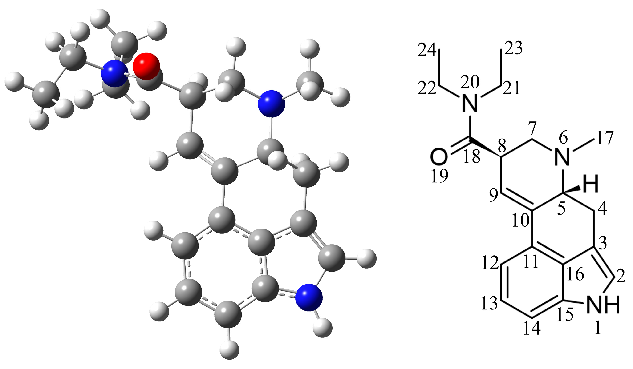
2.1. Selection of Appropriate Level of Theory

The first part of the theoretical prediction of spectra includes the selection of the appropriate level of theory. The structure optimization is carried out using the crystal structure deposited in the Cambridge Crystallographic Data Centre (CCDC) [18]. The alternative would be to select a compound structurally similar to the investigated one, usually some derivative or analog, and then determine the applicable theory level. Standard functionals, such as B3LYP, CAM-B3LYP, B3PW91, M06-2X, and M05-2X, perform the optimization reasonably well for most organic compounds, but the subtle differences in structural parameters determine the success in the prediction of spectra. The authors have selected these five functionals to optimize LSD structure, starting from the crystallographic one (CCDC number: 1208523) [19,20] (Figure 1). Before the optimization, it is necessary to remove the counter ions and co-crystalized solvent molecules or impurities. It should be kept in mind that impurities can influence the spectra of seized compounds, and that they can be important for determining their origin. The choice of basis set is also an important question to address. However, the 6-311++G(d,p) basis set is small, fast, and reasonably well performing for the optimization of organic compounds [21].
The structure of LSD (Figure 1) consists of an indole ring and conjugated single and double bonds, which limit the free rotation around the bonds and lead to a stable structure. After the optimization is completed, the obtained structures are compared to the crystallographic one by determining the bond lengths and angle deviations. For each functional, the structures underwent a conformational search along the C-C-C(O)-N(CH3)2 fragment, which is the only routable part of LSD. These comparisons are quantified by the mean absolute error (MAE) as the average absolute value of the difference between experimental and theoretical parameters. A lower MAE value shows a higher resemblance between experimental and theoretical structural parameters. Tables S1 and S2 contain the complete list of crystallographic and optimized bond lengths and angles, while the MAE values are shown in Table 1. When MAE values are compared, it should be kept in mind that the bond angles are more reliable than bond lengths. Although all MAE values for bond lengths are between 0.0423 and 0.0444 Å, the LSD structure optimized at B3LYP/6-311++G(d,p) level of theory had the lowest MAE value for bond lengths. The second lowest MAE value is for the M05-2X functional. In terms of bond angles, the M05-2X functional (2.70°) had the lowest MAE value, while the highest was for B3LYP (2.88°). Including dispersion interactions (B3LYP-D3BJ) lowers the MAE value for bond angles, but this value is still higher than the respective value for the M05-2X functional. This reasonably simple methodology leads to the conclusion that the M05-2X functional is suitable for the theoretical description of LSD structure and spectra prediction (Figure 1). The last section also checks applicability, in which two other LSD derivatives are optimized.
Sometimes, it is beneficial to understand the stability of the psychoactive compounds through analysis of intermolecular interactions. This is a more complex task, as it requires some knowledge of substituents’ electron donating/withdrawing properties, stereochemistry, and resonance. Still, it can be essential to determine the direction of signal shifts in IR, UV–VIS, and NMR spectra.

2.2. Experimental and Theoretical IR Spectra

The IR spectra are an indispensable tool for quickly determining the present substances in seized samples, especially if they are in appreciable quantities. There is no need to analyze the whole spectrum but only for several intense bands originating from carbonyl vibration or some other highly polar groups. Marking agents are often added to cover these bands or induce their shift through intermolecular bonding. The following section presents the comparison between the experimental and theoretical IR spectra of LSD and LSD tartrate. Some of the practical aspects are outlined.
The IR spectrum of LSD has been calculated during the optimization process. The vibrational spectrum can be visualized by the GaussView [22] program and further analyzed in the VEDA software [23]. As LSD is a low-symmetry molecule, all vibrations are active in the IR spectrum (141 normal modes). When the highly symmetric molecule is analyzed, it is beneficial to obtain both IR and Raman spectra because of the exclusion principle. In our case, the calculated wavenumbers were overestimated, and the scaling factor was calculated by comparing experimental and theoretical wavenumbers. The scaling factor can be obtained as the ratio between the observed and calculated wavenumbers of the same vibration, most commonly C=O vibration. The band belonging to this vibration in the experimental spectrum is positioned at 1625 nm, while in the theoretical spectrum, it is at 1747 cm−1 (Figure 2). The scaling factor calculated based on these values is 0.930. The experimental, scaled, and unscaled theoretical values are presented in Table S3. Theoretical values are shown only in the observed range (3500 to 500 cm−1). Figure 2 shows the experimental and scaled IR spectrum of LSD base and LSD tartrate.
The IR spectrum of LSD can be divided into three distinct regions. The first one is a high-frequency region between 3500 and 2500 cm−1 characterized by the peaks belonging to the N−H and C−H stretching vibrations. The N−H stretching vibration is located at 3200 cm−1 in the experimental spectrum [24,25]. In the theoretical spectrum, this band is positioned at 3446 cm−1. This difference is a consequence of the physical state of the sample, as the theoretical calculations were performed for the isolated molecule in a vacuum. The other prominent bands include the sp2 hybridized carbon atom C−H vibrations at 3097 and 3046 cm−1. The sp3 hybridized carbon atoms C−H vibrations can be assigned to the bands at 2920, 2883, 2839, 2810, and 2774 cm−1. These values are well reproduced in the theoretical spectrum (Table S3). As these vibrations are not particularly helpful for the LSD analysis, they are not discussed in detail. The most noticeable band located at 1625 cm−1 belongs to the C=O stretching vibration in both spectra. This band (between 1600 and 1630 cm−1) is typical for all the tertiary LSD amides and their analogs, although its position depends on the neighboring substituents [8,9,11,24]. Another intense band, assigned as C−C stretching vibration, is positioned at 1432/1379 cm−1 in the experimental/theoretical spectrum. The bands between 1600 and 1000 cm−1 can be attributed to mixed vibrational modes, including stretching and bending vibrations. Below 1000 cm−1, mostly bending and torsional vibrations are observed.
Various salts of LSD are often encountered at crime scenes. The spectra of LSD tartrate show peaks characteristic both for LSD base and salt (Figure 2b). The correction factor (0.921) calculated previously was used for the wavenumbers in the IR spectrum of LSD tartrate (Figure 2b) based on the optimized structure (at M05-2X/6-311++G(d,p) level of theory) shown in Figure S1. The optimization of LSD tartrate salt was performed for the 1:1 molar ratio between LSD and tartrate, as determined experimentally by NMR spectroscopy [8]. The C=O groups of LSD tartrate show two absorptions at 1716 and 1698 cm−1 in the experimental spectrum and 1705 and 1694 (scaled) cm−1 in the theoretical spectrum. These results prove the assumption that the selected level of theory and correction factor is appropriate for the description of LSD and its salts. High wavenumber values for the C=O stretching vibrations are due to the proximity of the α-hydroxyl group [24]. The position of the carboxyl group stretching vibration (1609 cm−1) of LSD is not significantly influenced by the presence of tartrate ions. Two vibrations characteristic for the carboxylate anion at 1604 and 1404 cm−1 in the experimental spectrum (1599 and 1406 cm−1 in theoretical) were also observed, both of which were partially overlaid with peaks of LSD [24]. The theoretical spectrum also contains bands associated with the tartrate ion at 1102, 1052, and 680 cm−1, corresponding to the peaks at 1109, 1065, and 690 cm−1 in the experimental spectrum [24]. The same methodology was applied to predict the IR spectra of two novel LSD analogs in the last section of the manuscript.

2.3. Experimental and Theoretical NMR Spectra

The chemical characterization of compounds through NMR spectra is necessary for structure determination except for when single-crystal XRD measurements are available. Before calculating NMR spectra, optimizing the structure in a solvent used in the experiment is beneficial. Optimizing TMS in the selected model and predicting its NMR spectrum is essential, as the values are shown relative to this compound (analogous to experiments).
The theoretical NMR spectra can be computed for any magnetically active nuclei, including 1H, 13C, 15N, and 17O. The 1H NMR spectra comparison to theoretical spectra can be ambiguous because of the multiplet structure and second-order effects, mainly if the compound contains aromatic moieties, which is the case with LSD. 1H NMR spectra can be well reproduced, such as in references [26,27,28], but in the following parts, only the 13C NMR spectrum of LSD will be explained. The proton-decoupled, 13C{1H}, NMR spectrum is much easier to reproduce, as only singlets are observed. Table 2 presents experimental, unscaled, and scaled theoretical values of 13C chemical shifts, relative to TMS of the LSD structure in chloroform. The atom numbering scheme follows Figure 1. The NMR spectrum was acquired, as explained in the Methodology section.
The experimental and theoretical results can be compared by calculating the correlation coefficient (R) and MEA value. As can be seen in Table 2, there is a high correlation between experimental and calculated values of 13C NMR with an MAE value of 14.9 ppm. The 13C NMR chemical shifts are usually overestimated, and the correction factor should be calculated. The correction factor for the given set of values is 0.848, including the possible solvent effect and rotation of specific bonds. Scaled values of theoretical shifts have the same correlation coefficient as experimental ones. However, the MAE is lowered to 2.0 ppm, which could include the uncertainty due to the solvent, temperature, and concentration dependence. The applicability of this correlation coefficient is probed in the last part of the article for the LSD analogs. A detailed analysis of the positions is not necessary for the prediction. However, it can be beneficial, especially if different substituents are added to the psychoactive substance backbone, and their effects can be followed. The following paragraph outlines some of the prominent carbon positions.
The lowest values of chemical shifts were obtained for the terminate carbon atoms in positions 22 and 24 (around 13 ppm in experimental and theoretical spectra). The chemical shift of C4 is reproduced within 0.5 ppm in the theoretical spectrum. CH2 group carbon atoms of diethylamine moiety have higher chemical shifts due to the proximity of nitrogen atoms (39 ppm on average). The same reason leads to the chemical shift of 42 ppm in the experimental and 39 ppm in the theoretical spectrum of the methyl group attached to a nitrogen atom. Carbon atoms in the aromatic core and parts with elongated delocalization have chemical shifts above 100 ppm in both spectra. Due to the similarity in the chemical shifts, the reproduction of this part can be difficult. However, in the case of LSD, the differences between the experiment and prediction were minimal. The highest chemical shift value was calculated for the carbonyl oxygen with a difference of 4 ppm. The same method is applied in this contribution to the LSD analogs to prove the applicability of the selected level of theory.

2.4. Experimental and Theoretical UV–VIS Spectra

The UV–VIS spectra are not helpful in the chemical characterization of substances due to the very broad peaks and spectra similarity for compounds that do not share the same structure. Electronic absorption of several functional groups can be assigned to the bands in the UV–VIS spectrum. However, substituents, elongated delocalization of π-electrons, solvent, pH, and temperature can influence their position. These spectra are essential in forensic analysis when the absorption wavelength is determined for the UV–VIS detectors coupled with chromatography columns, especially in liquid chromatography. Even when spectra are known, selecting wavelengths not shared with other compounds is of utmost interest, as this would induce an error in quantification. The UV–VIS spectrum of LSD consists of two wide peaks with maxima around 220 and 315 nm (Table 3), of which the one with a longer wavelength is used for quantification [29]. These spectra were recorded in water, as shown in the previous reference. A maximum at this wavelength is common for the compounds containing the aromatic core with the additional ring structures and electron-donating groups. There are slight deviations in the UV–VIS spectra of LSD’s analogs, especially when another carboxyl group is present [8,29]. The prediction of the wavelength for the novel LSD derivatives would be of the highest importance, especially when their quantification in seized samples is required. Several methods can be applied for the prediction of spectra, and they are discussed in the following paragraphs using the LSD’s electronic spectrum as an example.
The simplest method for the spectra prediction is the compound’s optimization in the desired solvent by the mentioned CPCM method (or any other suitable for the prediction). Then, the TD-DFT method is applied to calculate the electronic transitions. Ten transitions with the lowest energy (longest wavelength transitions) were calculated. In the studied example, two transitions with significant oscillator strength were observed. The first one is at 288 nm, significantly different from the experimental one. The second wavelength is well reproduced (226 nm). The first transition is assigned as HOMO→LUMO with an oscillator strength of 0.3793. The second can be denoted as HOMO→LUMO+2. The calculated energy values represent only the energy difference between the bottoms of the excited state and ground state energy surfaces, while the experimental spectrum contains the adjacent vibrational and rotational transitions responsible for the peak width. The bathochromic shift in the observed spectrum compared to the theoretical one is probably due to the stabilization effect of the solvent on the excited state. The HOMO, LUMO, and LUMO+1 densities of LSD are shown in Figure 3. The HOMO orbital is mainly localized on the indole core of LSD, covering the adjacent ring’s double C=C bond.
On the other hand, the LUMO orbital includes the indole moiety and the carbonyl group as the most polar group present in the structure. Most LSD molecules consist of carbon–carbon bonds, and this difference in electron density distribution only slightly changes the possible interactions with solvent molecules. The LUMO+1 is similar to the HOMO orbital and includes the same functional groups (indole ring). Based on these electron density distributions, the solvent molecules should stabilize the LUMO orbital more than the other two through interactions with the carbonyl group. These interactions would lead to the bathochromic shift of theoretically calculated values when a compound is surrounded by solvent molecules (315 vs. 288 nm).
The possible improvement of the previously discussed strategy can be the addition of explicit solvent molecules [30]. Solvent molecules form stabilization interactions (dipole–dipole and hydrogen bonds) with the polar groups in the molecule and lead to bathochromic/hypochromic shifts. It is suggested that solvent molecules are always placed around polar groups, as these are usually responsible for the transitions in visible and vacuum UV regions. Figure S3 shows the positions of the four water molecules added around LSD. These solvent molecules were introduced in the vicinity of polar groups, nitrogen atoms of an amino group, pyrrole and piperidine rings, and oxygen of the carbonyl group. Another water molecule was added above the aromatic ring, as O−H⋯π interactions are also responsible for the stabilization of the spectrum. A new electronic spectrum was also computed for ten transitions. The value of the longest wavelength transition is 291 nm, which is only 2 nm higher than the previously discussed value. The obtained values do not differ significantly, proving that the polarity of excited and ground states are similar and that the interactions with solvent molecules stabilize both states equally, as previously postulated through the electron density distribution analysis.
The third strategy is optimizing the first excited state (singlet or triplet, depending on the compound) and calculating the energy difference. The excited state is optimized by adding the optimization keyword in the input file and the TD-DFT. The electronic energy difference in Hartree is then recalculated to nm. The structure of the optimized first electronic state is shown in Figure S2. This figure shows no significant differences in the ground and excited state geometries. The obtained energy of 330 nm fairly well reproduces the experimental value. The difference of 10 to 15 nm falls within the experimental width of UV–VIS transitions of this type of molecule. Therefore, the authors suggest applying all these methods to predict the expected range of electronic transitions.

2.5. Experimental and Theoretical MS Spectra

Mass spectra and the fragmentation scheme are the final proof that LSD is found in the samples at the crime scene. A possible fragmentation pattern under the electron impact conditions for LSD has been extensively investigated in the literature [8,31,32,33]. The main fragments are positioned at 323, 221, 207, 181, and 72 m/z (Figure 4). These fragments were identified by the literature review of similar compounds [8,29,32,33] and were optimized at the M05-2X/6-311++G(d,p) level of theory to prove the applicability of theoretical considerations for the determination of the mass pattern. The optimized structures of fragments are shown in Figure 3, except for the molecular ion, which has a structure identical to that presented in Figure 1.
The fragment at 221 m/z represents a product of the diethylamide moiety removal upon ionization. In this fragment, the planarity of the six-membered ring is restored, along with its aromaticity, which makes this species completely planar and, therefore, stable. A methyl group is removed in the second step, and a fragment at 207 m/z is obtained. The extended π-electron delocalization is crucial for the stability of this fragment, although it is less stable than the previously described one due to the loss of the hyperconjugation effect of the methyl group. This is reflected in the lower intensity of the peak at 207 compared to the 221 m/z [8]. The peak at 181 m/z represents a fragment formed when the CH2=CH-C(O)-N(C2H5)2 fragment was removed. This step creates a stable species with a four-membered ring instead of a six-membered ring. The extended delocalization, together with restored planarity, leads to the increased stability of the fragment. Besides all of the mentioned effects, lone pairs of electrons on nitrogen additionally stabilize the structure through interactions such as LP(N)→π*(C-C), as calculated in the second-order perturbation theory and NBO analysis [21,34]. The last abundant fragment is common for all of the diethylamide derivatives of LSD. It is suggested that this fragment is formed when the C=O group is separated from the moiety mentioned in the first step. Complex fragmentation schemes of LSD and its analogs are shown elsewhere. Still, the theoretical methods are a valuable tool for predicting stability and intermolecular interactions governing the stability of the formed ions, radicals, and neutral species.

2.6. Case Study of Real Samples Containing LSD

The seized sample was complex, as seen in the chromatogram (Figure S4). The peak that could be potentially identified as LSD was among the last ones in the chromatogram at 21 min. The mass spectrum shows the fragmentation peaks of the compound (lower figure). The prominent peaks at 323, 221, 207, 181, and 72 m/z coincide nicely with the previously analyzed mass spectrum. The most abundant species in the spectrum were correctly identified and optimized previously. This analysis proves that the theoretical methods can identify novel LSD analogs.

2.7. LSD Analogues, the Applicability of the Results

The methodology presented in the paper can be applied to similar compounds. It is important to “mimic” the experimental conditions throughout the theoretical calculations. For the optimization of the crystallographic structure, common functionals should be applied along with a reasonably large basis set. The IR spectrum should be calculated for an isolated molecule. The UV–VIS and NMR spectra prediction should be performed on structures optimized in a solvent. The practical application depends mainly on the available data for similar compounds. The obtained level of theory is examined on the LSD analogs in this section. In the paper by Monte and coworkers [35], two d-Lysergic acid amides were synthesized, and the single crystal X-ray analysis determined their structure. These analogs differ from LSD in the size of the alkyl group of the amide part (analog 1 (A1)—2-pentyl, analog 2 (A2)—2-hexyl). The optimized structures of these compounds at the M05-2X/6-311++G(d,p) level of theory are presented in Figure 5, while their bond lengths and angles are shown in Tables S4–S7. The MAE values for the bond lengths are 0.0176 (A1) and 0.0207 Å (A2). The bond angles were also well reproduced, with the MAEs equal to 0.83 (A1) and 1.08 (A2). Low MAE values for the bond lengths and angles prove that the determined level of theory optimizes crystallographic structure adequately. The spectral characterization is expected to follow the previously discussed procedure, although 13C NMR spectra were not provided.
On the other hand, the 13C NMR spectral prediction was performed for two commonly encountered analogs of LSD, namely 1-propionyl-d-lysergic acid diethylamide (1P-LSD) [8] and 1-butanoyl-d-lysergic acid diethylamide (1B-LSD) [29]. The experimental, unscaled, and scaled (by the previously determined factor of 0.848) theoretical chemical shifts are shown in Tables S8 and S9. Optimized structures of 1P-LSD and 1B-LSD are also presented in Figure 5. These results show a high correlation coefficient (0.999) between experimental and theoretical values in the case of both LSD analogs. The values of chemical shifts were overestimated with the MAE values of 15.7 (1P-LSD) and 14.7 (1B-LSD) ppm, similar to the results shown in Table 2. After the correction, the MAE values were lowered significantly to 2.5 (1P-LSD) and 2.4 (1B-LSD) ppm. The 13C NMR spectra of these two analogs were similar to those of the LSD base. Propionyl group carbon atoms give rise to signals at 11.6 (CH3), 31.8 (CH3), and 170.3 (C=O) ppm in the theoretical spectrum. The significant difference in the theoretical chemical shift was calculated for C3 (LSD: 112.4 ppm and 1P-LSD: 119.7 ppm) due to the presence of the propionyl group, which is consistent with the experimental results [8]. In the case of the butanoyl group, the chemical shifts in the theoretical spectrum are comparable to previously listed ones, 13.8 (CH3), 21.9 (CH2), 40 (CH2), and 169.3 (C=O) ppm. These results prove the assumption that the selected level of theory, along with the correction factor, can be used to predict the 13C NMR spectrum of novel LSD analogs.

3. Materials and Methods

3.1. Spectra Acquisition

The experimental IR, NMR, and GC-MS spectra were obtained from an online database (https://www.swgdrug.org/, accessed on 1 December 2022) and used without further modifications. This database contains spectrum of seized drugs analyzed by the standard procedures, as further explained. The IR spectra were recorded for the LSD base and LSD tartrate in the KBr matrix at mass ratios 3:150 mg and 4:150 mg (substance: KBr). The NMR spectrum was obtained in chloroform and with TMS as standard. A blotter paper was soaked in methanol for four hours in the dark. Then, 1 μL of sample solution was injected into a GC-MS system for the analysis. The emission current was 200 μA, scan time of 1 s with a scan range between 29 and 600 m/z. The ion source temperature was maintained at 220 °C. A fused silica capillary DB-1 column was used (30 m × 0.25 mm, film thickness 0.25 μm). The initial temperature was held for 2 min at 80 °C and later followed by a ramp to 310 °C at 20 °C min−1. The injector and transfer line temperatures were 280 and 300 °C, and the carrier gas was helium in constant flow mode at a rate of 1.2 mL min−1.

3.2. Theoretical Methods

The structure of LSD was optimized starting from the crystalographic structure of lysergic acid diethylamide o-iodobenzoate with removed counterion [19,20] in the Gaussian Program package [36]. Several standard functionals (B3LYP, CAM-B3LYP, B3PW91, M05-2X, and M06-2X) [37,38,39,40,41,42] in conjunction with 6-311++G(d,p) basis set [43] were applied for the optimization to find a suitable model that well represents the experimental structure. The effects of dispersion corrections were analyzed in the case of the B3LYP-D3BJ functional [44]. The optimizations were performed without any geometrical constraints, and the absence of imaginary frequencies showed that the minima on the energy surface were found. The charge and multiplicity for the neutral species were 0 and 1. The conformational research along the C-C-C(O)-N(CH3)2 fragment was performed for each optimized structure, and the minima on each potential surface for a specific functional were compared. The bond lengths and angles are read from the GausView [22] or any other program to visualize quantum chemistry data. The vibrational spectrum was examined based on the potential energy distribution (PED) analysis implemented in the FCART version 7.0 software [45]. The preparation of UV–VIS and NMR spectra of LSD requires solvent use, such as water and chloroform. Therefore, the optimization was performed in these two solvents using the Conductor-like Polarizable Continuum (CPCM) model. Thus, one water molecule was positioned in four positions close to the polar groups of LSD in the case of UV–VIS spectra prediction. This allowed for quantification and determination of intramolecular interactions crucial for correctly predicting spectra. These optimizations were performed at the same level of theory. The Time-Dependent (TD-DFT) approach was used to calculate electronic transitions in water [46,47]. The nuclear magnetic resonance (NMR) spectra were obtained by the Gauge Independent Atomic Orbital (GIAO) approach [48,49].

4. Conclusions

LSD and its newly obtained analogs have been commonly found at crime scenes, and their analysis is a constant task for forensic investigators. Five functionals (B3LYP, CAM-B3LYP, B3PW91, M05-2X, and M06-2X) in conjunction with a 6-311++G(d,p) basis set have been employed for the optimization of structure starting from a crystallographic LSD structure. The lowest mean absolute error (MAE) was calculated for the structure optimized at the M05-2X/6-311++G(d,p) level of theory with MAE of bond lengths and angles being 0.0436 Å and 2.7°. The correction factor (0.848) for IR wavenumbers was calculated by comparing the experimental and theoretical position of the C=O stretching vibration wavenumber. The rest of the bands were also well reproduced. The 13C NMR spectrum was assigned, and a high correlation factor (0.999) was obtained for the experimental and theoretical data, with a mean absolute error of 2.0 ppm. Several methods for predicting UV–VIS spectrum were shown, with the optimization of ground and first excited state leading to the difference of 15 nm in the experimental and theoretical values. The most abundant mass fragments a″ 221′ 207, 181, and 72 m/z were optimized. Their stability was explained by the restored planarity of the fragment and formation of the six- and four-membered rings with extended delocalization. These results coincided adequately with the mass spectrum of the seized real sample. Mentioned level of theory was applied for the optimization of two LSD analogs with 2-pentyl and 2-hexyl moieties. MAE values for the bond lengths and angles were around 0.02 Å and 1°, respectively. The 13C NMR spectra of two common analogs, 1-propionyl-d-lysergic acid diethylamide (1P-LSD) and 1-butanoyl-d-lysergic acid diethylamide (1B-LSD), were predicted with correlation coefficients of 0.999 and MAE values lower than 2.5 ppm. These results prove the assumption that theoretical methods could be applied to the structural analysis of novel psychedelics based on the available spectra from the seized samples.

Supplementary Materials

The following supporting information can be downloaded at: https://www.mdpi.com/article/10.3390/app13052984/s1, Table S1. Experimental and theoretical (different functionals with 6-311++G(d,p) basis set) bond lengths (in Å) of LSD; Table S2. Experimental and theoretical (different functionals with 6-311++G(d,p) basis set) bond angles (in °) of LSD.; Table S3. The assigned vibrations of LSD (at M05-2X/6-311++G(d,p) level of theory); Figure S1: Optimized structure of LSD tartrate (at M05-2X/6-311++G(d,p) level of theory); Figure S2: Optimized structure of the first excited state of LSD (at M05-2X/6-311++G(d,p) level of theory); Figure S3: Optimized structures (at M05-2X/6-311++G(d,p) level of theory) of LSD with one water molecule; Figure S4: GC-MS data of the seized sample; Table S4: Experimental and theoretical (at M05-2X/6-311++G(d,p) level of theory) bond lengths (in Å) of A1; Table S5: Experimental and theoretical (at M05-2X/6-311++G(d,p) level of theory) bond angles (in °) of A1; Table S6: Experimental and theoretical (at M05-2X/6-311++G(d,p) level of theory) bond lengths (in Å) of A2; Table S7: Experimental and theoretical (at M05-2X/6-311++G(d,p) level of theory) bond lengths (in °) of A2; Table S8: Experimental and theoretical (at M05-2X/6-311++G(d,p) level of theory) chemical shifts of 1P-LSD; Table S9: Experimental and theoretical (at M05-2X/6-311++G(d,p) level of theory) chemical shifts of 1B-LSD. Scheme S1: The atom numbering scheme for LSD.

Author Contributions

Conceptualization, D.D. and J.D.; methodology, D.D. and D.M.; software, D.D., Z.M. and D.M.; validation, J.D., M.M. and D.M.; formal analysis, J.D.; investigation, J.D.; resources, D.D.; data curation, M.M.; writing—original draft preparation, J.D., M.M. and D.D.; writing—review and editing, D.M, Z.M. and D.D.; visualization, J.D.; supervision, Z.M. and D.D.; project administration, D.D.; funding acquisition, Z.M. and D.D. All authors have read and agreed to the published version of the manuscript.

Funding

This research was funded by the Ministry of Education, Science, and Technological Development of the Republic of Serbia, grant numbers 451-03-68/2022-14/200146 and 451-03-68/2022-14/200378.

Institutional Review Board Statement

Not applicable.

Informed Consent Statement

Not applicable.

Data Availability Statement

Not applicable.

Conflicts of Interest

The authors declare no conflict of interest. The funders had no role in the design of the study; in the collection, analyses, or interpretation of data; in the writing of the manuscript; or in the decision to publish the results.

Sample Availability

Samples of the compounds are available from the authors.

References

  1. dos Santos, R.G.; Osório, F.L.; Crippa, J.A.S.; Hallak, J.E.C. Classical hallucinogens and neuroimaging: A systematic review of human studies. Neurosci. Biobehav. Rev. 2016, 71, 715–728. [Google Scholar] [CrossRef] [PubMed]
  2. Dos Santos, R.G.; Osório, F.L.; Crippa, J.A.S.; Riba, J.; Zuardi, A.W.; Hallak, J.E.C. Antidepressive, anxiolytic, and antiaddictive effects of ayahuasca, psilocybin and lysergic acid diethylamide (LSD): A systematic review of clinical trials published in the last 25 years. Ther. Adv. Psychopharmacol. 2016, 6, 193–213. [Google Scholar] [CrossRef] [PubMed] [Green Version]
  3. Libânio Osório Marta, R.F. Metabolism of lysergic acid diethylamide (LSD): An update. Drug Metab. Rev. 2019, 51, 378–387. [Google Scholar] [CrossRef] [PubMed]
  4. Bouso, J.C.; dos Santos, R.G.; Alcázar-Córcoles, M.Á.; Hallak, J.E.C. Serotonergic psychedelics and personality: A systematic review of contemporary research. Neurosci. Biobehav. Rev. 2018, 87, 118–132. [Google Scholar] [CrossRef] [PubMed]
  5. dos Santos, R.G.; Bouso, J.C.; Alcázar-Córcoles, M.Á.; Hallak, J.E.C. Efficacy, tolerability, and safety of serotonergic psychedelics for the management of mood, anxiety, and substance-use disorders: A systematic review of systematic reviews. Expert Rev. Clin. Pharmacol. 2018, 11, 889–902. [Google Scholar] [CrossRef] [PubMed]
  6. Favretto, D.; Frison, G.; Maietti, S.; Ferrara, S.D. LC-ESI-MS/MS on an ion trap for the determination of LSD, iso-LSD, nor-LSD and 2-oxo-3-hydroxy-LSD in blood, urine and vitreous humor. Int. J. Legal Med. 2007, 121, 259–265. [Google Scholar] [CrossRef]
  7. da Cunha, K.F.; Kahl, J.M.M.; Fiorentin, T.R.; Oliveira, K.D.; Costa, J.L. High-sensitivity method for the determination of LSD and 2-oxo-3-hydroxy-LSD in oral fluid by liquid chromatography-tandem mass spectrometry. Forensic Toxicol. 2022, 40, 322–331. [Google Scholar] [CrossRef]
  8. Brandt, S.D.; Kavanagh, P.V.; Westphal, F.; Stratford, A.; Elliott, S.P.; Hoang, K.; Wallach, J.; Halberstadt, A.L. Return of the lysergamides. Part I: Analytical and behavioural characterization of 1-propionyl- d -lysergic acid diethylamide (1P-LSD). Drug Test. Anal. 2016, 8, 891–902. [Google Scholar] [CrossRef] [Green Version]
  9. Brandt, S.D.; Kavanagh, P.V.; Westphal, F.; Elliott, S.P.; Wallach, J.; Colestock, T.; Burrow, T.E.; Chapman, S.J.; Stratford, A.; Nichols, D.E.; et al. Return of the lysergamides. Part II: Analytical and behavioural characterization of N 6 -allyl-6-norlysergic acid diethylamide (AL-LAD) and (2’ S,4’ S )-lysergic acid 2,4-dimethylazetidide (LSZ). Drug Test. Anal. 2017, 9, 38–50. [Google Scholar] [CrossRef] [PubMed] [Green Version]
  10. Brandt, S.D.; Kavanagh, P.V.; Westphal, F.; Elliott, S.P.; Wallach, J.; Stratford, A.; Nichols, D.E.; Halberstadt, A.L. Return of the lysergamides. Part III: Analytical characterization of N 6 -ethyl-6-norlysergic acid diethylamide (ETH-LAD) and 1-propionyl ETH-LAD (1P-ETH-LAD). Drug Test. Anal. 2017, 9, 1641–1649. [Google Scholar] [CrossRef] [PubMed] [Green Version]
  11. Brandt, S.D.; Kavanagh, P.V.; Twamley, B.; Westphal, F.; Elliott, S.P.; Wallach, J.; Stratford, A.; Klein, L.M.; McCorvy, J.D.; Nichols, D.E.; et al. Return of the lysergamides. Part IV: Analytical and pharmacological characterization of lysergic acid morpholide (LSM-775). Drug Test. Anal. 2018, 10, 310–322. [Google Scholar] [CrossRef] [Green Version]
  12. Halberstadt, A.L.; Klein, L.M.; Chatha, M.; Valenzuela, L.B.; Stratford, A.; Wallach, J.; Nichols, D.E.; Brandt, S.D. Pharmacological characterization of the LSD analog N-ethyl-N-cyclopropyl lysergamide (ECPLA). Psychopharmacology 2019, 236, 799–808. [Google Scholar] [CrossRef] [Green Version]
  13. Nichols, D.E. Hallucinogens. Pharmacol. Ther. 2004, 101, 131–181. [Google Scholar] [CrossRef]
  14. Grumann, C.; Henkel, K.; Stratford, A.; Hermanns-Clausen, M.; Passie, T.; Brandt, S.D.; Auwärter, V. Validation of an LC-MS/MS method for the quantitative analysis of 1P-LSD and its tentative metabolite LSD in fortified urine and serum samples including stability tests for 1P-LSD under different storage conditions. J. Pharm. Biomed. Anal. 2019, 174, 270–276. [Google Scholar] [CrossRef] [PubMed]
  15. Spálovská, D.; Maříková, T.; Kohout, M.; Králík, F.; Kuchař, M.; Setnička, V. Methylone and pentylone: Structural analysis of new psychoactive substances. Forensic Toxicol. 2019, 37, 366–377. [Google Scholar] [CrossRef] [Green Version]
  16. Wu, X.; Cañamares, M.V.; Kakoulli, I.; Sanchez-Cortes, S. Chemical Characterization and Molecular Dynamics Simulations of Bufotenine by Surface-Enhanced Raman Scattering (SERS) and Density Functional Theory (DFT). J. Phys. Chem. Lett. 2022, 13, 5831–5837. [Google Scholar] [CrossRef] [PubMed]
  17. Hosseinian, A.; Vessally, E.; Bekhradnia, A.; Nejati, K.; Rahimpour, G. Benzoylethanamine drug interaction with the AlN nanosheet, nanotube and nanocage: Density functional theory studies. Thin Solid Film. 2017, 640, 93–98. [Google Scholar] [CrossRef]
  18. Groom, C.R.; Bruno, I.J.; Lightfoot, M.P.; Ward, S.C. IUCr The Cambridge Structural Database. Acta Crystallogr. Sect. B Struct. Sci. Cryst. Eng. Mater. 2016, 72, 171–179. [Google Scholar] [CrossRef]
  19. Baker, R.W.; Chothia, C.; Pauling, P.; Weber, H.P. Molecular Structures of Hallucinogenic Substances: Lysergic Acid Diethylamide, Psilocybin, and 2,4,5-Trimethoxyamphetamine. Mol. Pharmacol. 1973, 9, 23–32. [Google Scholar] [PubMed]
  20. Baker, R.W.; Chothia, C.; Pauling, P.; Weber, H.P. Molecular Structure of LSD. Science 1972, 178, 614–615. [Google Scholar] [CrossRef] [PubMed]
  21. Shobana, D.; Sudha, S.; Ramarajan, D.; Dimić, D. Synthesis, crystal structure, spectral characterization and Hirshfeld surface analysis of (E)-N′-(3-ethoxy-4-hydroxybenzylidene)-4-fluorobenzohydrazide single-crystal—A novel NLO active material. J. Mol. Struct. 2022, 1250, 131856. [Google Scholar] [CrossRef]
  22. Dennington, R.; Todd, K.; Millam, J. Gauss View; Semichem Inc.: Shawnee, KS, USA, 2009. [Google Scholar]
  23. Jamroz, M.H. Vibrational Energy Distribution Analysis VEDA 4, Warsaw, 2004–2010. Available online: https://smmg.pl/software/veda (accessed on 1 December 2022).
  24. Mesley, R.J.; Evans, W.H. Infrared identification of lysergide (LSD). J. Pharm. Pharmacol. 2011, 21, 713–720. [Google Scholar] [CrossRef] [PubMed]
  25. Dimić, D.S.; Kaluđerović, G.N.; Avdović, E.H.; Milenković, D.A.; Živanović, M.N.; Potočňák, I.; Samoľová, E.; Dimitrijević, M.S.; Saso, L.; Marković, Z.S.; et al. Synthesis, Crystallographic, Quantum Chemical, Antitumor, and Molecular Docking/Dynamic Studies of 4-Hydroxycoumarin-Neurotransmitter Derivatives. Int. J. Mol. Sci. 2022, 23, 1001. [Google Scholar] [CrossRef] [PubMed]
  26. Dimić, D.; Milenković, D.; Ilić, J.; Šmit, B.; Amić, A.; Marković, Z.; Dimitrić Marković, J. Experimental and theoretical elucidation of structural and antioxidant properties of vanillylmandelic acid and its carboxylate anion. Spectrochim. Acta Part A Mol. Biomol. Spectrosc. 2018, 198, 61–70. [Google Scholar] [CrossRef] [PubMed]
  27. Dimić, D.; Milenković, D.; Marković, Z.; Marković, J.D. Structural and spectral analysis of 3-metoxytyramine, an important metabolite of dopamine. J. Mol. Struct. 2017, 1134, 226–236. [Google Scholar] [CrossRef]
  28. Avdović, E.H.; Dimić, D.S.; Dimitrić Marković, J.M.; Vuković, N.; Radulović, M.Đ.; Živanović, M.N.; Filipović, N.D.; Đorović, J.R.; Trifunović, S.R.; Marković, Z.S. Spectroscopic and theoretical investigation of the potential anti-tumor and anti-microbial agent, 3-(1-((2-hydroxyphenyl)amino)ethylidene)chroman-2,4-dione. Spectrochim. Acta Part A Mol. Biomol. Spectrosc. 2019, 206, 421–429. [Google Scholar] [CrossRef]
  29. Brandt, S.D.; Kavanagh, P.V.; Westphal, F.; Stratford, A.; Elliott, S.P.; Dowling, G.; Wallach, J.; Halberstadt, A.L. Return of the lysergamides. Part V: Analytical and behavioural characterization of 1-butanoyl-d-lysergic acid diethylamide (1B-LSD). Drug Test. Anal. 2019, 11, 1122–1133. [Google Scholar] [CrossRef] [PubMed]
  30. Dimić, D. The importance of specific solvent–solute interactions for studying UV–VIS spectra of light-responsive molecular switches. Comptes Rendus Chim. 2018, 21, 1001–1010. [Google Scholar] [CrossRef]
  31. Bellman, S.W. Mass Spectral Identification of Some Hallucinogenic Drugs. J. AOAC Int. 1968, 51, 164–175. [Google Scholar] [CrossRef]
  32. Clark, C.C. The Differentiation of Lysergic Acid Diethylamide (LSD) from N -Methyl- N -Propyl and N -Butyl Amides of Lysergic Acid. J. Forensic Sci. 1989, 34, 12674J. [Google Scholar] [CrossRef]
  33. Nakahara, Y.; Niwaguchi, T. Studies on Lysergic Acid Diethylamide and Related Compounds. I. Synthesis of d-N6-Demethyl-lysergic Acid Diethylamide. Chem. Pharm. Bull. 1971, 19, 2337–2341. [Google Scholar] [CrossRef] [Green Version]
  34. Shobana, D.; Sudha, S.; Ramarajan, D.; Ristivojević, N.; Rakić, A.; Dimić, D. Structural, spectroscopic (IR, Raman, and NMR), quantum chemical, and molecular docking analysis of (E)-2-(2,5-dimethoxybenzylidene)hydrazinecarbothioamide and its dimers. J. Mol. Struct. 2022, 1247, 131277. [Google Scholar] [CrossRef]
  35. Monte, A.P.; Marona-Lewicka, D.; Kanthasamy, A.; Sanders-Bush, E.; Nichols, D.E. Stereoselective LSD-like Activity in a Series of d-Lysergic Acid Amides of (R)- and (S)-2-Aminoalkanes. J. Med. Chem. 1995, 38, 958–966. [Google Scholar] [CrossRef]
  36. Frisch, M.J.; Trucks, G.W.; Schlegel, H.B.; Scuseria, G.E.; Robb, M.A.; Cheeseman, J.R.; Scalmani, G.; Barone, V.; Petersson, G.A.; Nakatsuji, H.; et al. Gaussian 09, Revision A.02; Gaussian, Inc.: Wallingford, CT, USA, 2016. [Google Scholar]
  37. Becke, A.D. Density-functional thermochemistry. III. The role of exact exchange. J. Chem. Phys. 1993, 98, 5648. [Google Scholar] [CrossRef] [Green Version]
  38. Becke, A.D. Density-functional exchange-energy approximation with correct asymptotic behavior. Phys. Rev. A 1988, 38, 3098–3100. [Google Scholar] [CrossRef]
  39. Zhao, Y.; Schultz, N.E.; Truhlar, D.G. Design of Density Functionals by Combining the Method of Constraint Satisfaction with Parametrization for Thermochemistry, Thermochemical Kinetics, and Noncovalent Interactions. J. Chem. Theory Comput. 2006, 2, 364–382. [Google Scholar] [CrossRef]
  40. Zhao, Y.; Truhlar, D.G. The M06 suite of density functionals for main group thermochemistry, thermochemical kinetics, noncovalent interactions, excited states, and transition elements: Two new functionals and systematic testing of four M06-class functionals and 12 other functionals. Theor. Chem. Acc. 2008, 120, 215–241. [Google Scholar] [CrossRef] [Green Version]
  41. Yanai, T.; Tew, D.P.; Handy, N.C. A new hybrid exchange–correlation functional using the Coulomb-attenuating method (CAM-B3LYP). Chem. Phys. Lett. 2004, 393, 51–57. [Google Scholar] [CrossRef] [Green Version]
  42. Perdew, J.P.; Burke, K.; Wang, Y. Generalized gradient approximation for the exchange-correlation hole of a many-electron system. Phys. Rev. B-Condens. Matter Mater. Phys. 1996, 54, 16533–16539. [Google Scholar] [CrossRef] [PubMed] [Green Version]
  43. Dunning, T.H. Gaussian basis sets for use in correlated molecular calculations. I. The atoms boron through neon and hydrogen. J. Chem. Phys. 1989, 90, 1007. [Google Scholar] [CrossRef]
  44. Grimme, S.; Ehrlich, S.; Goerigk, L. Effect of the damping function in dispersion corrected density functional theory. J. Comput. Chem. 2011, 32, 1456–1465. [Google Scholar] [CrossRef] [PubMed]
  45. Munos, R.A.; Panchenko, Y.N.; Koptev, G.S.; Stepanov, N.F. Program for calculating distribution of potential energy in internal coordinates. J. Appl. Spectrosc. 1970, 12, 428–429. [Google Scholar] [CrossRef]
  46. Jacquemin, D.; Preat, J.; Perpète, E.A. A TD-DFT study of the absorption spectra of fast dye salts. Chem. Phys. Lett. 2005, 410, 254–259. [Google Scholar] [CrossRef]
  47. Jacquemin, D.; Perpète, E.A. Ab initio calculations of the colour of closed-ring diarylethenes: TD-DFT estimates for molecular switches. Chem. Phys. Lett. 2006, 429, 147–152. [Google Scholar] [CrossRef]
  48. Zieliński, R.; Szymusiak, H. Application of Dft B3Lyp/Giao and B3Lyp/Csgt Methods for Interpretation of Nmr Spectra of Flavonoids. Pol. J. Food Nutr. Sci. 2003, 53, 157–162. [Google Scholar]
  49. Bohmann, J.A.; Weinhold, F.; Farrar, T.C. Natural chemical shielding analysis of nuclear magnetic resonance shielding tensors from gauge-including atomic orbital calculations. J. Chem. Phys. 1997, 107, 1173. [Google Scholar] [CrossRef]
Figure 1. Optimized structure of LSD (at M05-2X/6-311++G(d,p) level of theory) with the atom numbering scheme.
Applsci 13 02984 g001
Figure 2. Experimental and theoretical (at M05-2X/6-311++G(d,p) level of theory) IR spectra of (a) LSD base and (b) LSD tartrate.
Applsci 13 02984 g002
Figure 3. Electron density distribution of HOMO, LUMO, and LUMO+1 orbitals of the optimized structures of LSD (at M05-2X/6-311++G(d,p) level of theory, isovalue = 0.02).
Applsci 13 02984 g003
Figure 4. Optimized structures (at M05-2X/6-311++G(d,p) level of theory) of the most abundant fragments in the mass spectrum of LSD.
Applsci 13 02984 g004
Figure 5. Optimized structures (at M05-2X/6-311++G(d,p) level of theory) of several analogs of LSD.
Applsci 13 02984 g005
Table 1. Mean absolute errors for the bond lengths and angles for structures optimized with various functionals in conjunction with 6-31++G(d,p) level of theory.
B3LYPB3LYP-D3BJCAM-B3LYPB3PW91M05-2XM06-2X
Bond lengths (Å)0.04230.04300.04440.04390.04360.0437
Bond angles (°)2.882.822.832.852.702.72
Table 2. Experimental and theoretical (at M05-2X/6-311++G(d,p) level of theory) 13C chemical shifts of LSD in chloroform.
Carbon AtomExperimental (ppm)Calculated, Unscaled (ppm) Calculated, Scaled (ppm)
C2413.113.511.4
C2214.815.513.2
C427.332.827.8
C2339.944.737.9
C2140.246.039.0
C1742.046.839.7
C843.948.541.1
C756.060.851.5
C563.271.260.3
C14109.7130.9110.9
C3110.8132.6112.4
C12112.6133.4113.2
C2118.3139.3118.0
C9119.7146.2123.9
C13123.3146.4124.1
C16126.3149.0126.3
C11128.1152.4129.2
C15134.0155.4131.8
C10136.3166.3141.0
C18170.9196.4166.5
R0.9990.999
MAE (ppm)14.92.0
Table 3. Experimental and theoretical (at M05-2X/6-311++G(d,p) level of theory) electronic transitions of LSD.
Experimental [nm]CPCM [nm]CPCM + 1 Molecule Water (nm)Excited State Optimization (nm)
314288
(HOMO→LUMO, 96%, f = 0.3948)
291
(HOMO→LUMO, 96%, f = 0.3793)
330
(HOMO→LUMO)
222225
(HOMO→LUMO+1,
32%, f = 0.2307)
226
(HOMO→LUMO+2,
37%, f = 0.1816)
Disclaimer/Publisher’s Note: The statements, opinions and data contained in all publications are solely those of the individual author(s) and contributor(s) and not of MDPI and/or the editor(s). MDPI and/or the editor(s) disclaim responsibility for any injury to people or property resulting from any ideas, methods, instructions or products referred to in the content.

