Article

Potential Effects of Photobiomodulation Therapy on Human Dental Pulp Stem Cells

1
Department of Restorative Dentistry, School of Dentistry, University of Jordan, Amman 11942, Jordan
2
Faculty of Pharmacy and Medical Sciences, Petra University, Amman 11196, Jordan
3
Cell Therapy Center, University of Jordan, Amman 11942, Jordan
4
Oral Biology, Surgery and Biomedical Engineering, University at Buffalo, Buffalo, NY 14214, USA
*
Authors to whom correspondence should be addressed.
These authors contributed equally to this work.
Appl. Sci. 2024, 14(1), 124; https://doi.org/10.3390/app14010124
Submission received: 14 October 2023 / Revised: 9 December 2023 / Accepted: 13 December 2023 / Published: 22 December 2023

Abstract

:
Background: A significant clinical technology enabling the use of stem cells in dentistry is Photobiomodulation (PBM). The current study aimed to investigate various PBM treatment variables on human dental pulp stem cell proliferation, survival, and changes in cell cycle progression. Methods: Human third molar samples were collected, and human Dental Pulp Stem Cells (hDPSCs) were isolated, expanded, seeded, and cultured in media containing 0, 1, 5, or 10% FBS. PBM treatments using an 810 nm diode laser at various doses were performed 24, 48, and 72 h after seeding. Cell proliferation and apoptosis were assessed. A two-way ANOVA test was performed among all treatment groups, followed by multiple comparison tests (Tukey’s test) performed between each treated group and its control untreated group. Results: After 24 h, only cells cultured in 10% FBS showed a significant (p < 0.005) increase in the proliferation rate of cells treated with T7 (0.4 W × 10 s). After 48 h, hDPSCs cultured in both 5% and 10% FBS showed a significant (p < 0.005) increase in the proliferation rate of cells treated with T7 (0.4 W × 10 s) as compared to the untreated control. After 72 h, only cells cultured in 10% FBS showed a significant increase in the proliferation rate of the cells treated with T9 (1 W × 3 s) (p < 0.005) and T7 (0.4 W × 10 s) (p < 0.00005). Conclusions: Low-power laser therapy at a wavelength of 810 nm induced hDPSC proliferation at the following parameters: power output 0.4 W, irradiance 0.5 W/cm2, fluence 0.416 J/cm2, exposure time 10 s.

1. Introduction

The use of mesenchymal stem cells (MSCs) to replace conventional therapeutic regimes in tissue engineering and regenerative medicine has been the subject of growing interest in the medical and dental fields [1]. Regenerative medicine has been focusing on directing the differentiation of stem cells into lineage-specific, functional cells that can promote tissue repair and organization [2]. With the advent of modern tissue engineering concepts and the discovery of dental stem cells, stem cell therapy and pulp tissue regeneration have been evolving for years to replace the shortcomings of dental materials and the pitfalls of traditional therapeutic approaches in dentistry [3].
Mesenchymal stem cells (MSCs) are multipotent cells that can be derived from various tissues including bone marrow, dental pulp, adipose tissue, and umbilical cord, to name a few. These cells possess the ability to self-renew and differentiate into multiple cell types depending on the specific in vitro conditions provided [4]. Dental pulp stem cells (DPSCs) are an excellent source for pulp regeneration [5]. It was demonstrated in the literature that different types of extracellular matrix (ECM) scaffolds—with and without growth factors—can induce the odontogenic differentiation of DPSCs [5,6]. Enamel, which is the only mineralized tooth tissue derived from the dental epithelium, does not have any regenerative capabilities. However, the other mineralized periodontal and dental tissues, including dentin, pulp, cementum, periodontal ligament, and alveolar bone, are formed from neural crest-derived dental ectomesenchyme and have varying degrees of regenerative capability. This capability is believed to be partially due to the presence of stem cells [7]. All dental tissues are subject to traumatic injuries and caries infection [3]. Current clinical dentistry focuses on restorative and endodontic therapies that have several limitations based on eventual biomaterial failure [2,8]. Therefore, dental bioengineering research has focused extensively on pulp regeneration and dentinogenesis to understand and clinically promote the formation and mineralization of the pulp–dentin complex [9]. The reparative capacity for dentin has important implications, especially with the current emphasis on minimally invasive conservative interventional strategies. However, the progress in these bioengineering approaches in vitro has not been actively implemented into clinical practice.
One of the major novel technologies capable of modulating stem cell proliferation and differentiation with potent clinical implications in dentistry is laser photobiomodulation (PBM). Lasers have been popular in clinical dentistry and can be used for both surgical and non-surgical applications based on the power used. High-power lasers involve using high power levels to vaporize or ablate tissues, promote blood coagulation to stop bleeding, and disinfect tissues, primarily relying on the photothermal effects [10]. In contrast, lasers at low power involve using electromagnetic or photochemical processes to achieve therapeutic benefits without causing cellular damage or death [11]. Using the appropriate dose, low-power lasers have been noted to stimulate biological cells to invoke physiological responses to improve local blood circulation, regulate cell functions, and promote immunological functions, wound healing, hair growth, and tissue regeneration while reducing pain and inflammation [4,10,11]. The role of PBM therapy in dental and oral tissue regeneration is gaining much attention as a non-invasive biophysical modality when used in conjunction with growth factors and scaffolds [2,4,8].
Despite their popular clinical usage, there is surprisingly little information on the precise biological responses and safety of PBM parameters, potentially contributing to the inconsistencies in its clinical efficacy [11]. Moreover, the complexity of the PBM dose demonstrates a biphasic dose–response (Arndt–Schultz) curve where low doses appear to have beneficial therapeutic effects, while higher doses are harmful (phototoxic) [12,13]. This specific phenomenon is potentially attributed to excessive inadvertent dosing that does not cause harm but appears to negate potential therapeutic benefits. A major lack of consistency in the critical parameters defining PBM therapy dosing, namely fluence, irradiance, and time, is a lack of attention to the individual photon energies at given wavelengths which will hinder the development of robust PBM treatment protocols [14]. Moreover, previous studies demonstrated that when the cultured cells are grown in a culture medium supplemented with reduced fetal bovine serum (FBS) concentrations (nutritional-deficient culture media), the growth rate of the cell cultures is diminished, which makes the growth effect of PBM therapy on such cultured cells noticeable [4].
Thus, a critical need in this field of PBM for pulp regeneration is to investigate precise laser dose thresholds for clinical use. Thus, controlling critical parameters affecting the therapeutic PBM variables will evoke certain biological responses. This motivated the current study, where we investigated various PBM treatment variables on human pulp stem cell proliferation, survival, and changes in cell cycle progression.

2. Materials and Methods

2.1. Human Sample Collection

Samples were collected from healthy donors aged 18, 19, and 21 years, following approval from the Institutional Review Board Cell Therapy Center, (IRB/06/2018). Human dental pulp stem cells (hDPSCs) extracted from human third molars of the same donor were pooled together and considered to be one biological sample. In the current study, there were three different biological samples (from three different donors).

2.2. Dental Pulp Stem Cell Culture and Expansion

HDPSCs were isolated by using the explant method, as described previously [15]. Briefly, the disinfection of teeth was performed for 5 min incubation time, washing the extracted teeth with phosphate-buffered saline (PBS, Gibco, Waltham, MA, USA) containing 10% penicillin/streptomycin and 1% amphotericin B three times; following that, pulp tissue was collected from teeth by excavating them with a handpiece. Then, the pulp was fragmented into very small pieces, and the derived pieces were cultured in a 6-well culture plate with alpha minimum essential medium (MEM) medium (a-MEM, Gibco, Waltham, MA, USA) supplemented with 10% FBS (Hyclone, Conshohocken, PA, USA), 2 mM L-glutamine, 100 mg/mL streptomycin, 100 units/mL penicillin, and 0.25 mg/mL amphotericin B (All Invitrogen, Waltham, MA, USA). The derived cells were incubated in a CO2 incubator at 37 °C, then the culture medium was changed two times per week until the monolayer reached 70–80% confluence. The derived cells were observed under an inverted microscope (Zeiss, Oberkochen, Germany).

2.3. Characterization of hDPSCs

2.3.1. Expression of MSC Surface Markers

For the expression of mesenchymal stem cell (MSC) surface markers, hDPSCs at passage 3 (P3) were collected using 0.25% trypsin EDTA (Gibco, Waltham, MA, USA). Then, the cells were stained with the human MSC characterization kit (BD stem flow kit, BD Biosciences, Franklin Lakes, NJ, USA) containing the following fluorescein-labeled antibodies: FITC CD90, PE-CD44 PerCP-Cy5.5 CD105, APC CD73, and PE-negative cocktail (which contains the following hematopoietic stem cell markers: HLA-DR, CD19, CD45, CD19, CD11b, and CD34), as well as their isotype controls as per the manufacturer’s recommendations. Following that, the cells were centrifuged at 300× g for 5 min, followed by re-suspension with phosphate-buffered saline (PBS). The expression profile was analyzed by FACS DIVA software version 7, using FACS Canto II (BD, Biosciences, Franklin Lakes, NJ, USA).

2.3.2. Multi-Lineage Differentiation

For multi-genic differentiation potential, osteogenic and adipogenic differentiation were evaluated as previously described [16]. Briefly, the derived DPSCs were induced for osteogenic and adipogenic differentiation for 21 days.
Following 21 days of adipogenic and osteogenic differentiation, an Oil red stain (Sigma, Oliver Township, MI, USA) was used to detect the formation of oil droplets, and an Alizarin red stain (Sigma, Oliver Township, MI, USA) was used to detect the formation of calcium deposits.

2.4. Photobiomodulation Therapy

PBM treatments were performed using a GaAlAs laser diode at 810 nm (QuickLase, Ramsgate-Kent, UK) under a standardized setup of parameters, as described previously (Khan et al., 2015 [11]) (Table 1).

2.5. Cell Proliferation Assay (MTT Assay)

A total of 6000 h DPSCs at passage 5 (P5) were seeded into a 96-well plate (TPP, Trasadingen, Switzerland) and cultured in media containing 0, 1, 5, or 10% FBS. After 24 h of seeding, cells were divided into nine experimental groups other than the control group (no laser treatment). Each monolayer culture in each group received laser treatment once according to different laser parameters (power and time). Laser treatments were performed in continuous wave (CW) mode at 10 mm from the monolayer culture with media removed during treatments. The laser probe was continuously moving in a slow circular motion on top of the cells, with the beam perpendicular to cell monolayers with minimal ambient light.
Culture media were added to the treated cells as follows; 1, 5, or 10% FBS. Proliferation was measured at 24, 48, and 72 h using Cell-Titer 96R Non-Radioactive Cell Proliferation Assay (Promega, Madison, WI, USA).

2.6. Cell Death (Apoptosis or Necrosis) Assay

To determine the cytotoxic effect of laser treatment on hDPSCs, an apoptosis assay was performed. First, hDPSCs were seeded in 6-well plates with a seeding density of 2 × 10⁵ cells/well for 24 h. Next, cells in groups T6 to T9 were exposed to laser treatments in various serum conditions for 48 h (Table 1). Cells were then trypsinized, centrifuged, collected, and washed with PBS. After that, cells were centrifuged and stained with Annexin-V-FITC and propidium iodide (PI) according to the manufacturer’s instructions (apoptosis kit, Bioscience, Rolla, MO, USA). Stained samples were kept on ice in the dark for 10 min before analysis using BD FACS Canto II flow cytometer instrument (BD, Franklin Lakes, NJ, USA). Data were analyzed and interpreted using FACS DIVA software version 8 (BD Biosciences, Franklin Lakes, NJ, USA).

2.7. Cell Cycle

To determine the effect of PBM on hDPSC growth, cell-cycle analysis was performed to examine the effect of laser treatment on cell-cycle progression. First, hDPSCs were seeded in 6-well plates with a seeding density of 2 × 10⁵ cells/well for 24 h. Next, cells in groups T6 to T9 were exposed to laser treatments in various serum conditions for 48 h (Table 1). Cells were then trypsinized, centrifuged, collected, and washed with PBS. After 48 h, cells were harvested and pelleted by centrifugation, then cells were fixed by adding 200 µL of 100% absolute methanol for 30 min. Following fixation, cells were centrifuged and washed twice with cold PBS. Then, cells were stained with 300 µL of fluorochrome solution containing 4 µg/mL PI and 2 µg/mL RNASe A (Invitrogen, San Diego, CA, USA) for 30 min in the dark at room temperature; following that, 200 µL of cold PBS were added to the samples. Finally, samples were analyzed with FACS DIVA software version 8, using BD FACS Canto II flow cytometer instrument (BD, NJ, USA). Data interpretation was performed using Flowlogic software version 7.3 (Inivai, Melbourne, Australia).

2.8. Statistical Analysis

All experiments were run in triplicate in three different biological samples of DPSCs (n = 3). The results are expressed as means ± standard deviations. The results were analyzed by GraphPad Prism (Version 9, Boston, MA, USA) and Microsoft Windows Excel to determine the statistical differences among all assays. For the cell proliferation assay (MTT assay), cell cycle, and cell death analysis, using the software SPSS Statistics for Windows, Version 16.0 (SPSS Inc., Chicago, IL, USA). A two-way ANOVA test was performed among all treatment groups, followed by multiple comparison tests (Tukey’s test) performed between each treated group and its control untreated group; significance assumed for (p < 0.05).

3. Results

3.1. Characterization of hDPSCs

The morphological appearance of the derived DPSCs is very similar to MSC appearance; DPSCs possess fibroblastic-like morphology and a spindle-to-elongated cell body shape, Figure 1A.
Following the International Society for Cellular Therapy (ISCT) guidelines, we first characterized our primary cells for surface marker expression using FACS and their differentiation potential into multilineages [17]. We observed that hDPSC-derived cells demonstrated a high expression level of MSC surface markers, CD44, CD73, CD105, and CD90, and were negative for the expression of hematopoietic stem cell markers (Negative cocktail: CD19, CD34, CD11b, CD45, and HLA-DR) (Figure 1B,C).

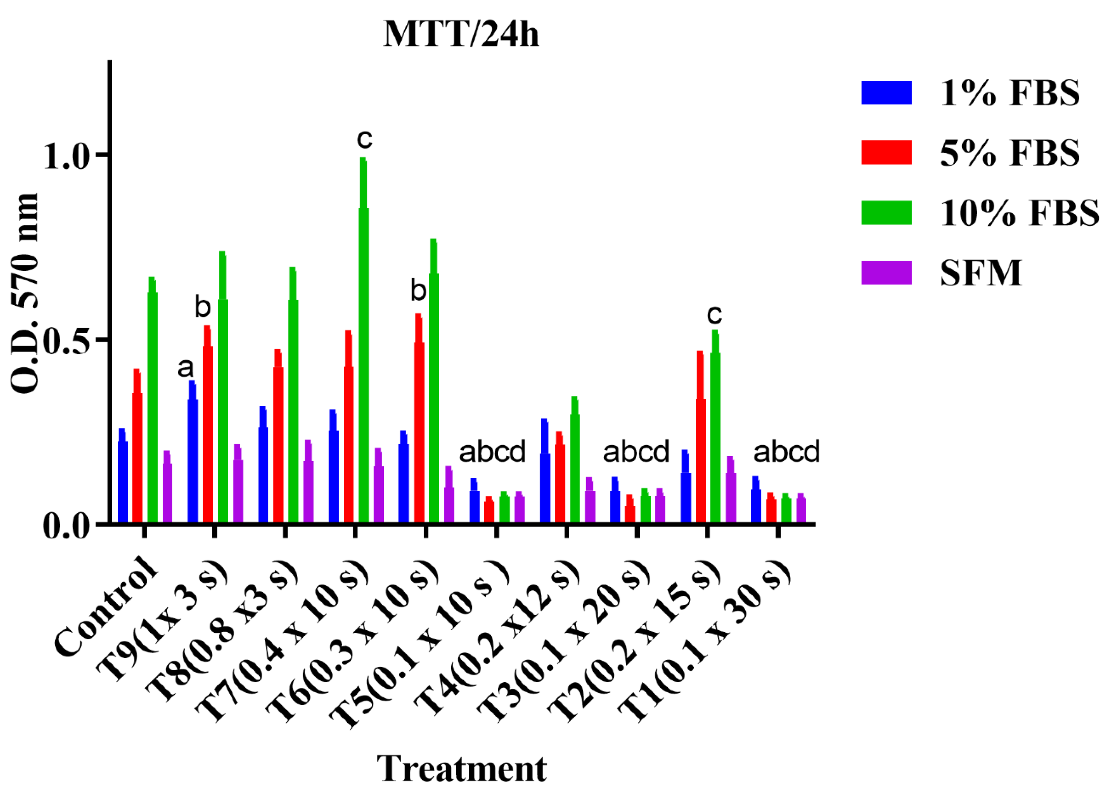
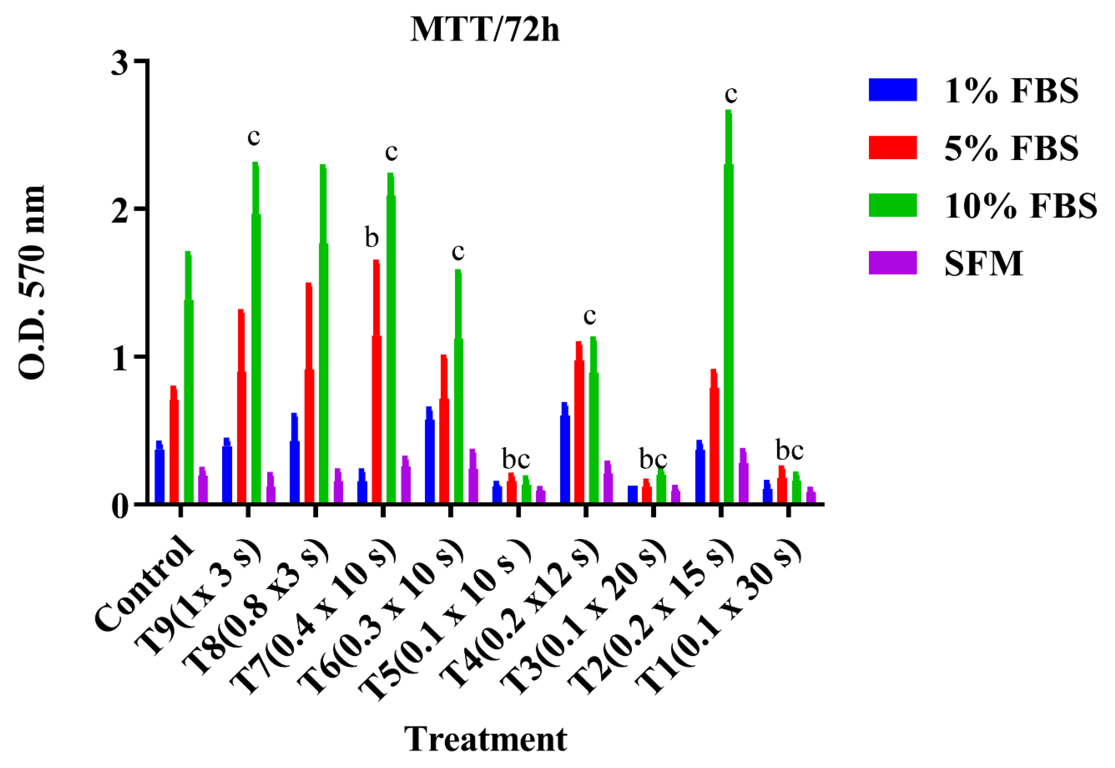
3.2. Cell Proliferation Assay (MTT)

The effects of various treatment (laser power and exposure time) conditions on DPSC proliferation under different serum conditions were assessed at 24, 48, and 72 h. At 24 h, cells cultured in 1% FBS in the T1 group showed a significant difference when compared to the control untreated group (p < 0.05) (Figure 2).
For 5% FBS, cells treated with the treatments T5 (0.1 × 10 s), T3 (0.1 × 20 s), and T1 (0.1 × 30 s) showed a significant decrease in the cell growth rate when compared to the control untreated group (p < 0.05). For 10% FBS, a significant increase in the proliferation rate of cells treated with T7 (0.4 × 10 s) was detected (p < 0.05), whereas the following treatments showed a statistically significant decrease in the proliferation rate of treated cells: T1 (0.1 × 10 s), T2 (0.2 × 15 s), T3 (0.1 × 20 s), T4 (0.2 × 12 s), and T5 (0.1 × 10 s) (p < 0.05). Cells cultured in serum-free media (SFM) did not show any significant difference among treated groups.
After 48 h, DPSCs cells cultured in 5% FBS showed a significant increase in the proliferation rate of cells treated with T7 (0.4 × 10 s) and T6 (0.3 × 10 s) was observed when cells compared to the control untreated group ((p < 0.05) and (p < 0.05), respectively), while T5 (0.1 × 10 s), T3 (0.1 × 20 s), and T1 (0.1 × 30 s) showed a significant decrease in the cell growth rate when compared to the control untreated group (p < 0.00005). For 10% FBS, a significant increase in the proliferation rate of cells treated with T7 (0.4 × 10 s) was detected (p < 0.05), whereas the following treatments showed a statistically significant decrease in the proliferation rate of treated cells: T1 (0.1 × 10 s), T2 (0.2 × 15 s), T3 (0.1 × 20 s), T4 (0.2 × 12 s), and T5 (0.1 × 10 s) (p < 0.05). For 1% FBS and SFM, no significant difference was observed among treated groups, Figure 3.
After 72 h, cells cultured in 5% FBS showed a significant decrease in the proliferation rate of cells treated with T6 (0.3 × 10 s) (p < 0.05), T5 (0.1 × 10 s) (p < 0.05), T3 (0.1 × 20 s (p < 0.05), T2 (0.2 × 15 s) (p < 0.05), and T1 (0.1 × 30 s) (p < 0.05), compared to the control untreated group. For 10% FBS, T9 (1 × 3 s) (p < 0.05) and T7 (0.4 × 10 s) (p < 0.05) showed a significant increase in the proliferation rate of the treated cells, whereas the following treatment showed a significant decrease in the growth rate of treated cells: T5 (0.1 × 10 s) (p < 0.05), T4 (0.2 × 12 s) (p < 0.05), T3 (0.1 × 20 s) (p < 0.05), T2 (0.2 × 15 s) (p < 0.05), and T1 (0.1 × 30 s) (p < 0.05). For 1% FBS and SFM, no significant difference was observed among treated groups, Figure 4.

3.3. Cell Death Analysis
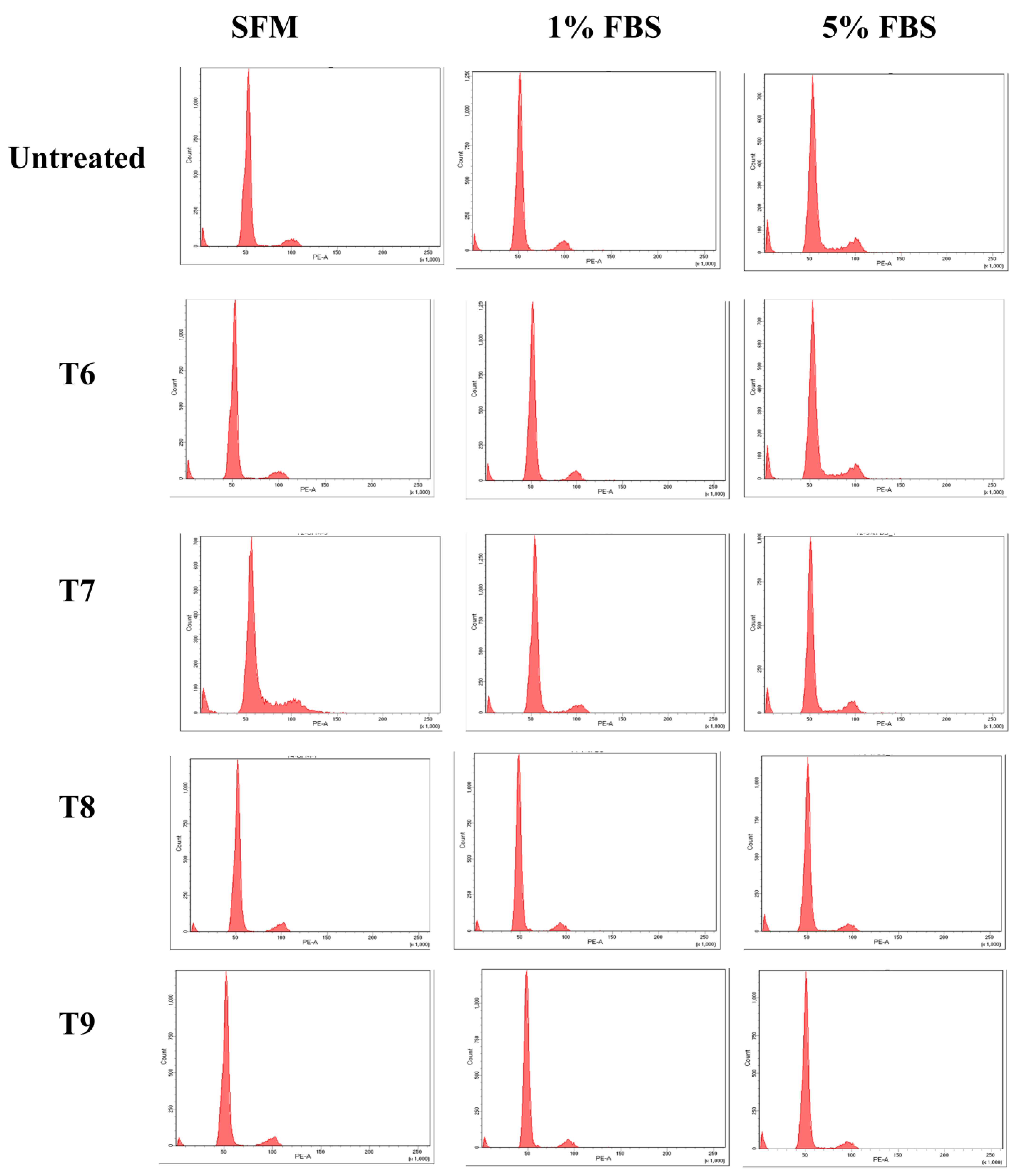
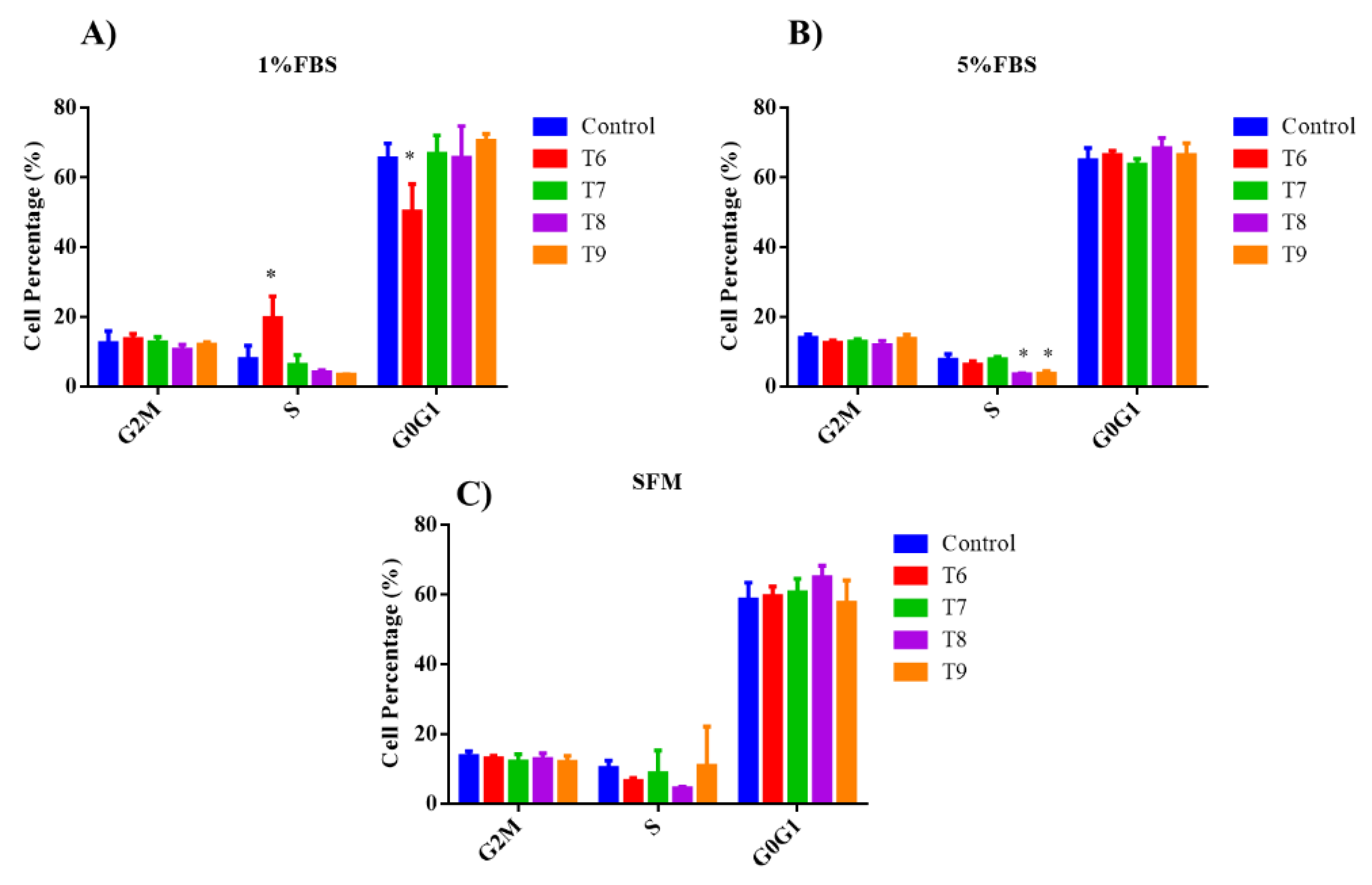
Cell death analysis for apoptosis or necrosis was performed in hDPSCs treated with various laser treatments (Groups T6 to T9) cultured under different serum (0, 1, or 5%) conditions after 48 h using flow cytometry. These groups were selected according to their data in the previous experiment as treatment groups from T6 to T9 showed the highest and most significant proliferation rates when compared to their control groups (p < 0.05).
For cell death analysis, interestingly, none of our treatment groups demonstrated any significant differences compared to the untreated control group. Remarkably, no significant difference in the percentage of healthy and apoptotic/necrotic cells was detected among all treatment groups (T6 to T9) compared to the control untreated group, Figure 5.

3.4. Cell Cycle

To examine if PBM treatments impact cell cycle progression, hDPSCs treated with various laser treatments (Groups T6 to T9) cultured under different serum (0, 1, or 5%) conditions were analyzed after 48 h of treatment by flow cytometry, Figure 6. As shown in Figure 7, our data analysis demonstrated that there is a significant increase in the number of cells arrested in the S phase for DPSCs treated with T6 and cultured in 1% FBS compared to their control untreated cells (p < 0.05). Meanwhile, for cells cultured in 5% FBS, cells treated with T8 and T9 showed a significant decrease in the number of cells in the S phase compared to their control untreated group (p < 0.05), concluding that these parameters were able to enhance the progression of treated cells in the cell cycle to the second stage (G2M). Moreover, it was noted there were no significant changes in individual cell cycle phases in any of the PBM-treated groups when cells were cultured in SFM.

4. Discussion

There have been recent reports on the accumulating evidence for the use of photobiomodulation (PBM) therapy in stimulating several biological responses in dental pulp stem cells to regenerate dentin [8,18,19,20]. However, these reviews pointed to a major lack of consistency in the critical parameters defining the PBM dosing that has generated uncertainties and inconsistencies in clinical outcomes. Due to a lack of information regarding a well-defined set of parameters concerning laser PBM’s effect on hDPSCs, the current study examined the key parameters such as the power output, time, irradiance (power density), and fluence (energy density). This study aimed to bridge the gap in knowledge by investigating the effects of PBM treatments on hDPSC proliferation, cell death, and cell cycle to outline optimal parameters for clinical regenerative protocols. We generated and characterized primary cell culture studies from human-extracted teeth and performed studies in varying serum conditions from normal (10% serum) versus stress-induced (0 to 5% serum) to mimic clinical scenarios of cells following pulp damage.
The present study revealed that the 810 nm wavelength at 0.4 W for 10 s was more effective in stimulating hDPSC cell growth when compared to other treatment settings. These results were consistent in 5% and 10% FBS after 24, 48, and 72 h. Optimizing parameters in this preliminary study required various settings to standardize variables as indicated by Khan and Arany’s analysis and as stated by “Arndt-Schultz Law” [11,12,13]. For instance, although T6 and T9 yielded the same dose (0.3125 J/cm2), the biological response of the cells behaved differently; after 48h, DPSCs cultured in 5% FBS in T6 (0.3 × 10 s) showed a significant increase in the proliferation rate of cells when cells compared to the control untreated group, while after 72 h, cells in the same group T6 showed a significant decrease in the proliferation rate compared to the control untreated group, and cells in group T9 (1 × 3 s) showed a significant increase in the proliferation rate of the treated cells. These current findings, which demonstrate the non-reciprocity effect of laser photobiomodulation on cells, confirm previous reports that demonstrated the non-linear effect of laser photobiomodulation; even if the energy density is the same (inverse linear relationship between irradiance and time), the changes in biological endpoint will not necessarily be equal [21].
Regarding wavelength, our findings are in line with previous studies, where an infrared 810 nm wavelength laser demonstrated the capability for cell biostimulation and proliferation on hDPSCs [2,17,18,19]. Infrared light was chosen because its penetration through tissue is maximal in this wavelength range due to lower scattering and absorption by tissue chromophores [21]. The effectiveness of photobiomodulation on the target tissues is dependent on a combination of parameters such as wavelength, laser power, power density (irradiance), energy density (fluence), and exposure time [22]. The current study aimed to standardize the different variables to use them as a starting point for further studies. Laser power of 0.4 W for 10 s resulted in significant cell proliferation in comparison to other treated and control groups for cells cultured in 10% FBS. As demonstrated in Table 1, the power density was 0.5 W/cm2 and the energy density was 0.416 J/cm2. These results, although disagreeing with other results which revealed that the energy density ranging between 1 and 5 J/cm2 is optimal to achieve an optimal biological effect in different cells [22,23], cannot be compared due to the differences in the studies’ settings. Moreover, the power density of 0.5 W/cm2, which was optimal in our study for cell proliferation, was considered the optimal therapeutic dose in other studies, although with different settings and purposes [24,25].
Although continuous mode rather than pulsed mode was used in this study, the apoptosis assay, which was performed to determine the cell death modality of hDPSCs treated with different laser treatments for 48 h by flow cytometry, did not show any significant difference compared to the control untreated groups, and the number of healthy cells was similar among all treated groups and their control untreated groups. The credit for minimizing the phototoxic effect of the laser dose and preventing thermal damage to the treated cells goes to a previous communication between the authors and Dr. Arany regarding the burning of the cells. The continuous steady motion during treatment kept the distance between the probe fiber tip and the cell layer in the bottom of a clear plate well constant at 1 cm, minimizing the induction of any thermal detrimental phototoxic effects on the treated cells [11,14,26]. The current study precisely demonstrated the mode of laser application and the parameters utilized to facilitate comparison with any future studies and upcoming results. since it is well established that the bio-stimulatory effect of low-power laser is influenced by several combined parameters [27]. Recent studies in the field of photobiomodulation therapy on stem cells are shifting toward understanding the precise molecular mechanism involved in the process [2]. Identifying the mode of action, the molecular mechanism, and the side effects before transferring the in vitro studies to clinical practice is fundamental. Tissue engineering and regenerative therapy will replace and bypass the limitations of the conventional therapeutic regimes in both medical and dental fields [1,2,27]. Moreover, it would be interesting in the future to test photobiomodulation in combination with other compounds such as ozone application [28] and probiotics [29] to evaluate possible combined therapeutic strategies.
The limitations of the current study are as any other in vitro studies which do not accurately recapitulate the pathophysiology of human cells in vivo. More sophisticated investigations should be performed in the future to lead to a deeper insight into this era. The wide variety of laser devices in the dental market forms another challenge with clinical implications. Further optimization will therefore be required to achieve the optimal effects desired. Nonetheless, the detailed mechanisms underlying the photobiomodulation process remain undetermined and require further research to demonstrate the direct link between photobiomodulation and stem cell biology.

5. Conclusions

Despite the study limitations, it was possible to conclude that treating hDPSCs by infrared wavelength (810 nm) at the given parameters (0.4 W, 10 s, 0.5 W/cm2, 0.416 J/cm2) and study setting showed no cell necrosis and induced cell proliferation. Further studies still need to be performed to better optimize these parameters and laser photobiomodulation protocols. However, this study, which is performed for the first time in our stem cell center in Jordan, is certainly a brick in the era of tissue engineering related to the regenerative pulp therapy field.

Author Contributions

Conceptualization, A.A.A.-A.; Methodology, A.A.A.-A., D.A. and N.A.; Investigation, D.A., S.Z. and M.I.; Study design, A.A.A.-A., D.A. and H.J.; Writing the original draft, A.A.A.-A. and D.A.; Supervision, writing—review and editing, A.A.A.-A., A.A. and P.A. All authors have read and agreed to the published version of the manuscript.

Funding

This research received no external funding.

Institutional Review Board Statement

Human third molar samples were collected from healthy donors (18, 19, and 21 years), according to the guidelines of the Declaration of Helsinki and according to the Institutional Review Board (IRB) guidelines from the Cell Therapy Center/The University of Jordan (IRB/06/2018) and approved on 13 March 2018.

Informed Consent Statement

All human participants gave their informed consent before their tooth donation.

Data Availability Statement

The datasets used and/or analyzed during the current study are available from the corresponding author upon reasonable request. The data are not publicly available due to restrictions for privacy.

Acknowledgments

The authors would like to express their acknowledgment to Jamil Al-Kayyali and Fadi Nahhab for their assistance in laser device utilization.

Conflicts of Interest

The authors declare no conflict of interest.

References

  1. Tuby, H.; Maltz, L.; Oron, U. Low-level laser irradiation (LLLI) promotes proliferation of mesenchymal and cardiac stem cells in culture. Lasers Surg. Med. 2007, 39, 373–378. [Google Scholar] [CrossRef] [PubMed]
  2. Arany, P.R.; Cho, A.; Hunt, T.D.; Sidhu, G.; Shin, K.; Hahm, E.; Huang, G.X.; Weaver, J.; Chen, A.C.-H.; Padwa, B.L.; et al. Photoactivation of endogenous latent transforming growth factor–β1 directs dental stem cell differentiation for regeneration. Sci. Transl. Med. 2014, 6, 238. [Google Scholar] [CrossRef] [PubMed]
  3. Huang, G.T.-J. Dental pulp and dentin tissue engineering and regeneration–advancement and challenge. Front. Biosci. 2011, 3, 788. [Google Scholar] [CrossRef] [PubMed]
  4. Eduardo, F.D.P.; Bueno, D.F.; de Freitas, P.M.; Marques, M.M.; Passos-Bueno, M.R.; Eduardo, C.D.; Zatz, M. Stem cell proliferation under low-intensity laser irradiation: A preliminary study. Lasers Surg. Med. 2008, 40, 433–438. [Google Scholar] [CrossRef] [PubMed]
  5. Ravindran, S.; Huang, C.-C.; George, A. Extracellular matrix of dental pulp stem cells: Applications in pulp tissue engineering using somatic MSCs. Front. Physiol. 2016, 4, 395. [Google Scholar] [CrossRef]
  6. Marrelli, M.; Falisi, G.; Apicella, A.; Apicella, D.; Amantea, M.; Cielo, A.; Bonanome, L.; Palmieri, F.; Santacroce, L.; Giannini, S.; et al. The behavior of dental pulp stem cells on different types of innovative mesoporous and nanoporous silicon scaffolds with different functionalizations of the surfaces. J. Biol. Regul. Homeost. Agents 2015, 29, 991–997. [Google Scholar]
  7. Dannan, A. Dental-derived stem cells and whole tooth regeneration: An overview. J. Clin. Med. Res. 2009, 1, 63–71. [Google Scholar] [CrossRef]
  8. Kitamura, C.; Nishihara, T.; Terashita, M.; Tabata, Y.; Jimi, E.; Washio, A.; Hirata, S. Regeneration approaches for dental pulp and periapical tissues with growth factors, biomaterials, and laser irradiation. Polymers 2011, 3, 1776–1793. [Google Scholar] [CrossRef]
  9. Goldberg, M.; Smith, A.J. Cells and extracellular matrices of dentin and pulp: A biological basis for repair and tissue engineering. Crit. Rev. Oral Biol. Med. 2004, 15, 13–27. [Google Scholar] [CrossRef]
  10. Shen, C.C.; Yang, Y.C.; Chiao, M.T.; Chan, S.C.; Liu, B.S. Low-level laser stimulation on adipose-tissue-derived stem cell treatments for focal cerebral ischemia in rats. Evid. Based Complement. Altern. Med. 2013, 2013, 594906. [Google Scholar] [CrossRef]
  11. Khan, I.; Tang, E.; Arany, P. Molecular pathway of near-infrared laser phototoxicity involves ATF-4 orchestrated ER stress. Sci. Rep. 2015, 5, 10581. [Google Scholar] [CrossRef] [PubMed]
  12. Huang, Y.Y.; Chen, A.C.; Carroll, J.D.; Hamblin, M.R. Biphasic dose response in low-level light therapy. Dose-Response 2009, 7, 9–27. [Google Scholar] [CrossRef] [PubMed]
  13. Huang, Y.Y.; Sharma, S.K.; Carroll, J.; Hamblin, M.R. Biphasic dose response in low-level light therapy–an update. Dose-Response 2011, 9, 602–618. [Google Scholar] [CrossRef] [PubMed]
  14. Khan, I.; Arany, P. Photobiomodulation therapy promotes expansion of epithelial colony forming units. Photomed. Laser Surg. 2016, 34, 550–555. [Google Scholar] [CrossRef] [PubMed]
  15. Tanikawa, D.Y.S.; Pinheiro, C.C.G.; Almeida, M.C.A.; Oliveira, C.R.; Coudry, R.D.; Rocha, D.L.; Bueno, D.F. Deciduous dental pulp stem cells for maxillary alveolar reconstruction in cleft lip and palate patients. Stem Cells Int. 2020, 2020, 6234167. [Google Scholar] [CrossRef] [PubMed]
  16. Jafar, H.; Abuarqoub, D.; Ababneh, N.; Hasan, M.; Al-Sotari, S.; Aslam, N.; Kailani, M.; Ammoush, M.; Shraideh, Z.; Awidi, A. HPL promotes osteogenic differentiation of stem cells in 3D scaffolds. PLoS ONE 2019, 14, e0215667. [Google Scholar] [CrossRef]
  17. Dominici, M.; Blanc, K.L.; Mueller, I.; Slaper-Cortenbach, I.; Marini, F.C.; Krause, D.S.; Deans, R.J.; Keating, A.; Prockop, D.J.; Horwitz, E.M. Minimal criteria for defining multipotent mesenchymal stromal cells. The International Society for Cellular Therapy position statement. Cytotherapy 2006, 8, 315–317. [Google Scholar] [CrossRef]
  18. Borzabadi-Farahani, A. Effect of low-level laser irradiation on proliferation of human dental mesenchymal stem cells; a systemic review. J. Photochem. Photobiol. B Biol. 2016, 162, 577–582. [Google Scholar] [CrossRef]
  19. Gholami, L.; Afshar, S.; Mahmoudi, R.; Arkian, A.A.; Parsamanesh, G.; Rad, M.R.; Baghaei, K. Evaluation of the effect of near-infra-red photobiomodulation on buccal fat pad-derived stem cells. Int. J. Dent. Oral Sci. 2020, 7, 960–967. [Google Scholar]
  20. Santos, L.T.O.; Santos, L.O.; Guedes, C.D. LASERTERAPIA NA ODONTOLOGIA: Efeitos e aplicabilidades. Sci. Gen. 2021, 2, 29–46. [Google Scholar]
  21. Keshri, G.K.; Kumar, G.; Sharmak, M.; Bora, K.; Kumar, B.; Gupta, A. Photobiomodulation effects of pulsed-NIR laser (810 nm) and LED (808±3 nm) with identical treatment regimen on burn wound healing: A quantitative label-free global proteomic approach. J. Photochem. Photobiol. 2021, 6, 100024. [Google Scholar] [CrossRef]
  22. de Freitas, L.F.; Hamblin, M.R. Proposed mechanisms of photobiomodulation or low-level light therapy. IEEE J. Sel. Top. Quantum Electron. 2016, 22, 348–364. [Google Scholar] [CrossRef] [PubMed]
  23. Zein, R.; Selting, W.; Hamblin, M.R. Review of light parameters and photobiomodulation efficacy: Dive into complexity. J. Biomed. Opt. 2018, 23, 120901. [Google Scholar] [CrossRef] [PubMed]
  24. Kocherova, I.; Bryja, A.; Błochowiak, K.; Kaczmarek, M.; Stefańska, K.; Matys, J.; Grzech-Leśniak, K.; Dominiak, M.; Mozdziak, P.; Kempisty, B.; et al. Photobiomodulation with red and near-infrared light improves viability and modulates the expression of mesenchymal and apoptotic-related markers in human gingival fibroblasts. Materials 2021, 14, 3427. [Google Scholar] [CrossRef] [PubMed]
  25. Babuccu, C.; Keklikoğlu, N.; Baydoğan, M.; Kaynar, A. Cumulative effect of low-level laser therapy and low-intensity pulsed ultrasound on bone repair in rats. Int. J. Oral Maxillofac. Surg. 2014, 43, 769–776. [Google Scholar] [CrossRef] [PubMed]
  26. Schimberg, A.S.; Heldens, G.T.N.; Klabbers, T.M.; Grunsven, A.C.v.E.-V.; Verdaasdonk, R.M.; Takes, R.P.; Wellenstein, D.J.; Broek, G.B.v.D. Thermal effects of CO2, KTP, and blue lasers with a flexible fiber delivery system on vocal folds. J. Voice 2022, in press. [CrossRef] [PubMed]
  27. El Gammal, Z.H.; Zaher, A.M.; El-Badri, N. Effect of low-level laser-treated mesenchymal stem cells on myocardial infarction. Lasers Med. Sci. 2017, 32, 1637–1646. [Google Scholar] [CrossRef]
  28. Scribante, A.; Gallo, S.; Pascadopoli, M.; Frani, M.; Butera, A. Ozonized gels vs chlorhexidine in non-surgical periodontal treatment: A randomized clinical trial. Oral Dis. 2023, in press. [CrossRef]
  29. Amini, F.; Rezvani, M.B.; Bakhtiari, R.; Ghomsheh, E.T. Effects of dental pulp stem cell preconditioning on osteogenesis using conditioned media of probiotics bacteria. Avicenna J. Med. Biotechnol. 2023, 15, 76–83. [Google Scholar] [CrossRef]
Figure 1. Characterization of Dental Pulp Stem Cells (DPSCs). (A) Morphological appearance of DPSCs at day 7 of primary extraction. (B) Flow cytometric histograms for the analysis of the expression of mesenchymal stem cell (MSC) surface marker expression on the derived hDPSCs; flow cytometric histograms show that DPSCs are positive for CD90, CD44, CD73, and CD105, and negative for expression of hematopoietic stem cells surface markers (Negative cocktail = CD19, CD34, CD11b, CD45, and HLA-DR). Grey histograms represent the isotype control for each marker, while red histograms represent the expression of the marker. (C) Adipogenic and osteogenic differentiation of the derived DPSCs (Scale Bar = 10 µm).
Applsci 14 00124 g001
Figure 2. Cell proliferation assay (MTT) was performed on different low-power laser treatment groups after 24 h of laser treatment. A two-way ANOVA test was used to calculate the significant difference of the treated groups compared to the control untreated group. a: significance of 1% FBS, b: significance of 5% FBS, c: significance of 10% FBS, d: significance of SFM (p < 0.05). O.D. = optical density.
Applsci 14 00124 g002
Figure 3. Cell proliferation assay (MTT) was performed on different low-power laser treatment groups after 48 h of laser treatment. A two-way ANOVA test was used to calculate the significant difference of the treated groups compared to the control untreated group. b: significance of 5% FBS, c: significance of 10% FBS (significance: p < 0.05). O.D. = optical density.
Applsci 14 00124 g003
Figure 4. Cell proliferation assay (MTT) was performed on different low-power laser treatment groups after 72 h of laser treatment. A two-way ANOVA test was used to calculate the significant difference of the treated groups compared to the control untreated group. b: significance of 5% FBS, c: significance of 10% FBS (significance: p < 0.05). O.D. = optical density.
Applsci 14 00124 g004
Figure 5. Cell death analysis of DPSCs treated with various PBM parameters under different serum conditions for 48 h. Samples were stained with Annexin/PI and analyzed by flow cytometry.
Applsci 14 00124 g005
Figure 6. Flow cytometric analysis of the cell cycle of dental pulp stem cells (DPSCs) treated with various PBM parameters under different serum conditions, 1% FBS, 5% FBS, and serum-free medium (SFM), for 48 h. Samples were prepared by staining cells with Propidium Iodide (PI) and analyzed by flow cytometry.
Applsci 14 00124 g006
Figure 7. Statistical analysis of cell cycle phases (G2M, S, and G0G1) of DPSCs treated with different parameters of PBM under different serum conditions (A) 1% FBS, (B) 5% FBS, and (C) serum-free medium (SFM) for 48 h. A two-way ANOVA test was used for statistical analysis. (* p < 0.05).
Applsci 14 00124 g007
Table 1. Photobiomodulation therapy (810 nm) was used in the treatment of hDPSCs (6-well and 96-well plates) at different laser power levels and different exposure time intervals, after 24 h of seeding to perform MTT assay (groups T1–T9) and apoptosis assay (groups T6–T9).
Treatment GroupPower (W)Time (s)Energy (J)Spot Size (cm2)Irradiance (W/cm2)Surface Area (cm2) (6 or 96-Well Plate)Fluence (J/cm2)
T10.13030.80.1251.086 2.76
T20.21530.80.251.0862.76
T30.12020.80.1251.0861.84
T40.2122.40.80.251.0862.21
T50.11010.80.1251.0860.92
T60.31030.80.3759.60.3125
T70.41040.80.59.60.416
T80.832.40.819.60.25
T91330.81.259.60.3125
Disclaimer/Publisher’s Note: The statements, opinions and data contained in all publications are solely those of the individual author(s) and contributor(s) and not of MDPI and/or the editor(s). MDPI and/or the editor(s) disclaim responsibility for any injury to people or property resulting from any ideas, methods, instructions or products referred to in the content.

