Article

The Influence of Cytokinin on the Multiplication Efficiency and Genetic Stability of Scutellaria baicalensis Regenerants in In Vitro Culture Conditions

by
Magdalena Dyduch-Siemińska
and
Jacek Gawroński
*
Department of Genetics and Horticultural Plant Breeding, Institute of Plant Genetics, Breeding and Biotechnology, University of Life Sciences in Lublin, Akademicka 15 Street, 20-950 Lublin, Poland
*
Author to whom correspondence should be addressed.
Appl. Sci. 2024, 14(11), 4791; https://doi.org/10.3390/app14114791
Submission received: 30 April 2024 / Revised: 27 May 2024 / Accepted: 29 May 2024 / Published: 31 May 2024
(This article belongs to the Special Issue Advances in Breeding in Agricultural and Animal Science)

Abstract

:
The efficiency and method of regeneration in in vitro culture conditions depend primarily on the plant growth regulators (PGRs) used. Even growth regulators belonging to one group may have different effects, stimulating the process of direct or indirect organogenesis, thus possibly disturbing the genetic stability among regenerants. The main aim of this study was to identify the genetic stability of Scutellaria baicalensis regenerates obtained by in vitro culture method using start codon targeted (ScoT) markers. S. baicalensis nodal explants were regenerated on MS medium supplemented with kinetin (KIN) at concentrations of 0.25, 0.5, 1.0, and 2.0 mg × dm−3 or benzylaminopurine (BAP)—0.25, 0.5, 1.0, and 2.0 mg × dm−3. The effects of the number of propagated shoots, length, number of nodes, and fresh mass of regenerants were assessed. Moreover, the genetic stability of the regenerants was analyzed using start codon targeted (SCoT) markers. Direct shoot organogenesis was observed on an MS medium containing kinetin, while indirect shoot induction occurred on an MS medium supplemented with BAP. The highest average number of shoots (3.6) was achieved for the MS + KIN medium at a concentration of 0.25 and 5.8 for the MS + BAP 1.0 medium. The average length and average number of nodes were the highest on the MS + BAP 0.25 medium (50.0 and 6.0, respectively), while the lowest values of these features were observed on the MS + KIN 2.0 medium (40.3 and 4.9, respectively). A total of 111 amplified bands were exhibited by SCoT primers. Three of the analyzed primers revealed four unique genotype-specific markers. The average percentage of polymorphism obtained was 36.7%. The analysis of genetic similarity revealed a high level of genetic similarity between the donor plant and regenerants obtained on MS “0” (medium without the addition of phytohormones). A slightly lower value of genetic similarity was observed for regenerants obtained by direct organogenesis (MS + KIN medium at all concentrations). Indirect shoot organogenesis observed on the MS + BAP medium (all concentrations) resulted in the highest differentiation, both in relation to the donor plant and MS “0” regenerants. The results of our work indicate that, in the case of S. baicalensis, the maintenance of genetic stability depends primarily on the presence of the cytokinin type in the medium.

1. Introduction

The valuable properties of medicinal plants have been appreciated for many centuries worldwide, and research on their active compounds is being conducted on an increasingly large scale. Scutellaria baicalensis Georgi (Baikal skullcap) is one of the plants utilized in modern conventional and herbal medicine [1,2,3]. The properties of biologically active compounds in this plant have allowed for its wide applications, ranging from treating common colds to addressing significant contemporary issues such as the global SARS-CoV-2 pandemic and cancer [4]. The origins of its use trace back to Central and East Asia. The Chinese have been using the dried roots of this medicinal plant (Scutellariae baicalensis radix) for over 2000 years in traditional medicine, known as Huang-Qin [5,6]. It contains biologically active substances, mainly flavonoids [7]. The genus Scutellaria belongs to the family Lamiaceae, and according to various sources, it includes approximately 360 species. S. baicalensis is a perennial herb that reaches heights in a range of 25–60 cm. It forms straight stems covered with small lanceolate leaves and has a small, short rhizome with a taproot with numerous lateral roots. The flowers are blue-violet in color [8]. Its broad pharmacological effects have led to attempts to propagate this species using in vitro tissue culture techniques. Plant tissue culture techniques are widely used for the rapid and efficient propagation of various medicinal plant species [9]. This technique is an alternative method for the clonal multiplication of plants when conventional methods are not effective enough. Micropropagation can provide large numbers of uniform plants, using only a small fragment of the parent plant as the primary explant. They provide invaluable assistance in conventional plant improvement methods, enabling the production of renewable biomass, typically with a high capacity for the biosynthesis of desired compounds [10,11,12]. These techniques also contribute to the conservation of endangered and endemic medicinal plant species [13]. So far, research on the in vitro micropropagation of the Scutellaria genus has focused on species, such as S. orientalis [14,15], S. alpine [16,17], S. araxiensis [18], S. barbata, and S. racemosa [19]. They indicate the possibility of regeneration by both indirect and direct organogenesis. For S. baicalensis, efficient regeneration took place from various types of explants, in the medium enriched with kinetin and 1-Naphthaleneacetic acid (NAA), NAA and 6-Benzylaminopurine (BAP) [20,21], and also in the presence of thidiazuron (TDZ) [22,23]. At the same time, the process of clonal multiplication can generate genetically uniform plants or, through somaclonal variation, lead to their diversification [24]. Molecular markers based on the DNA sequence are the most commonly used approach to detect the genetic stability/variability of plants grown in vitro [25].
Start codon targeted polymorphism markers are molecular tools used to analyze differences in DNA sequences. The method utilizing SCoT markers involves the amplification of random DNA fragments using short primers (15–16 nucleotides) that flank the translation start codon—ATG. Since their development, SCoT markers have been applied in population genetics and genetic diversity analysis in a wide range of plants, like cereals, legumes, fruit and vegetable crops, medicinal plants, forest trees, ornamental plants, and other economically important plants [26,27,28,29]. This type of marker has been used to analyze the genetic fidelity of tissue culture-raised plants, which is essential before its commercialization, particularly for elite and superior genotypes, where the regenerant is expected to be true to type [24,30,31,32]. Publications strictly on Scutellaria baicalensis, both those relating to the micropropagation technique and the assessment of genetic diversity using genetic markers, are very rare. An effective system for the in vitro propagation of S. baicalensis was elaborated by Stojakowska et al., 1999 [20]. The analysis of genetic variability in the studied species has, so far, been presented on RAPD [33,34], ISSR [35], and SSR [36] markers. Therefore, this work may expand knowledge regarding the studied species.
The main objective of this study was to identify the genetic stability of Baikal skullcap regenerants obtained on the medium enriched with different types of cytokinin in in vitro cultures using SCoT markers. This allowed us to determine whether and to what extent the application of plant growth regulators influenced the genetic stability of the regenerants by affecting the regeneration process of the studied species. Additionally, the impact of the applied growth regulators on selected culture parameters is presented. The results of this study contribute to elucidating the genetic identity of Baikal skullcap regenerants and enable the selection of a regeneration method that meets the set research objectives.

2. Materials and Methods

2.1. Plant Material and Culture Conditions

The analysis of shoot multiplication efficiency was conducted using a medium developed by Murashige and Skoog (MS) [37], supplemented with agar–agar (8.0 g × dm−3) (Sigma-Aldrich, St. Louis, MO, USA). The medium was enriched with kinetin (KIN—cytokinin containing furfuryl moiety) at concentrations of 0.25, 0.5, 1.0, 2.0 mg × dm3, marked accordingly as MS + KIN 0.25, MS + KIN 0.5, MS + KIN 1, MS + KIN 2, or 6-benzylaminopurine (BAP—cytokinin with an aromatic benzyl group) at a concentration of 0.25, 0.5, 1.0, 2.0 mg × dm3 marked accordingly as MS + BAP 0.25, MS + BAP 0.5, MS + BAP 1, MS + BAP 2. MS medium without phytohormones served as the control (MS “0”). Before autoclaving at 121 °C for 20 min, the pH was set at 5.8. The medium was prepared in glass jars with a total volume of 0.5 dm3, and the volume of the medium was 0.05 dm3. A single S. baicalensis plant was used to initiate the in vitro culture, which was also used for molecular analyses, referred to as the donor plant (DP) (Figure 1A). The plant came from Experimental Farm of the University of Life Sciences in Lublin. Preparation of plant material to start in vitro culture was carried out according to the methodology provided by Gawroński and Dyduch-Siemińska [38] with modifications. Briefly, shoots were washed under tap water for an hour, rinsed for 15 min in distilled water, then sterilized with 70% ethanol for 15 s and 1% sodium hypochlorite for 5 min; they were subsequently washed three times (5 min each) in sterile distilled water. The nodal segments (5–7 mm) were excised and placed on MS medium (“0”) to stabilize the culture (Figure 1B,C). The prepared medium was used to initiate the culture: under aseptic conditions using a laminar flow hood, shoots from the in vitro culture (MS “0”) were cut into segments, and the obtained nodal explants were transferred to jars with 8 explants each. For all substrate variants and the control, 6 jars were analyzed in each of three replicates. The explants were cultured under 16 h light photoperiod (40 μmol m−2 s−1) in growth room at 21 °C ± 2 °C. After 4 weeks, the number of propagated shoots, their average length (mm), average number of nodes, fresh weight of the regenerants (g), and the intensity of callus tissue formation were recorded. The analysis of variance (ANOVA), Duncan’s test, and Pearson correlation coefficient were carried out in Statistica 13.1 software, and the significant difference between each treatment method was determined with a significance level of 5% (p ≤ 0.05).

2.2. DNA Extraction

We extracted DNA from a fragment of a shoot with leaves of the donor plant and plants regenerated on the tested medium. From each tested culture medium, plants were randomly selected, from which DNA isolation was carried out according to the CTAB method [39] in two replicates. The total yields of DNA as well as DNA purity were estimated using a Nanodrop 2000 spectrophotometer (Thermo Scientific, Waltham, MA, USA). Next, a working concentration of 25 ng μL−1 was prepared and stored at 4 °C for further analyses.

2.3. SCoT Analysis

Thirty SCoT markers were preliminarily tested for their suitability for genetic analysis of S. baicalensis. Primers that reproducibly amplified clearly separated bands were selected for further analysis. DNA was amplified using thermocycler (Biometra GmbH) with twelve SCoT primers (Table 1), in a total volume of 10 µL, which contained the following: 25 ng of genomic DNA as a template, 1 × PCR buffer with 1.5 mM MgCl2, 1 μM dNTPs, 0.8 μM of oligonucleotide primer, and 0.5 U of Taq DNA Polymerase (Fermentas). The annealing temperatures provided in Table 2 were used to program a thermocycler for amplification. PCR amplification with the SCoT primers was repeated twice. The amplified bands were resolved on 1.5% (w/v) agarose electrophoresis made in 1.0 × TBE buffer and stained with ethidium bromide. Molecular weight markers of 200–10,000 bp NZYDNA Ladder III (NZYTech) were loaded along with the amplified DNA samples to determine the size of the amplified product, which was visualized and photographed using gel documentation system GeneSnap ver. 7.09 (SynGene, Bangalore, India).

2.4. DNA Data Analysis

Each SCoT fragment was scored as present (1) or absent (0) for each of the 10 DNA samples, excluding the weak and blurred bands, thus generating a binary data matrix. A locus was considered polymorphic if more than one band at the same position was detected for all individuals of the material. The genetic similarity index (SI) values between pairs of samples were estimated according to Dice’s formula [40]. The similarity matrix was used to construct a dendrogram using the UPGMA (Unweighted Pair Group Method of Arithmetic Averages) method in PAST software 4.03 [41] in order to cluster the samples in related groups based on their similarities.

3. Results and Discussion

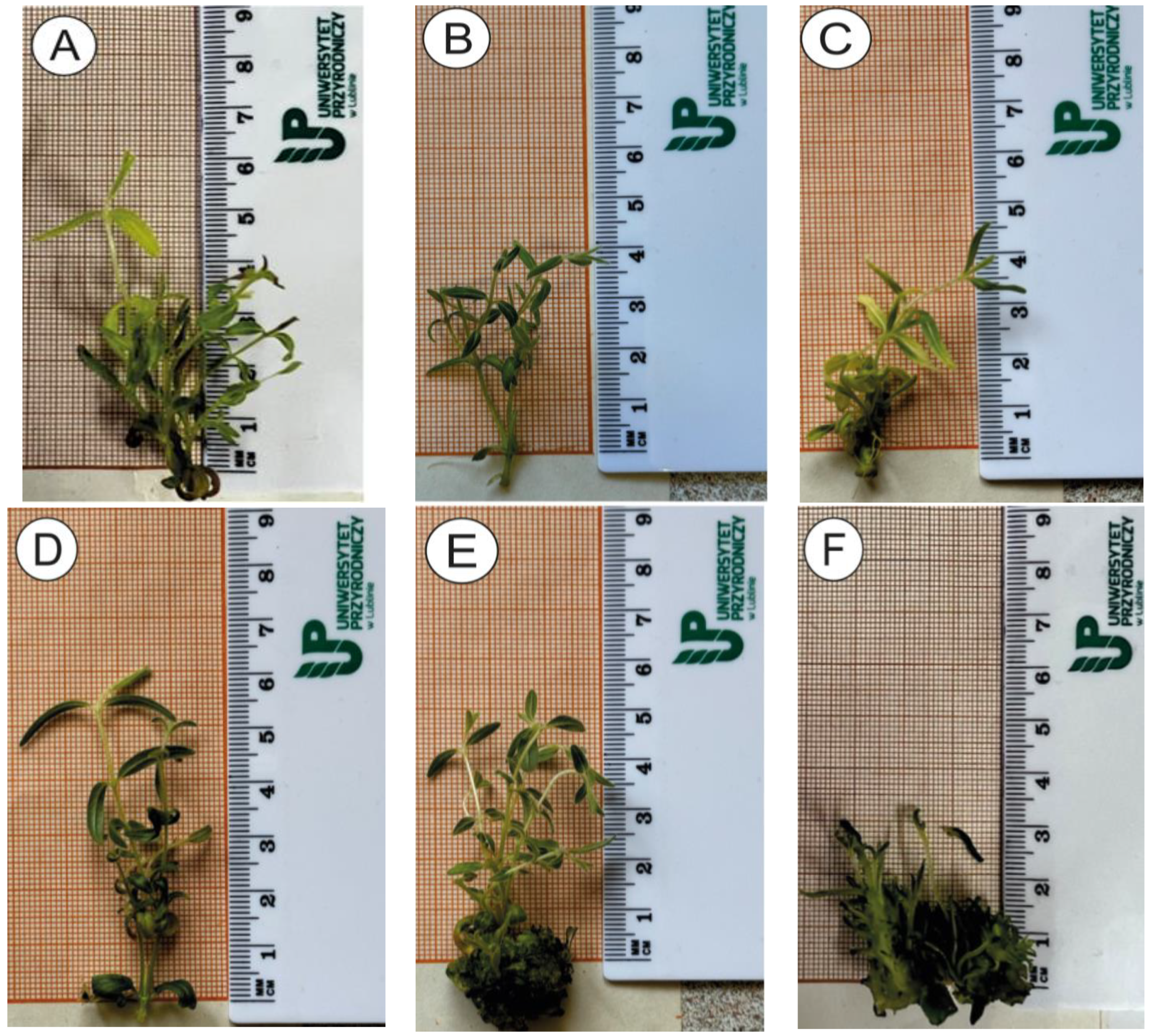
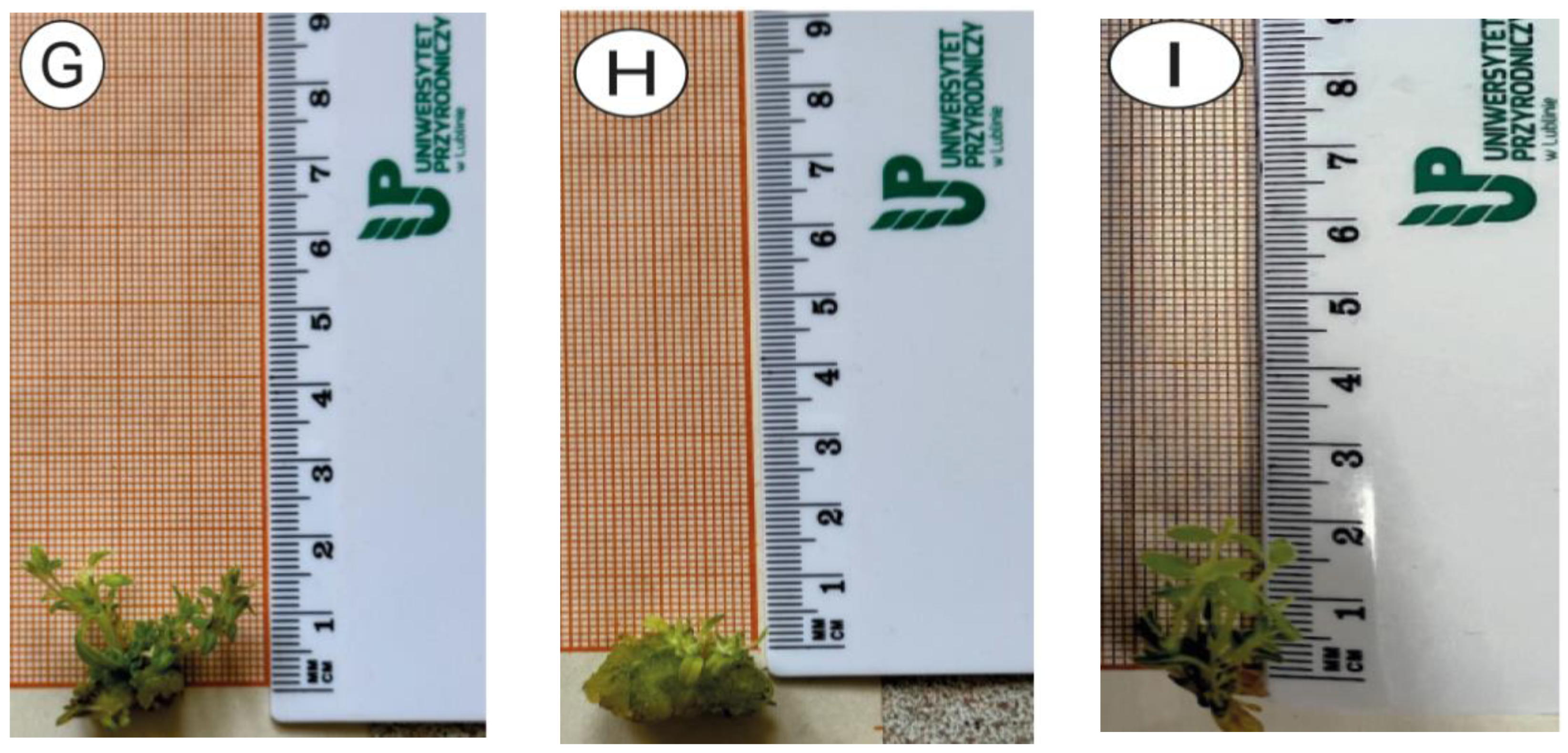
3.1. In Vitro Culture Analysis

The conducted study revealed a significant impact of the applied medium variants on the regeneration of nodal explants of Baikal skullcap (Table 3, Figure 2). The highest mean number of shoots (5.8) of the obtained regenerants was acquired on the MS + BAP 1 medium. The lowest mean number of shoots was obtained on the MS + KIN 2 (2.1). For the observed feature, no statistically significant differences were found on media with different KIN concentrations. These differences were also insignificant in relation to the MS “0” medium. On the medium with BAP at 0.25 and 0.5 mg/dm3, the mean number of shoots was 2.7 and was also statistically insignificant. Increasing the BAP concentration resulted in an increase in the trait value to 5.8 (MS + BAP 1) and 4.3 (MS + BAP 2), respectively, but no statistically significant differences between them were proven. Regenerants from the MS + BAP medium exhibited a well-developed callus (that formed at the base of the shoot) and the highest mean weight. The composition of the medium had a fundamental influence on the observed mode of regeneration. The MS medium with the addition of kinetin initiated the process of direct shoot organogenesis. On the other hand, the process of indirect shoot organogenesis was stimulated by the MS medium with the addition of all concentrations of BAP. Since the regeneration process through this pathway occurs in two stages—the first involving the formation of callus tissue and the second involving the regeneration of shoots by some of its cells—high values were obtained for the presence of calluses and the mass of regenerants in this medium. In particular, the values of the latter were the highest on the MS + BAP 2 medium (statistically significant compared to the other media), which was due to the simultaneous presence of callus tissue and shoots in the structure of the regenerants (Figure 2H). Gharari et al. [18] pointed out the high effectiveness of BAP in combinations with various concentrations of TDZ, IBA, and NAA in the regeneration of Scutellaria araxensis, concluding that the best effect in indirect shoot organogenesis was achieved with the combination of BAP and TDZ. According to a study by Ozdemir et al. [14] on S. orientalis subsp. bicolor, the callus could be regenerated in the presence of BAP alone or in combination with NAA. However, an increase in the concentration of BAP had an inhibitory effect on its mass. In a study by Stojakowska et al. [20] concerning S. baicalensis, the combination of BAP and NAA promoted shoot organogenesis and allowed them to obtain 1.4 to 8.5 shoots per explant. In addition, this combination of phytohormones caused the simultaneous formation of callus tissue. Such a response of explants in the present study occurred as a result of the independent action of BAP at concentrations from 0.25 to 2 mg/dm3 in the medium, and the number of shoots produced varied from 2.7 to 5.8. Various combinations of BAP and NAA in the MS medium were also investigated by Kwiecień et al. [42] in Scutellaria brevibracteata subsp. subvelutina. These authors concluded that BAP concentrations in a range of 0.5–3 mg/dm3 in combination with NAA did not result in statistically significant differences in both fresh and dry biomass. According to Hwang et al. [43], high efficiency in shoot formation through callus tissue could be achieved but only when it was obtained in media containing NAA in combination with KIN or BAP at a concentration of 0.5 or 1.0 mg/dm3. The results of our study partially confirmed this observation, with respect to the MS + BAP medium at all concentrations, where indirect shoot organogenesis was observed (as an effect of BAP alone), while they were contradictory in relation to the MS + KIN medium. The reason for these discrepancies could be the interaction of the applied PGRs with the components of the medium, as pointed out by Petrasek et al. [44]. In addition, it is necessary to consider the interactions between the genotype, type, and PGR concentration, as postulated by Dyduch-Siemińska [45] and confirmed by the results of Gharari et al. [46], where shoot organogenesis through the callus stage allowed for obtaining a very high number of shoots, reaching up to 18 per explant in S. araxensis, and for the species we studied, S. baicalensis, the maximum was 5.8. At the same time, it is worth noting that different skullcap species, i.e., Scutellaria araxensis [18], Scutellaria baicalensis [20], Scutellaria bornmuelleri [46], and Scutellaria orientalis subsp. bicolor [14], exhibited a similar response to the applied MS medium without phytohormones. In the presence of this medium, there was no callus tissue formation, which was also observed in the present study. Considering the mean number of nodes, it should be noted that this feature reached the highest value on the MS + BAP 0.25 (6.0) medium and the lowest on the MS + KIN 0.25 (2.1) medium. The values of this feature are strictly dependent on the length of the shoot, which, for the above-mentioned media combinations, were 50.0 and 13.1 mm, respectively. On the MS + BAP 2 medium, few shoots up to 5.0 mm long and very numerous shoot buds were observed, which were not able to regenerate the shoot during the test, and the mean number of nodes per shoot could not be assessed. In the work of Grzegorczyk-Karolak [47], the length of regenerated shoots ranged from 2 to 4 cm after 6 weeks of culture, depending on the medium applied. In the case of culture multiplication, the number of explants used to establish it is important. It may be high only in the case of the regeneration of a large number of shoots per explant as well as a large number of nodes per shoot. Achieving this goal is possible due to the highly positive correlation between these characteristics (0.97—Table 4). Therefore, based on the conducted research, the MS + KIN 2 medium should be considered the most optimal for the cloning process (direct organogenesis). However, in order to obtain differentiated material in the indirect organogenesis process, the MS + BAP 0.25 medium should be considered the best. In the analysis of the correlation coefficient for the analyzed characteristics, the positive impact of the formation of callus tissue on both the number of shoots per explant (0.71) and the mass of regenerants (0.78) should be distinguished. The correlation coefficient values between the remaining characteristics turned out to be statistically insignificant.

3.2. Molecular Analysis

Numerous publications on the genus Scutellaria have indicated the possibility of obtaining a large number of regenerants, as presented above. However, there are only a few studies that have assessed the quality of regenerants in terms of their genetic homogeneity using commonly applied molecular marker systems, while those that utilized the repeatable SCoT marker system are unique. The present study belongs to the latter group. Rai [48] reported that SCoT markers have been used to detect somaclonal changes and assess the genetic homogeneity of many plant species grown in tissue cultures. Most micropropagation methods involving axillary shoot proliferation showed 100% monomorphism, whereas plants regenerated through the callus exhibited a polymorphism [28,29,44,45]. This study analyzed twelve SCoT primers, and their sequences are listed in Table 1. Products of electrophoresis with SCoT-16,24,28 primers are shown in Figure 3. All primers generated a total of 111 products (averaging 9.25 bands per primer), of which 37 were identified as polymorphic bands, averaging 3.08 bands per primer (Table 5). The percentage of polymorphisms obtained was in the range of 9.0–71.4%, while the average value was 36.7%. Primer SCoT-75 produced 16% of the polymorphic bands. Primers, SCoT-16, SCoT-50, SCoT-71, and SCoT-75 generated polymorphism above 50%. Monomorphic bands were amplified by all primers, with SCoT-4 generating the most, i.e., 11 bands. A total of 61 monomorphic bands were obtained, with an average of 5.08 monomorphic bands per primer. Specific bands were also detected in the analysis of three primers (SCoT-4, SCoT-33, SCoT-75). Specific bands were obtained for genotypes regenerated on the medium with MS + BAP 2, MS + KIN 2, and for donor plants, and their sizes were 2900 bp, 1300, 900 bp, and 1200 bp, respectively. The size of all products obtained ranged from 200 to 8000 bp. The largest product was generated by primer SCoT-75, while the smallest was generated by SCoT-12.
Based on the polymorphism identified using the SCoT technique, the genetic similarity between the examined regenerants was determined using Dice’s formula, developed by Nei and Li [40]. In the genetic similarity analysis and dendrogram creation, all four analyzed concentrations were taken into account for BAP and KIN, respectively. The tested regenerants from the medium without phytohormones showed high genetic similarity to the donor plant, reaching a level of 0.84 (Table 6). This allowed us to conclude that in this regeneration variant, the obtained shoots were free from significant somaclonal variations. This level of genetic similarity in a study by Grzegorczyk-Karolak et al. [47] indicated that true-to-type regenerants of Salvia bulleyana were obtained. Based on Figure 4, it can be observed that regenerants from the MS “0” medium were grouped together with the donor plant. Due to the high value of the genetic similarity of regenerants obtained in the KIN medium, both in relation to DP and MS “0”, regenerants from the MS + KIN medium were added to the dendrogram as the next ones. Low similarity values were observed between the regenerants from the MS + BAP medium and the donor plant as well as MS “0” regenerants (Table 6), as a result of which regenerants from the MS + BAP medium were included last in the formed cluster. The presence of somaclonal variation among regenerants originating from a single donor plant was indicated by Rathore et al. [49] and Rawat et al. [50]. Due to the genetic heterogeneity of callus tissue, shoots/plants regenerated through it may exhibit variation at the molecular level [47,51,52]. The observed low level of genetic similarity regenerants obtained through indirect shoot organogenesis in the medium containing BAP in relation to donor plants was consistent with reports by other authors, such as Zayova et al. [53] and Saravanan et al. [54]. According to some authors [55,56], BAP, as well as other growth regulators such as 2,4-dichlorophenoxy acetic acid (2,4-D) and naphthaleneacetic acid (NAA), may be responsible for the occurrence of variability. Moreover, the balance between cytokinins and auxins may influence the process of callus tissue formation and development [57], and the genetic variability obtained through it can be utilized for the selection of desirable traits in breeding programs [58,59,60,61]. However, such variability is undesirable in the commercial-scale multiplication of a wide range of crops. As Bairu et al. [24] argued, the genetic stability of plants obtained in vitro depends on the regeneration method applied. In such cases, a method that allows for maintaining high genetic integrity should be employed. According to Rai [48], direct shoot regeneration should ensure that true-to-type regenerants are obtained. As stated by Krishna et al. [30], the method of the clonal propagation of plants through adventitious and axillary bud explants is a technique that maintains genetic stability. As regards the analyzed study, direct organogenesis observed on a medium with kinetin resulted in a high genetic similarity, particularly to plants from the MS “0” medium, which are a source of explants for cultures grown on the MS + KIN medium. However, as reported by Ferreira et al. [25], kinetin present in the medium may also contribute to generating variability within somaclones. With respect to the work analyzed, this was reflected in the lower genetic similarity obtained in the MS + KIN medium than the MS “0” in relation to DP plants. The reason for such a situation, as indicated by Gao et al. [62], could be attributed to the influence of PGRs that increase the multiplication rate and induce adventitious shoots or existing genetic variation in cultured cells or tissues [31].

4. Conclusions

The results of our study indicate that in the case of Scutellaria baicalensis, maintaining genetic stability is primarily dependent on the presence of the cytokinin type in the medium. The utilized plant growth regulators determined different pathways of plants’ regeneration under in vitro conditions. BAP, at all concentration tested, by stimulating the shoot regeneration process through the callus stage, caused high genetic variability in regenerants in relation to the donor plant. On the other hand, the process of direct organogenesis induced by the addition of kinetin ensured higher genetic stability. However, it should be noted that, in addition to the aforementioned factors, the composition of the medium, culture conditions, number of subcultures, type of explant, and plant genotype can also influence the expression of polymorphisms. The results of this study enable the optimization of S. baicalensis micropropagation conditions, depending on the expected direction of regenerant utilization. The existence of variation between regenerants, although unfavorable from the perspective of commercial micropropagation, provides an opportunity to select specific genotypes. In the case of the species studied, this may involve a high content of bioactive metabolites. In the next stage of research, we plan to adapt the regenerants to in vivo conditions and then isolate and evaluate the content of biologically active substances.

Author Contributions

Conceptualization, J.G. and M.D.-S.; methodology, J.G.; software, M.D.-S.; validation, J.G. and M.D.-S.; formal analysis, M.D.-S.; investigation, J.G.; resources, J.G.; data curation, M.D.-S.; writing—original draft preparation, J.G.; writing—review and editing, M.D.-S.; visualization, J.G.; supervision, M.D.-S.; project administration, M.D.-S.; funding acquisition, J.G. All authors have read and agreed to the published version of the manuscript.

Funding

The work was funded from the statutory activity of the University of Life Sciences in Lublin UP/RGH/6/022.

Institutional Review Board Statement

Not applicable.

Informed Consent Statement

Not applicable.

Data Availability Statement

The original contributions presented in the study are included in the articlel, further inquiries can be directed to the corresponding author.

Conflicts of Interest

The authors declare no conflicts of interest.

References

  1. Wang, Z.-L.; Wang, S.; Kuang, Y.; Hu, Z.-M.; Qiao, X.; Ye, M. A comprehensive review on phytochemistry, pharmacology, and flavonoid biosynthesis of Scutellaria baicalensis. Pharm. Biol. 2018, 56, 465–484. [Google Scholar] [CrossRef]
  2. Zhao, T.; Tang, H.; Xie, L.; Zheng, Y.; Ma, Z.; Sun, Q.; Li, X. Scutellaria baicalensis Georgi. (Lamiaceae): A review of its traditional uses, botany, phytochemistry, pharmacology and toxicology. J. Pharm. Pharmacol. 2019, 71, 1353–1369. [Google Scholar] [CrossRef]
  3. Song, J.-W.; Long, J.-Y.; Xie, L.; Zhang, L.-L.; Xie, Q.-X.; Chen, H.-J.; Deng, M.; Li, X.-F. Applications, phytochemistry, pharmacological effects, pharmacokinetics, toxicity of Scutellaria baicalensis Georgi. And its probably potential therapeutic effects on COVID-19: A review. Chin. Med. 2020, 15, 102. [Google Scholar] [CrossRef]
  4. Liu, H.; Ye, F.; Sun, Q.; Liang, H.; Li, C.; Lu, R.; Huang, B.; Tan, W.; Lai, L. Scutellaria baicalensis extract and baicalein inhibit replication of SARS-CoV-2 and its 3C-like protease in vitro. J. Enzym. Inhib. Med. Chem. 2021, 36, 497–503. [Google Scholar] [CrossRef]
  5. Zhao, Q.; Chen, X.-Y.; Martin, C. Scutellaria baicalensis, the golden herb from the garden of Chinese medicinal plants. Sci. Bull. 2016, 61, 1391–1398. [Google Scholar] [CrossRef]
  6. Wang, D.; Luo, X.; Huang, Y.; Wang, M.; Xia, Z. Combined magnetic molecularly imprinted polymers with a ternary deep eutectic solvent to purify baicalein from the Scutellaria baicalensis Georgi by magnetic separation. Microchem. J. 2020, 157, 105–109. [Google Scholar] [CrossRef]
  7. Olennikov, D.N.; Chirikova, N.K.; Tankhaeva, L.M. Phenolic compounds of Scutellaria baicalensis Georgi. Russ. J. Bioorg. Chem. 2010, 37, 816–842. [Google Scholar] [CrossRef]
  8. Karpińska, E. Anti-inflammatory and anticancer properties of Scutellaria baicalensis Georgi. Post Fitoter. 2010, 4, 215–223. [Google Scholar]
  9. Debnath, S.C. Zeatin overcomes thidiazuron-induced inhibition of shoot elongation and promotes rooting in strawberry culture in vitro. J. Hortic. Sci. Biotech. 2006, 81, 349–354. [Google Scholar] [CrossRef]
  10. Efferth, T. Biotechnology applications of plant callus cultures. Engineering 2019, 5, 50–59. [Google Scholar] [CrossRef]
  11. Espinosa-Leal, C.A.; Puente-Garza, C.A.; García-Lara, S. In vitro plant tissue culture: Means for production of biological active compounds. Planta 2018, 248, 1–18. [Google Scholar] [CrossRef] [PubMed]
  12. Kikowska, M.; Turowska, N.; Thiem, B. Czy kultury in vitro gatunków roślin chronionych mogą być źródłem surowców do badań fitochemicznych i biologicznych? Farm. Współczesna 2019, 12, 210–217. [Google Scholar]
  13. Coelho, C.; Bocca, A.L.; Casadevall, A. The tools for virulence of Cryptococcus neoformans. Adv. Appl. Microbiol. 2014, 87, 1–41. [Google Scholar] [CrossRef]
  14. Ozdemir, F.A.; Yildirim, M.U.; Kahriz, P.P. Micropropagation of endemic Scutellaria orientalis L. subsp. bicolor using modified MS medium & TDZ. Emir. J. Food Agric. 2015, 27, 818–824. [Google Scholar] [CrossRef]
  15. Ozdemir, F.A.; Kilic, O.; Atalan, E. In vitro callus propagation and antibacterial activities of callus an edible endemic and medicinal plant Scutellaria orientalis L. subsp. bicolor. Prog. Nutr. 2016, 18, 81–86. [Google Scholar]
  16. Zakaria, I.A.; Sherman, S.; Vaidya, B.; Joshee, N. In vitro propagation and synseed mediated short-term conservation of Scutellaria alpine L. and Scutellaria altissima L. Plant Cell Cult. Micropropag. 2020, 16, e163. [Google Scholar] [CrossRef]
  17. Grzegorczyk-Karolak, I.; Kuźma, Ł.; Wysokińska, H. In vitro cultures of Scutellaria alpina as a source of pharmacologically active metabolites. Acta Physiol. Plant. 2016, 38, 7. [Google Scholar] [CrossRef]
  18. Gharari, Z.; Bagheri, K.; Sharafi, A. High-frequency adventitious shoot organogenesis from in vitro stem explants of Scutellaria araxensis Grossh. BioTechnologia 2022, 103, 143–151. [Google Scholar] [CrossRef]
  19. Brearley, T.; Vaidya, B.; Joshee, N. Cytokinin, Carbon Source, and Acclimatization Requirements for In Vitro Propagation of Scutellaria barbata D. Don and Scutellaria racemose Pers. Am. J. Plant Sci. 2014, 5, 3662–3672. [Google Scholar] [CrossRef]
  20. Stojakowska, A.; Malarz, J.; Kohlmünzer, J. Micropropagation of Scutellaria baicalensis Georgi. Acta Soc. Bot. Pol. 1999, 68, 103–107. [Google Scholar] [CrossRef]
  21. Hwang, I.-T.; Lee, J.-J.; Lee, J.; Paik, S.-W.; Kim, Y.-H. Production of Baicalin, Baicalein, and Wogonin on Plant Tissue Culture of Scutellaria baicalensis. Korean J. Plant Resour. 2015, 28, 526–532. [Google Scholar] [CrossRef]
  22. Trivedi, M.; Trivedi, R.K.; Guag, Z.C.; Guo, G.; Zheng, G. Hormone-Induced Indirect Regeneration Protocol for Scutellaria baicalensis Georgi (Huang-qin). J. Crop Improv. 2011, 25, 550–559. [Google Scholar] [CrossRef]
  23. Li, H.; Murch, S.J.; Saxena, P.K. Thidiazuron-induced de novo shoot organogenesis on seedlings, etiolated hypocotyls and stem segments of Huang-qin. Plant Cell Tissue Organ Cult. (PCTOC) 2000, 62, 169–173. [Google Scholar] [CrossRef]
  24. Bairu, M.W.; Kane, M.E. Physiological and developmental problems encountered by in vitro cultured plants. Plant Growth Regul. 2011, 63, 101–103. [Google Scholar] [CrossRef]
  25. Ferreira, M.d.S.; Rocha, A.d.J.; Nascimento, F.d.S.; Oliveira, W.D.d.S.; Soares, J.M.d.S.; Rebouças, T.A.; Morais Lino, L.S.; Haddad, F.; Ferreira, C.F.; Santos-Serejo, J.A.d.; et al. The Role of Somaclonal Variation in Plant Genetic Improvement: A Systematic Review. Agronomy 2023, 13, 730. [Google Scholar] [CrossRef]
  26. Marakli, S. A Brief Review of Molecular Markers to Analyse Medically Important Plants. Int. J. Life Sci. Biotechnol. 2018, 1, 29–36. [Google Scholar] [CrossRef]
  27. Clapa, D.; Hârța, M. Establishment of an Efficient Micropropagation System for Humulus lupulus L. cv. Cascade and Confirmation of Genetic Uniformity of the Regenerated Plants through DNA Markers. Agronomy 2021, 11, 2268. [Google Scholar] [CrossRef]
  28. Thakur, J.; Dwivedi, M.D.; Sourabh, P.; Uniyal, P.L.; Pandey, A.K. Genetic Homogeneity Revealed Using SCoT, ISSR and RAPD Markers in Micropropagated Pittosporum eriocarpum Royle—An Endemic and Endangered Medicinal Plant. PLoS ONE 2016, 11, e0159050. [Google Scholar] [CrossRef] [PubMed]
  29. Sathish, D.; Vasudevan, V.; Theboral, J.; Elayaraja, D.; Appunu, C.; Siva, R.; Manickavasagam, M. Efficient direct plant regeneration from immature leaf roll explants of sugarcane (Saccharum officinarum L.) using polyamines and assessment of genetic fidelity by SCoT markers. In Vitro Cell. Dev. Biol.-Plant 2018, 54, 399–412. [Google Scholar] [CrossRef]
  30. Krishna, H.; Alizadeh, M.; Singh, D.; Singh, U.; Chauhan, N.; Eftekhari, M.; Sadh, R.K. Somaclonal variations and their applications in horticultural crops improvement. 3 Biotech 2016, 6, 54. [Google Scholar] [CrossRef]
  31. Rai, M.K. Somaclonal variation in improvement of agricultural crops: Recent progress. In Agricultural Biotechnology: Latest Research and Trends; Srivastava, D.K., Thakur, A.K., Kumar, P., Eds.; Springer: Singapore, 2021; pp. 129–146. [Google Scholar]
  32. Arora, K.; Rai, M.K.; Sharma, A.K. Tissue culture mediated biotechnological interventions in medicinal trees: Recent progress. Plant Cell Tissue Organ Cult. (PCTOC) 2022, 150, 267–287. [Google Scholar] [CrossRef]
  33. Su, S.; He, C.-M.; Li, L.-C.; Chen, K.-J.; Zhou, T.-S. Genetic characterization and phytochemical analysis of wild and cultivated populations of Scutellaria baicalensis. Chem. Biodivers. 2008, 5, 1353–1363. [Google Scholar] [CrossRef]
  34. Shao, A.-J.; Li, X.; Huang, L.-Q.; Lin, S.-F.; Chen, J. RAPD analysis of Scutellaria baicalensis from different germplasms. Zhongguo Zhong Yao Za Zhi 2006, 31, 452–455. [Google Scholar]
  35. Bai, C.; Wen, M.; Zhang, L.; Li, G. Genetic diversity and sampling strategy of Scutellaria baicalensis germplasm resources based on ISSR. Genet. Resour. Crop Evol. 2013, 60, 1673–1685. [Google Scholar] [CrossRef]
  36. Yuan, Y.; Long, P.; Jiang, C.; Li, M.; Huang, L. Development and characterization of simple sequence repeat (SSR) markers based on a full-length cDNA library of Scutellaria baicalensis. Genomics 2015, 105, 61–67. [Google Scholar] [CrossRef] [PubMed]
  37. Murashige, T.; Skoog, F.A. Revised medium for rapid growth and bioassays with tobacco tissue culture. Physiol. Plant. 1962, 15, 473–497. [Google Scholar] [CrossRef]
  38. Gawroński, J.; Dyduch-Siemińska, M. Potential of In Vitro Culture of Scutellaria baicalensis in the Formation of Genetic Variation Confirmed by ScoT Markers. Genes 2022, 13, 2114. [Google Scholar] [CrossRef] [PubMed]
  39. Doyle, J.J.; Doyle, J.L. A Rapid DNA Isolation Procedure for Small Quantities of Fresh Leaf Tissue. Phytochem. Bull. 1987, 19, 11–15. [Google Scholar]
  40. Nei, M.; Li, W. Mathematical model for studying genetic variation in terms of restriction endonucleases. Proc. Natl. Acad. Sci. USA 1979, 76, 5269–5273. [Google Scholar] [CrossRef]
  41. Hammer, Ø.; Harper, D.A.T.; Ryan, P.D. Past: Paleontological statistics software package for education and data analysis. Palaeontol. Elect. 2001, 4, 9. [Google Scholar]
  42. Kwiecień, I.; Łukaszyk, A.; Miceli, N.; Taviano, M.F.; Davì, F.; Kędzia, E.; Ekiert, H. In Vitro Cultures of Scutellaria brevibracteata subsp. subvelutina as a Source of Bioactive Phenolic Metabolites. Molecules 2023, 28, 1785. [Google Scholar] [CrossRef]
  43. Hwang, H.D.; Kwon, S.H.; Murthy, H.N.; Yun, S.W.; Pyo, S.S.; Park, S.Y. Temporary immersion bioreactor system as an efficient method for mass production of in vitro plants in horticulture and medicinal plants. Agronomy 2022, 12, 346. [Google Scholar] [CrossRef]
  44. Petrasek, J.; Hoyerova, K.; Motyka, V.; Hejatko, J.; Dobrev, P.; Kaminek, M.; Vankova, R. Auxins and Cytokinins in Plant Development 2018. Int. J. Mol. Sci. 2019, 20, 909. [Google Scholar] [CrossRef]
  45. Dyduch-Siemińska, M. A fast and effective protocol for obtaining genetically diverse stevia (Stevia rebaudiana Bertoni) regenerants through indirect organogenesis. Agron. Sci. 2021, 76, 47–62. [Google Scholar] [CrossRef]
  46. Gharari, Z.; Bagheri, K.; Sharafi, A.; Danafar, H. Thidiazuron induced efficient in vitro organogenesis and regeneration of Scutellaria bornmuelleri: An important medicinal plant. In Vitro Cell. Dev. Biol.-Plant 2019, 55, 133–138. [Google Scholar] [CrossRef]
  47. Grzegorczyk-Karolak, I.; Kuźma, Ł.; Wysokińska, H. The influence of cytokinins on proliferation and polyphenol accumulation in shoot cultures of Scutellaria altissima L. Phytochem. Lett. 2017, 20, 449–455. [Google Scholar] [CrossRef]
  48. Rai, M.K. Start codon targeted (SCoT) polymorphism marker in plant genome analysis: Current status and prospects. Planta 2023, 257, 34. [Google Scholar] [CrossRef] [PubMed]
  49. Rathore, M.S.; Chikara, J.; Mastan, S.G.; Rahman, H.; Anand, K.G.V.; Shekhawat, N.S. Assessment of genetic stability and instability of tissue culture-propagated plantlets of Aloe vera L. by RAPD and ISSR markers. Appl. Biochem. Biotechnol. 2011, 165, 1356–1365. [Google Scholar] [CrossRef] [PubMed]
  50. Rawat, J.M.; Rawat, B.; Mehrotra, S.; Chandra, A.; Nautiyal, S. ISSR and RAPD based evaluation of genetic fidelity and active ingredient analysis of regenerated plants of Picrorhiza kurroa. Acta Physiol Plant 2013, 35, 1797–1805. [Google Scholar] [CrossRef]
  51. Mo, X.-Y.; Long, T.; Liu, Z.; Lin, H.; Liu, X.-Z.; Yang, Y.-M.; Zhang, H.-Y. AFLP analysis of somaclonal variations in Eucalyptus globulus. Biol. Plant. 2009, 53, 741–744. [Google Scholar] [CrossRef]
  52. Mizia, P.; Kwolek, D.; Ilnicki, T. DNA stability contrasts with chromosome variability in Allium fistulosum calli. Acta Biol. Cracoviensia Ser. Bot. 2014, 56, 66–72. [Google Scholar] [CrossRef]
  53. Zayova, E.; Vassilevska, I.R.; Kraptchev, B.; Stoeva, D. Somaclonal variations through indirect organogenesis in eggplant (Solanum melongena L.). Biol. Divers. Conserv. 2010, 3, 1–5. [Google Scholar] [CrossRef]
  54. Saravanan, S.; Sarvesan, R.; Vinod, M.S. Identification of DNA elements involved in somaclonal variants of Rauvolfia serpentine (L.) arising from indirect organogenesis as evaluated by ISSR analysis. Indian J. Sci. Technol. 2011, 4, 1241–1245. [Google Scholar] [CrossRef]
  55. Sun, S.; Zhong, J.; Li, S.; Wang, X. Tissue culture-induced somaclonal variation of decreased pollen viability in torenia (Torenia fournieri Lind.). Bot. Stud. 2013, 54, 36. [Google Scholar] [CrossRef] [PubMed]
  56. Sales, E.K.; Butardo, N.G. Molecular analysis of somaclonal variation in tissue culture derived bananas using MSAP and SSR markers. Int. J. Biol. Vet. Agric. Food Eng. 2014, 8, 63–610. [Google Scholar]
  57. Corpes, R.S.; Santos, A.S. Influência dos reguladores de crescimento 2, 4-D e BAP associados aos agentes solidificantes ágar e Phytagel na indução de calos de Crinum americanum L. (Amaryllidaceae). Res. Soc. Dev. 2021, 10, e299101220378. [Google Scholar] [CrossRef]
  58. Ramírez-Mosqueda, M.A.; Iglesias-Andreu, L.G. Indirect organogenesis and assessment of somaclonal variation in plantlets of Vanilla planifolia Jacks. Plant Cell Tissue Organ Cult. (PCTOC) 2015, 123, 657–664. [Google Scholar] [CrossRef]
  59. Liberatore, C.M.; Rodolfi, M.; Beghè, D.; Fabbri, A.; Ganino, T.; Chiancone, B. In vitro leaf-derived organogenesis and somaclonal variant detection in Humulus lupulus L. In Vitro Cell. Dev. Biol. Plant 2020, 56, 865–874. [Google Scholar] [CrossRef]
  60. Novikova, T.I.; Asbaganov, S.V.; Ambros, E.V.; Zaytseva, Y.G. TDZ-induced axillary shoot proliferation of Rhododendron mucronulatum Turcz and assessment of clonal fidelity using DNA-based markers and flow cytometry. In Vitro Cell. Dev. Biol. Plant 2020, 56, 307–317. [Google Scholar] [CrossRef]
  61. Roux, N.; Chase, R.; Van den Houwe, I.; Chao, C.P.; Perrier, X.; Jacquemoud-Collet, J.P.; Sardos, J.; Rouard, M. Somaclonal variation in clonal crops: Containing the bad, exploring the good. In Mutation Breeding, Genetic Diversity and Crop Adaptation to Climate Change; Sivasankar, S., Ellis, N., Jankuloski, L., Eds.; CABI: Wallingford, UK, 2021; pp. 355–365. [Google Scholar]
  62. Gao, X.; Yang, D.; Cao, D.; Ao, M.; Sui, X.; Wang, Q.; Kimatu, J.N.; Wang, L. In Vitro Micropropagation of Freesia hybrida and the Assessment of Genetic and Epigenetic Stability in Regenerated Plantlets. J. Plant Growth Regul. 2010, 29, 257–267. [Google Scholar] [CrossRef]
Figure 1. Initiation and stabilization of culture—(A) donor plant; (B) primary explants on MS “0” medium; (C) four-week-old S. baicalensis culture.
Applsci 14 04791 g001
Figure 2. Regenerants from different type of medium after 4 weeks of the culture—(A)—MS + KIN 0.25; (B)—MS + KIN 0.5; (C)—MS + KIN 1.0; (D)—MS + KIN 2.0; (E)—MS + BAP 0.25; (F)—MS + BAP 0.5; (G)—MS + BAP 1.0; (H)—MS + BAP 2.0; (I)—MS “0”.
Applsci 14 04791 g002aApplsci 14 04791 g002b
Figure 3. Band profile of the regenerants from MS medium with different PGR’s generated by the SCoT-16,24,28 primers. M- DNA banding pattern of NZYDNA Ladder VIII (5000 bp; 3000 bp; 2000 bp; 1400 bp; 1000 bp; 800 bp; 600 bp; 400 bp—from top to bottom).
Applsci 14 04791 g003
Figure 4. Dendrogram representing the genetic relationship among donor plant and regenerants from MS medium with different PGRs generated based on SCoT marker analysis.
Applsci 14 04791 g004
Table 1. SCoT primer sequences.
No. Starter Numer Sequence (5′-3′)
1SCoT-4CAACAATGGCTACCACCT
2SCoT-12ACGACATGGCGACCAACG
3SCoT-14ACGACATGGCGACCACGC
4SCoT-16ACCATGGCTACCACCGAC
5SCoT-24CACCATGGCTACCACCAT
6SCoT-28CCATGGCTACCACCGCCA
7SCoT-30CCATGGCTACCACCGGCG
8SCoT-33CCATGGCTACCACCGCAG
9SCoT-46ACAATGGCTACCACTGAG
10SCoT-50ACAATGGCTACCACTGGG
11SCoT-71CCATGGCTACCACCGCCG
12SCoT-75CCATGGCTACCACCGGAG
Table 2. PCR amplification protocols using SCoT primers.
StepTemperatureTime
Initial denaturation94 °C3 min
No. of cycles = 35 cycles
Denaturation 94 °C1 min
Annealing 50 °C1 min
Extension 72 °C2 min
Final extension 72 °C5 min
Table 3. Evaluation of in vitro culture status of Baikal skullcap after 4 weeks of culture on different MS medium variants.
Medium
Variant
Mean Number of Shoots per ExplantMean Shoot
Length (mm)
Mean Number of Nodes per Shoot Calli Formation **Average Weight of Regenerants (g)
MS “0”2.4
D *
22.0
CD
2.4
C
10.09
E
MS + KIN 0.253.6
B–D
13.1
DE
2.1
C
10.18
DE
MS + KIN 0.52.4
D
29.6
BC
3.5
BC
10.14
E
MS + KIN 13.0
B–D
25.9
CD
3.0
C
10.19
DE
MS + KIN 22.1
D
40.3
AB
4.9
AB
10.22
DE
MS + BAP 0.252.7
CD
50.0
A
6.0
A
20.35
CD
MS + BAP 0.52.7
CD
25.5
CD
2.9
C
30.43
C
MS + BAP 15.8
A
15.0
DE
2.6
C
40.88
B
MS + BAP 24.3 ***
A–C
5.0 ***
E
0.0
D
41.18
A
*—means in columns followed by the same letter do not differ significantly at the 5% level of probability; **—score on the valuation scale, where: 4—large size callus >2.0 cm, 3—medium size callus 2.0–1.0 cm, 2—small size callus <1.0 cm, 1—no callus; ***—the number/length of shoots above 0.5 cm, a large (uncountable) number of shoots below this value was also observed.
Table 4. Correlation matrix between the characteristics of regenerants in in vitro culture conditions.
CharacteristicsMean Number of Shoots per ExplantMean Shoot
Length
Mean Number of Nodes per Shoot Calli FormationAverage Weight of Regenerants
Mean number of shoots per explant1.00−0.59 ns−0.49 ns0.71 * 0.30 ns
Mean shoot
Length
-1.000.97 *−0.28 ns−0.09 ns
Mean number of nodes per shoot 1.00−0.26 ns−0.19 ns
Calli formation 1.000.78 *
Average weight of regenerants 1.00
*—significant at the 5% level of probability; ns—not significant.
Table 5. Assessment of genetic diversity of S. baicalensis using SCoT markers—summary of results.
Name of Starter Number of PCR Product% of PolymorphismSize Range (bp)
Total Polymorphic BandsMonomorphic BandsSpecyfic Bands
SCoT-415211213.3600–7600
SCoT-121138027.3200–4200
SCoT-141019010.0410–4000
SCoT-16642066.7930–6700
SCoT-24725028.6770–6000
SCoT-281111009.0470–7600
SCoT-30523040.0400–2100
SCoT-33935133.3750–3300
SCoT-461147036.3450–6200
SCoT-50844050.0300–5800
SCoT-71752071.4450–4100
SCoT-751164154.5300–8000
Total11137614-200–8000
Mean9.253.085.080.3336.7-
Table 6. Matrix of genetic similarity between regenerants from MS medium with different PGRs and donor plant obtained on the basis of SCoT markers.
DPMS “0”MS + KIN MS + BAP
DP1.00.840.760.58
MS “0” 1.00.800.47
MS + KIN 1.00.67
MS + BAP 1.0
Disclaimer/Publisher’s Note: The statements, opinions and data contained in all publications are solely those of the individual author(s) and contributor(s) and not of MDPI and/or the editor(s). MDPI and/or the editor(s) disclaim responsibility for any injury to people or property resulting from any ideas, methods, instructions or products referred to in the content.

