Review

Role of Kir4.1 Channel in Auditory Function: Impact on Endocochlear Potential and Hearing Loss

1
Bioacoustics Research Laboratory, Department of Neuroscience-DNS, University of Padova, 35128 Padova, Italy
2
Phoniatrics and Audiology Unit, Department of Neuroscience-DNS, University of Padova, 31100 Treviso, Italy
3
Section of Otorhinolaryngology, Department of Neuroscience-DNS, University of Padova, 35128 Padova, Italy
4
Section of Human Anatomy, Department of Neuroscience-DNS, University of Padova, 35121 Padova, Italy
*
Authors to whom correspondence should be addressed.
These authors contributed equally to this work.
Appl. Sci. 2024, 14(12), 4985; https://doi.org/10.3390/app14124985
Submission received: 8 May 2024 / Revised: 28 May 2024 / Accepted: 6 June 2024 / Published: 7 June 2024
(This article belongs to the Special Issue Hearing Loss: From Pathophysiology to Therapies and Habilitation)

Abstract

:

Featured Application

Featured Application: The functioning of the auditory system is crucially tied to the endocochlear potential. Hearing loss can result from impairment in the structures that are responsible for its preservation. Understanding and describing the role of different ionic channels in potassium recycling can provide the foundation for future pharmacological research.

Abstract

Hearing loss can result from impairments in structures that support endocochlear potential, as they play a crucial role in the transduction and transmission of auditory waves. This aspect has been the subject of several studies to date. In our review, the role of ion transport channels and pumps involved in hearing function has been highlighted, emphasizing how important the Kir4.1 channel is in maintaining the endocochlear potential. The Kir4.1 channel, a member of the inwardly rectifying potassium channel (Kir) family, plays a key role in the regulation of cell electrical activity and potassium ion homeostasis. The cochlear expression of these channels is at the level of the intermediate cells of the vascular stria, in the root cells of the outer sulcus, and in the glial cells of the spiral ganglion. In development, its expression demonstrates its involvement in the progression of pathologies related to potassium channel dysfunction, and its activation in the stria vascularis is directly related to the generation of endocochlear potential. Kir4.1 is fundamental in stabilizing the resting membrane potential of cells and modulating their excitability, as it facilitates a greater influx of potassium into cells compared to efflux when the membrane potential is negative. Mutations in the K+ channel gene KCNJ10 (Kir4.1) have been associated with several disorders, with the most significant studies on EAST/SeSAME syndrome and Pendred syndrome. Recent research has explored the metabolic importance of potassium channel changes associated with stria vascularis degeneration in the progression of age-related hearing loss. Furthermore, in ototoxicity studies, the Kir4.1 channel has been shown to have the ability to compensate for the deficiency of other K+ channels, as it maintains the cochlear homeostasis by correcting the imbalanced K+ concentration.

1. Introduction

The World Health Organization reports that hearing impairment is a common clinical condition, with more than 5% of the world’s population experiencing impaired hearing loss that necessitates rehabilitation: around 430 million adults and more than 30 million children appear to be affected [1]. Hearing loss or deafness has several causes and forms, with distinct pathophysiology and degrees of impairment. It should be noted that according to epidemiological studies, hearing loss is becoming more prevalent with age, and it is estimated that 1 in 10 people will experience hearing loss or deafness by 2050 [1]. Despite the knowledge about the wide range of possible causes, for more than 40% of cases, the etiology remains unknown. Estimates suggest that 2 to 3 out of 1000 newborns have a significant hearing disability at birth and over 50% of these cases have a genetic origin. Communication difficulties and a lower quality of life could result from mutations in nuclear or mitochondrial genes, leading to hearing loss [2,3,4,5]. About 75% of the genetic etiology diagnosed results in non-syndromic conditions, while the remaining 25% includes syndromic conditions or chromosomal abnormalities [6]. Hearing loss can be classified into two broad types: conductive or sensorineural hearing loss. Conductive hearing loss occurs when the pathological process affects the middle or external ear, while sensorineural hearing loss happens when the pathology affects the inner ear [1,2,3,4,5,6]. The sensorineural hearing loss may arise from either the cochlea or any of the retrocochlear auditory structures. The sound is converted into electrical signals by cochlear hair cells (sound transduction) to travel to the brainstem through the cochlear nerve [5,7]. Auditory function is primarily ensured by proper inner ear homeostasis, which is determined by maintaining a high positive endocochlear potential (+80–100 mV) and the correct pH of the endolymph. The latter are ensured by the physiological functioning of the cochlear tissues involved in the recirculation of potassium, sodium, and calcium ions [5]. Severe deep recessive congenital autosomal hearing loss in many populations is caused by mutations in the GJB2 gene encoding connexin 26 (gap junction) [8]. EAST/SeSAME syndrome and Pendred syndrome are examples of altered K+ flows that can occur when the KCNQ genes encoding for potassium channels are altered. Today, there is an urgent need to investigate the proper functioning of transport channels, ion pumps, and cellular junctions because there is a high percentage of individuals who are affected by changes in endocochlear potential [4,5,6]. A critical and narrative review of the literature was conducted to verify the current level of knowledge on the role of potassium channels in the function and development of the auditory system. In particular, it has been a priority to raise awareness about Kir4.1 and its correlated pathways, clarifying their mechanisms, and fostering the development of therapeutic goals for medical treatments.

2. Materials and Methods

To find the relevant literature in this state-of-the-art review, searches were carried out on electronic databases Scopus, PubMed, and EMBASE from database inception to 10 April 2024. The reference list was obtained by searching for the MeSH terms and free text words reported in Appendix A. Other relevant articles were identified by thoroughly reviewing the included articles. Upon removing duplicates, all titles and abstracts have been reviewed independently by Dr. S. Fracaro and Dr. F. Hellies, evaluating the full texts of articles according to the inclusion criteria. The reviewers of the literature reached a consensus after discussing with all authors. The inclusion criteria listed below determined the eligibility for studies: (i) the role that potassium channels play in auditory function; (ii) presenting data on a clinical study or translational research (animal models); (iii) the function and alterations of Kir4.1 in cochlear tissue; (iv) an explanation of the primary cause associated with Kir4.1. The criteria for exclusion were (i) a lack of relevant involvement of the K channel; (ii) no original studies, such as recommendations, letters, editorials, conference papers, and book chapters; (iii) studies not written in English.

3. Results

Data Collection and Screening

The total number of titles retrieved was 76, consisting of 35 from PubMed, 22 from Scopus, and 19 from Web of Science. After excluding records that were not written in English, books, and duplicates, 54 titles were screened. The inclusion/exclusion criteria were met by full-text screening of these articles, which resulted in the exclusion of 27 studies and the addition of 32 records (articles, books, and websites) as potentially relevant to the topic. A total of 59 records were considered eligible for this review.

4. Discussion

4.1. Auditory Function

The outer ear picks up sound waves that are then transmitted through the tympanic membrane to the middle ear, where the ossicles amplify them to reach the inner ear (cochlea). The mechanical energy of waves is converted into electrical signals by the cochlear sensorial tissue [9,10,11,12]. The cochlea is a spiral auditory sense organ composed of three fluid-filled compartments: the scala tympani, the scala media, and the scala vestibuli. In the scala tympani and scala vestibuli, perilymph flows exhibit higher sodium and lower potassium levels. However, in scala media, the endolymph displays a higher potassium concentration and lower levels of calcium and sodium, ensuring a positive endocochlear potential (EP) (Figure 1).
The concentration of K+ ions in the endolymph is responsible for maintaining it [13]. The vibration induced by sounds causes the stereocilia to deflect from the apical membrane of hair cells. This deflection led to the opening of the K+ mechanosensitive channels located at the top of their stereocilia, which are in contact with the endolymph. The entry of endolymphatic K+ into hair cells leads to their depolarization. By opening calcium channels and exocytosing glutamate neurotransmitters, mechanical signals transform into electrical transmission. The signal is directed toward the nerve endings of the auditory nerve and the nuclei of the central nervous system [9,10,11,14]. The action of EP leads to an increase in sensitivity for cochlear hair cells by enhancing the potential generated on their basolateral membrane. After acoustic stimulation, the ions pass through a gap junction pathway and specific channels [9,10,11,14,15].
It is widely documented that K+ is produced from the basal region of hair cells and transported back from the perilymph to the endolymph by means of tissues of the cochlear lateral wall (including the spiral ligament and stria vascularis) [14,16]. The spiral ligament comprises fibrocytes, while the stria vascularis is made up of two epithelial layers: one marginal cell layer and another layer consisting of intermediate and basal cells. The intrastrial space is the region between these two layers [13,14,16] (Figure 2).
The circulation of potassium ions in the cochlea is believed to be mediated by five components: (1) the perilymph receives K+ from hair cells through their basolateral K+ channels; (2) the cells that support hair cells are responsible for absorbing K+; (3) epithelial cells on the basilar membrane; (4) the spiral ligament; (5) the stria vascularis. Finally, potassium is released into the endolymphatic fluid through marginal cells of the stria vascularis [14]. Recent studies have demonstrated that each component has its own set of ion transport devices and plays a specific role in the cochlear circulation of K+. When potassium recycling is interrupted, it results in decreased EP, which ultimately leads to deafness [14].

Overview of Potassium Channels

Potassium channels, involved in the regulation of inflow and ionic efflux in cell membranes, are diffused in the human organism and structurally consist of a domain of the formation of pores assigned to the ion transport and a regulatory domain that detects the various stimulations [17]. Potassium channels, which have several transmembrane helices that cross the lipid double layer, are divided into four classes based on their structure and function, as reported in Table 1 [18,19].
In particular, in the inner ear, there are several potassium channels involved in K+ recirculation, among which
  • Kir channels are responsible for maintaining the cochlear EP [14];
  • Are voltage-gated channels Kv 7.1 (KCNQ1) and Kv 7.4 (KCNQ4), whose mutations have been associated with hearing loss, such as DFNA2 [15]. The resting potential and membrane potential in internal hair cells are regulated by these channels. When opened, they allow for the release of K+ from the cell, which supports the maintenance of the membrane potential of the internal ciliate cell, which is fundamental for hearing transduction [20];
  • Large-conductance calcium-activated potassium channels, named BK channels, are capable of allowing significant K+ efflux due to the binding of Ca2+ ions to specific sites in the channel protein, which can hyperpolarize the cell membrane and regulate cellular excitability. BK channels play a role in the transduction of the auditory signal in hair cells by responding to changes in sound intensity [21].
Other proteins involved in auditory function include Na+K+Cl2− and Na+K+ATPase pumps. Among the proteins mentioned, firstly, there is NKCC1, which is found in the marginal cells of the stria vascularis. It transports sodium, potassium, and chloride ions across the cell membrane at once. It is involved in the maintenance mechanisms of ionic homeostasis necessary for sensory cell functionality by regulating the concentration of potassium in the scala media [9]. Na+K+ pumps, on the other hand, are enzymes expressed along the vascularis stria; they have a role in the maintenance of EP by transporting, against the gradient of concentrations, the sodium ions out of the cell and the potassium ions inside [9].

4.2. Kir Channel Family

The inward rectifying potassium channels (Kir) are so called because they display a change in conductance based on the cell’s polarization state. The entry of current into the cell is facilitated by their higher conductance during hyperpolarization than during depolarization. Kir channel conductance is dependent on the extracellular concentration of K+ since changes in extracellular K+ levels cause a displacement of the current outward [22]. The typical structure of Kir channels is composed of multiple subunits assembled together, with minor structural distinctions between various subtypes and species. Kir channels have been reported to be present in a wide range of tissues, including cardiomyocytes, endothelial cells, erythrocytes, neurons, glial cells, and epithelial cells [11,14]. In the channels there are hydrophobic transmembrane segments, M1 and M2, which cross the cell membrane and are crucial for the formation of the ionic pore inside which the selection loop is the region that enables the selective passage of ions through the channel. Hydrophilic amino-(N−) and carboxylic (C−) terminals, respectively, located on the intracellular and extracellular sides of the membrane, are involved in the regulation of channel activity and its interaction with other proteins and regulatory factors. The gating region plays a role in regulating channel opening and closing in response to specific cellular stimuli, like changes in membrane potential or chemical modulator binding [23]. The Kir family consists of seven subfamilies that are classified by their molecular structure and electrophysiological properties and are grouped into four groups based on their biophysical characteristics as follows: (a) classic Kir channels (including the subfamily Kir2.x); (b) Kir channels with protein G (Kir3.x); (c) K+ channels sensitive to ATP (Kir6.x); (d) K+ transport channels (Kir1.x, Kir4.x, Kir5.x, and Kir7.x) [13].

4.3. Kir4.1 Channel

4.3.1. Kir4.1 Channel Expression

The KCNJ10 gene encodes the Kir4.1 channel, expressed in glial cells of the brain and kidney, but also in the inner ear, particularly in the lateral cochlear wall, spiral ganglion cells, and supporting cells in the organ of Corti [22,23,24] (Figure 2).
This channel, also known as inactivable internally rectified potential 4.1, is an important component of the ion channels in the cochlea, crucial to maintaining ion homeostasis and for the generation and maintenance of the cochlear EP, critical for sound transduction in the inner ear [11,14,22,23,24,25,26,27,28]. The loss of endocochlear potential and degeneration of inner ear structures result in profound deafness and decreased acoustic responses in mice that lack KCNJ10 [14,29]. In particular, intermediate cells express the Kir4.1 channel at their apical membrane [14]. Cells in stria vascularis are divided into three types: marginal, basal, and intermediate. The marginal cells form a layer that continuously extends to the endolymph, while the basal cell layer borders the spiral ligament that is filled with perilymph [11]. Between the two layers, intermediate cells and capillaries are scattered. In these cells, there are multiple ion transporters and channels that could be the reason for the creation of EP and an endolymphatic high [K+]. Between marginal cells and the basal cells along the stria vascularis in the lateral wall of the cochlea in the inner ear, there is an intrastrial space with a low [K+] and a high positive potential (more than the EP of 10–15 mV). Kir4.1 is expressed in the apical membranes of intermediate cells, where it maintains the equilibrium of potassium ion concentration and generates the transmembrane potential [14,28,30]. Kir4.1’s role in potassium recycling is confirmed by its expression in root cells [31,32].
Furthermore, the Kir4.1 channel is found in supporting cells, primarily in glial cells that provide metabolic support, near outer hair cells and spiral ganglion neurons, contributing to the balance of extracellular potassium ions necessary for hair cell function and hearing transduction, in addition to potassium recirculation in the cochlea [28,33]. In detail, Kir4.1 in glial cells plays a significant role in maintaining resting potential and regulating ionic homeostasis in the extracellular environment near neurons. Hair cells release neurotransmitters that stimulate cochlear ganglion neurons, which are bipolar sensory neurons. Signals are sent by these neurons to the cochlear nucleus, which, through the auditory neural pathway, reach the central auditory system. More than two satellite glial cells typically surround cochlear ganglion neurons, as in other types of sensory and autonomous ganglia. It has been found by electron microscope imaging that the satellite glial cells in the cochlear ganglion wrap the soma of the neurons in the ganglion with multiple layers of myelin sheaths, where Kir4.1 is present [34]. Regarding the subcellular localization of Kir4.1, satellite glial cells have been suggested to be able to absorb K+ ions that ganglion neurons release during excitation [14,30].

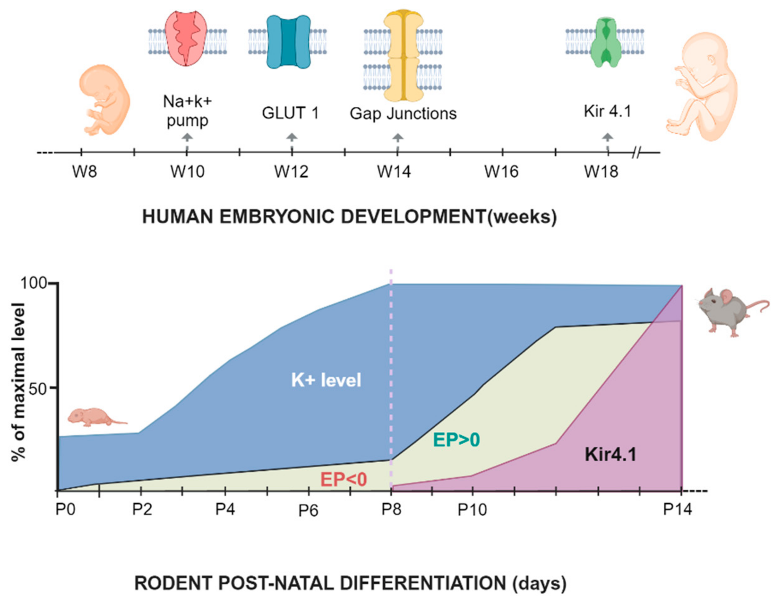
4.3.2. Embryonal and Postnatal Development

Several authors reported that in the stria vascularis, KCNJ10 is the only Kir channel expressed in intermediate cells [11,35,36]. The development of intermediate cells shows that KCNJ10 channels play a functional role in the formation of the endocochlear potential, as indicated by the time course of KCNJ10 expression in the rat model [11].
The cochlear lateral wall is where Kir4.1 expression is developed and it is linked to the generation of EP in the inner ear, as is stated in the literature. This can be inferred from both the gradual positivity of EP and the increase in the ion potassium concentration in the endolymph after birth [28]. Kir4.1 was the only member of the Kir channel family in the rat animal model that has been specifically identified in the basolateral membrane of marginal cells, not base or intermediate cells [28]. The Kir4.1 immunoreactivity was not observed from postnatal day 1 (P1) to P5 in rats [28]. Similar results were reported in mice deficient in H+-ATPase, when the stria vascularis is still in an immature state and no EP can be recorded [35]. At P8, the endolymphatic [K+] level reaching the adult positive level leads to a weak Kir4.1 immunoreactivity in the stria vascularis, which then progresses to a positive EP (it is noteworthy that the immunoreactivity appeared simultaneously in all turns of the cochlea in the stria vascularis). Kir4.1’s expression increased rapidly during the next few days, peaking at P14 when it exhibits a high expression, similar to that of adult rats. Furthermore, Kir4.1 developmental expression in satellite cells surrounding ganglion neurons appears to be equivalent to that in marginal cells [28]. The authors conclude that Kir4.1’s time course is closely associated with EP elevation, but not endolymphatic-wise [K+], which implies that Kir4.1 is involved in EP generation [28] (Figure 3).
The expression of potassium channels was also investigated in human fetal cochleae during week 9 to week 18 (W9–W18) [7]. The sodium–potassium pump Na+/K+ATPase was found to be positive in the cochlear duct epithelium at W10 and exhibited an increase in expression in the lateral wall epithelium at W12. In parallel, KCNQ1 appeared at W10 and W12 in the epithelium of the lateral wall of the basement membrane. It is noteworthy that at W12, melanocytes that derive from the neural crest migrated into the cochlea and reached the basement membrane of the lateral wall epithelium, becoming intermediate cells of the stria vascularis. In the following two weeks, the positivity of sodium–potassium pump expression was extended to marginal cells, the inner and outer hair cells, and epithelial cells in the outer sulcus, while KCNQ1 became expressed in the Reissner’s membrane and the outer sulcus cells, and at W16, also to the marginal cells. In parallel, the SLC2A1 channel, responsible for the expression of GLUT1, was detectable from W12 to W18 in the apical membrane of epithelial cells between the organ of Corti and the outer sulcus root cells. The developmental expression of KCNQ1 and the sodium–potassium pump Na+/K+ATPase suggests that both K+ uptake and secretion could be mediated by marginal cells as early as W16–W18 [7]. KCNJ10, or Kir4.1, plays a significant role in the ability of stria vascularis melanocytes to release K+ ions into the intrastrial space and contribute to the generation of EP. Its expression in melanocytes did not occur until the last stage of the investigation of W18; in addition, it took from W16 to W18 to become expressed in the epithelial cells of the outer sulcus, including root cells (Figure 3). From W14 to W18, the gap junction proteins GJB2/CX26 and GJB6/CX30 were also expressed in the cells in the outer sulcus, but not in the spiral ligament. These two pieces of evidence suggest that it is likely that the EP has not yet been generated at this stage [7].
In contrast, in guinea pigs, KCNJ10 channel expression began to appear in intermediate striated cells at embryonic day 50 (E50) [37,38]. The low expression of KCNJ10 has been observed to begin at E30, increases at E45, and remains unchanged until adulthood. In particular, at E40, the KCNJ10 channel protein was expressed in spiral ganglion satellite cells of the basal turn, and at E45, it exhibited a gradient of expression from the base to the apex of the cochlear spiral. In the myelin sheath of the cochlear nerve, the KCNJ10 protein was abundant between E40 and E45, but it slowly disappeared with age. The altered development of marginal cells, leading to impaired KCNJ10 expression, was confirmed in adult mice with C-Raf−/− genotypes: hearing loss appears to be correlated with a marked downregulation of KNCJ10, despite the presence of normal stria vascularis morphology and the absence of impairment of the sodium–potassium pump and KCNQ1 [39]. Further evidence was demonstrated in mice treated with the intermediate cell-specific tyrosinase promoter and under the control of the diphtheria toxin [40]. In their adulthood, these mice experienced the consequences of losing their endocochlear potential and profound deafness. Until 3 weeks of age, Kir4 expression was not observed in striated vascular melanocytes in those mice, but then a minimal level of Kir4.1 expression was observed. Furthermore, in all striated vascular cells, excess melanin granules were shown, indicating that striatal homeostasis had been disrupted [40]. Remarkably, regarding the KCNJ10 expression in the root cells of the outer sulcus, during cochlear development, it looks comparable to the expression patterns of pendrin in the outer sulcus of the adult mouse cochlea. In adultness, the pendrin colocalizes with the KCNJ5 expression in the outer sulcus of the mouse cochlea, showing the contribution of this potassium channel to pH buffering of the endolymph [41]. The correlation with the embryonic expression of KNCJ10 provides a conspicuous link between the two proteins and provides a new point of view on the etiology of sensorineural hearing loss in Pendred syndrome [7].

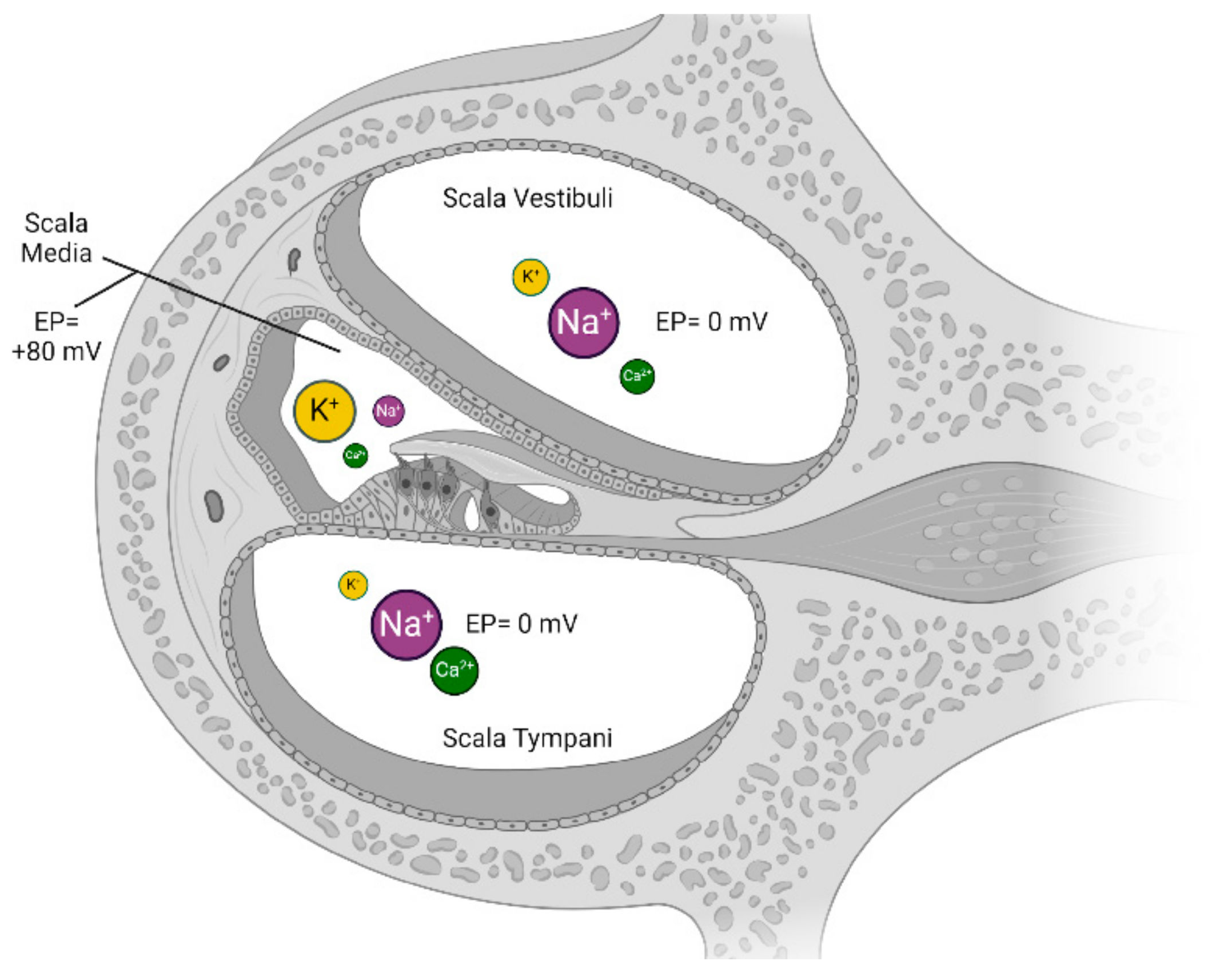
4.3.3. Endocochlear Potential

The process of generating EP in the stria vascularis is part of a cycle that involves different components of the organ of Corti. To describe this process, it is appropriate to begin with the presence of potassium from the perilymph in the interstitial space of the spiral ligament.
Through channels expressed on the basal part of their membrane, such as the Na+/K+/2Cl co-transporter (NKCC1), the 3Na+/2K+ATPase [18], and the inwardly rectifying potassium channel Kir5.1 [42], potassium is absorbed into the cytosol of the basal cells. From this, through a dense network of gap junctions (connexins 26, 30, 31, and 43) [15], K+ is transferred to the basal cells. The presence of tight junctions in the basal and marginal cell layers allows the formation of two physical barriers, which prevent the loss of potential created in the intrastrial space of the stria vasculature [15].
Basal cells are, in turn, connected to intermediate cells through extensive gap junctions. The fibrocytes, basal cells, and intermediate cells behave electrically as a single cellular entity due to the network of gap junctions, and the combination of these three cell types forms the first entity of the “two-cell model” of the stria vascularis [43].
The intermediate cells are immersed in the intrastrial space, which is a fluid with a very low potassium concentration (1–2 mM). The difference in K+ concentration between the cytosol of intermediate cells (150 mM) connected to fibrocytes and basal cells and the interstitial space causes the Kir4.1 channels to activate, ultimately allowing the passage of K+ ions out of the intermediate cells [15].
Kir4.1 channels mediate potassium efflux in intermediate cells of the stria vascularis, rather than influx as they typically do, due to the electrochemical gradients of potassium present in these cells. More specifically,
  • Marginal cells actively pump K+ ions into their cytoplasm to maintain a high intracellular K+ concentration;
  • This generates a high gradient of K+ between the marginal and intermediate cells, with the intermediate cells having lower intracellular K+;
  • Furthermore, the intermediate cells have a relatively negative membrane potential compared to the marginal cells;
  • This double electrochemical gradient (chemical for K+ and electrical for the membrane potential) causes K+ to spontaneously efflux through the Kir4.1 channel into the intrastrial space.
Therefore, the driving force for the efflux of K+ from the intermediate cells is the combination of a concentration gradient (high K+ intracellularly in the adjacent marginal cells) and an electrical potential gradient favoring the exit of positively charged K+ ions.
At this stage, the Kir4.1 channels fulfill their fundamental function in generating the EP.
Finally, marginal cells pump K+ into their interior through ATP-dependent channels on their basal membrane, keeping the K+ low in the intrastrial space. In their apical part, which is in direct contact with the endolymph, they present the potassium channels KCNQ1/KCNE1, which release the captured potassium into the endolymph, ready to be taken up by hair cells [15]. Marginal cells also form the second entity of the ‘two-cell’ model (Figure 4).
The movement of potassium ions through passive voltage-sensitive channels such as Kir4.1 makes it the most suitable candidate for depolarizing auditory cells and, consequently, the key ion in the generation of EP [15]. If sodium ions (Na+) had been utilized for depolarizing hair cells, this would have necessitated the consumption of ATP to activate its channels and thus the movement into the cells. A significant amount of ATP would have been required, necessitating a great deal of vascularization in the region of the hair cells. This would have led to undesirable vibrations around the hair cells caused by the heartbeat transmitted through the capillaries. These vibrations would have interfered with the ability to perceive sounds, generating significant disruptions in mechanical sound transduction. To avoid this risk, significant ATP exchange through the vessels takes place in the vascular stria, which is far from the hair cells, generating an ion-based EP through passive channels. In this way, the stria vascularis can be compared to a ‘remote power plant” that fuels the sense of hearing.

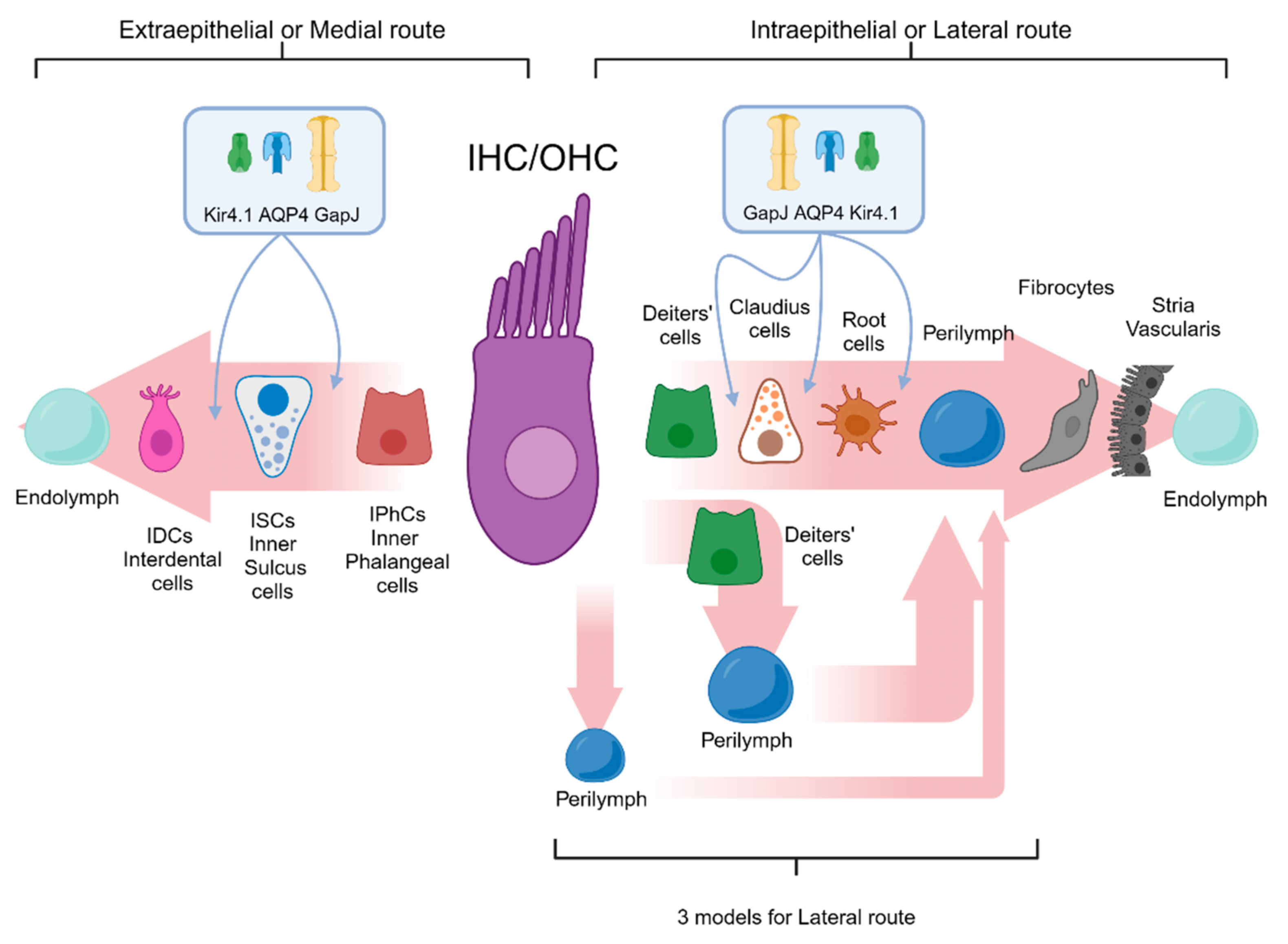
4.3.4. K+ Cochlear Cycle

The stereocilia of hair cells are equipped with mechanosensitive channels. These channels are connected to prestin, a protein that, when mechanically stimulated, opens the K+ channels, inducing an influx of ions into the hair cell due to the large concentration gradient [44].
It should be noted that the potential in the vestibular endolymph is only about +5 mV, while as seen in the cochlea duct, it can reach up to +100 mV due to the EP. This large potential makes the cochlear hair cells much more sensitive to the opening of potassium channels. Once potassium depolarizes the cell and triggers the release of calcium ions, it exits from the basolateral portion of the hair cells through a series of channels present in the membrane such as KCNQ4 and BK in OHCs and KNCQ4, BK, and Ik5 in IHCs. When KCNQ4 is absent, cells degenerate [45]. At this point, the potassium recycling phase begins, which can be divided into two different pathways: the intraepithelial or lateral route and the extraepithelial or medial route of K+ recycling [46] (Figure 5).
The intraepithelial or lateral route, which serves as the primary potassium recycling route, warrants further examination. According to Zdebik et al., three models could elucidate the recycling process [15]:
  • Potassium recycling through open perilymph (first pathway): The potassium efflux from the hair cells is directed into the interstitial space between the inner hair cells and supporting cells, as well as between the outer hair cells and Deiters’ cells. Subsequently, potassium is introduced into the perilymph and captured by the fibrocytes of the spiral ligament without the involvement of other cells in its entry into the perilymph.
  • Potassium recycling buffered (second pathway): The potassium introduced into the interstitial space, released from the OHCs or IHCs, is promptly recaptured by Deiters’ cells or supporting cells, which mediate its release into the perilymph.
  • Potassium recycling via gap junctions (third pathway): Once the IHCs or OHCs release potassium ions into the interstitial space, it is immediately recaptured by Deiters’ cells and supporting cells and subsequently transported back to the stria vascularis via a dense network of gap junctions.
Moreover, observational evidence suggests that, in addition to Deiters’ and Claudius cells, outer sulcus cells contribute to the following factors: (1) Ionic homeostasis is maintained, as the root cells, along with other cells of the cochlear epithelium, are implicated in maintaining appropriate potassium concentrations in the perilymph and endolymph for normal auditory function. (2) There is a role in potassium “recycling”, and in this case the root cells may participate in the “recycling” of potassium in the cochlear epithelium. This process involves the removal of K+ from the extracellular environment in close proximity to auditory hair cells and its subsequent reintroduction into the perilymph. (3) Kir4.1 channels are expressed—the root cells express Kir4.1 channels, which may mediate potassium fluxes across cellular membranes. These channels are responsible for the resting conductance of root cells and may facilitate the movement of K+ across the cell membrane. (4) There is a role in maintaining the endocochlear potential, and the potassium transport mediated by root cells may contribute to the generation and maintenance of the endocochlear potential, a critical component for auditory transduction. (5) There is an interconnection via gap junctions, and the root cells are interconnected via gap junctions that allow the passage of molecules such as Lucifer yellow. This interconnection enables root cells to collaborate on ion transport and homeostasis maintenance [31,32].
Once potassium reaches the outer sulcus cells, it is exchanged with the fibrocytes of the stria vascularis. Kir4.1 is expressed by various cells surrounding the IHCs and OHCs, and it appears that, aided by other channels such as aquaporin-4 (AQP4), it is involved in the recycling and ionic homeostasis of supporting cells [46,47].
The extraepithelial or medial route follows the potassium flow that, leaving the IHCs, is directed into the interstitial space, where it is subsequently gathered by the inner pillar cells and inner phalangeal cells, which have the Kir4.1 channel expressed in their membrane. In inner sulcus cells in close proximity to the organ of Corti, there is an expression of both Kir4.1 and aquaporin-4 (AQP4) channels [46]. Additionally, a series of gap junctions between these cells facilitate ion exchanges in the area surrounding the basal part of the hair cell. Progressing into the inner sulcus, a series of cells equipped with a gap junction and Kir4.1 facilitate the subsequent removal of K+ towards the interdental cells while releasing the K+ stored in the interstitial space between the inner sulcus cells and the fibrocytes present. Potassium returns to flow into the endolymph from both the central and lateral interdental cells in the region where the tectorial membrane attaches. The interdental cells also express a combination of Kir4.1 and AQP4 [47]. In summary, Kir4.1 and AQP4 play complementary roles in K+ recycling in the cochlea, with Kir4.1 mediating ion flux and AQP4 facilitating the water flow necessary for osmotic balance. Their expression and regulation are important for maintaining ionic homeostasis and preventing hearing loss [46,47].

4.3.5. Pathophysiology

The KCNJ10 gene is located on chromosome 1q22-23. Mutations can lead to the synthesis of altered Kir4.1, which can result in hearing loss. To date, it is well documented that it is associated with the development of two syndromes: EAST/SeSAME syndrome and Pendred syndrome [14].
In 2009, Bockenhauer and Scholl independently identified a rare autosomal recessive syndrome named “EAST/SeSAME syndrome”, derived from EAST (epilepsy, ataxia, sensorineural deafness, tubulopathy) and SeSAME (seizures, sensorineural deafness, ataxia, intellectual disability, and electrolyte imbalance) diseases [48,49]. As defined by the acronym, it is characterized by seizures, sensorineural deafness, ataxia, intellectual disability, and electrolyte imbalance. It can be identified by sequencing the KCNJ10 gene: it has been documented in all patients with SeSAME coming from four different families, missense or nonsense mutations that affected both alleles. According to evidence, these mutations result in a loss of function in K+ channels that are related to glia in the brain, in the spinal cord, on the basolateral membrane of the distal nephron, and in the cochlea, where, at this specific site, it is involved in the endolymph homeostasis [49,50]. The medical literature has reported 16 mutations associated with different clinical characteristics observed even within family members: the most debilitating features are epilepsy and ataxia; seizures generally occur at the beginning of childhood; muscle tone increases with age; and the degree of hearing impairment ranges from mild to severe [48,51,52].
Autosomal recessive syndromic deafness caused primarily by SLC26A4 mutations was described in KCNJ10 mutations or deficiencies that could also be associated with an enlarged vestibular aqueduct (EVA) and Pendred syndrome (PDS)/DNFB4 [29,53]. Hearing loss and a goiter are common symptoms of Pendred syndrome, which is caused by mutations in genes that encode ion transporters and pumps that regulate the pH and ionic composition of the endolymph. These alterations can cause distal renal tubular acidosis, deafness, or non-syndromic early-onset severe-to-profound hearing loss, which is often associated with EVA. About one third of patients experience a fluctuating hearing loss due to enlarged vestibular aqueducts, whether they are syndromic or non-syndromic. The fluctuations may be linked to endolymphatic hydrops (an excessive accumulation of endolymph in the cochlea and the vestibular system), but it is still unclear what exactly leads to hearing impairment and the generation of sudden drops [5].
In 2009, 89 patients were screened for a clinical diagnosis of EVA/PS and were known carriers of a single mutation of the SLC26A4 coding sequence [54]. In SLC26A4−/− mice, the KCNJ10 protein was not expressed, resulting in their lack of endocochlear potential. Intermediate cells protect the stria vascularis themselves from free radical damage by converting CO2 to HCO3 and generating glutathione and melanin pigment [55,56]. The loss of pendrin, which allows the transport of HCO3 into the endolymph, may result in the alkalinization of intrastriatal spaces. Thus, the loss of the KCNJ10 protein in strial intermediate cells can be caused by changes in cytosolic pH and free radical stress. Cytosolic pH and free radicals control the function and expression of other K+ channels, indicating the metabolic status of the cell [28,49,51]. Deafness may be directly caused by the suppression of the KCNJ10 channel in intermediate strial cells, which is crucial for generating EP, in SLC26A4−/− mice and patients with Pendred syndrome [29,53,55,57].
Yang et al. [48] have shown that the combination of two mutations, SLC26A4 and KCNJ10, was specifically associated with inner ear dysfunction [54]. The mutations in SLC26A4 lead to hypofunction or haploinsufficiency. The striatal expression of KCNJ10 protein, in patients affected by EVA and Pendred syndrome (EVA/PS), is lowered by the combination of SLC26A4 haploinsufficiency and oxidative stress. Due to the decrease in KCNJ10 expression, marginal cells in the stria vascularis receive less K+. Consequently, these cells decrease the rate of K+ secretion. This self-limiting mechanism could be the reason for intermittent recovery of hearing thresholds in patients with EVA/PS, which results in fluctuating and progressive hearing loss [50]. New insights into the pathogenesis of EVA/PS can be gained from this paradigm, as this suggests that if the strial expression of KCNJ10 can be maintained, controlling endolymph pH or limiting oxidative stress through medical therapy may be a way to prevent hearing loss in some individuals with the EVA/PS phenotype.
The genetic basis of EVA in patients with hearing loss (HL) was determined by investigating genomic DNA mutations using a custom panel of HL genes that included 237 genes related to hearing loss (HL) or a clinical exome [50]. The study by Baldyga and colleagues reported that 74% of patients investigated for inner ear malformations and hearing loss had causal genetic variants: two pathogenic variants were found in the SLC26A4 gene in approximately half of the patients, and one of these patients had a Caucasian EVA haplotype (CEVA) well; patients with only one pathogenic variant in the SLC26A4 gene were often diagnosed with CEVA; novel pathogenic variants of the EYA1 gene causing cochlear hypoplasia were identified in two individuals with phenotypes that correspond to branchio-oto-renal spectrum disorder (BOR); and a new variant with unknown significance of CHD7 was discovered in a patient; no pathogenic mutations in the KCNJ10 and FOXI1 genes were identified [58]. Landa et al. reported similar findings ten years ago, concluding that there is no significant link between these two genes and the onset of Pendred syndrome with EVA [59]. In a study with 32 Chinese patients with pathogenic SLC26A4 mutations, no potential pathogenic mutations were identified in the FOXI1 or KCNJ10 genes: amino acid changes were not present in either of the FOXI1 mutations, c.279G.A (p.R93R) or c.1044T.C (p.Y348Y), or the KCNJ10 mutation, c.1053C.T (p.D351D) [60]. Jonard et al. found that 25 patients with unilateral hearing impairment and ipsilateral EVA did not have mutations in FOXI1 and KCNJ10 genes [61]. In conclusion, it is uncertain whether digenic inheritance of PS can occur due to single allele mutations in both KCNJ10 and SLC26A4.
Suzumoto et al. described four adult patients with EAST/SeSAME syndrome with homozygous missense mutation p.Ala167Val (identified in two siblings) and homozygous frameshift mutations p.Asn232Glnfs*14 and p.Gly275Valfs*7 (both born from consanguineous parents, first cousins). These mutation predictions were made using in silico modeling and bioinformatics, which indicated that patients who had truncated mutations had more serious consequences: tubulopathy and neurological symptoms associated with severe epilepsy [62].
In an in vitro study, Kir4.1 has been characterized by new mutations that appear linked to EAST/SeSAME syndrome [25,63]. Radiotracer efflux and inside–out membrane patch clamping were performed on COSm6 cells expressing homomeric Kir4.1 or heteromeric Kir4.1/Kir5.1 channels, to investigate the functional significance of the reported mutations in the patients. Although all mutations affected channel function, the underlying mechanisms were not the same. R65P, T164I, and R297C played a crucial role in pH sensing and pore gating by causing an alkaline shift in pH sensitivity. A disruption occurred in the intersubunit salt bridge in R297C. G77R may experience changes in channel structure or gating due to the introduction of a positive charge in the bilayer. A167V had no impact on channel properties, but could cause decreased surface expression in A167V/R297C. These data match those obtained in Xenopus oocytes, in which the K current levels of all disease-associated mutant channels, such as R65P, G77R, and A167V/R297C, were lowered, with a range of a 1 to 23% difference compared to the wild-type level [25]. In order to imitate the heterozygous state that exists among R199X, C140R, and G77R, what has been investigated is the coexpression of mutant and wild-type subunits that results in a decrease in currents to 55, 40, and 20% of wild-type levels, suggesting that carriers of these mutations may have an abnormal phenotype [25]. Meanwhile, the coexpression of Kir4.1 mutations with Kir5.1 resulted in a reduction in currents that was at least as significant as when the mutants were expressed alone. The currents of all mutants except R199X were significantly reduced when pH was reduced from approximately 7.4 to 6.8, but wild-type channels remained unaffected [25]. The authors suggest that these mutations provide an explanation for the molecular defects that make up the EAST/SeSAME syndrome. Their conclusion is that the loss of channel function for the disease-associated mutant Kir41 channels could be attributed to altered pH gating, which could have significant physiological consequences [25,63].
By using mutant mice that lack the KCNJ10 K+ channel, how it generates EP and delivers K+ to marginal cells was evaluated. The measurement of EP and endolymphatic K+ was performed on homozygous (−/−) mutant mice, with wild-type (+/+) and heterozygous (−/+) littermates being the controls. The KCNJ10 channel and other transport pathways are responsible for the generation of EP and the establishment of differences in K concentration across the channel, making it an important mechanism for marginal delivery of K+ to the strial marginal cells [43]. The marked anatomical anomalies present in the inner ear of the Kir4.1 knockout mouse indicate that the pathology matches the spatial and temporal distribution of Kir4.1. Kir4.1 was suggested to be an essential component of the stria vascularis and is the main K+ channel in glial cells surrounding spiral ganglion neurons and axons [33].
Finally, recent research has shown that Kir4.1 plays a role in progressive hearing loss caused by aging in the CBA/caj mouse model and in human temporal bone. Changes in structure and function have been observed in intermediate cells of the vascular stria, the outer sulcus of the cochlear lateral wall, and the satellite cells of the spiral ganglion. In particular, a significant decrease or alterations in the expression of the kir4.1 potassium channel have been identified. The data indicate that progressive hearing loss was caused by the degeneration/dysfunction of ion channels, which provides new insights into understanding metabolism and pathophysiological processes related to aging [64].
In vitro studies have shown that aminoglycoside antibiotics target Kir4.1 channels, which could have an impact on potassium transport and lead to an imbalance of homeostasis in various tissues, providing an explanation for the ototoxicity effect of these commonly used antibiotics [65,66]. In both in vitro and in vivo mouse models, Kir4.1 was demonstrated to act as a replacement for deficient K+ channels in supporting cells to rectify the imbalance in K+ concentration [66].

5. Conclusions

The Kir4.1 channel has an objective role in maintaining cochlear fluid homeostasis and endocochlear potential. To date, the relationship between deafness and mutations of the gene coding for kir4.1 has been demonstrated in two syndromes, Pendred and EAST/SeSAME, which are closely tied to the homeostasis of cochlear fluids. In several knockout mouse studies on the development of the auditory system, it has been consistently shown that the absence or deficiency of Kir4.1 channel expression affects the generation of endocochlear potential. Recent investigations suggested that an impairment of the Kir4.1 channel function might be linked to progressive hearing loss in the elderly. This is a consequence of the deterioration of the physiological condition of the vascular stria and/or spiral ligament (both involved in the K+ cycle). The extensive clinical and experimental evidence, which encompasses the complicated network of ion channels and pumps, and the gap and tight junctions between cells in different tissue layers, does not allow for a clear overview that provides effective suggestions for directing therapies aimed at resolving the aforementioned auditory pathologies. Our view is that the most likely drugs are those that can regulate blood pH or prevent cell damage, for instance, avoiding the production of reactive oxygen substances and the accumulation of free radicals. It has been shown in studies on ototoxicity that aminoglycosides can inhibit the function of the Kir4.1 channel. The imbalance in potassium homeostasis is inevitable when Kir4.1 is silenced, as it acts as a channel that compensates for the deficiency of other potassium channels. In this context, it is feasible to develop new therapies that increase gene expression to prevent damage from Kir4.1 deficiency. Preventing changes in the homeostasis of cochlear fluids would be crucial for the maintenance of endocochlear potential.

Author Contributions

Conceptualization, L.A. and G.A.; methodology, S.F. (Silvia Fracaro) and F.H.; data curation, S.F. (Silvia Fracaro) and F.H.; writing—original draft preparation, L.A., G.A., S.F. (Silvia Fracaro) and F.H.; writing—review and editing, L.A., G.A., S.F. (Silvia Fracaro), F.H., S.F. (Sebastiano Franchella), G.M. and D.B.; visualization, L.A., G.A., S.F. (Silvia Fracaro), F.H., G.M., D.B., S.F. (Sebastiano Franchella) and E.Z.; supervision, L.A. and G.A.; funding acquisition, L.A. All authors have read and agreed to the published version of the manuscript.

Funding

This research was funded by MIUR, grant number PRIN-2022YRBE8B.

Acknowledgments

Figures were created under license using BioRender.com, https://biorender.com/.

Conflicts of Interest

The authors declare no conflicts of interest.

Appendix A

Each database’s query box was filled with specific search details as follows:

Appendix A.1. PubMed

(“Hearing Loss”[Mesh] OR “Deafness”[Mesh]) AND (( “Ear, Inner/abnormalities”[Mesh] OR “Ear, Inner/anatomy and histology”[Mesh] OR “Ear, Inner/pathology”[Mesh] OR “Ear, Inner/physiology”[Mesh] OR “Ear, Inner/physiopathology”[Mesh]) OR (“Cochlea/abnormalities”[Mesh] OR “Cochlea/anatomy and histology”[Mesh] OR “Cochlea/pathology”[Mesh] OR “Cochlea/physiology”[Mesh] OR “Cochlea/physiopathology”[Mesh]) OR (“Stria Vascularis”[Mesh]) OR (“Spiral Ligament of Cochlea”[Mesh])) AND (“Kcnj10 (channel)” [Supplementary Concept] OR “Kir4.1”)
https://pubmed.ncbi.nlm.nih.gov/ (accessed on 29 May 2024)

Appendix A.2. Scopus

(TITLE-ABS-KEY (“Hearing Loss” OR “Deafness”) AND TITLE-ABS-KEY (“Inner ear” OR “Cochlea” OR “Stria Vascularis” OR “Spiral Ligament”) AND TITLE-ABS-KEY (“anatomy” OR “histology” OR “pathology” OR “physiology” OR “physiopathology”) AND TITLE-ABS-KEY (“kcnj10” OR “kir4.1”))
https://www.scopus.com/ (accessed on 29 May 2024)

Appendix A.3. Web of Science

“Hearing Loss” OR “Deafness” (All Fields) and “Inner ear” OR “Cochlea” OR “Stria Vascularis” OR “Spiral Ligament” (All Fields) and “anatomy” OR “histology” OR “pathology” OR “physiology” OR “physiopathology” (All Fields) and “kcnj10” OR “kir4.1” (All Fields)
https://www.webofscience.com/ (accessed on 29 May 2024)

References

  1. Deafness and Hearing Loss. Available online: https://www.who.int/news-room/fact-sheets/detail/deafness-and-hearing-loss (accessed on 17 April 2024).
  2. Busi, M.; Castiglione, A.; Taddei Masieri, M.; Ravani, A.; Guaran, V.; Astolfi, L.; Trevisi, P.; Ferlini, A.; Martini, A. Novel Mutations in the SLC26A4 Gene. Int. J. Pediatr. Otorhinolaryngol. 2012, 76, 1249–1254. [Google Scholar] [CrossRef] [PubMed]
  3. Guaran, V.; Astolfi, L.; Castiglione, A.; Simoni, E.; Olivetto, E.; Galasso, M.; Trevisi, P.; Busi, M.; Volinia, S.; Martini, A. Association between Idiopathic Hearing Loss and Mitochondrial DNA Mutations: A Study on 169 Hearing-Impaired Subjects. Int. J. Mol. Med. 2013, 32, 785–794. [Google Scholar] [CrossRef] [PubMed]
  4. Bernardinelli, E.; Roesch, S.; Simoni, E.; Marino, A.; Rasp, G.; Astolfi, L.; Sarikas, A.; Dossena, S. Novel POU3F4 Variants Identified in Patients with Inner Ear Malformations Exhibit Aberrant Cellular Distribution and Lack of SLC6A20 Transcriptional Upregulation. Front. Mol. Neurosci. 2022, 15, 999833. [Google Scholar] [CrossRef] [PubMed]
  5. Korver, A.M.H.; Smith, R.J.H.; Van Camp, G.; Schleiss, M.R.; Bitner-Glindzicz, M.A.K.; Lustig, L.R.; Usami, S.-I.; Boudewyns, A.N. Congenital Hearing Loss. Nat. Rev. Dis. Primer 2017, 3, 16094. [Google Scholar] [CrossRef] [PubMed]
  6. Yoshimura, H.; Okubo, T.; Shinagawa, J.; Nishio, S.-Y.; Takumi, Y.; Usami, S.-I. Epidemiology, Aetiology and Diagnosis of Congenital Hearing Loss via Hearing Screening of 153913 Newborns. Int. J. Epidemiol. 2024, 53, dyae052. [Google Scholar] [CrossRef]
  7. Locher, H.; de Groot, J.C.M.J.; van Iperen, L.; Huisman, M.A.; Frijns, J.H.M.; Chuva de Sousa Lopes, S.M. Development of the Stria Vascularis and Potassium Regulation in the Human Fetal Cochlea: Insights into Hereditary Sensorineural Hearing Loss. Dev. Neurobiol. 2015, 75, 1219–1240. [Google Scholar] [CrossRef] [PubMed]
  8. Franz, L.; Incognito, A.; Gallo, C.; Turolla, L.; Scquizzato, E.; Cenedese, R.; Matarazzo, A.; Savegnago, D.; Zanatta, P.; Genovese, E.; et al. Audiological Phenotypes of Connexin Gene Mutation Patterns: A Glance at Different GJB2/GJB6 Gene Mutation Profiles. Children 2024, 11, 194. [Google Scholar] [CrossRef] [PubMed]
  9. Lang, F.; Vallon, V.; Knipper, M.; Wangemann, P. Functional Significance of Channels and Transporters Expressed in the Inner Ear and Kidney. Am. J. Physiol.-Cell Physiol. 2007, 293, C1187–C1208. [Google Scholar] [CrossRef] [PubMed]
  10. Jagger, D.J.; Forge, A. Connexins and Gap Junctions in the Inner Ear—It’s Not Just about K+ Recycling. Cell Tissue Res. 2015, 360, 633–644. [Google Scholar] [CrossRef] [PubMed]
  11. Hibino, H.; Nin, F.; Tsuzuki, C.; Kurachi, Y. How Is the Highly Positive Endocochlear Potential Formed? The Specific Architecture of the Stria Vascularis and the Roles of the Ion-Transport Apparatus. Pflug. Arch.-Eur. J. Physiol. 2010, 459, 521–533. [Google Scholar] [CrossRef] [PubMed]
  12. Casale, J.; Kandle, P.F.; Murray, I.V.; Murr, N.I. Physiology, Cochlear Function. In StatPearls; StatPearls Publishing: Treasure Island, FL, USA, 2024. [Google Scholar]
  13. Rask-Andersen, H.; Liu, W.; Erixon, E.; Kinnefors, A.; Pfaller, K.; Schrott-Fischer, A.; Glueckert, R. Human Cochlea: Anatomical Characteristics and Their Relevance for Cochlear Implantation. Anat. Rec. 2012, 295, 1791–1811. [Google Scholar] [CrossRef] [PubMed]
  14. Chen, J.; Zhao, H.-B. The Role of an Inwardly Rectifying K+ Channel (Kir4.1) in the Inner Ear and Hearing Loss. Neuroscience 2014, 265, 137–146. [Google Scholar] [CrossRef] [PubMed]
  15. Zdebik, A.A.; Wangemann, P.; Jentsch, T.J. Potassium Ion Movement in the Inner Ear: Insights from Genetic Disease and Mouse Models. Physiology 2009, 24, 307–316. [Google Scholar] [CrossRef] [PubMed]
  16. Takiguchi, Y.; Sun, G.; Ogawa, K.; Matsunaga, T. Long-Lasting Changes in the Cochlear K+ Recycling Structures after Acute Energy Failure. Neurosci. Res. 2013, 77, 33–41. [Google Scholar] [CrossRef] [PubMed]
  17. MacKinnon, R. Potassium Channels. FEBS Lett. 2003, 555, 62–65. [Google Scholar] [CrossRef] [PubMed]
  18. Buckingham, S.D.; Kidd, J.F.; Law, R.J.; Franks, C.J.; Sattelle, D.B. Structure and Function of Two-Pore-Domain K+ Channels: Contributions from Genetic Model Organisms. Trends Pharmacol. Sci. 2005, 26, 361–367. [Google Scholar] [CrossRef] [PubMed]
  19. Kuang, Q.; Purhonen, P.; Hebert, H. Structure of Potassium Channels. Cell. Mol. Life Sci. 2015, 72, 3677–3693. [Google Scholar] [CrossRef] [PubMed]
  20. Robbins, J. KCNQ Potassium Channels: Physiology, Pathophysiology, and Pharmacology. Pharmacol. Ther. 2001, 90, 1–19. [Google Scholar] [CrossRef] [PubMed]
  21. Pyott, S.J.; Duncan, R.K. Chapter Ten—BK Channels in the Vertebrate Inner Ear. In International Review of Neurobiology; Contet, C., Ed.; Big on Bk; Academic Press: Cambridge, MA, USA, 2016; Volume 128, pp. 369–399. [Google Scholar]
  22. Bazard, P.; Frisina, R.D.; Acosta, A.A.; Dasgupta, S.; Bauer, M.A.; Zhu, X.; Ding, B. Roles of Key Ion Channels and Transport Proteins in Age-Related Hearing Loss. Int. J. Mol. Sci. 2021, 22, 6158. [Google Scholar] [CrossRef] [PubMed]
  23. Fakler, B.; Ruppersberg, J.P. Functional and Molecular Diversity Classifies the Family of Inward-Rectifier K+ Channels. Cell Physiol. Biochem. 1996, 6, 195–209. [Google Scholar] [CrossRef]
  24. Parrock, S.; Hussain, S.; Issler, N.; Differ, A.-M.; Lench, N.; Guarino, S.; Oosterveld, M.J.S.; Keijzer-Veen, M.; Brilstra, E.; van Wieringen, H.; et al. KCNJ10 Mutations Display Differential Sensitivity to Heteromerisation with KCNJ16. Nephron Physiol. 2013, 123, 7–14. [Google Scholar] [CrossRef] [PubMed]
  25. Williams, D.M.; Lopes, C.M.B.; Rosenhouse-Dantsker, A.; Connelly, H.L.; Matavel, A.; O-Uchi, J.; McBeath, E.; Gray, D.A. Molecular Basis of Decreased Kir4.1 Function in SeSAME/EAST Syndrome. J. Am. Soc. Nephrol. JASN 2010, 21, 2117–2129. [Google Scholar] [CrossRef] [PubMed]
  26. Sodium-Potassium-Chloride Cotransport. Available online: https://journals.physiology.org/doi/epdf/10.1152/physrev.2000.80.1.211 (accessed on 29 April 2024).
  27. Strepay, D.; Olszewski, R.T.; Nixon, S.; Korrapati, S.; Adadey, S.; Griffith, A.J.; Su, Y.; Liu, J.; Vishwasrao, H.; Gu, S.; et al. Transgenic Tg(Kcnj10-ZsGreen) Fluorescent Reporter Mice Allow Visualization of Intermediate Cells in the Stria Vascularis. Sci. Rep. 2024, 14, 3038. [Google Scholar] [CrossRef]
  28. Hibino, H.; Horio, Y.; Inanobe, A.; Doi, K.; Ito, M.; Yamada, M.; Gotow, T.; Uchiyama, Y.; Kawamura, M.; Kubo, T.; et al. An ATP-Dependent Inwardly Rectifying Potassium Channel, KAB-2 (Kir4. 1), in Cochlear Stria Vascularis of Inner Ear: Its Specific Subcellular Localization and Correlation with the Formation of Endocochlear Potential. J. Neurosci. Off. J. Soc. Neurosci. 1997, 17, 4711–4721. [Google Scholar] [CrossRef] [PubMed]
  29. Wangemann, P.; Itza, E.M.; Albrecht, B.; Wu, T.; Jabba, S.V.; Maganti, R.J.; Ho Lee, J.; Everett, L.A.; Wall, S.M.; Royaux, I.E.; et al. Loss of KCNJ10 Protein Expression Abolishes Endocochlear Potential and Causes Deafness in Pendred Syndrome Mouse Model. BMC Med. 2004, 2, 30. [Google Scholar] [CrossRef] [PubMed]
  30. Korrapati, S.; Taukulis, I.; Olszewski, R.; Pyle, M.; Gu, S.; Singh, R.; Griffiths, C.; Martin, D.; Boger, E.; Morell, R.J.; et al. Single Cell and Single Nucleus RNA-Seq Reveal Cellular Heterogeneity and Homeostatic Regulatory Networks in Adult Mouse Stria Vascularis. Front. Mol. Neurosci. 2019, 12, 316. [Google Scholar] [CrossRef] [PubMed]
  31. Jagger, D.J.; Nevill, G.; Forge, A. The Membrane Properties of Cochlear Root Cells Are Consistent with Roles in Potassium Recirculation and Spatial Buffering. JARO—J. Assoc. Res. Otolaryngol. 2010, 11, 435–448. [Google Scholar] [CrossRef] [PubMed]
  32. Gu, S.; Olszewski, R.; Taukulis, I.; Wei, Z.; Martin, D.; Morell, R.J.; Hoa, M. Characterization of Rare Spindle and Root Cell Transcriptional Profiles in the Stria Vascularis of the Adult Mouse Cochlea. Sci. Rep. 2020, 10, 18100. [Google Scholar] [CrossRef] [PubMed]
  33. Rozengurt, N.; Lopez, I.; Chiu, C.-S.; Kofuji, P.; Lester, H.A.; Neusch, C. Time Course of Inner Ear Degeneration and Deafness in Mice Lacking the Kir4.1 Potassium Channel Subunit. Hear. Res. 2003, 177, 71–80. [Google Scholar] [CrossRef]
  34. Tang, X.; Schmidt, T.M.; Perez-Leighton, C.E.; Kofuji, P. Kir4.1 Is Responsible for the Native Inward Potassium Conductance of Satellite Glial Cells in Sensory Ganglia. Neuroscience 2010, 166, 397. [Google Scholar] [CrossRef] [PubMed]
  35. Lorente-Cánovas, B.; Ingham, N.; Norgett, E.E.; Golder, Z.J.; Karet Frankl, F.E.; Steel, K.P. Mice Deficient in H+-ATPase A4 Subunit Have Severe Hearing Impairment Associated with Enlarged Endolymphatic Compartments within the Inner Ear. Dis. Model. Mech. 2013, 6, 434–442. [Google Scholar] [CrossRef] [PubMed]
  36. Koh, J.-Y.; Affortit, C.; Ranum, P.T.; West, C.; Walls, W.D.; Yoshimura, H.; Shao, J.Q.; Mostaert, B.; Smith, R.J.H. Single-Cell RNA-Sequencing of Stria Vascularis Cells in the Adult Slc26a4−/− Mouse. BMC Med. Genom. 2023, 16, 133. [Google Scholar] [CrossRef] [PubMed]
  37. Jin, Z.; Ulfendahl, M.; Järlebark, L. Spatiotemporal Loss of K+ Transport Proteins in the Developing Cochlear Lateral Wall of Guinea Pigs with Hereditary Deafness. Eur. J. Neurosci. 2008, 27, 145–154. [Google Scholar] [CrossRef] [PubMed]
  38. Jin, Z.; Wei, D.; Järlebark, L. Developmental Expression and Localization of KCNJ10 K+ Channels in the Guinea Pig Inner Ear. Neuroreport 2006, 17, 475–479. [Google Scholar] [CrossRef] [PubMed]
  39. De Iriarte Rodríguez, R.; Magariños, M.; Pfeiffer, V.; Rapp, U.R.; Varela-Nieto, I. C-Raf Deficiency Leads to Hearing Loss and Increased Noise Susceptibility. Cell. Mol. Life Sci. 2015, 72, 3983–3998. [Google Scholar] [CrossRef]
  40. Kim, H.J.; Gratton, M.A.; Lee, J.-H.; Perez Flores, M.C.; Wang, W.; Doyle, K.J.; Beermann, F.; Crognale, M.A.; Yamoah, E.N. Precise Toxigenic Ablation of Intermediate Cells Abolishes the “Battery” of the Cochlear Duct. J. Neurosci. Off. J. Soc. Neurosci. 2013, 33, 14601–14606. [Google Scholar] [CrossRef] [PubMed]
  41. Cazals, Y.; Bévengut, M.; Zanella, S.; Brocard, F.; Barhanin, J.; Gestreau, C. KCNK5 Channels Mostly Expressed in Cochlear Outer Sulcus Cells Are Indispensable for Hearing. Nat. Commun. 2015, 6, 8780. [Google Scholar] [CrossRef] [PubMed]
  42. Hibino, H.; Higashi-Shingai, K.; Fujita, A.; Iwai, K.; Ishii, M.; Kurachi, Y. Expression of an Inwardly Rectifying K+ Channel, Kir5.1, in Specific Types of Fibrocytes in the Cochlear Lateral Wall Suggests Its Functional Importance in the Establishment of Endocochlear Potential. Eur. J. Neurosci. 2004, 19, 76–84. [Google Scholar] [CrossRef] [PubMed]
  43. Marcus, D.C.; Wu, T.; Wangemann, P.; Kofuji, P. KCNJ10 (Kir4.1) Potassium Channel Knockout Abolishes Endocochlear Potential. Am. J. Physiol.-Cell Physiol. 2002, 282, C403–C407. [Google Scholar] [CrossRef] [PubMed]
  44. Ashmore, J. Cochlear Outer Hair Cell Motility. Physiol. Rev. 2008, 88, 173–210. [Google Scholar] [CrossRef] [PubMed]
  45. Spitzmaul, G.; Rías, E.; Dionisio, L.; Spitzmaul, G.; Rías, E.; Dionisio, L. Potential Mechanisms of Hearing Loss Due to Impaired Potassium Circulation in the Organ of Corti. In Updates on Hearing Loss and its Rehabilitation; IntechOpen: London, UK, 2023; ISBN 978-1-83769-777-9. [Google Scholar]
  46. Eckhard, A.; Gleiser, C.; Rask-Andersen, H.; Arnold, H.; Liu, W.; Mack, A.; Müller, M.; Löwenheim, H.; Hirt, B. Co-Localisation of K(Ir)4.1 and AQP4 in Rat and Human Cochleae Reveals a Gap in Water Channel Expression at the Transduction Sites of Endocochlear K(+) Recycling Routes. Cell Tissue Res. 2012, 350, 27–43. [Google Scholar] [CrossRef]
  47. Eckhard, A.; Löwenheim, H. Water Regulation in the Cochlea: Do Molecular Water Channels Facilitate Potassium-Dependent Sound. Transduction? HNO 2014, 62, 423–431. [Google Scholar] [CrossRef] [PubMed]
  48. Bockenhauer, D.; Feather, S.; Stanescu, H.C.; Bandulik, S.; Zdebik, A.A.; Reichold, M.; Tobin, J.; Lieberer, E.; Sterner, C.; Landoure, G.; et al. Epilepsy, Ataxia, Sensorineural Deafness, Tubulopathy, and KCNJ10 Mutations. N. Engl. J. Med. 2009, 360, 1960–1970. [Google Scholar] [CrossRef] [PubMed]
  49. Scholl, U.I.; Choi, M.; Liu, T.; Ramaekers, V.T.; Häusler, M.G.; Grimmer, J.; Tobe, S.W.; Farhi, A.; Nelson-Williams, C.; Lifton, R.P. Seizures, Sensorineural Deafness, Ataxia, Mental Retardation, and Electrolyte Imbalance (SeSAME Syndrome) Caused by Mutations in KCNJ10. Proc. Natl. Acad. Sci. USA 2009, 106, 5842–5847. [Google Scholar] [CrossRef] [PubMed]
  50. Scholl, U.I.; Dave, H.B.; Lu, M.; Farhi, A.; Nelson-Williams, C.; Listman, J.A.; Lifton, R.P. SeSAME/EAST Syndrome--Phenotypic Variability and Delayed Activity of the Distal Convoluted Tubule. Pediatr. Nephrol. Berl. Ger. 2012, 27, 2081–2090. [Google Scholar] [CrossRef] [PubMed]
  51. Freudenthal, B.; Kulaveerasingam, D.; Lingappa, L.; Shah, M.A.; Brueton, L.; Wassmer, E.; Ognjanovic, M.; Dorison, N.; Reichold, M.; Bockenhauer, D.; et al. KCNJ10 Mutations Disrupt Function in Patients with EAST Syndrome. Nephron Physiol. 2011, 119, P40–P48. [Google Scholar] [CrossRef]
  52. Celmina, M.; Micule, I.; Inashkina, I.; Audere, M.; Kuske, S.; Pereca, J.; Stavusis, J.; Pelnena, D.; Strautmanis, J. EAST/SeSAME Syndrome: Review of the Literature and Introduction of Four New Latvian Patients. Clin. Genet. 2019, 95, 63–78. [Google Scholar] [CrossRef] [PubMed]
  53. Wangemann, P. The Role of Pendrin in the Development of the Murine Inner Ear. Cell. Physiol. Biochem. 2011, 28, 527–534. [Google Scholar] [CrossRef] [PubMed]
  54. Yang, T.; Gurrola, J.G.; Wu, H.; Chiu, S.M.; Wangemann, P.; Snyder, P.M.; Smith, R.J.H. Mutations of KCNJ10 Together with Mutations of SLC26A4 Cause Digenic Nonsyndromic Hearing Loss Associated with Enlarged Vestibular Aqueduct Syndrome. Am. J. Hum. Genet. 2009, 84, 651–657. [Google Scholar] [CrossRef]
  55. Wangemann, P.; Nakaya, K.; Wu, T.; Maganti, R.J.; Itza, E.M.; Sanneman, J.D.; Harbidge, D.G.; Billings, S.; Marcus, D.C. Loss of Cochlear HCO3- Secretion Causes Deafness via Endolymphatic Acidification and Inhibition of Ca2+ Reabsorption in a Pendred Syndrome Mouse Model. Am. J. Physiol.-Ren. Physiol. 2007, 292, F1345–F1353. [Google Scholar] [CrossRef]
  56. Singh, R.; Wangemann, P. Free Radical Stress-Mediated Loss of Kcnj10 Protein Expression in Stria Vascularis Contributes to Deafness in Pendred Syndrome Mouse Model. Am. J. Physiol.-Ren. Physiol. 2008, 294, F139–F148. [Google Scholar] [CrossRef] [PubMed]
  57. Wangemann, P. Mouse Models for Pendrin-Associated Loss of Cochlear and Vestibular Function. Cell. Physiol. Biochem. 2013, 32, 157–165. [Google Scholar] [CrossRef] [PubMed]
  58. Bałdyga, N.; Oziębło, D.; Gan, N.; Furmanek, M.; Leja, M.L.; Skarżyński, H.; Ołdak, M. The Genetic Background of Hearing Loss in Patients with EVA and Cochlear Malformation. Genes 2023, 14, 335. [Google Scholar] [CrossRef] [PubMed]
  59. Landa, P.; Differ, A.-M.; Rajput, K.; Jenkins, L.; Bitner-Glindzicz, M. Lack of Significant Association between Mutations of KCNJ10 or FOXI1 and SLC26A4 Mutations in Pendred Syndrome/Enlarged Vestibular Aqueducts. BMC Med. Genet. 2013, 14, 85. [Google Scholar] [CrossRef] [PubMed]
  60. Chen, K.; Wang, X.; Sun, L.; Jiang, H. Screening of SLC26A4, FOXI1, KCNJ10, and GJB2 in Bilateral Deafness Patients with Inner Ear Malformation. Otolaryngol. Head Neck Surg. 2012, 146, 972–978. [Google Scholar] [CrossRef]
  61. Jonard, L.; Niasme-Grare, M.; Bonnet, C.; Feldmann, D.; Rouillon, I.; Loundon, N.; Calais, C.; Catros, H.; David, A.; Dollfus, H.; et al. Screening of SLC26A4, FOXI1 and KCNJ10 Genes in Unilateral Hearing Impairment with Ipsilateral Enlarged Vestibular Aqueduct. Int. J. Pediatr. Otorhinolaryngol. 2010, 74, 1049–1053. [Google Scholar] [CrossRef] [PubMed]
  62. Suzumoto, Y.; Columbano, V.; Gervasi, L.; Giunta, R.; Mattina, T.; Trimarchi, G.; Capolongo, G.; Simeoni, M.; Perna, A.F.; Zacchia, M.; et al. A Case Series of Adult Patients Affected by EAST/SeSAME Syndrome Suggests More Severe Disease in Subjects Bearing KCNJ10 Truncating Mutations. Intractable Rare Dis. Res. 2021, 10, 95–101. [Google Scholar] [CrossRef] [PubMed]
  63. Sala-Rabanal, M.; Kucheryavykh, L.Y.; Skatchkov, S.N.; Eaton, M.J.; Nichols, C.G. Molecular Mechanisms of EAST/SeSAME Syndrome Mutations in Kir4.1 (KCNJ10). J. Biol. Chem. 2010, 285, 36040–36048. [Google Scholar] [CrossRef] [PubMed]
  64. Liu, T.; Li, G.; Noble, K.V.; Li, Y.; Barth, J.L.; Schulte, B.A.; Lang, H. Age-Dependent Alterations of Kir4.1 Expression in Neural Crest-Derived Cells of the Mouse and Human Cochlea. Neurobiol. Aging 2019, 80, 210–222. [Google Scholar] [CrossRef] [PubMed]
  65. Morán-Zendejas, R.; Delgado-Ramírez, M.; Xu, J.; Valdés-Abadía, B.; Aréchiga-Figueroa, I.A.; Cui, M.; Rodríguez-Menchaca, A.A. In Vitro and in Silico Characterization of the Inhibition of Kir4.1 Channels by Aminoglycoside Antibiotics. Br. J. Pharmacol. 2020, 177, 4548–4560. [Google Scholar] [CrossRef] [PubMed]
  66. Choi, J.S.; Ahn, Y.J.; Lee, S.; Park, D.J.; Park, J.; Ha, S.M.; Seo, Y.J. Role of Kir4.1 Channels in Aminoglycoside-Induced Ototoxicity of Hair Cells. BioMed Res. Int. 2023, 2023, 4191999. [Google Scholar] [CrossRef]
Figure 1. The cochlear anatomy and composition of fluids in the ear. The scala media is filled with the endolymph, which contains 150 mM potassium, 1 mM sodium, and almost no calcium (0.02 mM), resulting in a highly positive endocochlear potential (+80 mV). The perilymph fills the scala vestibuli and the scala tympani; similarly to the cerebrospinal fluid, it is rich in sodium (140 mM) and poor in potassium (5 mM) and calcium (1.2 mM), resulting in a potential near 0 mV.
Applsci 14 04985 g001
Figure 2. The detailed structure of the organ of Corti and the stria vascularis.
Applsci 14 04985 g002
Figure 3. The expression and activation of Kir4.1 genes in embryonic and postnatal cochlear development are depicted in diagrams. The embryonic development of humans is documented from week 8 (W8) to week 18 (18); the arrows show the time at which the expression of the RNA of different proteins was detected in the cochlear tissue [7]. In rodent models, it has been documented that Kir4.1 expression (pink color) occurs postnatally and correlates with the endocochlear potential generation (green) but not with the potassium ion concentration (blue) [28].
Applsci 14 04985 g003
Figure 4. The concept picture explains how the endocochlear potential in the stria vascularis is generated according to the two-cell model. The Kir channels release a symbolic flux of potassium ions that fall in a downward direction, indicating the establishment of an electrical potential of approximately 100 mV. This visual depiction demonstrates the directionality of the flow of K+ ions from intermediate cells to marginal cells. The tight junctions in the layers of intermediate and marginal cells, which are shown in the respective two walls, ensure that the surrounding intrastrial space is electrically isolated. Concentrations of K+ are depicted before the basal cell layer (first wall of tight junction), within the intrastrial space, and after the marginal cell layer (second wall of tight junction). Gap junctions, NKCC1, the Na+/K+ATPase pump, and other K+ ion channels have also been reported in the respective cell layers. 1st 2CM: first layer of two-cell model; 2nd 2CM: second layer of two-cell model.
Applsci 14 04985 g004
Figure 5. The schematic representation represents the two pathways involved in potassium recycling within the cochlea. (1) The intraepithelial or lateral route, proper for the outer hair cell (OHC), can follow three distinct pathways. The first is a non-mediated route that directly transfers potassium to the perilymph. The second pathway facilitated by Deiters’ cells is towards the perilymph. The third pathway is more complex; it involves several cell types, such as Deiters’ cells, Claudius cells, and root cells, which ultimately lead to the perilymph adjacent to fibrocytes. After that, all potassium is transported to the stria vascularis. Gap junctions (GapJ) are heavily relied upon by the third pathway. (2) The extraepithelial or medial route, which is proper for the inner hair cell (IHC), encompasses interdental cells (IDCs), inner sulcus cells (ISCs), and interphalangeal cells (IPhCs). The transport of potassium in the endolymph occurs through an interconnected network of gap junctions. The image shows the types of ion channels that reside within the aforementioned cells, including Kir channels, aquaporin-4 (AQP4), and gap junctions.
Applsci 14 04985 g005
Table 1. Vertebrate K+ channel classification [18,19].
AcronymsChannel TypeTM Number 1
Kirinwardly rectifying2
K2Ptandem pore domain4
Kvvoltage-gated6
Kligandligand-gated channels2 or 6
1 TM = transmembrane helices.
Disclaimer/Publisher’s Note: The statements, opinions and data contained in all publications are solely those of the individual author(s) and contributor(s) and not of MDPI and/or the editor(s). MDPI and/or the editor(s) disclaim responsibility for any injury to people or property resulting from any ideas, methods, instructions or products referred to in the content.

