Article

Retained Placenta as a Potential Source of Mastitis Pathogens in Dairy Cows

1
Department of Veterinary Sciences, Animal and Veterinary Research Centre (CECAV), Associate Laboratory for Animal and Veterinary Sciences (AL4AnimalS), School of Agricultural and Veterinary Sciences, University of Trás-os-Montes and Alto Douro (UTAD), 5000-801 Vila Real, Portugal
2
Animal Reproduction Department, National Institute of Agronomic Research (INIA-CSIC), Puerta De Hierro Avenue s/n, CP, 28040 Madrid, Spain
3
Department of Veterinary Medicine, School of Biomedical and Health Sciences, Universidad Europea de Madrid, C/Tajo s/n, Villaviciosa De Odón, 28670 Madrid, Spain
*
Author to whom correspondence should be addressed.
Appl. Sci. 2024, 14(12), 4986; https://doi.org/10.3390/app14124986
Submission received: 1 May 2024 / Revised: 29 May 2024 / Accepted: 5 June 2024 / Published: 7 June 2024
(This article belongs to the Section Agricultural Science and Technology)

Abstract

:

Featured Application

This study identifies potential mastitis pathogens, in the uterine fluid of dairy cows presenting placental retention, that may be widespread in confined farm environments.

Abstract

(1) Background: Retained placenta (RP) and mastitis are relevant diseases in dairy cows. This study mainly aimed to evaluate the contamination of uterine fluid by mastitis pathogens in dairy cows presenting RP. (2) Methods: Uterine fluids were sampled at RP diagnosis (89 ± 15 h after calving) from 5 primiparous and 10 (parity: 2–5) multiparous cows. The real-time PCR methodology was used to identify 15 mastitis and uterine pathogens. Results were analyzed using multivariate logistic regression, including the factors fever and parity. (3) Results: The prevalence of Escherichia coli was 93.3% (95% CI: 70.2–98.9%); Staphylococcus spp. (93.3%; 95% CI: 70.2–98.9%), yeasts (92.9%; 95% CI: 68.5–98.7%), Trueperella pyogenes/Peptoniphilus indolicus (80.0%; 95% CI: 54.8–93.0%), Streptococcus uberis (78.6%; 95% CI: 52.4–92.4%) and Streptococcus dysgalactiae (57.1%; 95% CI: 32.6–78.6%) comprised the largest proportions of pathogens in uterine contamination. Strep. uberis was related to the presence of fever (relative risk: 1.6; 95% CI: 1.0–2.8; p = 0.05). (4) Conclusions: Dairy cows with RP can be a relevant source of mastitis pathogens in farms. Only Strep. uberis was linked to clinical signs of infection. A high proportion of yeasts was observed in uterine fluids. Further research is needed to evaluate the real impact of RP on mastitis prevalence in dairy herds.

1. Introduction

Bovine uterine infections and mastitis are two of the most important causes of economic losses in dairy farms [1,2]. Environmental bacteria play an important role in the contamination of uterine lumen from the vagina through to the cervical canal in the immediate postpartum period, and can lead to uterine infections [3]. Bacteria causing uterine infections after calving come from the feces and from a contaminated environment, with the establishment of an infection depending on the animal’s immune status, bacterial load and the pathogenicity of the invading microorganisms [4,5,6].
Retained placenta (RP) is a reproductive–metabolic disease [7], defined as a failure to expel fetal membranes within 12–24 h after calving. The persistence of placental membranes and lochia is a source of nutrients for bacterial growth. The economic consequence of RP, in addition to direct veterinary costs and impaired milk production, is the increased risk for further metabolic diseases and enhanced incidence of metritis and endometritis [8]. RP enhances the severity of infection, as it is the perfect medium for bacterial growth [6]. The interactions among pathogenic, opportunist and microbiome species form a complex and highly dynamic process [2,9,10,11].
Uterine microbiota is indeed more diverse than once was thought, and due to the significant impact of the related reproductive disorders, its characterization has long been a focus of research. The techniques have included traditional culture-based techniques and more advanced molecular ones, such as real-time polymerase chain reaction (RT-PCR), denaturing gradient gel electrophoresis (DGGE) or ribosomal RNA clone libraries [12,13,14]. More recently, next-generation sequencing (NGS) technologies (sequencing of the rrs gene encoding 16S rRNA), allowing researchers to conduct in-depth sequencing and facilitating the analysis of thousands of sequences per sample, permit the description of previous unculturable bacteria of the complex uterine bacterial community [15,16,17,18].
The establishment of uterine microbiota is a normal and dynamic postpartum process. Less uterine microbial diversity and microbial dysbiosis with an increased abundance of several bacterial species such as Bacteroides, Fusobacterium, Trueperella and Porphyromonas contributes to uterine infection [19]. Trueperella pyogenes, Escherichia coli, Bacillus spp. and Streptococcus uberis are the bacteria most frequently isolated from the uterine content in cows presenting clinical endometritis [2]. Most of these bacteria are also mastitis pathogens. The etiological agents of mastitis include a variety of Gram-positive and Gram-negative bacteria, and can be environmental (e.g., E. coli, Enterococcus spp., coagulase-negative Staphylococcus (CNS), Strep. uberis, Serratia marcescens) or contagious (e.g., Staphylococcus aureus, Streptococcus agalactiae, Mycoplasma spp.) [1]. In a previous study, Strep. uberis was simultaneously observed in both the genital tract (vaginal and uterine secretions) and udder, in 82.6% of cases [20]. The carriage of Mycoplasma bovigenitalium from the lochia of postpartum cows to their udder was also reported [21].
Nonetheless, bacterial data from uterine samples of primiparous and multiparous dairy cows presenting RP are limited (e.g., [4,22,23]). There is also scarce information on the relationship among bacterial species present in the uterine lumen during RP and the clinical signs observed, such as fever. We hypothesized that dairy cattle with RP may be a source of mastitis pathogens. Therefore, we aimed at evaluating the presence of mastitis pathogens in dairy cows with RP, using RT-PCR, and determining if the severity of the disease in terms of the presence of fever related to certain species. The agreement between RT-PCR and bacterial culture was also assessed.

2. Materials and Methods

2.1. Animals, Study Design and Sample Collection

Between 2020 and 2021, uterine fluid samples were taken, at 89 ± 15 h after eutocic calving, from 5 primiparous and 10 (parity: 2–5) multiparous Holstein-Friesian cows presenting with RP from seven dairy farms located in North Portugal. RP was diagnosed via a visualization of fetal membranes hanging from the vulva >24 h after calving and their unsuccessful removal after a brief tracking procedure conducted to confirm cotyledon–caruncle attachment. Transrectal palpation was avoided to minimize the risk of further contamination for sampling. Prior to sampling, rectal temperature was measured. Fever was considered if cows had a rectal temperature ≥39.5 °C, depression and anorexia. The cows presented healthy udders, and no antimicrobial treatment was carried out, at least from calving to sample collection.
Uterine fluid samples were collected at the time of diagnosis of RP using a sterilized uterine swab (Ref.: 17214/2950; Minitub Ibérica S.L., Barcelona, Spain). Prior cleaning and disinfection of the vulvar area were carried out. A pipette and pre-perforated cap were introduced into the uterine lumen in accordance with the manufacturer’s instructions. These samples were transferred to a sterile AMIES transport medium to avoid dehydration until they arrived at an accredited (NP EN ISO/IEC 17025) laboratory (Segalab SA, Porto, Portugal). Each sample was immediately refrigerated and delivered to the laboratory within 24 h after collection.

2.2. Gene Detection by Polymerase Chain Reaction

Samples were analyzed by RT-PCR for S. aureus, Staphylococcus spp. (including all major coagulase-negative staphylococci), Strep. agalactiae, Strep. dysgalactiae, Strep. uberis, E. coli, Enterococcus spp. (including E. faecalis and E. faecium), Klebsiella oxytoca/K. pneumoniae, Serratia marcescens, Corynebacterium bovis, T. pyogenes/Peptoniphilus indolicus, Mycoplasma spp., Mycoplasma bovis, yeasts, Prototheca spp. and the beta-lactamase (BlaZ) gene.
Briefly, microorganisms were characterized using Applied Biosystems™ VetMAX™ MastiType Multi Kit (ThermoFisher Scientific Inc., Waltham, MA, USA), which enables the detection of mastitis-causing pathogens using RT-PCR amplification using the DNA methodology, unique to each pathogen, based on the extraction of its DNA, in four separate PCR reactions. The results were interpreted, reported, and stored using Animal Health VeriVet Software available on ThermoFisher Cloud. All procedures were carried out in accordance with the manufacturer’s instructions [24].

2.3. Bacterial Culture

Upon laboratory arrival, the uterine fluid samples were immediately and individually inoculated into three different mediums, Columbia Agar containing 5% sheep blood, Columbia CNA Agar containing 5% sheep blood (Gram-positive bacteria) and MacConkey Agar (Gram-negative bacteria), and then subsequently incubated at 37 °C for 24 h (max. 48 h) in aerobic conditions. Obtained isolates were identified through the VITEK®2 Gram Positive (GP) or Gram Negative (GN) identification card (BioMérieux S.A., Marcy l’Étoile, France) [25]. All procedures were carried out in accordance with the manufacturer’s instructions.

2.4. Statistical Analyses

Results were analyzed via multivariate logistic regression to determine the association between the presence/absence of each pathogen and the factors fever and parity. Differences between groups were evaluated using a likelihood ratio test. Relative risk (RR) was determined.
Cohen’s Kappa coefficient was used to test the range agreement between both methods of bacterial determination (RT-PCR and bacterial culture) and interpreted according to the level of agreement reported by McHugh (2012) [26].
A significance level of 0.05 was used to evaluate differences between groups. JMP® version Pro 16 statistical software was used for all analyses.

3. Results

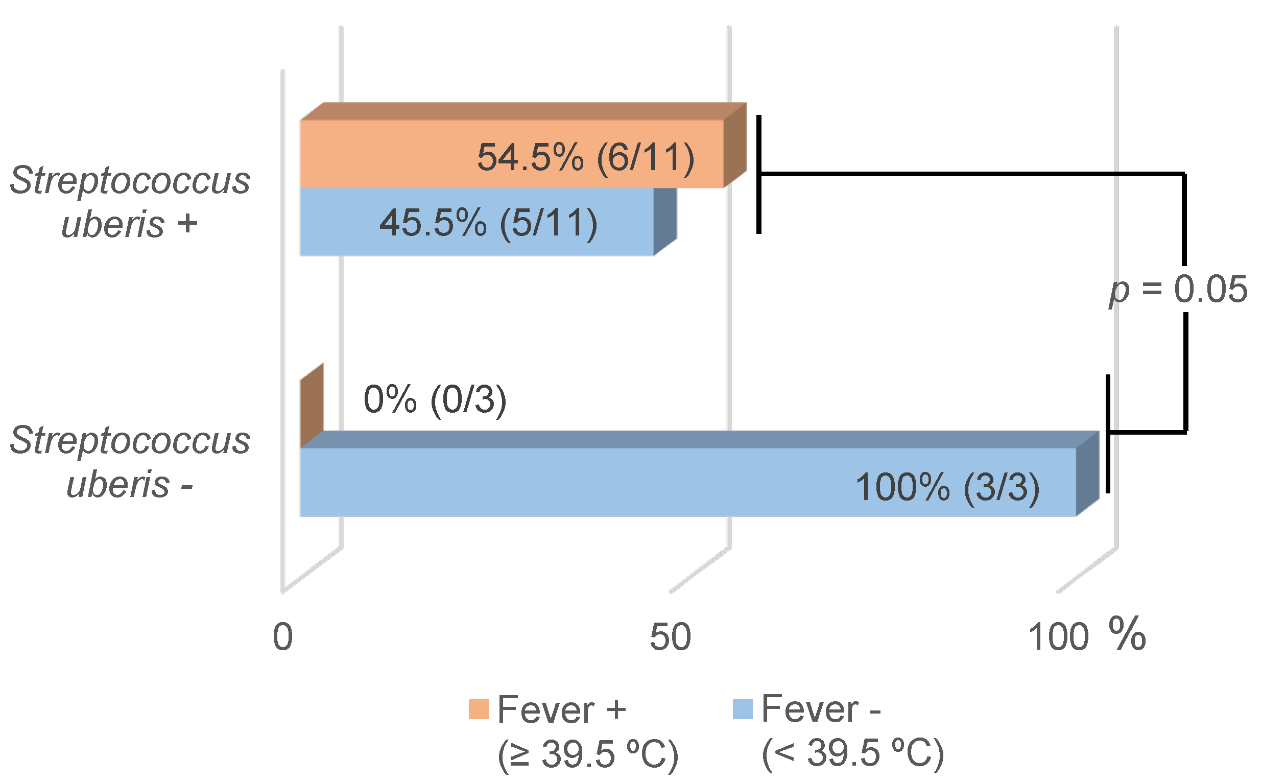
3.1. Identification of Mastitis Pathogens and BlaZ Gene via RT-PCR and Their Association with Parity and Fever

The microorganisms identified using RT-PCR are described in Table 1. At least three species of mastitis pathogens or groups were detected in each cow. No significant association was observed between parity and fever with any pathogen, or with the blaZ gene (p > 0.05), except for Strep. uberis.
The following mastitis pathogens, M. bovis, S. aureus, Prototheca spp., C. bovis, K. oxytoca/K. pneumoniae and S. marcescens, were not detected via RT-PCR.
Fever was detected in 54.5% of the 11 cows with Strep. uberis in their uterine fluid. In the absence of this bacterium, no animal had fever (Figure 1). Therefore, the presence of Strep. uberis in the uterine lumen of cows with RP was revealed as a significant risk factor for fever occurrence (RR = 1.6; 95% CI: 1.0–2.8; p = 0.05).

3.2. Identification of Mastitis Pathogens with Bacterial Culture and Agreement between Methodologies

In total, 28 bacterial isolates were obtained, from which 46.4% (13/28) were confirmed as E. coli through conventional bacterial culture, whereas 25.0% (7/28) were identified as Strep. uberis, and only 3.6% (1/28) were identified as Strep. dysgalactiae and Enterococcus faecalis. Additionally, Strep. mutans, Strep. porcinus, Strep. pseudoporcinus, Hafnia alvei, Proteus vulgaris and E. fergusonii were isolated.
Except for E. coli (substantial agreement; p < 0.01), the agreement between RT-PCR and the bacterial culture was fair (Strep. uberis; p < 0.05) or not significant (Enterococcus spp. and Strep. dysgalactiae; p > 0.05; Table 2).

4. Discussion

In this study, 60% (9/15) of the mastitis pathogens that could be identified via RT-PCR MastiType Multi Kit were detected in the uterine fluid of cows with RP. Of these pathogens, 40% (6/15) were isolated in more than half of the cows. The microorganisms were mainly (66.7%; 6/9) environmental mastitis pathogens (E. coli, Staphylococcus spp., Strep. uberis, Enterococcus spp., T. pyogenes/P. indolicus, yeasts). As previously mentioned, when the cervix opens at calving, environmental bacteria and yeast (e.g., vaginal and fecal) contaminate the uterus, usually up to two weeks after calving [27].
Strep. dysgalactiae, Strep. agalactiae and Mycoplasma spp. are considered contagious mastitis pathogens [28,29], i.e., their main route of transmission occurs from udder (mammary gland) to udder through milking procedures carried out with contaminated milking equipment. However, environmental spread also occurs, and these pathogens can be classified as both environmental and contagious pathogens (see the review by Cobirka et al. [29]). Mycoplasma spp., e.g., M. bovigenitalium and M. californicum (9.3%; 3/32), have also been detected previously in vaginal [30] and milk samples [31] from dairy cows.
In cattle, uterine microbiota is established within 20 min after calving [17]. At that time, the decomposing placental membrane and lochia provide an optimal intrauterine environment for microorganisms’ colonization and growth [32,33]. In turn, the discharge of uterine secretions contributes to the spread of (normal and pathogenic) bacteria. It was observed by Bicalho et al. [34] that the total bacteria load in the vagina was similar at the day of calving in cows with or without RP. Nonetheless, a tendency (p = 0.06) on day 3 postpartum and on day 7 postpartum of a significantly larger (p < 0.001) total bacteria load (bacterial/archaeal 16S rDNA gene), in cows with RP rather than in healthy cows, was observed in their study [34]. These differences highlight the relevance of environmental bacteria spreading to RP animals.
In our study, E. coli and Staphylococcus spp. were the bacteria most frequently detected in the uterine fluid. Likewise, E. coli has been reported by Dohmen et al. [4] as the most prevalent bacteria in the uterus, in early postpartum in cows with or without RP. Moreover, it was observed by Bicalho et al. [35] that cows with RP were 45 times more likely to present E. coli during the first three days postpartum than cows without it (odds ratio = 44.8; p < 0.01). E. coli is also the most frequently found Gram-negative pathogen causing acute clinical mastitis during early lactation in high-producing dairy cows. It invades the udder through the teats, proliferates, and induces a local and systemic acute phase response that may cause irreversible tissue damage in the mammary gland, and severe clinical symptoms, sometimes even leading to the animal’s death [1,36].
E. coli, as with other microorganisms, is a ubiquitous and opportunistic pathogen that depends on intrinsic virulent factors [37], bacterial load [38], and immune, metabolic and oxidative status to cause disease [6,34]. During the transition period and mainly around parturition, the increased energy demand favors the incidence of metabolic diseases, compromising immunity and resistance to infections by pathogenic and opportunistic agents. The condition of RP increases the occurrence of postpartum infectious diseases via uterine colonization and the development of E. coli and other microorganisms [9,39]. In the same way, the negative energy balance during this period impairs the immune response in the mammary gland of lactating dairy cows, increasing the susceptibility to mastitis [40,41,42] and RP [39]. Furthermore, oxidative stress has also been associated with negative energy balance [43,44] and metabolic disruptions [45] in dairy cows. It was suggested that the link between RP and mastitis may be related to the weakening of the immune system [46], through peripheral leukocyte impairment [47]. Moreover, the chronological conjugation of these factors with bacterial load, including bacterial virulent factors [35], can have a relevant impact on the incidence of these diseases. Nonetheless, the common possible source for RP and mastitis does not rule out the possibility that RP plays a key role in aggravating mastitis through causing an increased level of contamination in the environment of the udder of affected cows, regarding E. coli and other bacteria.
Suriyasathaporn et al. [48] observed a tendency for cows with RP to have an increased risk of presenting clinical mastitis (risk ratio = 2.88; 95% CI = 0.96–8.64; p = 0.06) than cows without RP. The risk of developing severe mastitis was higher (RR = 5.4) in cows with RP than that in healthy cows. Also, cows with RP up to five days after calving were more likely to present clinical mastitis up to 120 days postpartum (odds ratio = 9.45; 95% IC = 8.62–10.27; p < 0.001) than cows without RP [49]. A large epidemiological study addressing environmental, bacterial and host factors is required to evaluate the relationship between RP and mastitis in more depth.
The Staphylococcus spp. group mainly comprises CNS with more than 50 bacterial species, with S. chromogenes, S. epidermidis, S. haemolyticus, S. simulans and S. xylosus being the most common CNS milk pathogens [50]. Large numbers of this species are also found in the environment and can partially explain the high proportion of Staphylococcus spp. (93.3%) observed in our study as a result of contamination through the vagina in the early postpartum period. On the other hand, in recent years, CNS have been considered the most prevalent microorganisms identified in subclinical mastitis in dairy cows [51].
A high proportion of yeasts (92.9% of the cows) were detected in our study. However, detection does not mean that colonization occurs. We hypothesize that the birth channel is just the route of transmission from the environment (or vagina) to the udder. Yeats are common in the environment, on the skin, in feces and even in the vagina microbiota. They are also opportunist microorganisms (e.g., Candida albicans, C. krusei, C. tropicalis), and uterine colonization is usually related to cows with specific conditions such as infertile cows, repeat breeders, cows with persistent endometritis, prolonged intra-uterine antibiotherapy, and immunosuppression [33,52]. Nonetheless, a yeast prevalence between 21 and 33% from cervicovaginal fluids of Holstein cows (n = 176) was previously reported by Garoussi et al. [53]. In that study, no statistically different prevalence was observed between cows with reproductive disorders (including those with RP) and non-pregnant or pregnant healthy cows. A significant prevalence of yeast contamination from the mammary glands of dairy cows has been reported in some countries such as Mexico (25.8%; 282/1095; [54]) and Algeria (10.2%; 114/1121; [55]). In a recent and large study (Japan) [56], from 18,915 bulk tank milk samples in 1,088 bovine farms, 184 isolates of yeast species were detected. Pichia kudriavzevii (n = 97 isolates), Kluyveromyces marxianus (n = 28), C. akabanensis (n = 21), C. parapsilosis (n = 13) and P. cactophila (n = 12) were isolated, highlighting their relative importance (1% of the samples) in mastitis prevalence. However, contamination of bulk milk by environmental saprophytes yeasts can also occur. Therefore, more attention is required to establish the impact of yeasts on reproductive health in dairy cows.
In our study, similar intrauterine prevalence was observed for T. pyogenes/P. indolicus (80.0%; 95% CI: 54.8−93.0%) and Strep. uberis (78.6%; 95% CI: 52.4−92.4%). It has been previously observed by Ballas et al. [10] that the T. pyogenes prevalence correlates (p < 0.001) with that of Peptoniphilus spp. on day 3 postpartum in uterine fluid, with T. pyogenes prevalence reaching its maximum (52.5%; 64/122) on day 15 [10]. A similar pattern was also found by Wagener et al. [2]. Regarding the differences in T. pyogenes prevalence between both studies, it is necessary to investigate if the presence of RP increases the risk of T. pyogenes contamination during the first days postpartum, and if the physical presence of the placenta membrane enhances that risk. Mastitis caused by this bacterium has been related to summer mastitis [28,57]. This may be explained by the increased number of vectors (Hydrotaea irritans) able to transmit T. pyogenes [58].
Strep. uberis is considered a major mastitis pathogen with high genetic variability in the environment and animal udders [59]. It is also a bacterium that frequently colonizes the uterus postpartum, and a dynamic interaction between Strep. uberis and T. pyogenes has been described [2]. Strep. uberis is also a commensal microorganism of the bovine gut [60]. Thus, transmission to the udder via the digestive tract and feces is considered one of the most relevant routes of udder contamination [61], and the same is probably true for uterine contamination. Interestingly, 54.5% of the cows positive for Strep. uberis presented fever, whereas this clinical sign was not observed in any other cow. We did not find any study on this issue. Strep. uberis mastitis is often sudden in terms of its onset, and produces a hard, swollen quarter, with large white clots in milk, and sometimes with a high, or very high, body temperature [62]. In fact, this infection in dairy cows causes major signs of clinical mastitis, in some cases associated with systemic reactions [63]. Strep. uberis is recognized as an inducer of fever due to its involvement of systemic cytokines [64,65]. Apparently, according to our study, similar pathogenesis may occur when the infection is intrauterine.
Strep. dysgalactiae is other relevant contagious mastitis pathogen with previous scarce detection in the uterus, during the early postpartum period [66,67]. In contrast, we detected it in a high proportion of cases (57.1%; 8/14) in our study. There is evidence that Strep. dysgalactiae can persist for more than one year on the environment in dairy farms [63]. Hence, the importance of Strep. dysgalactiae in the puerperal uterus should be evaluated.
Finally, as expected, except for E. coli, a fair (Strep. uberis; k = 0.34; p < 0.05) or non-significant agreement (Enterococcus spp., Strep. dysgalactiae; p > 0.05) was observed between RT-PCR and bacterial culture methodologies, confirming the latter as useful especially for challenging bacterial cultures (e.g., Mycoplasma). In fact, the better performance of a multiplex PCR assay than that of bacterial cultures considering mastitis pathogens has previously been reported [68,69]. Despite its high sensitivity and specificity, RT-PCR can cause an increase in false positives due to the contamination of samples. Moreover, false negatives can appear due to small changes in the primer sites of new strains [14]. The substantial agreement (K = 0.64; p < 0.01) between RT-PCR and bacterial culture methods for E. coli observed in the present study was probably due to the high prevalence of this bacteria in the uterus detected via both methodologies (14/15 and 13/15 contaminated cows, respectively).
A limitation of this study was the reduced number of animals studied and that we only worked with cows with RP, without previous or posterior udder health evaluation. It was therefore not possible to associate RP with mastitis. However, overall, a large proportion of mastitis pathogens were detected in uterine fluids, supporting our hypothesis that cows with RP may serve as sources of intramammary infection, indirectly. More in-depth and larger comparative studies are required to evaluate the real impact of uterine secretions on mastitis prevalence during the postpartum period. Factors such as the environmental bacterial load on the animal surface and facilities (e.g., milking room and machines; calving parks), bacterial virulence and animal susceptibility should be taken into consideration for a holistic approach.

5. Conclusions

We found the same bacterial species that cause mastitis in the uterine fluid of cows presenting RP, which suggests that uterine secretions from cows with RP may be a relevant contamination source of mastitis pathogens. Therefore, although further studies should be carried out to better understand the extent of this relationship, specific care should be implemented for the udders of dairy cows with RP.

Author Contributions

Conceptualization, methodology, formal analysis, and data curation were performed by D.R., S.A., A.F.-N. and J.S. writing—original draft preparation; writing—review and editing were performed in equal proportions by D.R., S.A., A.F.-N., G.M. and J.S. All authors have read and agreed to the published version of the manuscript.

Funding

This work was supported by projects with the grant numbers UIDB/00772/2020 and LA/P/0059/2020, funded by the Portuguese Foundation for Science and Technology (FCT).

Institutional Review Board Statement

This study was approved by the Institutional Review Board of the University of Trás-os-Montes and Alto Douro (protocol code DOC_FP.8895-847PA38542; 2020-08-25) and conducted in accordance with the Declaration of Helsinki.

Informed Consent Statement

Informed consent was obtained from all farmers and the laboratory involved in the study.

Data Availability Statement

The raw data presented in this study are available on request from the corresponding author.

Acknowledgments

We would like to express our gratitude to all farmers, to Ana Paula Peixoto and to Nuno Tavares for helping us by giving us a supply of cases, and for their help in the collection of samples. We also thank Segalab laboratory for their work in RT-PCR and conventional bacterial culture, which made this study possible.

Conflicts of Interest

The authors declare no conflicts of interest.

References

  1. Cheng, W.N.; Han, S.G. Bovine Mastitis: Risk Factors, Therapeutic Strategies, and Alternative Treatments—A Review. Asian-Australas. J. Anim. Sci. 2020, 33, 1699–1713. [Google Scholar] [CrossRef] [PubMed]
  2. Wagener, K.; Grunert, T.; Prunner, I.; Ehling-Schulz, M.; Drillich, M. Dynamics of Uterine Infections with Escherichia coli, Streptococcus uberis and Trueperella pyogenes in Post-Partum Dairy Cows and Their Association with Clinical Endometritis. Vet. J. 2014, 202, 527–532. [Google Scholar] [CrossRef] [PubMed]
  3. Sheldon, I.M.; Lewis, G.S.; LeBlanc, S.; Gilbert, R.O. Defining Postpartum Uterine Disease in Cattle. Theriogenology 2006, 65, 1516–1530. [Google Scholar] [CrossRef] [PubMed]
  4. Dohmen, M.J.W.; Joop, K.; Sturk, A.; Bols, P.E.J.; Lohuis, J.A.C.M. Relationship between Intra-Uterine Bacterial Contamination, Endotoxin Levels and the Development of Endometritis in Postpartum Cows with Dystocia or Retained Placenta. Theriogenology 2000, 54, 1019–1032. [Google Scholar] [CrossRef]
  5. Silva, J.C.C.; Siqueira, L.C.; Rodrigues, M.X.; Zinicola, M.; Wolkmer, P.; Pomeroy, B.; Bicalho, R.C. Intrauterine Infusion of a Pathogenic Bacterial Cocktail Is Associated with the Development of Clinical Metritis in Postpartum Multiparous Holstein Cows. J. Dairy Sci. 2023, 106, 607–623. [Google Scholar] [CrossRef] [PubMed]
  6. Sheldon, I.M.; Cronin, J.; Goetze, L.; Donofrio, G.; Schuberth, H.-J. Defining Postpartum Uterine Disease and the Mechanisms of Infection and Immunity in the Female Reproductive Tract in Cattle1. Biol. Reprod. 2009, 81, 1025–1032. [Google Scholar] [CrossRef] [PubMed]
  7. Mahnani, A.; Sadeghi-Sefidmazgi, A.; Ansari-Mahyari, S.; Ghorbani, G.-R. Assessing the Consequences and Economic Impact of Retained Placenta in Holstein Dairy Cattle. Theriogenology 2021, 175, 61–68. [Google Scholar] [CrossRef] [PubMed]
  8. LeBlanc, S.J. Postpartum Uterine Disease and Dairy Herd Reproductive Performance: A Review. Vet. J. 2008, 176, 102–114. [Google Scholar] [CrossRef]
  9. Han, Y.K.; Kim, I.H. Risk Factors for Retained Placenta and the Effect of Retained Placenta on the Occurrence of Postpartum Diseases and Subsequent Reproductive Performance in Dairy Cows. J. Vet. Sci. 2005, 6, 53. [Google Scholar] [CrossRef] [PubMed]
  10. Ballas, P.; Gabler, C.; Wagener, K.; Drillich, M.; Ehling-Schulz, M. Streptococcus uberis Strains Originating from Bovine Uteri Provoke Upregulation of Pro-Inflammatory Factors mRNA Expression of Endometrial Epithelial Cells In Vitro. Vet. Microbiol. 2020, 245, 108710. [Google Scholar] [CrossRef] [PubMed]
  11. Paiano, R.B.; Moreno, L.Z.; Gomes, V.T.M.; Parra, B.M.; Barbosa, M.R.; Sato, M.I.Z.; Bonilla, J.; Pugliesi, G.; Baruselli, P.S.; Moreno, A.M. Assessment of the Main Pathogens Associated with Clinical and Subclinical Endometritis in Cows by Culture and MALDI-TOF Mass Spectrometry Identification. J. Dairy Sci. 2022, 105, 3367–3376. [Google Scholar] [CrossRef] [PubMed]
  12. Kaeberlein, T.; Lewis, K.; Epstein, S.S. Isolating “Uncultivable” Microorganisms in Pure Culture in a Simulated Natural Environment. Science 2002, 296, 1127–1129. [Google Scholar] [CrossRef] [PubMed]
  13. Santos, T.M.A.; Gilbert, R.O.; Bicalho, R.C. Metagenomic Analysis of the Uterine Bacterial Microbiota in Healthy and Metritic Postpartum Dairy Cows. J. Dairy Sci. 2011, 94, 291–302. [Google Scholar] [CrossRef]
  14. Compton, S.R. PCR and RT-PCR in the Diagnosis of Laboratory Animal Infections and in Health Monitoring. J. Am. Assoc. Lab. Anim. Sci. 2020, 59, 458–468. [Google Scholar] [CrossRef] [PubMed]
  15. Vidal, S.; Kegler, K.; Posthaus, H.; Perreten, V.; Rodriguez-Campos, S. Amplicon Sequencing of Bacterial Microbiota in Abortion Material from Cattle. Vet. Res. 2017, 48, 64. [Google Scholar] [CrossRef] [PubMed]
  16. Machado, V.S.; Oikonomou, G.; Bicalho, M.L.S.; Knauer, W.A.; Gilbert, R.; Bicalho, R.C. Investigation of Postpartum Dairy Cows’ Uterine Microbial Diversity Using Metagenomic Pyrosequencing of the 16S rRNA Gene. Vet. Microbiol. 2012, 159, 460–469. [Google Scholar] [CrossRef] [PubMed]
  17. Jeon, S.J.; Vieira-Neto, A.; Gobikrushanth, M.; Daetz, R.; Mingoti, R.D.; Parize, A.C.B.; de Freitas, S.L.; da Costa, A.N.L.; Bicalho, R.C.; Lima, S.; et al. Uterine Microbiota Progression from Calving until Establishment of Metritis in Dairy Cows. Appl. Environ. Microbiol. 2015, 81, 6324–6332. [Google Scholar] [CrossRef] [PubMed]
  18. Wang, Y.; Wang, J.; Li, H.; Fu, K.; Pang, B.; Yang, Y.; Liu, Y.; Tian, W.; Cao, R. Characterization of the Cervical Bacterial Community in Dairy Cows with Metritis and during Different Physiological Phases. Theriogenology 2018, 108, 306–313. [Google Scholar] [CrossRef] [PubMed]
  19. Çömlekcioğlu, U.; Jezierska, S.; Opsomer, G.; Pascottini, O.B. Uterine Microbial Ecology and Disease in Cattle: A Review. Theriogenology 2024, 213, 66–78. [Google Scholar] [CrossRef] [PubMed]
  20. Savov, N.; Buchvarova, I. Relationship between infections of the genitalia and udder in cows. Vet. Med. Nauki 1977, 14, 43–46. [Google Scholar]
  21. Itoh, M.; Furuoka, M.; Baba, Y.; Saitoh, T.; Hata, E.; Hirano, Y.; Senna, K.; Yamada, K. Relationship between Genital Carriage and Udder Infection with Mycoplasma bovigenitalium in Dairy Farms. Jpn. J. Vet. Res. 2021, 69, 83–87. [Google Scholar] [CrossRef]
  22. Bicalho, R.C.; Cheong, S.H.; Galvão, K.N.; Warnick, L.D.; Guard, C.L. Effect of Twin Birth Calvings on Milk Production, Reproductive Performance, and Survival of Lactating Cows. J. Am. Vet. M. Ass. 2007, 231, 1390–1397. [Google Scholar] [CrossRef] [PubMed]
  23. Azawi, O.; Rahawy, M.; Hadad, J. Bacterial Isolates Associated with Dystocia and Retained Placenta in Iraqi Buffaloes. Reprod. Domest. Anim. 2008, 43, 286–292. [Google Scholar] [CrossRef] [PubMed]
  24. de Koff, E.M.; van Baarle, D.; van Houten, M.A.; Reyman, M.; Berbers, G.A.M.; van den Ham, F.; Chu, M.L.J.N.; Sanders, E.A.M.; Bogaert, D.; Fuentes, S. Mode of Delivery Modulates the Intestinal Microbiota and Impacts the Response to Vaccination. Nat. Commun. 2022, 13, 6638. [Google Scholar] [CrossRef] [PubMed]
  25. Wallet, F.; Loïez, C.; Renaux, E.; Lemaitre, N.; Courcol, R.J. Performances of VITEK 2 Colorimetric Cards for Identification of Gram-Positive and Gram-Negative Bacteria. J. Clin. Microbiol. 2005, 43, 4402–4406. [Google Scholar] [CrossRef] [PubMed]
  26. McHugh, M.L. Interrater Reliability: The Kappa Statistic. Biochem. Med. 2012, 22, 276–282. [Google Scholar] [CrossRef]
  27. Várhidi, Z.; Csikó, G.; Bajcsy, Á.C.; Jurkovich, V. Uterine Disease in Dairy Cows: A Comprehensive Review Highlighting New Research Areas. Vet. Sci. 2024, 11, 66. [Google Scholar] [CrossRef] [PubMed]
  28. Rzewuska, M.; Kwiecień, E.; Chrobak-Chmiel, D.; Kizerwetter-Świda, M.; Stefańska, I.; Gieryńska, M. Pathogenicity and Virulence of Trueperella pyogenes: A Review. Int. J. Mol. Sci. 2019, 20, 2737. [Google Scholar] [CrossRef] [PubMed]
  29. Cobirka, M.; Tancin, V.; Slama, P. Epidemiology and Classification of Mastitis. Animals 2020, 10, 2212. [Google Scholar] [CrossRef] [PubMed]
  30. Hazelton, M.S.; Morton, J.M.; Bosward, K.L.; Sheehy, P.A.; Parker, A.M.; Dwyer, C.J.; Niven, P.G.; House, J.K. Mycoplasma Species in Vaginas of Dairy Cows before and after Exposure to Bulls and Their Association with Conception. J. Dairy Sci. 2020, 103, 11795–11805. [Google Scholar] [CrossRef] [PubMed]
  31. Gioia, G.; Addis, M.F.; Santisteban, C.; Gross, B.; Nydam, D.V.; Sipka, A.S.; Virkler, P.D.; Watters, R.D.; Wieland, M.; Zurakowski, M.J.; et al. Mycoplasma Species Isolated from Bovine Milk Collected from US Dairy Herds between 2016 and 2019. J. Dairy Sci. 2021, 104, 4813–4821. [Google Scholar] [CrossRef] [PubMed]
  32. Eiler, H.; Fecteau, K.A. Retained Placenta. In Current Therapy in Large Animal Theriogenology; Elsevier: Amsterdam, The Netherlands, 2007; pp. 345–354. ISBN 978-0-7216-9323-1. [Google Scholar]
  33. Saini, P.; Singh, M.; Kumar, P. Fungal Endometritis in Bovines. Open Vet. J. 2019, 9, 94. [Google Scholar] [CrossRef] [PubMed]
  34. Bicalho, M.L.S.; Santin, T.; Rodrigues, M.X.; Marques, C.E.; Lima, S.F.; Bicalho, R.C. Dynamics of the Microbiota Found in the Vaginas of Dairy Cows during the Transition Period: Associations with Uterine Diseases and Reproductive Outcome. J. Dairy Sci. 2017, 100, 3043–3058. [Google Scholar] [CrossRef] [PubMed]
  35. Bicalho, M.L.S.; Machado, V.S.; Oikonomou, G.; Gilbert, R.O.; Bicalho, R.C. Association between Virulence Factors of Escherichia coli, Fusobacterium necrophorum, and Arcanobacterium pyogenes and Uterine Diseases of Dairy Cows. Vet. Microbiol. 2012, 157, 125–131. [Google Scholar] [CrossRef] [PubMed]
  36. Khatun, M.; Sørensen, P.; Ingvartsen, K.L.; Bjerring, M.; Røntved, C.M. Effects of Combined Liver and Udder Biopsying on the Acute Phase Response of Dairy Cows with Experimentally Induced E. coli Mastitis. Animal 2013, 7, 1721–1730. [Google Scholar] [CrossRef] [PubMed]
  37. Aditya, A.; Julianingsih, D.; Tabashsum, Z.; Alvarado-Martinez, Z.; Tung, C.-W.; Wall, M.; Biswas, D. Dominance of Diarrheagenic E. coli Virulent Types in Integrated Crop–Livestock Farms and Their Antibiotic Resistance Patterns. Zoonotic Dis. 2024, 4, 11–21. [Google Scholar] [CrossRef]
  38. Hansen, A.-M.; Chaerkady, R.; Sharma, J.; Díaz-Mejía, J.J.; Tyagi, N.; Renuse, S.; Jacob, H.K.C.; Pinto, S.M.; Sahasrabuddhe, N.A.; Kim, M.S.; et al. The Escherichia coli Phosphotyrosine Proteome Relates to Core Pathways and Virulence. PLoS Pathog. 2013, 9, e1003403. [Google Scholar] [CrossRef] [PubMed]
  39. Dervishi, E.; Zhang, G.; Hailemariam, D.; Dunn, S.M.; Ametaj, B.N. Occurrence of Retained Placenta Is Preceded by an Inflammatory State and Alterations of Energy Metabolism in Transition Dairy Cows. J. Animal Sci. Biotechnol. 2016, 7, 26. [Google Scholar] [CrossRef] [PubMed]
  40. Moyes, K.M.; Drackley, J.K.; Morin, D.E.; Rodriguez-Zas, S.L.; Everts, R.E.; Lewin, H.A.; Loor, J.J. Mammary Gene Expression Profiles during an Intramammary Challenge Reveal Potential Mechanisms Linking Negative Energy Balance with Impaired Immune Response. Physiol. Genom. 2010, 41, 161–170. [Google Scholar] [CrossRef] [PubMed]
  41. Hillreiner, M.; Flinspach, C.; Pfaffl, M.W.; Kliem, H. Effect of the Ketone Body Beta-Hydroxybutyrate on the Innate Defense Capability of Primary Bovine Mammary Epithelial Cells. PLoS ONE 2016, 11, e0157774. [Google Scholar] [CrossRef] [PubMed]
  42. Swartz, T.H.; Bradford, B.J.; Mamedova, L.K. Connecting Metabolism to Mastitis: Hyperketonemia Impaired Mammary Gland Defenses during a Streptococcus uberis Challenge in Dairy Cattle. Front. Immunol. 2021, 12, 700278. [Google Scholar] [CrossRef] [PubMed]
  43. Tsuchiya, Y.; Kawahara, N.; Kim, Y.-H.; Ichijo, T.; Sato, S. Changes in Oxidative Stress Parameters in Healthy and Diseased Holstein Cows during the Transition Period in Yamagata Prefecture, Japan. J. Vet. Med. Sci. 2020, 82, 955–961. [Google Scholar] [CrossRef] [PubMed]
  44. Malledevarahalli Chandrappa, S.; Pascottini, O.B.; Opsomer, G.; Meineri, G.; Martino, N.A.; Banchi, P.; Vincenti, L.; Ricci, A. Circulating and Endometrial Cell Oxidative Stress in Dairy Cows Diagnosed with Metritis. Theriogenology 2023, 198, 217–223. [Google Scholar] [CrossRef] [PubMed]
  45. Turk, R.; Koledić, M.; Maćešić, N.; Benić, M.; Dobranić, V.; Đuričić, D.; Cvetnić, L.; Samardžija, M. The Role of Oxidative Stress and Inflammatory Response in the Pathogenesis of Mastitis in Dairy Cows. Mljekarstvo 2017, 67, 91–101. [Google Scholar] [CrossRef]
  46. Zdunczyk, S.; Ahlers, D.; Grunert, E. The relationship between bovine clinical mastitis at the time of parturition and retained placenta. Dtsch. Tierarztl. Wochenschr. 1992, 99, 386–389. [Google Scholar] [PubMed]
  47. Schukken, Y.H.; Erb, H.N.; David Smith, R. The Relationship between Mastitis and Retained Placenta in a Commercial Population of Holstein Dairy Cows. Prev. Vet. Med. 1988, 5, 181–190. [Google Scholar] [CrossRef]
  48. Suriyasathaporn, W.; Schukken, Y.H.; Nielen, M.; Brand, A. Low Somatic Cell Count: A Risk Factor for Subsequent Clinical Mastitis in a Dairy Herd. J. Dairy Sci. 2000, 83, 1248–1255. [Google Scholar] [CrossRef] [PubMed]
  49. Ghavi Hossein-Zadeh, N.; Ardalan, M. Cow-Specific Risk Factors for Retained Placenta, Metritis and Clinical Mastitis in Holstein Cows. Vet. Res. Commun. 2011, 35, 345–354. [Google Scholar] [CrossRef] [PubMed]
  50. Vanderhaeghen, W.; Piepers, S.; Leroy, F.; Van Coillie, E.; Haesebrouck, F.; De Vliegher, S. Identification, Typing, Ecology and Epidemiology of Coagulase Negative Staphylococci Associated with Ruminants. Vet. J. 2015, 203, 44–51. [Google Scholar] [CrossRef] [PubMed]
  51. Addis, M.F.; Maffioli, E.M.; Ceciliani, F.; Tedeschi, G.; Zamarian, V.; Tangorra, F.; Albertini, M.; Piccinini, R.; Bronzo, V. Influence of Subclinical Mastitis and Intramammary Infection by Coagulase-Negative Staphylococci on the Cow Milk Peptidome. J. Proteom. 2020, 226, 103885. [Google Scholar] [CrossRef] [PubMed]
  52. Silva, L.P.; Ramos, S.Á.; Cavalcanti Neto, C.C.; Dos Santos, T.M.C.; De Oliveira, J.A.C.; Dos Santos, M.T.; Da Silva, J.M.; Brito Neto, J.S.; Montaldo, Y.C. Vaginal Microbiota of Nulliparous and Multiparous Cows and Their Resistance to Antimicrobials. Med. Vet. 2019, 13, 474. [Google Scholar] [CrossRef]
  53. Garoussi, M.T.; Khosrave, A.R.; Havareshti, P. Mycoflora of Cervicovaginal Fluids in Dairy Cows with or without Reproductive Disorders. Mycopathologia 2007, 164, 97–100. [Google Scholar] [CrossRef] [PubMed]
  54. Zaragoza, C.; Cervantes Olivares, R.A.; Ducoing Watty, A.E.; de la Peña Moctezuma, A.; Villa Tanaca, L. Yeasts Isolation from Bovine Mammary Glands under Different Mastitis Status in the Mexican High Plateu. Rev. Iberoam. Micol. 2011, 28, 79–82. [Google Scholar] [CrossRef] [PubMed]
  55. Ksouri, S.; Djebir, S.; Hadef, Y.; Benakhla, A. Survey of Bovine Mycotic Mastitis in Different Mammary Gland Statuses in Two North-Eastern Regions of Algeria. Mycopathologia 2015, 179, 327–331. [Google Scholar] [CrossRef] [PubMed]
  56. Toyotome, T.; Matsui, S. Analysis of Prototheca and Yeast Species Isolated from Bulk Tank Milk Collected in Tokachi District, Japan. J. Dairy Sci. 2022, 105, 8364–8370. [Google Scholar] [CrossRef] [PubMed]
  57. Ishiyama, D.; Mizomoto, T.; Ueda, C.; Takagi, N.; Shimizu, N.; Matsuura, Y.; Makuuchi, Y.; Watanabe, A.; Shinozuka, Y.; Kawai, K. Factors Affecting the Incidence and Outcome of Trueperella pyogenes Mastitis in Cows. J. Vet. Med. Sci. 2017, 79, 626–631. [Google Scholar] [CrossRef] [PubMed]
  58. Chirico, J.; Jonsson, P.; Kjellberg, S.; Thomas, G. Summer Mastitis Experimentally Induced by Hydrotaea irritans Exposed to Bacteria. Med. Vet. Entomol. 1997, 11, 187–192. [Google Scholar] [CrossRef] [PubMed]
  59. Wald, R.; Baumgartner, M.; Gutschireiter, J.; Bazzanella, B.; Lichtmannsperger, K.; Wagner, M.; Wittek, T.; Stessl, B. Comparison of the Population Structure of Streptococcus uberis Mastitis Isolates from Austrian Small-Scale Dairy Farms and a Slovakian Large-Scale Farm. J. Dairy Sci. 2020, 103, 1820–1830. [Google Scholar] [CrossRef] [PubMed]
  60. Reyes, J.; Rodriguez-Lecompte, J.C.; Blanchard, A.; McClure, J.T.; Sánchez, J. Molecular Variability of Streptococcus uberis Isolates from Intramammary Infections in Canadian Dairy Farms from the Maritime Region. Can. J. Vet. Res. 2019, 83, 168–176. [Google Scholar] [PubMed]
  61. Fessia, A.S.; Odierno, L.M. Potential Factors Involved in the Early Pathogenesis of Streptococcus uberis Mastitis: A Review. Folia Microbiol. 2021, 66, 509–523. [Google Scholar] [CrossRef] [PubMed]
  62. Blowey, R.; Edmondson, P. Bactoscan and Total Bacterial Count (TBC). In Mastitis Control in Dairy Herds; CABI: Wallingford, UK, 2010; pp. 171–183. [Google Scholar] [CrossRef]
  63. Abd El-Aziz, N.K.; Ammar, A.M.; El Damaty, H.M.; Abd Elkader, R.A.; Saad, H.A.; El-Kazzaz, W.; Khalifa, E. Environmental Streptococcus uberis Associated with Clinical Mastitis in Dairy Cows: Virulence Traits, Antimicrobial and Biocide Resistance, and Epidemiological Typing. Animals 2021, 11, 1849. [Google Scholar] [CrossRef] [PubMed]
  64. Bannerman, D.D.; Paape, M.J.; Goff, J.P.; Kimura, K.; Lippolis, J.D.; Hope, J.C. Innate Immune Response to Intramammary Infection with Serratia marcescens and Streptococcus uberis. Vet. Res. 2004, 35, 681–700. [Google Scholar] [CrossRef] [PubMed]
  65. Tassi, R.; McNeilly, T.N.; Fitzpatrick, J.L.; Fontaine, M.C.; Reddick, D.; Ramage, C.; Lutton, M.; Schukken, Y.H.; Zadoks, R.N. Strain-Specific Pathogenicity of Putative Host-Adapted and Nonadapted Strains of Streptococcus uberis in Dairy Cattle. J. Dairy Sci. 2013, 96, 5129–5145. [Google Scholar] [CrossRef]
  66. Muskens, J.; van Maanen, C.; Mars, M.H. Dairy Cows with Metritis: Coxiella burnetii Test Results in Uterine, Blood and Bulk Milk Samples. Vet. Microbiol. 2011, 147, 186–189. [Google Scholar] [CrossRef] [PubMed]
  67. Kronfeld, H.; Kemper, N.; Hölzel, C.S. Vaginal and Uterine Microbiomes during Puerperium in Dairy Cows. Agriculture 2022, 12, 405. [Google Scholar] [CrossRef]
  68. Ashraf, A.; Imran, M.; Yaqub, T.; Tayyab, M.; Shehzad, W.; Thomson, P.C. A Novel Multiplex PCR Assay for Simultaneous Detection of Nine Clinically Significant Bacterial Pathogens Associated with Bovine Mastitis. Mol. Cell. Probes 2017, 33, 57–64. [Google Scholar] [CrossRef] [PubMed]
  69. Imam, T.; Horsman, S.; Wood, B.; Grewar, J.D.; Langhorne, C.; Price, R.; Wood, C.; Henning, J.; Gibson, J.S. Assessment of Sensitivity and Specificity of Bacterial Culture and the VetMAXTM Mastitype Multi Kit in Detecting Streptococcus uberis and Escherichia coli in Milk Samples from Dairy Cows with Clinical Mastitis in Subtropical Australia. Soc. Sci. Res. Netw. 2024. preprint. [Google Scholar] [CrossRef]
Figure 1. Relationship between Strep. uberis and presence of fever according to logistic regression (n = 14).
Applsci 14 04986 g001
Table 1. Association between parity and fever in terms of the prevalence of microorganisms and the beta-lactamase (BlaZ) gene isolated with RT-PCR from uterine fluid samples of cows with RP.
Bacteria Identified by RT-PCR % Infected Cows (x/X)95% CIParityFever
Escherichia coli93.3 (14/15)70.2–98.90.120.28
Staphylococcus spp. 93.3 (14/15)70.2–98.90.120.28
Yeasts *92.9 (13/14)68.5–98.70.340.18
Trueperella pyogenes/Peptoniphilus indolicus80.0 (12/15)54.8–93.01.000.30
Streptococcus uberis *78.6 (11/14)52.4–92.40.850.05
Streptococcus dysgalactiae *57.1 (8/14)32.6–78.60.620.30
Enterococcus spp.46.7 (7/15)24.8–69.90.710.83
Mycoplasma spp.40.0 (6/15)19.8–64.30.260.50
BlaZ gene *28.6 (4/14)11.7–54.70.600.75
Streptococcus agalactiae6.7 (1/15)1.2–29.80.110.14
95% CI: 95% confidence interval. * The result was inconclusive in one cow.
Table 2. Agreement in bacterial identification between RT-PCR and bacterial culture methods.
BacteriaAgreement between MethodsCohen’s Kappa CoefficientNumber of Isolates BC- and RT-PCR+ (1)
E. coli (n = 15)93.3%0.64 (substantial) **1
Strep. uberis (n = 14)64.3%0.34 (fair) *5
Enterococcus spp. (n = 15)60.0%0.15 (slight)6
Strep. dysgalactiae (n = 14)50.0%0.11 (slight)7
** p < 0.01; *: p < 0.05. (1) Number of isolates negative on bacterial culture (BC) but positive on RT-PCR. The inverse occurrence (RT-PCR—and bacterial culture +) was not observed.
Disclaimer/Publisher’s Note: The statements, opinions and data contained in all publications are solely those of the individual author(s) and contributor(s) and not of MDPI and/or the editor(s). MDPI and/or the editor(s) disclaim responsibility for any injury to people or property resulting from any ideas, methods, instructions or products referred to in the content.

