Article

Pathogens in the Food Chain: Escherichia coli Strains in Raw Milk Originating from Ewes Treated for Mastitis with Various Therapeutic Protocols

by
Konstantina Fotou
1,
Georgios Rozos
1,2,
Konstantinos Zaralis
2,
Aikaterini Dadamogia
1,
Elisavet Stavropoulou
3,
Panagiotis Demertzis
4,
Konstantoula Akrida-Demertzi
4,*,
Athina Tzora
1 and
Chrysoula (Chrysa) Voidarou
1,*
1
Laboratory of Animal Health, Food Hygiene and Quality, Department of Agriculture, School of Agriculture, University of Ioannina, 47100 Arta, Greece
2
Laboratory of Animal Husbandry and Nutrition, Department of Agriculture, University of Western Macedonia, 53100 Florina, Greece
3
Laboratory of Hygiene and Environmental Protection, Department of Medicine, Democritus University of Thrace, Dragana, 68100 Alexandroupolis, Greece
4
Food Chemistry Laboratory, Section of Industrial and Food Chemistry, Department of Chemistry, University of Ioannina, 45110 Ioannina, Greece
*
Authors to whom correspondence should be addressed.
Appl. Sci. 2024, 14(13), 5383; https://doi.org/10.3390/app14135383
Submission received: 29 March 2024 / Revised: 8 June 2024 / Accepted: 17 June 2024 / Published: 21 June 2024
(This article belongs to the Special Issue Food Contamination: Sources, Detection, and Monitoring)

Abstract

:
Dairy products from ovine milk are very popular in the Mediterranean countries and are gaining a large portion of the market in EU countries and worldwide. EU legislation permits the dairy processing of raw ovine milk under certain conditions. To study the ecology and prevalence of E. coli in raw ewes’ milk and assess thus the public health risk, samples of milk were taken from 75 different sheep farms in the rural area of Epirus, Greece. The initial sampling was conducted in clinically healthy animals which were noted as controls (group A). From the same farms, samples were taken from animals with clinical mastitis and before treatment (group B). For therapeutic purposes, to some animals, a combination of penicillin and streptomycin was administrated (group C1), or tetracycline (group C2), or enrofloxacin (group C3). Finally, samples of raw milk were taken from the C groups, on the first day after the withdrawal period of the antibiotics used, when the milk is permitted to enter the food chain. In total, 97 isolates of Escherichia coli were recovered from all groups. Analysis revealed an impressive increase in E. coli strains in the milk of group B (39.33%) with respect to group A (5%). Even after treatment, although the prevalence was decreased, it was still found to be higher in the C groups than group A. E. coli O157:H7 strains absent from group A were detected in all other groups in relatively low occurrence rates with respect to other “O” serotypes but non-O157:H7 strains. Virulence factors such as the production of toxins (32.60% for serotoxin 1 and 18.47% for serotoxin 2) and hemolysin (42.39%) as well as biofilm formation capacity (52.17% of the total) and ESDL production (43.47% of the total) were also studied. All strains were also tested for susceptibility against 12 antibiotics by the MIC method and the results showed a high prevalence of resistance and multi-resistance. The presence of various resistant strains to antibiotics and pathogenic “O” serotype strains in the milk when it can enter the food chain again is an alarming conclusion.

1. Introduction

In Greece, as in most Balkan and Mediterranean countries, ewes’ milk has a significant financial importance. According to the EU, 57.7% of the milk delivered to the dairies in Greece originates from ewes [1]. Ovine milk and particularly ovine dairy products have been important staple foods since antiquity. Ovine species are perfectly adapted to the Mediterranean climate and besides their milk, they also provide meat, skin, and wool, contributing thus significantly to the survival of the local populations in historical as well as contemporary times. As far as the safety of these dairy products is concerned, their consumption has been associated with various diseases [2,3,4], and this is not a surprise, given that milk is an excellent medium for the growth of almost all pathogens and that the hygienic status of many farms is often not acceptable. At the same time, there is a growing demand and preference for raw ovine milk and dairy products made from such milk [5]. This movement combines these products which are usually produced by local small-scale dairies, with attractive concepts such as “natural”, “green”, “organic”, “authentic taste”, and “traditional” [6,7]. In these establishments, however, the hygienic status is often compromised, thus increasing the odds of products detrimental to public health.
Milk, per se, is a complex matrix of ingredients and an excellent medium for bacteria, pathogenic or not. It has been argued that milk has its own defense systems against bacteria, such as its microbiota whose populations act antagonistically against pathogenic intruders [8,9]. But even these defense systems succumb to various factors such as the environmental temperature, the environmental spoilage, the welfare of the animals, the sanitation of the milking apparatus and of the milking process, and the health of the udders and of the animals, to name a few [10,11,12,13,14,15,16,17,18]. Contaminated milk in the bulk tank is a common finding [19,20,21].
Milking is the initial and most crucial stage of the dairy food chain since bacteria such as Escherichia coli can contaminate the milk and compromise the quality and safety of the final products [22,23]. E. coli O157:H7 is a serotype of special significance due to its high virulence but other serotypes are also of high concern, such as the—so-called—“big six” (O26, O45, O103, O111, O121, and O145), owing their nickname to their association with severe foodborne illness [24].
Although EU legislation (EC 852/2004, EC 853/2004) enforces pasteurization [25,26] in raw milk, it permits under certain conditions the processing of raw milk without any thermal treatment, based on the total bacterial count (TBC), the absence of Brucella species from the milking animals and the ripening of the curd for a period greater than two months. Yet none of these conditions ensures the absence of pathogenic E. coli from the final dairy products. Many pathogens can survive for weeks or even months in dairy products. Salmonella spp, Staphylococcus aureus, Listeria mocytogenes, Coxiella brunetti, and Escherichia coli have been isolated from raw milk as well as from cheeses made from raw milk [27,28,29,30,31,32,33,34].
Clinical mastitis is a serious problem in milk-producing flocks and the major cause of “milk drop syndrome” [35]. E. coli is involved in the pathogenesis of clinical mastitis along with other pathogens [36]. In the early stages of mastitis, it is possible that milk from unnoticed sick animals may end up in the same bulk tank as the milk from the healthy ones. After successful treatment with antibiotics when the clinical signs have vanished, it is possible that some pathogens may remain in the udder contaminating the milk. From the perspective of food safety, it is of utmost importance to investigate the diversity and prevalence of the hazardous E. coli “O” serotypes in milk from healthy udders, in milk from udders suffering from mastitis, and in milk after the withdrawal period of the antibiotics used to treat mastitis, when milk enters the food chain again, and these are the aims of this study (Figure 1).

2. Materials and Methods

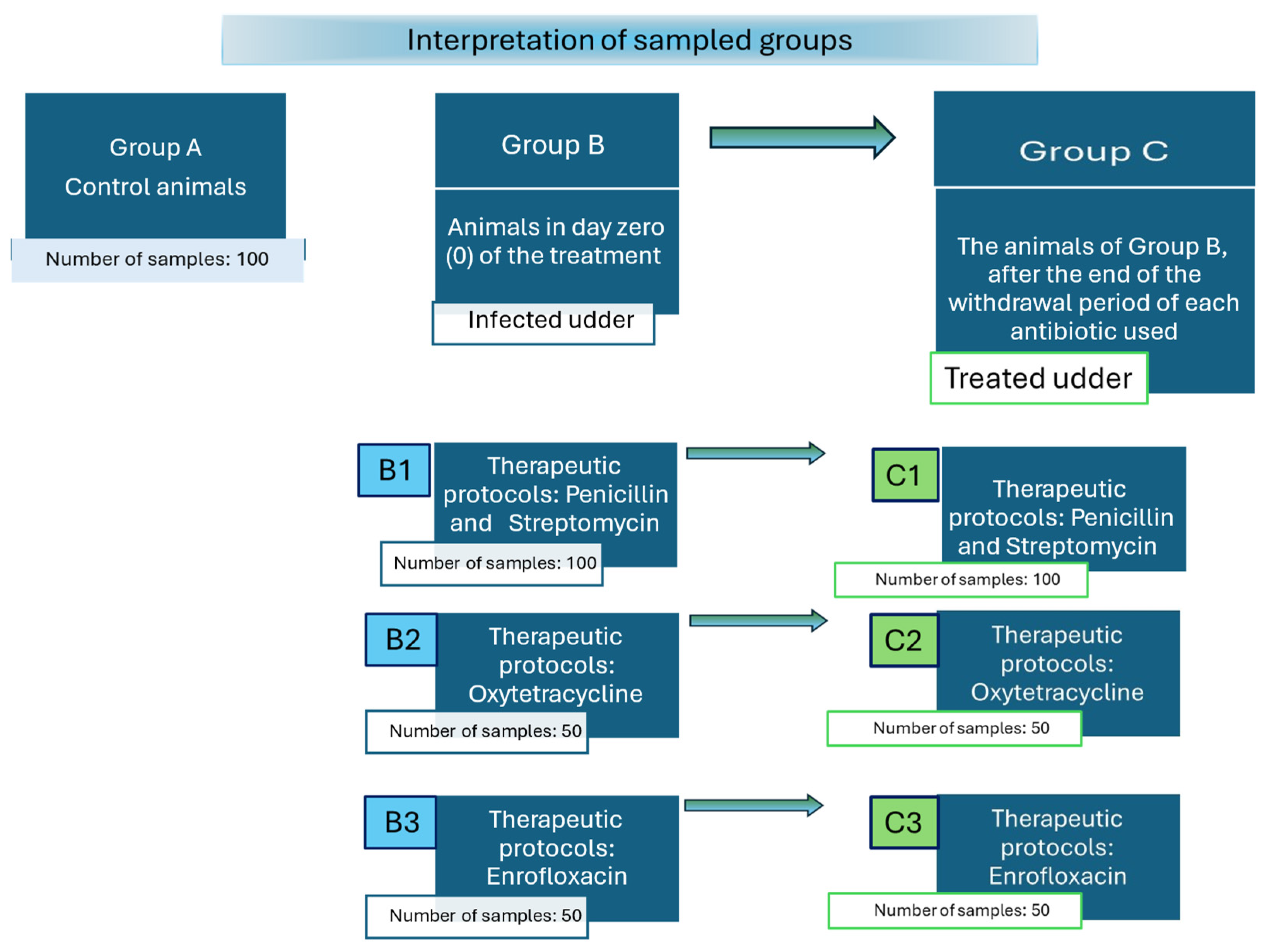
2.1. Description of Sampled Animal Groups

The study was conducted in 75 sheep farms located in the region of Epirus, Greece. All farms were situated in a lowland area, sharing pastures with similar vegetation. The size of the flocks was 100–300 animals of a local breed (“boutsikes”), and all used the same pastures and followed similar farming practices such as vaccinations, antiparasitic treatments, use of milking machines, administration of concentrated feeds, etc. Samples of milk were collected from healthy sheep with healthy udders (referred exclusively to as the control group-“martyrs”), from sheep with clinical mastitis before treatment (clinical evaluation points: color and temperature of udders, pain, change in color and viscosity of milk, decreased milk production and California Mastitis Test category of grade 2 and higher), and from the previous animals after treated for mastitis. All animals were in the middle of their milking period and were randomly selected using tables of random numbers based on their ear tag numbers. The animals suffering from mastitis were kept away from the healthy ones under strict hygienic conditions. Diagnosis and treatments were carried out by professional veterinarians. It is important to clarify that identifying the responsible microorganism in clinical practice is usually not feasible, cost-effective, or practical due to the remoteness of the farms and the mountainous terrain. Most veterinarians in Greece determine the treatment regimen empirically, based on clinical signs and the therapeutic history of the flock (efficacy of previous treatments, success rates, etc.). Three groups of animals were formed (Figure 1): (a) control animals with clinically healthy udders—group A—control; (b) animals suffering from mastitis, before treatment (infected udder)—group B; and (c) the animals of group B, at the first day after the end of the withdrawal period of each antibiotic used (treated udder)—group C. The treated ewes were divided into three subgroups, receiving different drastic compounds. The therapeutic protocols used included the following antibiotic substances: (1) penicillin and streptomycin, (2) oxytetracycline, and (3) enrofloxacin (Table 1). On the first day after the end of the withdrawal period when the milk can be given to the dairies for processing, samples were taken from the same animals, classified into three subgroups (C1 for penicillin and streptomycin; C2 for oxytetracycline; and C3 for enrofloxacin). It should be noted that most of the milk from these farms is supplied to local dairies, while a smaller portion is reserved by the producers’ families for making artisan cheeses for their own consumption. In total, 500 samples of milk were analyzed, originating from the above groups of ewes. Milk samples were collected following a procedure that included cleaning the udder with lukewarm water, discarding the first three streams of milk, and disinfecting the teat ends with an alcohol solution (70% absolute alcohol). All samples were immediately transported to the laboratory within one hour of reception and kept at 4 °C for a maximum of 24 h before analysis.

2.2. Methodology: Isolation and Phenotypic Characterization of Escherichia coli

Each sample underwent thorough mixing in its sampling vial via 20–25 inversion movements. Subsequently, within 3 min of agitation, 10 mL of the sample was drawn using a sterile pipette and transferred to a dilution bottle containing 90 mL of homogenization liquid (Buffered Peptone Water, BPW, BioMerieux, Marcy l’Etoile, France), resulting in a 1:10 dilution. Decimal dilutions were subsequently carried out up to a dilution factor of 10−8, using Ringer’s solution. A variety of selective and differential plating media has been used for the recovery of E coli isolates. The rationale for adopting this approach was to support the growth of both starved and unstarved E. coli cells. The following commercial media were used: Violet Red Bile Glucose Agar (VRBGA, Merck KGaA, Darmstadt, Germany), Endo Agar (Merck KGaA, Darmstadt, Germany), Ssorbitol McConkey agar containing 2.5 mg/L potassium tellurite and 0.05 mg/L cefixime (CT-SMAC, Merck KGaA, Darmstadt, Germany), CT-SMAC additionally supplemented with 50 mg/L 5-Brom-4-chlor-indolyl-beta-D-glucuronid cyclohexylammonium-salt (Merck KGaA, Darmstadt, Germany), CHROMagar™ E.coli, EC O157:H7 ChromoSelect Agar, modified and supplemented with 0.25 mL of 1% potassium tellurite solution/L (Merck KGaA, Darmstadt, Germany), CHROMagar™ O157 (CHROMagar Microbiology, Paris, France), supplemented with 2.5 mg/L tellurite and 2.5 mg/L cefsulodin, CHROMagar™ STEC (with 50 mg/L STEC supplement), and CHROMagar STEC supplemented with 1 μg/mL cefotaxime. Τhe inoculated different solid media were incubated aerobically at 37 °C for 24–32 h.
Additionally, apart from employing standard procedures with chromogenic agar media, as described above, an enrichment protocol was implemented that consisted of two steps. Initially, 25 mL of homogenized sample is transferred to a sterile container containing 225 mL of modified Trypticase Soy Broth with the addition of 20 mg/L novobiocin (mTSB, Merck KGaA, Darmstadt, Germany). After 24 h of incubation at 37 °C, the second step involves the after-enrichment process, where a fraction of 100 μL of the 24 h TCS culture is inoculated onto sorbitol MacConkey agar (CT-SMAC) and supplemented with enrichment agents cefixime (0.05 mg/L) and potassium tellurite (2.5 mg/L), CHROMagar™ STEC (with 50 mg/L STEC supplement), and CHROMagar STEC supplemented with 1 μg/mL cefotaxime, followed by incubation at 37 °C for 24 h [37,38,39].

Bio-Typing: By Conventional Biochemical Tests and Platform VITEK 2 System

After incubation, presumptive E. coli isolates were enumerated and subjected to phenotypic bio-typing procedures. These procedures involved a series of tests aimed at detecting specific metabolic characteristics, such as Gram staining, motility testing, cytochrome oxidase and catalase production, Triple Sugar Iron (TSI) Agar, and subsequently, the APIE 20E system (Biomerieux SA, Marcy l’Etoile, France) and the VITEK 2 automated microbiology system (bioMérieux, Marcy l’Etoile, France), all conducted according to the manufacturer’s instructions. The latter system, employing the Vitek 2® ID-GN (Gram-negative, ID Card No. 21311) specific identification cards, performs comprehensive mapping of the biochemical properties of the isolates, alongside a reference strain E. coli ATCC 25922 utilized as a control. The confidence level was assessed at various levels, including excellent, very good, good, species, genus, low, and non-identification [40,41].

2.3. Sero-Typing: Serological Identification of E. coli Isolates

E. coli isolates were serotyped for the O157 and H7 antigens by slide agglutination using commercially available single SSI® antisera E. coli and the corresponding pool systems against all recognized E. coli O antigens (O1 to O188) and H antigens (H1 to H56) (SSI Diagnostica, Hillerød, Denmark).
This was carried out in accordance with the manufacturer’s instructions. Options such as using either boiled culture or live culture, or varying nutrient media and incubation temperatures were employed to induce the isolates to express the presence of the O antigen or their H flagellar.

2.4. Vero-Typing: Detection of Verocytotoxins Producing E. coli Serotypes

The presence of verocytotoxins (VT1 and VT2) produced by various E. coli serotypes was assessed using the Oxoid™ Reverse passive latex agglutination (VET-RPLA Toxin Detection Kit (Thermo Scientific™, Waltham, MA, USA). Each isolate was inoculated into CA-YE broth and incubated at 37 °C for 18–20 h with vigorous shaking (120–150 oscillations per minute). Following incubation, the culture was centrifuged at 4000 rpm for 20 min at 4 °C, and the supernatant was collected for the verocytotoxin assay. Each assay, conducted in triplicate, followed the manufacturer’s instructions precisely [42].

2.5. Phenotypic Evaluation of Virulence Markers and Exoenzymes Production

2.5.1. Hemolysin Production

Hemolysin, also known as alpha-hemolysin, is a cytolytic toxin secreted by certain strains of E. coli. The evaluation of alpha-hemolysin production was conducted through the plate hemolysis method, utilizing blood agar plates. E. coli isolates were inoculated onto BD™ Columbia Agar with 5% Sheep Blood (Becton Dickinson, Heidelberg, Germany) and incubated aerobically overnight at 37 °C. Hemolysin production was indicated by the presence of a zone of complete clearance (halo) of erythrocytes surrounding the colony when observed against transmitted light. The extent of hemolysis (halo) was categorized as either positive (+) or negative (−) to denote hemolysin production and non-production, respectively [41,43].

2.5.2. Serum Resistance

Overnight cultures of E. coli isolates, cultivated at 37 °C on BD™ Columbia Agar with 5% Sheep Blood, were suspended in Hank’s Balanced Salt Solution (HBS) containing 0.02% Phenol Red, filtered to 1000 mL through a membrane filter (0.45 µm pore size). Then, a bacterial suspension (0.05 mL) was incubated with 10% human serum (0.05 mL) at 37 °C for 180 min. Subsequently, 10 μL samples were extracted and spread onto 5% Sheep Blood agar plates, which were then incubated at 37 °C for 18 h to determine viable counts. Bacterial resistance to serum bactericidal activity was quantified as the percentage of surviving bacteria after 180 min of serum incubation relative to the initial count at time = 0. Bacteria were classified as serum-sensitive if the viable count decreased to 1% of the initial value (scored as negative), and as resistant if more than 90% of organisms survived after 180 min (scored as positive) [44].

2.5.3. Gelatinase Production Test

The gelatin hydrolysis test (gelatinase activity) was conducted utilizing Gelatin Agar Media (contains: 10 g/L gelatin, 5 g/L tryptone, 1 g/L glucose, 2.5 g/L yeast extract, and 20 g/L agar; final pH 7.2 ± 0.2). The plates were inoculated with the tested isolate and incubated at 37 °C for 24 h. Subsequently, the plates were flooded with a solution of mercuric chloride (150 g/L HgCl2 in 20% v/v HCl). The appearance of opacity in the medium and the presence of transparent circles around the colonies were interpreted as positive indicators of gelatinase production [43,45].

2.5.4. Phenotypic Assessment Detection of Lipase, Caseinase, Lecithinase, and Amylase

The isolates were screened to assess the secretion of various virulence markers, including lipase, caseinase, lecithinase, and amylase enzymes, using a culture-based agar plate assay. These virulence factors, when secreted, are known to play roles in host tissue invasion through direct damage, complement system inactivation, cytotoxicity, and other mechanisms. Overnight cultures of each isolate were activated and streaked onto agar media containing specific substrates suitable for each enzyme activity.
-
The lipolytic activity (lipase production) of E. coli isolates was assessed using Tween 80-agar media (composed of 15 mL/L Tween 80, 5 g/L tryptone, 2.5 g/L yeast extract, 5 g/L NaCl, and 20 g/L agar; pH 7.2 ± 0.2). The plates were spot inoculated with the isolates and incubated aerobically for 24–48 h at 37 °C. The presence of clear zones (halos) around the colonies, following staining with methyl red solution (0.2 g/L methyl red in 95% ethanol), indicated the presence of lipolytic activity (lipase production) [46].
-
The production of caseinase from the isolates was detected using Skimmed Milk Agar (SMA, HiMedia Laboratories GmbH, Modautal, Germany). The isolates were streaked onto SMA plates and then incubated aerobically for 24 h at 37 °C. The presence of transparent zones around the colonies indicated caseinase production [42].
-
To assess lecithinase activity, isolates were cultured on an Egg Yolk Agar base (HiMedia Laboratories GmbH, Modautal, Germany) supplemented with 2.5% yolk. Incubation was conducted for 18–48 h initially, with further incubation for up to 7 days to account for potential delayed lipase activity. The plates were then examined for the presence of opaque zones around the colonies to confirm lecithinase production [42].
-
For amylolytic enzymes, the E. coli isolates were streaked onto Starch Agar media plates containing 10 g/L soluble starch, 5 g/L tryptone, 3 g/L yeast extract, and 20 g/L agar at pH 7.2 ± 0.2. The plates were then incubated aerobically for 24–48 h at 37 °C. Following incubation, the plates were flooded with potassium iodide solution and allowed to react for 10 min. The presence of transparent zones surrounding the colonies indicated amylase production [47,48].

2.6. Biofilm Formation Assay

E. coli isolates, after being cultured overnight in Luria Bertani broth (LB, Merck KGaA, Darmstadt, Germany), were diluted 1:100 in M9 minimal medium enriched with 0.2% glycerol and a mineral mixture (1.16 mM MgSO4, 2 µM FeCl3, 8 µM CaCl2, and 16 µM MnCl2). These cultures were incubated again overnight at 37 °C. Subsequently, they were further diluted 1:100 in the same supplemented M9 medium, and 150 µL samples were dispensed in triplicate into a sterile 96-well microtiter plate (Costar® 3370, Corning, NY, USA). E. coli ATCC 25922 served as a positive control for each experiment, while the M9 medium without any bacteria functioned as the negative control. After a 24 h incubation period at 30 °C, the wells were thoroughly washed three times with distilled water, dried at 37 °C for 30 min, and then stained with 0.1% (w/v) crystal violet for 2 min. Following another set of three washes and drying, the crystal violet was dissolved in 150 µL of an 80:20 (v/v) ethanol/acetone solution. Absorbance was measured at 595 nm and the biofilm production was categorized as none, weak, moderate, or strong based on previously published criteria. The optical density of the negative control (ODc) served as a baseline. Isolates were classified as non-biofilm producers if their optical density was less than or equal to the ODc (−), weak biofilm producers if their optical density was above the ODc but less than or equal to twice the ODc (+), moderate producers if their optical density was more than twice but less than or equal to four times the ODc (++), and strong biofilm producers if their optical density exceeded four times the ODc (+++). This procedure was repeated three times for accuracy [49,50].

2.7. Antimicrobial Susceptibility Testing

2.7.1. Determination of Minimum Inhibitory Concentrations (MICs) for E. coli Isolates

A single colony from each E. coli isolate was initially inoculated into 5 mL of Luria-Bertani medium (LB broth) and then incubated overnight (~20 h) at 37 °C. Fifteen different concentrations of each antibiotic were prepared in Mueller–Hinton Broth using a stock solution via serial dilution: 0.03, 0.06, 0.12, 0.25, 0.50, 1, 2, 4, 8, 16, 32, 64, 128, 256, and 512 μg/mL. The isolates were tested for susceptibility to Ampicillin (AMP), Amikacin (AM), Tetracycline (TET), Chloramphenicol (CHL), Gentamicin (GEN), Streptomycin (STR), Trimethoprim/Sulfamethoxazole (TRI/SU), Nalidixic acid (NAL), Cephalothin (CEP), Cefotaxime (CEF), Sulfathiazole (SUF), and Ciprofloxacin (CIP).
Subsequently, 50 μL of each antibiotic solution was added to individual wells of a 96-well microplate. Then, 50 µL of the bacterial suspension (106–107 CFU/mL) was dispensed into each well, where the various dilutions of antibiotics had been placed. The microplate was incubated aerobically at 37 °C for 24–32 h. The Minimum Inhibitory Concentration (MIC) was recorded as the lowest concentration of the antibiotic that halted the growth of the tested isolates. The results were categorized as sensitive, intermediate, or resistant based on the standardized table provided by NCCLS 2000, CLSI 2018, and EUCAST 2013 [51,52,53]. E. coli ATCC 25922 and Staphylococcus aureus ATCC 6538 were used as quality controls. The experiment was repeated three times. Multidrug-resistant (MDR) E. coli was defined as bacteria that were resistant to at least three different classes of antimicrobials.

2.7.2. Detection of Extended-Spectrum Beta-Lactamase (ESBL) Producers Phenotype in E. coli Isolates

All isolates were assessed for extended-spectrum beta-lactamase (ESBL) production according to the criteria established by the CA-SFM/EUCAST [54,55], using the Combined Disc Synergy Method with MAST IDTM ESβL Detection Discs (Mast Group, Bootle, UK). The MAST Extended IDTM ESβL Detection Discs consist of discs containing cefpodoxime 30 µg, cefpodoxime 30 µg with clavulanic acid 10 µg, ceftazidime 30 µg, ceftazidime 30 µg with clavulanic acid 10 µg, cefotaxime 30 µg, and cefotaxime 30 µg with clavulanic acid 10 µg. A suspension of the test isolates in distilled water, equivalent in density to a McFarland 0.5 opacity standard, was prepared. This suspension was evenly spread across the surface of Mueller–Hinton agar plates (Merck KGaA, Darmstadt, Germany), using a sterile swab. One of each MAST IDTM ESβL Detection Disc was then placed onto the inoculated medium using sterile forceps, ensuring even spacing. The plates were aerobically incubated at 35–37 °C for 18–20 h. Any observed zones of inhibition were measured and recorded. The diameter of the inhibition zones for cefpodoxime, ceftazidime, and cefotaxime was compared to those of the respective combination discs containing clavulanic acid. An increase in zone diameter of ≥5 mm in the presence of clavulanic acid from any or all sets of MAST IDTM ESβL Detection Discs indicates the presence of ESBL in the tested E. coli isolates [56,57,58].

2.8. Statistical Analysis

Odds Ratio and a chi-square test of independence were used for a statistical significance level of 0.05. The Fisher exact test was employed to assess whether there was a non-uniform distribution of each serotype across various clinical and environmental sources. Prevalence rates and resistant phenotypes of the isolates were compared among the groups. Statistical analysis was conducted using SPSS software (version 20.0; SPSS, Chicago, IL, USA), with statistical significance set at a p-value below 0.05.

3. Results

Clarification: We would like to clarify that, for the sake of simplicity and economy, the subgroups (B1, B2, and B3) within group B are not mentioned in some tables. Instead, only the central subgroup, referred to as B, is listed. These animals exhibited clinical mastitis but had not yet started treatment, so they were considered as one group (B). Subsequently, three subgroups with different therapeutic approaches were created depending on the drug of choice (B1, B2, and B3). These three groups correspond to the three subgroups of the main group C (C1, C2 and C3), respectively.
Table 2 illustrates the distribution of the different types of E. coli isolated from raw milk among different groups and subgroups. The occurrence of the microorganism (total) in the milk from the healthy udders (group A, 5%) is significantly lower compared to milk from udders affected by mastitis (group B, 29.50%), which is very high, indicating a substantial difference (OR = 12.32, CI 95% 4.73–32.08, p < 0.001). Likewise, the prevalence of E. coli in the milk from animals treated with antibiotics (total of groups C1 + C2 + C3 = 17.00%) is lower than in the mastitis group B (OR = 2.29, CI 95% 1.39–3.81, p = 0.0013), but increased with respect to the controls (OR = 5.36, CI 95% 2.01–14.26, p = 0.0008).
E. coli O157:H7 sorbitol (−), absent in the controls, shows a similar prevalence rate of 1.00–2.00% in all other groups. E. coli O157:H7 sorbitol (+) is absent in the controls as well as in the C3 group but present in all others with prevalence rates varying from 4% in group C2, to 0.50% in group C1, and to 0.50% in group B (clinical mastitis). The non-O157:H7 E. coli isolation rate was minimal in the control group (5%), excessively high in the mastitis group (27.50%), and reached intermediate values in the treatment groups: 8.00% in C1, 14.00% in C2, and 26.00% in C3 (Table 2). As for the relationship between the isolation frequencies of E coli O157:H7 strains [both sorbitol (–) and sorbitol (+)] with the isolation frequencies of non-O157:H7 E. coli strains, their distribution does not differ significantly among the different groups (p = 0.8172).
Table 3 and Table 4 display the metabolic and virulence characteristics of the isolated strains of non-O157 E. coli, as well as the sorbitol-negative (−) and sorbitol-positive (+) E. coli O157 strains, respectively. There was a diversity of these traits among the different strains as expected but it could not be associated with the groups. However, the strains of E. coli O157 H7 sorbitol (−) showed a higher uniformity since all of them were amylase and lecithinase producers, reacted positively to the gelatinase test, had a positive serum resistance reaction, and all produced the serotoxin (STX) 2, while none of them was a lipase producer.
Table 5 shows the resistance to antibiotics of the isolated E. coli strains in the different groups. The prevalence of the susceptible and resistant strains changed significantly during and after the treatments, from the B to the C groups. Spearman’s rank test revealed a statistically significant monotonicity (r values > 0.80, p < 0.001) among the sensitive and resistant strains in each group. However, in every group, the antibiotic substance used for the treatment of mastitis led to an increased prevalence of resistant strains and a reduced prevalence of sensitive ones. Table 5 also displays the distribution of extended-spectrum β-lactamase (ESBL) phenotype production, which did not display a consistent pattern. For example, in groups C1 and C2, the majority of strains did not demonstrate ESBL activity, whereas the opposite trend was observed in group C3.
Table 6 shows the resistance of the nine E. coli O157:H7 strains classified by group and by their reaction to sorbitol. All strains were found to be resistant to amoxicillin, and all were found to be sensitive to sulfonamides.
In Table 7 the capacity of biofilm formation of the isolated E. coli strains is presented. Most of these strains (52.17%) had the capacity to form biofilm. Of the 48 strains able to form a biofilm, 27 of them (56.25%) possessed this property in high intensity.
In total, 38 out of 92 strains (Figure 2) were classified as multi-resistant (41.30%), 18 of which were isolated from the B group (47.36% of the MDRs). This finding implies that the E. coli strains involved in mastitis acquired resistant features and became MDR (OR = 3.50, CI 95% 1.44–8.55, p = 0.0058).
Table 8 shows the number of non-O157:H7 E. coli strains, isolated from the different groups, representing many potentially pathogenic serotypes. For instance, all the so-called “big six” were present in the B groups and remained present—although with reduced prevalence—in the C groups. Mastitis significantly increased the prevalence of the “O” serotype strains (OR = 8.43, CI 95% 2.95–24.87, p < 0.0001) with respect to the controls. The treatments decreased this prevalence in the C group (OR = 0.44, CI 95% 0.27–0.74, p = 0.002) with respect to group B, but not with respect to the controls (OR = 3.75, CI 95% 1.27–11.02, p = 0.0165). The distribution of the “O” serotype strains varied significantly among the C subgroups (χ2 = 9.337, df = 2, p = 0.0092).

4. Discussion

Milk is the foundational raw material of the dairy food chain, and its quality is crucial for the safety of dairy products. As a rich and complex nutritional matrix, milk provides an excellent medium for nearly all microorganisms. The health of the udder and the production site of milk significantly impact its hygienic status, especially in cases of subclinical or clinical mastitis. When mastitis occurs, treatment with antibiotics typically follows, curing the udder, and after the withdrawal period, the milk can be safely consumed again. This research aims to study the prevalence of pathogenic E. coli strains on the first day after the end of the withdrawal period when the milk re-enters the food chain.
Table 2 and Table 8 display the prevalence of pathogenic E. coli strains across different experimental groups. Mastitis groups (B groups) notably harbor a high abundance of such strains, which, although significantly reduced, persist in milk after the withdrawal period (C groups). In group A, originating from healthy udders, the prevalence of E. coli isolates is 4%, a figure relatively lower or comparable to that reported by other researchers [24,59,60,61]. While some authors have reported higher prevalence rates (Ombarak et al., 2016, 74.4%; Condoleo et al., 2020, 61%), they often refer to bulk tank samples [2,24,61]. The reported prevalence of E. coli in samples from udders with clinical mastitis varies widely from 7.3% (Mork et al., 2007) to 55.7% (El Seedy et al., 2022), and thus our result of 39.33% should be considered within this range [62,63]. It is important to note that E. coli is not commonly regarded as a major causative agent for mastitis in ewes [64].
Pathogenic E. coli O157 was not found in group A (healthy udders), consistent with findings from other studies by researchers such as Sancak et al. (2015), El Malt et al. (2017), and von der Brom et al. (2020) [2,31,65]. It was detected in group B (4 strains, 2.67% of the samples) and maintained a similar occurrence (2–4%) in the C groups despite antibiotic treatment. Additionally, other pathogenic non-O157 E. coli isolates were isolated. The “big six” serotypes (O26, O45, O103, O111, O121, O145), known for their ability to cause severe illness, were present in all groups [66,67], along with other serotypes associated with illness such as O104 and O152 [68,69,70].
The classification of “O” serotypes is pivotal in epidemiology for linking cases to their source. Currently, a total of 176 “O” antigen structures of E. coli have been identified [71], and the conventional serotyping method, which involves agglutination specific to each serotype, continues to be utilized [72]. In this study, 15 different “O” serotypes were isolated, 10 of which are considered hazardous. Since our samples were directly obtained by milking the udder and the microorganisms showed a low occurrence rate, it suggests that the infected udder is the source of these serotypes. When mastitis occurs, the bacterial populations of the normal microflora are significantly disrupted, allowing various commensals like E. coli to enter the ecosystem. Although most serotype prevalence rates decreased after treatment, not all did. For instance, in subgroup B3, three strains of O111 were isolated, while in subgroup C3, the number increased to four. This indicates contamination of environmental origin during the withdrawal period. The duration of the withdrawal period depends on the antibiotic class, and while it ensures the absence of antibiotic metabolites in milk, it does not guarantee the return of bacterial microflora to its normal equilibrium. Consequently, various opportunistic pathogens and commensals may still be present in the milk consumed by humans.
The virulence of E. coli relies on various factors including adhesion, biofilm formation, nutrient acquisition, competition with other bacteria, toxin production, and evasion of host defense mechanisms [73]. Toxins such as cytotoxic necrotizing factor 1, autotransporter toxins, and alpha-hemolysin enhance E. coli virulence by targeting the cell’s structure, metabolism, or cytoplasmic membrane [74]. Each pathotype exhibits a distinct virulence profile determined by gene clusters located in the chromosome or mobile elements encoding specific proteins [75]. Table 4 illustrates the virulence traits of E. coli O157 strains. A comparison between sorbitol-negative and sorbitol-positive strains suggests that sorbitol-positive strains are slightly more virulent. In this category, all strains produce both toxins, and more strains demonstrate biofilm capacity and exhibit ESBL resistance compared to sorbitol-negative strains. Table 3 depicts the virulence traits of non-O157 E. coli isolates, indicating they are generally less virulent than O157 isolates. There are notable differences in virulent metabolic traits between O157 and non-O157 isolates. All O157 isolates, regardless of sorbitol reaction, produce amylase and lecithinase, test positive for gelatinase, and exhibit serum resistance. However, among non-O157 isolates, the presence of these traits varies among groups. It is important to note that not all non-O157 isolates are less pathogenic, as many possess all aforementioned virulence traits.
Biofilm formation is a significant virulent trait in discussions concerning E. coli, particularly due to the “restaurant hypothesis”. According to this theory, commensal E. coli colonizes the intestine as part of a biofilm microbiome, absorbing essential nutrients through cross-feeding from anaerobes that decompose polysaccharides. Interestingly, they also exhibit antagonistic behavior against individual planktonic E. coli cells [76,77,78]. In our study (Table 7), 52.17% of the strains, regardless of their “O” classification, demonstrated biofilm-forming abilities, with almost half of these strains exhibiting this capacity to the highest degree. The survival of such strains in the C groups (46.74%) can be attributed to the increased protection provided by biofilms, even against antibiotics [79,80,81,82].
Mastitis treatment in our study was administered by professional veterinarians. Empirical selection of antibiotics at the farm level was the norm, meaning all animals affected by mastitis received the same antibiotic based on the farm’s therapeutic history. Antibiotic susceptibility testing or other laboratory diagnostic methods were often avoided due to increased treatment costs, leading veterinarians to rotate antibiotics frequently to prevent the emergence of resistant strains. Farmers and farm personnel were experienced in recognizing clinical mastitis early and promptly calling veterinarians. They were also well informed about the importance of withdrawal periods, ensuring compliance to prevent milk rejection.
Our findings (Table 5) demonstrate varying resistance frequencies depending on the antibiotic class used, an expected observation, along with some unexpected discoveries. The twelve antibacterial substances utilized in this study belonged to five distinct classes based on their structure and mode of action. Group C1, receiving a combination of procainic benzylpenicillin and dihydrostreptomycin, showed less impact on β-lactam resistance, with increased resistance observed in strains against amoxicillin, which is structurally and functionally similar to procainic benzylpenicillin. Resistance to Cephalothin (CEP), a third-generation β-lactam, was less prevalent, while resistance to cefotaxime, a fourth-generation β-lactam, remained unaffected. It appears that as β-lactams evolve, resistance mechanisms that were effective against older members of the family have a diminishing or negligible impact on newer members. The increased resistance frequencies to both streptomycin and chloramphenicol may be attributed to a mechanism of cross-resistance, as these antibiotics have different structures but similar modes of action (interfering with the 70s ribosomal subunit). Table 5 also highlights the capacity of isolated E. coli strains to produce ESBL, with 43.47% of strains demonstrating this phenotype in vitro. However, the expression of this phenotype in vivo depends on other genetic and environmental parameters, which were apparently not favorable in our study, as evidenced by the relatively lower resistance frequencies against Cephalothin (CEP) and cefotaxime (CEF). ESBL genes are known to circulate in the environment, with some studies suggesting sheep as reservoirs for these genes [83]. A meta-analytical study by Bezabih et al. (2020) concluded that human intestinal carriage of ESBL E. coli globally increased by a factor of eight [84]. Our results align with those of Obaidat and Gharaibeh (2022) [85].
In group C2, animals were administered oxytetracycline. Interestingly, while the resistance frequency to tetracycline decreased—a paradoxical finding—the resistance frequency to gentamicin increased, while resistance to amikacin remained unaffected. These three substances share the same mode of action (binding to the 30s subunit of the ribosome) but have different structures. The increased resistance to gentamicin may be attributed to a cross-resistant effect, and a similar explanation could apply to amikacin, a newer medication that might be less susceptible to standard mechanisms of resistance. It is possible that strains resistant to oxytetracycline were eliminated by other mechanisms, such as antagonism with other bacteria, preventing their survival and subsequent counting.
Finally, animals in experimental group C3 were treated with enrofloxacin, resulting in a significant increase in resistance frequency to ciprofloxacin. Interestingly, the resistance rate to nalidixic acid, which shares the same mode of action with quinolones by inhibiting the gyrase, decreased. This phenomenon could be attributed to the possibility, similar to that seen with oxytetracycline, that resistant bacteria were unable to survive due to antagonism with other microorganisms.
All isolates demonstrated sensitivity to the combination of trimethoprim/sulfamethoxazole (TRI/SU) and sulfathiazole (SUF), likely because neither of these substances nor any structurally or functionally related antibiotics (which interfere with the metabolism of folic acid) were included in the therapeutic protocols of local veterinarians.
A total of 41.30% of the E. coli isolates were classified as multidrug-resistant (MDR) as they exhibited resistance to more than two classes of antibiotics. It is concerning that 20 out of 38 MDR strains (52.63%) were isolated from the C groups. Among O157 E. coli isolates, all four from group B were multidrug-resistant (Figure 2), while three out of six strains from the C groups also demonstrated multidrug resistance. Interestingly, the two strains not classified as MDR were sorbitol-positive.
While molecular methods offer greater accuracy and precision in microbial characterization, conventional methods remain valid and widely used for classifying E. coli isolates [86]. Bacterial identification using selective media is cost-effective and less complex, although it lacks the specificity and sensitivity of molecular biology techniques. Consequently, selective culture media are not highly reliable for identifying bacteria from diverse environments, and results must be verified using more accurate methods. Despite this limitation, selective culture media serve as a robust screening tool for preliminary bacteria selection before further molecular analyses.

5. Conclusions

  • Udders affected by clinical mastitis harbor a diverse range of pathogenic E. coli “O” serotypes, including O157, at a higher prevalence compared to healthy and treated udders.
  • These isolates exhibit virulent traits, such as toxin production, hemolysin production, antibiotic resistance, ESBL activity, and biofilm formation.
  • Antibiotic resistance is influenced by the therapeutic protocol employed, particularly the choice of antibiotic.
  • Even after the end of the withdrawal period, some strains of the E. coli “O” serotype remain in the milk, thus posing a serious threat to public health if this milk is consumed raw or if it enters the dairy process without pasteurization.
  • The aforementioned threat is exacerbated by the relatively high prevalence of antibiotic-resistant isolates.

Author Contributions

Conceptualization, K.F., C.V. and K.A.-D.; methodology, K.F., C.V., E.S. and G.R.; software, K.F., G.R., K.Z., A.D. and E.S.; validation, C.V., G.R., P.D., K.A.-D. and A.T.; formal analysis, K.F., G.R., K.Z. and A.D.; investigation, K.F., G.R., K.Z. and C.V.; resources, K.F., K.Z., A.D. and E.S.; data curation, K.F., G.R., K.Z. and E.S.; writing—original draft preparation, K.F., C.V., G.R. and E.S.; writing—review and editing, C.V., G.R., P.D., K.A.-D. and A.T.; visualization, C.V., P.D. and K.A.-D.; supervision, C.V., K.A.-D. and A.T.; project administration, C.V. and K.A.-D.; funding acquisition, K.A.-D. and P.D. All authors have read and agreed to the published version of the manuscript.

Funding

This research received no external funding.

Institutional Review Board Statement

Not applicable.

Informed Consent Statement

Not applicable.

Data Availability Statement

Data is contained within the article.

Conflicts of Interest

The authors declare no conflicts of interest.

References

  1. Available online: https://ec.europa.eu/eurostat/statistics (accessed on 25 March 2024).
  2. van den Brom, R.; de Jong, A.; van Engelen, E.; Heuvelink, A.; Vellema, P. Zoonotic risks of pathogens from sheep and their milk borne transmission. Small Rumin. Res. 2020, 189, 106123. [Google Scholar] [CrossRef] [PubMed]
  3. Baylis, C. Raw milk and raw milk cheeses as vehicles for infection by Verocytotoxin-producing Escherichia coli. Int. J. Dairy Technol. 2009, 62, 293–307. [Google Scholar] [CrossRef]
  4. Nespolo, C.; Brandelli, A. Characterization of cheeses produced with ovine and caprine milk and microbiological evaluation of processing areas in the dairy plant in Brazil. Int. Food Res. J. 2012, 19, 1713–1721. [Google Scholar]
  5. Colonna, A.; Durham, C.; Meunier-Goddik, L. Factors affecting consumers’ preferences for and purchasing decisions regarding pasteurized and raw milk specialty cheeses. J. Dairy Sci. 2011, 94, 5217–5226. [Google Scholar] [CrossRef] [PubMed]
  6. Lahne, J.; Trubek, A. “A little information excites us”. Consumer sensory experience of Vermont artisan cheese as active practice. Appetite 2014, 78, 129–138. [Google Scholar] [CrossRef] [PubMed]
  7. Hidalgo-Milpa, M.; Arriaga-Jordán, C.M.; Cesín-Vargas, A.; Espinoza-Ortega, A. Characterisation of consumers of traditional foods: The case of Mexican fresh cheeses. Br. Food J. 2016, 118, 915–930. [Google Scholar] [CrossRef]
  8. Quigley, L.; O’Sullivan, O.; Stanton, C.; Beresford, T.P.; Ross, R.P.; Fitzgerald, G.F.; Cotter, P.D. The complex microbiota of raw milk. FEMS Microbiol. Rev. 2013, 37, 664–698. [Google Scholar] [CrossRef]
  9. Lamas, A.; Sanjulián, L.; Cepeda, A.; Fente, C.; Regal, P. Milk Microbiota: A Source of Antimicrobial-Producing Bacteria with Potential Application in Food Safety. Proceedings 2021, 70, 11. [Google Scholar] [CrossRef]
  10. Carloni, E.; Petruzzelli, A.; Amagliani, G.; Brandi, G.; Caverni, F.; Mangili, P.; Tonucci, F. Effect of farm characteristics and practices on hygienic quality of ovine raw milk used for artisan cheese production in central Italy. Anim. Sci. J. 2015, 87, 591–599. [Google Scholar] [CrossRef]
  11. Baines, J. The role of machine milking in quality milk production and ensuring dairy cow welfare. Cattle Pract. 2010, 18, 48–52. [Google Scholar]
  12. Stefan, G.; Baraitareanu, S. Approaches of Milking Biosecurity and Milking Parlour Hygiene in Dairy Farms [Internet]. In Recent Developments on Mastitis—Treatment and Control [Working Title]; IntechOpen: London, UK, 2023. [Google Scholar] [CrossRef]
  13. Gonzalo, C. Milk hygiene in small ruminants: A review. Span. J. Agric. Res. 2018, 15, e05R02. [Google Scholar] [CrossRef]
  14. Tonamo, A.; Komlósi, I.; Varga, L.; Czeglédi, L.; Peles, F. Bacteriological Quality of Raw Ovine Milk from Different Sheep Farms. Animals 2020, 10, 1163. [Google Scholar] [CrossRef]
  15. Toghdory, A.; Ghoorchi, T.; Asadi, M.; Bokharaeian, M.; Najafi, M.; Ghassemi Nejad, J. Effects of Environmental Temperature and Humidity on Milk Composition, Microbial Load, and Somatic Cells in Milk of Holstein Dairy Cows in the Northeast Regions of Iran. Animals 2022, 12, 2484. [Google Scholar] [CrossRef]
  16. López-Carlos, M.A.; Hernández-Briano, P.; Aguilera-Soto, J.I.; Carrillo-Muro, O.; Medina-Flores, C.A.; Méndez-Llorente, F.; Aréchiga-Flores, C.F. Effect of milking hygiene, herd size, water hardness and temperature-humidity index on milk quality of dairy farms. Rev. Bras. Zootec. 2023, 52, e20210189. [Google Scholar] [CrossRef]
  17. Marcone, G.; Carnovale, F.; Arney, D.; De Rosa, G.; Napolitano, F. A simple method for on-farm evaluation of sheep welfare using animal-based indicators. Small Rumin. Res. 2022, 208, 106636. [Google Scholar] [CrossRef]
  18. Jiménez, L.; Perea, J.M.; Caballero-Villalobos, J.; Angón, E.; Cecchinato, A.; Amalfitano, N.; Oliete, B.; Arias, R. A Stochastic Frontier Approach to Study the Relationship between the Hygienic Quality of Bulk Tank Sheep Milk and Technical Efficiency of the Coagulation Process. Foods 2024, 13, 873. [Google Scholar] [CrossRef]
  19. Gonzalo, C.; Juárez, M.T.; García-Jimeno, M.C.; De La Fuente, L.F. Bulk tank somatic cell count and total bacterial count are affected by target practices and milking machine features in dairy sheep flocks in Castilla y León region, Spain. Small Rumin. Res. 2019, 178, 22–29. [Google Scholar] [CrossRef]
  20. Lianou, D.T.; Michael, C.K.; Vasileiou, N.G.C.; Petinaki, E.; Cripps, P.J.; Tsilipounidaki, K.; Katsafadou, A.I.; Politis, A.P.; Kordalis, N.G.; Ioannidi, K.S.; et al. Extensive Countrywide Field Investigation of Somatic Cell Counts and Total Bacterial Counts in Bulk-Tank Raw Milk in Sheep Flocks in Greece. Foods 2021, 10, 268. [Google Scholar] [CrossRef]
  21. Lianou, D.T.; Petinaki, E.; Cripps, P.J.; Gougoulis, D.A.; Michael, C.K.; Tsilipounidaki, K.; Skoulakis, A.; Katsafadou, A.I.; Vasileiou, N.G.C.; Giannoulis, T.; et al. Prevalence, Patterns, Association with Biofilm Formation, Effects on Milk Quality and Risk Factors for Antibiotic Resistance of Staphylococci from Bulk-Tank Milk of Goat Herds. Antibiotics 2021, 10, 1225. [Google Scholar] [CrossRef] [PubMed]
  22. Condoleo, R.; Giangolini, G.; Chiaverini, A.; Patriarca, D.; Scaramozzino, P.; Mezher, Z. Occurrence of Listeria monocytogenes and Escherichia coli in Raw Sheep’s Milk from Farm Bulk Tanks in Central Italy. J. Food Prot. 2020, 83, 1929–1933. [Google Scholar] [CrossRef]
  23. Bagel, A.; Sergentet, D. Shiga Toxin-Producing Escherichia coli and Milk Fat Globules. Microorganisms 2022, 10, 496. [Google Scholar] [CrossRef]
  24. Otero, V.; Sánchez, S.; Herrera-León, S.; Rodríguez-Calleja, J.-M.; Otero, A.; García-López, M.-L.; Santos, J.A. Detection and characterization of Shiga toxin-producing Escherichia coli (STEC) in bulk tank ewes’ milk and sheep farm environment. Small Rumin. Res. 2017, 154, 110–114. [Google Scholar] [CrossRef]
  25. Available online: https://eur-lex.europa.eu/eli/reg/2004/852/oj (accessed on 2 January 2024).
  26. Available online: https://eur-lex.europa.eu/legal-content/en/ALL/?uri=CELEX%3A32004R0853 (accessed on 24 March 2024).
  27. Valero, A.; Hernandez, M.; De Cesare, A.; Manfreda, G.; González-García, P.; Rodríguez-Lázaro, D. Survival kinetics of Listeria monocytogenes on raw sheep milk cured cheese under different storage temperatures. Int. J. Food Microbiol. 2014, 184, 39–44. [Google Scholar] [CrossRef]
  28. Yoon, Y.; Lee, S.; Choi, K.H. Microbial benefits and risks of raw milk cheese. Food Control 2016, 63, 201–215. [Google Scholar] [CrossRef]
  29. Condoleo, R.; Mezher, Z.; Marozzi, S.; Guzzon, A.; Fischetti, R.; Senese, M.; Sette, S.; Bucchini, L. Risk Assessment of Human Listeriosis from Semisoft Cheeses Made from Raw Sheep’s Milk in Lazio and Tuscany (Italy). Risk Anal. 2016, 37, 661–676. [Google Scholar] [CrossRef]
  30. Napoleoni, M.; Villa, L.; Barco, L.; Busani, L.; Cibin, V.; Lucarelli, C.; Tiengo, A.; Dionisi, A.M.; Conti, F.; Da Silva Nunes, F.R.; et al. A Strong Evidence Outbreak of Salmonella Enteritidis in Central Italy Linked to the Consumption of Contaminated Raw Sheep Milk Cheese. Microorganisms 2021, 9, 2464. [Google Scholar] [CrossRef]
  31. Cardozo, M.V.; Nespolo, N.; Delfino, T.C.; de Almeida, C.C.; Pizauro, L.J.L.; Valmorbida, M.K.; Pereira, N.; Avila, F.A.D. Raw milk cheese as a potential infection source of pathogenic and toxigenic food born pathogens. Food Sci. Technol. 2021, 41, 355–358. [Google Scholar] [CrossRef]
  32. Barandika, J.F.; Alvarez-Alonso, R.; Jado, I.; Hurtado, A.; García-Pérez, A.L. Viable Coxiella burnetii in hard cheeses made with unpasteurized milk. Int. J. Food Microbiol. 2019, 303, 42–45. [Google Scholar] [CrossRef]
  33. Basanisi, M.G.; La Bella, G.; Nobili, G.; Raele, D.A.; Cafiero, M.A.; Coppola, R.; Damato, A.M.; Fraccalvieri, R.; Sottili, R.; La Salandra, G. Detection of Coxiella burnetii DNA in sheep and goat milk and dairy products by droplet digital PCR in south Italy. Int. J. Food Microbiol. 2022, 366, 109583. [Google Scholar] [CrossRef]
  34. Condoleo, R.; Palumbo, R.; Mezher, Z.; Bucchini, L.; Taylor, R.A. Microbial risk assessment of Escherichia coli shiga-toxin producers (STEC) in raw sheep’s milk cheeses in Italy. Food Control. 2022, 137, 108951. [Google Scholar] [CrossRef]
  35. Giadinis, N.; Arsenos, G.; Tsakos, P.; Psychas, V.; Dovas, C.; Papadopoulos, E.; Karatzias, H.; Fthenakis, G. Milk-drop syndrome of ewes: Investigation of the causes in dairy sheep in Greece. Small Rumin. Res. 2012, 106, 33–35. [Google Scholar] [CrossRef]
  36. Gonzales-Barron, U.; Gonçalves-Tenório, A.; Rodrigues, V.; Cadavez, V. Foodborne pathogens in raw milk and cheese of sheep and goat origin: A meta-analysis approach. Curr. Opin. Food Sci. 2017, 18, 7–13. [Google Scholar] [CrossRef]
  37. Al-Gburi, N.M. Prevalence of Escherichia coli O157: H7 in camels fecal samples. J. Genet. Environ. Resour. Conserv. 2016, 4, 46–50. Available online: https://www.researchgate.net/publication/310477771_Prevalence_of_Escherichia_coli_O157H7_in_camels_fecal_samples (accessed on 24 March 2024).
  38. Bosilevac, J.M.; Gassem, M.A.; Al Sheddy, I.A.; Almaiman, S.A.; Al-Mohizea, I.S.; Alowaimer, A.; Koohmaraie, M. Prevalence of Escherichia coli O157:H7 and Salmonella in camels, cattle, goats, and sheep harvested for meat in Riyadh. J. Food Prot. 2015, 78, 89–96. [Google Scholar] [CrossRef]
  39. Tzschoppe, M.; Martin, A.; Beutin, L. A rapid procedure for the detection and isolation of enterohaemorrhagic Escherichia coli (EHEC) serogroup O26, O103, O111, O118, O121, O145 and O157 strains and the aggregative EHEC O104:H4 strain from ready-to-eat vegetables. Int. J. Food Microbiol. 2012, 152, 19–30. [Google Scholar] [CrossRef]
  40. Godbout-DeLasalle, F.; Higgins, R. Biotyping of clinical isolates of Escherichia coli of animal origin, using the Analytab API 20E system. Can. J. Vet. Res. 1986, 50, 418–421. [Google Scholar] [PubMed] [PubMed Central]
  41. Akinjogunla, O.J.; Akaka, B.C.; Okon, M.U.; Inyang, C.U.; Umoh, M.I.; Etukudo, I.U. Serological identification, virulence factors, antibiogram and plasmid profile of Escherichia coli serotypes in raw milk and pasteurized milk products. Sci. Afr. 2022, 17, e01314. [Google Scholar] [CrossRef]
  42. Akinjogunla, O.J.; Akaka, B.C.; Inyang, C.U. Epidemiological Investigation, Serotypes and Distribution of Verocytotoxigenic Escherichia coli (VTEC) in Raw Milk and Milk Products in Uyo, Nigeria. Niger. J. Biotechnol. 2020, 37, 10–20. [Google Scholar] [CrossRef]
  43. Vaish, R.; Pradeep, M.; Setty, C.R.; Kandi, V. Evaluation of Virulence Factors and Antibiotic Sensitivity Pattern of Escherichia coli Isolated from Extraintestinal Infections. Cureus 2016, 8, e604. [Google Scholar] [CrossRef]
  44. Orole, O.O.; Gambo, S.M.; Fadayomi, V.S. Characteristics of Virulence Factors and Prevalence of Virulence Markers in Resistant Escherichia coli from Patients with Gut and Urinary Infections in Lafia, Nigeria. Microbiol. Insights 2022, 15, 11786361221106993. [Google Scholar] [CrossRef]
  45. Sharma, S.; Bhat, G.K.; Shenoy, S. Virulence factors and drug resistance in Escherichia coli isolated from extraintestinal infections. Indian J. Med Microbiol. 2007, 25, 369–373. [Google Scholar] [CrossRef] [PubMed]
  46. Samad, M.Y.A.; Razak, C.A.; Salleh, A.B.; Yunus, W.Z.W.; Ampon, K.; Basri, M. A plate assay for primary screening of lipase activity. J. Microbiol. Methods 1989, 9, 51–56. [Google Scholar] [CrossRef]
  47. Amoozegar, M.A.; Malekzadeh, F.; Malik, K.A. Production of amylase by newly isolated moderate halophile, Halobacillus sp. strain MA-2. J. Microbiol. Methods 2003, 52, 353–359. [Google Scholar] [CrossRef]
  48. Akinjogunla, O.J.; Adenugba, I.T.; Inyang, U.O. Extracellular Hydrolytic Enzymes and Location of Multidrug Resistance. World J. Biomed. Res. 2017, 4, 50–60. [Google Scholar]
  49. Tremblay, Y.D.; Vogeleer, P.; Jacques, M.; Harel, J. High-throughput microfluidic method to study biofilm formation and host-pathogen interactions in pathogenic Escherichia coli. Appl. Environ. Microbiol. 2015, 81, 2827–2840. [Google Scholar] [CrossRef] [PubMed]
  50. Novais, A.; Vuotto, C.; Pires, J.; Montenegro, C.; Donelli, G.; Coque, T.M. Diversity and biofilm-production ability among isolates of Escherichia coli phylogroup D belonging to ST69, ST393 and ST405 clonal groups. BMC Microbiol. 2013, 13, 144. [Google Scholar] [CrossRef]
  51. Clinical and Laboratory Standard Institute (CLSI). Performance Standards for Antimicrobial Susceptibility Testing, 28th ed.; CLSI Document M100-28; Clinical and Laboratory Standard Institute: Wayne, PA, USA, 2018. [Google Scholar]
  52. European Committee on Antimicrobial Susceptibility Testing. The Breakpoint Tables for Interpretations of MICs and Zone Diameters; Version 3.0, Valid from 2013-01-01; EUCAST: Basel, Switzerland, 2013. [Google Scholar]
  53. National Committee for Clinical Laboratory Standards. Performance Standards for Antimicrobial Disk Susceptibility Tests; M2-A6; NCCLS: Villanova, PA, USA, 2000. [Google Scholar]
  54. European Committee on Antimicrobial Susceptibility Testing, EUCAST. Breakpoint Tables for Interpretation of MICs and Zone Diameters. 2016. Available online: http://www.eucast.org/fileadmin/src/media/PDFs/EUCAST_files/Breakpoint_tables/v_5.0_Breakpoint_Table_01.pdf (accessed on 2 January 2024).
  55. Halabi, M.K.; Lahlou, F.A.; Diawara, I.; El Adouzi, Y.; Marnaoui, R.; Benmessaoud, R.; Smyej, I. Antibiotic Resistance Pattern of Extended Spectrum Beta Lactamase Producing Escherichia coli Isolated From Patients with Urinary Tract Infection in Morocco. Front. Cell. Infect. Microbiol. 2021, 11, 720701. [Google Scholar] [CrossRef]
  56. CLSI. Performance Standards for Antimicrobial Susceptibility Testing (M100), 30th ed.; Clinical and Laboratory Standards Institute: Wayne, PA, USA, 2020. [Google Scholar]
  57. Sampah, J.; Owusu-Frimpong, I.; Aboagye, F.T.; Owusu-Ofori, A. Prevalence of carbapenem-resistant and extended-spectrum beta-lactamase-producing Enterobacteriaceae in a teaching hospital in Ghana. PLoS ONE 2023, 18, e0274156. [Google Scholar] [CrossRef]
  58. Owusu, F.A.; Obeng-Nkrumah, N.; Gyinae, E.; Kodom, S.; Tagoe, R.; Tabi, B.K.A.; Dayie, N.T.K.D.; Opintan, J.A.; Egyir, B. Occurrence of Carbapenemases, Extended-Spectrum Beta-Lactamases and AmpCs among Beta-Lactamase-Producing Gram-Negative Bacteria from Clinical Sources in Accra, Ghana. Antibiotics 2023, 12, 1016. [Google Scholar] [CrossRef]
  59. Friker, B.; Morach, M.; Püntener, S.; Cernela, N.; Horlbog, J.; Stephan, R. Assessing the microbiological quality of raw goats’ and ewes’ tank milk samples in Switzerland. Int. Dairy J. 2019, 102, 104609. [Google Scholar] [CrossRef]
  60. El Malt, L.M.; El Wanis, S.A.A.; El-Zamkan, M.A.; Hafoda, M.A. Molecular characterization of Escherichia coli isolated from raw sheep and goat milk. J. Agric. Sci. Vet. Med. 2017, 5, 127–138. Available online: http://www.ijasvm.com/currentissue.php (accessed on 2 January 2024).
  61. Ombarak, R.A.; Hinenoya, A.; Awasthi, S.P.; Iguchi, A.; Shima, A.; Elbagory, A.M.; Yamasaki, S. Prevalence and pathogenic potential of Escherichia coli isolates from raw milk and raw milk cheese in Egypt. Int. J. Food Microbiol. 2016, 221, 69–76. [Google Scholar] [CrossRef]
  62. Mørk, T.; Waage, S.; Tollersrud, T.; Kvitle, B.; Sviland, S. Clinical mastitis in ewes; bacteriology, epidemiology and clinical features. Acta Vet. Scand. 2007, 49, 23. [Google Scholar] [CrossRef]
  63. El-Seedy, F.R.; Raheel, I.; Hassan, W.H.; Ramadan, A.G. Antimicrobial and Virulence Characteristics of Escherichia coli Isolated from Mastitis and Endometritis Cases in Sheep and Goats. J. Vet. Med. Res. 2023, 30, 12–18. [Google Scholar] [CrossRef]
  64. Fotou, K.; Tzora, A.; Voidarou Ch Alexopoulos, A.; Plessas, S.; Avgeris, I.; Bezirtzoglou, E.; Akrida-Demertzi, K.; Demertzis, P.G. Isolation of microbial pathogens of subclinical mastitis from raw sheep’s milk of Epirus (Greece) and their role in its hygiene. Anaerobe 2011, 17, 315–319. [Google Scholar] [CrossRef]
  65. Sancak, Y.C.; Sancak, H.; Isleyici, O.; Durmaz, H. Presence of Escherichia coli O157 and O157:H7 in raw milk and van herby cheese. J. Vet. Res. 2015, 59, 511–514. [Google Scholar] [CrossRef]
  66. Chen, L.; Zhao, X.; Wu, J.; Liu, Q.; Pang, X.; Yang, H. Metabolic characterisation of eight Escherichia coli strains including “Big Six” and acidic responses of selected strains revealed by NMR spectroscopy. Food Microbiol. 2020, 88, 103399. [Google Scholar] [CrossRef]
  67. Beier, R.C.; Franz, E.; Bono, J.L.; Mandrell, R.E.; Fratamico, P.M.; Callaway, T.R.; Andrews, K.; Poole, T.L.; Crippen, T.L.; Sheffield, C.L.; et al. Disinfectant and antimicrobial susceptibility profiles of the big six non-O157 Shiga toxin-producing Escherichia coli strains from food animals and humans. J. Food Prot. 2016, 79, 1355–1370. [Google Scholar] [CrossRef]
  68. Detert, K.; Schmidt, H. Sporadic Detection of Escherichia coli O104:H4 Strain C227/11Φcu in the Edible Parts of Lamb’s Lettuce Cultured in Contaminated Agricultural Soil Samples. Microorganisms 2023, 11, 2072. [Google Scholar] [CrossRef]
  69. Amemiya, K.; Rozak, D.A.; Dankmeyer, J.L.; Dorman, W.R.; Marchand, C.; Fetterer, D.P.; Worsham, P.L.; Purcell, B.K. Shiga-Toxin-Producing Strains of Escherichia coli O104:H4 and a Strain of O157:H7, Which Can Cause Human Hemolytic Uremic Syndrome, Differ in Biofilm Formation in the Presence of CO2 and in Their Ability to Grow in a Novel Cell Culture Medium. Microorganisms 2023, 11, 1744. [Google Scholar] [CrossRef]
  70. Perez-Corrales, C.; Peralta-Barquero, V.; Duarte-Martinez, F.; Oropeza-Barrios, G. A 6-year retrospective study of clinical features, microbiology, and genomics of Shiga toxin-producing Escherichia coli in children who presented to a tertiary referral hospital in Costa Rica. Microbiol. Spectr. 2024, 12, e0305623. [Google Scholar] [CrossRef] [PubMed]
  71. Liu, B.; Furevi, A.; Perepelov, A.V.; Guo, X.; Cao, H.; Wang, Q.; Reeves, P.R.; Knirel, Y.A.; Wang, L.; Widmalm, G. Structure and genetics of Escherichia coli O antigens. FEMS Microbiol. Rev. 2019, 44, 655–683. [Google Scholar] [CrossRef]
  72. DebRoy, C.; Roberts, E.; Fratamico, P.M. Detection of O antigens in Escherichia coli. Anim. Heal. Res. Rev. 2011, 12, 169–185. [Google Scholar] [CrossRef] [PubMed]
  73. Croxen, M.A.; Law, R.J.; Scholz, R.; Keeney, K.M.; Wlodarska, M.; Finlay, B.B. Recent advances in understanding enteric pathogenic Escherichia coli. Clin. Microbiol. Rev. 2013, 26, 822–880. [Google Scholar] [CrossRef] [PubMed]
  74. Sarowska, J.; Futoma-Koloch, B.; Jama-Kmiecik, A.; Frej-Madrzak, M.; Ksiazczyk, M.; Bugla-Ploskonska, G.; Choroszy-Krol, I. Virulence factors, prevalence and potential transmission of extraintestinal pathogenic Escherichia coli isolated from different sources: Recent reports. Gut Pathog. 2019, 11, 10. [Google Scholar] [CrossRef] [PubMed]
  75. Pakbin, B.; Brück, W.M.; Rossen, J.W.A. Virulence Factors of Enteric Pathogenic Escherichia coli: A Review. Int. J. Mol. Sci. 2021, 22, 9922. [Google Scholar] [CrossRef] [PubMed]
  76. Doranga, S.; Conway, T. Nitrogen assimilation by E. coli in the mammalian intestine. mBio 2024, 15, e0002524. [Google Scholar] [CrossRef]
  77. Conway, T.; Cohen, P.S. Commensal and Pathogenic Escherichia coli Metabolism in the Gut. Microbiol. Spectr. 2015, 3, 343–362. [Google Scholar] [CrossRef] [PubMed]
  78. Foster-Nyarko, E.; Pallen, M.J. The microbial ecology of Escherichia coli in the vertebrate gut. FEMS Microbiol. Rev. 2022, 46, fuac008. [Google Scholar] [CrossRef]
  79. Algburi, A.; Zehm, S.; Netrebov, V.; Bren, A.B.; Chistyakov, V.; Chikindas, M.L. Subtilosin prevents biofilm formation by inhibiting bacterial quorum sensing. Probiotics Antimicrob. Proteins 2017, 9, 81–90. [Google Scholar] [CrossRef]
  80. Ciofu, O.; Rojo-Molinero, E.; Macià, M.D.; Oliver, A. Antibiotic treatment of biofilm infections. APMIS 2017, 125, 304–319. [Google Scholar] [CrossRef] [PubMed]
  81. Shree, P.; Singh, C.K.; Sodhi, K.K.; Surya, J.N.; Singh, D.K. Biofilms: Understanding the structure and contribution towards bacterial resistance in antibiotics. Med. Microecol. 2023, 16, 100084. [Google Scholar] [CrossRef]
  82. Hathroubi, S.; Mekni, M.A.; Domenico, P.; Nguyen, D.; Jacques, M. Biofilms: Microbial Shelters Against Antibiotics. Microb. Drug Resist. 2017, 23, 147–156. [Google Scholar] [CrossRef] [PubMed]
  83. Atlaw, N.A.; Keelara, S.; Correa, M.; Foster, D.; Gebreyes, W.; Aidara-Kane, A.; Harden, L.; Thakur, S.; Fedorka-Cray, P.J. Evidence of sheep and abattoir environment as important reservoirs of multidrug resistant Salmonella and extended-spectrum beta-lactamase Escherichia coli. Int. J. Food Microbiol. 2021, 363, 109516. [Google Scholar] [CrossRef] [PubMed]
  84. Bezabih, Y.M.; Sabiiti, W.; Alamneh, E.; Bezabih, A.; Peterson, G.M.; Bezabhe, W.M.; Roujeinikova, A. The global prevalence and trend of human intestinal carriage of ESBL-producing Escherichia coli in the community. J. Antimicrob. Chemother. 2020, 76, 22–29. [Google Scholar] [CrossRef] [PubMed]
  85. Obaidat, M.M.; Gharaibeh, W.A. Sheep and goat milk in Jordan is a reservoir of multidrug resistant extended spectrum and AmpC beta-lactamases Escherichia coli. Int. J. Food Microbiol. 2022, 377, 109834. [Google Scholar] [CrossRef]
  86. Osińska, A.; Korzeniewska, E.; Korzeniowska-Kowal, A.; Wzorek, A.; Harnisz, M.; Jachimowicz, P.; Buta-Hubeny, M.; Zieliński, W. The challenges in the identification of Escherichia coli from environmental samples and their genetic characterization. Environ. Sci. Pollut. Res. 2022, 30, 11572–11583. [Google Scholar] [CrossRef]
Figure 1. Interpretation of sampled groups—sources of testing milk samples.
Applsci 14 05383 g001
Figure 2. Mult-resistant strains of E. coli, isolated from studied experimental groups. Clarification: MDR is multidrug resistance, meaning resistance in more than 3 classes of antimicrobial agents; Non-MDR means resistance in less than 3 classes of antimicrobial agents. The experimental groups of animals are displayed on the horizontal axis. The interpretation of sampled groups, including the sources of milk samples, is presented in Figure 1.
Applsci 14 05383 g002
Table 1. Antimicrobial substances and therapeutic protocols in the different experimental groups. a BW: body weight; b qd: every day; c IM: intramuscular; d SC: subcutaneous.
Experimental GroupSubstanceTherapeutic Protocol
Group 1Penicillin/streptomycin
(Commercial preparation containing per mL 200,000 IU of procain penicillin G and 250 mg dihydrostreptomycine
1 mL/25 kg; BW a qd b IM c for 5 days
Group 2Oxytetracycline8 mg/kg BW; qd SC d or IM for 5 days
Group 3Enrofloxacin2.5–5 mg/kg qd SC for 5 days
Table 2. Number of isolated E. coli strains in different experimental groups and total viable counts in log CFU/mL ± SD.
Group A * Controls (Healthy Udders)
(n = 100) **
Group B
(Mastitis, before Treatment)
(n = 200)
Group C1
(Treatment Udders with Penicillin and Streptomycin, on the First Day after the End of the Withdrawal Period of Each Antibiotic Used)
(n = 100)
Group C2
(Treatment Udders with Oxytetracycline, on the First Day after the End of the Withdrawal Period of each Antibiotic Used)
(n = 50)
Group C3
(Treatment Udders with Enrofloxacin, on the First Day after the End of the Withdrawal Period of each Antibiotic Used)
(n = 50)
Total (n)
Non-E. coli O157:H73.77 ± 0.52
(n = 5) ***
1.91 ± 0.71
(n = 55)
0.87 ± 0.18
(n = 8)
1.99 ± 0.83
(n = 7)
1.19 ± 0.61
(n = 13)
88
E. coli O157:H7 Sorbitol (+)02.36
(n = 1)
1.90
(n = 1)
1.65 ± 0.35
(n = 2)
04
E. coli O157:H7 Sorbitol (−)02.29 ± 0.25
(n = 3)
1.98
(n = 1)
1.72
(n = 1)
2.61
(n = 1)
5
Total55910101498
*: The interpretation of sampled groups, including the sources of milk samples, is presented in Figure 1; **: number of animals (and samples) in the group; ***: number of strains isolated in the group.
Table 3. Metabolic and virulent traits of the non-O157:H7 E. coli strains, isolated in the different groups.
Metabolic/Virulent Traits *Group A**
(n = 5 ***)
Group B
(n = 55)
Group C1
(n = 8)
Group C2
(n = 7)
Group C3
(n = 13)
Lipase3 (60.00%) 19 (34.55%)3 (37.50%)2 (28.57%)1 (7.70%)
Caseinase2 (40.00%)18 (32.73%)3 (37.50%)4 (57.14%)9 (69.23%)
Amylase4 (80.00%)44 (80.00%)7 (87.50%)6 (85.71%)13 (100%)
Lecithinase2 (40.00%)10 (18.19%)3 (37.50%)2 (28.57%)2 (15.40%)
Gelatinase3 (60.00%)44 (80.00%)7 (87.50%)7 (100%)9 (69.23%)
Serum resistance1 (20.00%)31 (56.36%)8 (100%)7 (100%)12 (92.31%)
Hemolysin4 (80.00%)20 (36.37%)5 (62.50%)4 (57.14%)6 (46.15%)
Serotoxin 10 (0%)17 (30.91%)6 (75.00%)4 (57.14%)3 (23.07%)
Serotoxin 22 (40.00%)8 (14.55%)0 (0%)3 (42.85%)4 (30.77%)
*: Production or detection; **: The interpretation of sampled groups, including the sources of milk samples, is presented in Figure 1; ***: number of strains isolated in the group.
Table 4. Metabolic and virulent traits of the E. coli O157:H7 isolates from the different animal groups studied.
Metabolic/
Virulent Traits *
Sorbitol (−)Sorbitol (+)
Strain no 1 (Group B) **Strain no 2 (Group B)Strain no 3 (Group B)Strain no 4 (Group C1)Strain no 5 (Group C2)Strain no 6 (Group C3)Strain no 7 (Group B) Strain no 8 (Group C1)Strain no 9 (Group C2)Strain no 10 (Group C2)
LipaseNoNoNoNoNoNoNoNoNoNo
CaseinaseNoNoNoYesNoNoYesNoYesNo
AmylaseYesYesYesYesYesYesYesYesYesYes
LecithinaseYesYesYesYesYesYesYesNoNoYes
GelatinaseYesYesYesYesYesYesYesYesYesYes
Serum resistanceYesYesYesYesYesYesYesYesYesYes
HemolysinYesNoNoNoYesYesNoYesyesYes
Serotoxin 1NoYesYesYesYesYesYesYesYesYes
Serotoxin 2YesYesYesYesYesYesYesYesYesYes
ESBL phenotypeYesYesNoNoYesYesYesYesYesYes
Biofilm formationstrongweakstrongmoderateNostrongweakweakstrongNo
*: Production or detection, accordingly; **: Enumeration of E. coli isolate and its corresponding experimental group designation. The interpretation of sampled groups, including the sources of milk samples, is presented in Figure 1.
Table 5. The distribution of antimicrobial resistance phenotypes of the isolated E. coli strains in the different groups by mode of action of the tested antibacterial substances.
Antimicrobial AgentsExperimental Group
B1
(n = 30) a
C1
(n = 10)
B2
(n = 13)
C2
(n = 9)
B3
(n = 13)
C3
(n = 14)
S bIRSIRSIRSIRSIRSIR
Mechanism of action: Inhibition of cell wall synthesis
AMP c125131182-113-62-111-13
CEP25325328235-4823104-
CEF25-59-112-18-112-114--
Mechanism of action: Binding at the 30s ribosomal subunit
AM30--10--13--9--13--1-1
TET2217514625612625329
GEN273-91-94-34294-842
Mechanism of action: Binding at the 50s ribosomal subunit of the 70s ribosome
CHL2316622103-63-103-1031
STR2127415724216724527
Mechanism of action: Inhibition of the tetrahydrofolic acid synthesis
SUL30--10--131-9--13--14--
TRI/SU30--10--13--9--13--14--
Mechanism of action: Inhibition of DNA gyrase
NAL19-118-27-66-37-69-5
CIP2721721103-621103-347
Production of extended spectrumβ-lactamase-ESBLs phenotypes
Yes8494510
No2264584
a: number of E. coli strains isolated in the experimental group. The interpretation of sampled groups, including the sources of milk samples, is presented in Figure 1; b: S, I, R for susceptible, intermediate, and resistant strains, respectively, according to the NCCLS 2000, CLSI 2018, and EUCAST 2013 values for MIC; c: Ampicillin (AMP), Amikacin (AM), Tetracycline (TET), Chloramphenicol (CHL), Gentamicin (GEN), Streptomycin (STR), Trimethoprim/Sulfamethoxazole (TRI/SU), Nalidixic acid (NAL), Cephalothin (CEP), Cefotaxime (CEF), Sulfathiazole (SUF), and Ciprofloxacin (CIP).
Table 6. Resistance to antimicrobial agents classified by mode of action of the E. coli O157:H7 strains isolated from the different experimental groups.
Antimicrobial AgentsTracking/Labeling of E. coli O157:H7 Isolates
Strain
no 1
B * (−) **
Strain
no 2
B (−)
Strain
no 3
B (−)
Strain
no 4
C1 (−)
Strain
no 5
C3 (−)
Strain
no 6
B (+)
Strain
no 7
C1 (+)
Strain
no 8
C2 (+)
Strain
no 9
C2 (+)
Mechanism of action: Inhibition of cell wall synthesis ***
AMP aR ^RRRRRRRR
CEPSSISIRIRS
CEFRSSSSSSSS
Mechanism of action: Binding at the 30s ribosomal subunit
AMSSRSRSSSS
TETRRRRRRSRS
GENIIISIISSI
Mechanism of action: Binding at the 50s ribosomal subunit of the 70s ribosome
CHLRSSSSSSSI
STRRIRRRSISR
Mechanism of action: Inhibition of the tetrahydrofolic acid synthesis
TRI/SUSSSSSSSSS
SULSSSSSSSSS
Mechanism of action: Inhibition of DNA gyrase
NALSRSSSRSRS
CIPSIRSRISRS
Number of classes in which the strains were found resistant
334343132
*: Experimental group. The interpretation of sampled groups, including the sources of milk samples, is presented in Figure 1; ** (−) or (+) for E. coli O157:H7 sorbitol (−) or E. coli O157:H7 sorbitol (+), respectively; ^: R for resistant, I for intermediate resistant, and S for susceptible; ***: classes of antibiotics against which the E. coli O157:H7 strains were found resistant. Resistance in more than two classes classifies a strain as multi-resistant (MDR). a: Ampicillin (AMP), Amikacin (AM), Tetracycline (TET), Chloramphenicol (CHL), Gentamicin (GEN), Streptomycin (STR), Trimethoprim/Sulfamethoxazole (TRI/SU), Nalidixic acid (NAL), Cephalothin (CEP), Cefotaxime (CEF), Sulfathiazole (SUF), and Ciprofloxacin (CIP).
Table 7. Biofilm activity of the isolated E. coli strains in the different experimental groups.
Group− *+ **+++++Total
B1 ***14241030
C1212510
B2821213
C23-339
B3914216
C38-1514
Total446152792
*: (−) not recorded biofilm activity; ** (weak, +), (moderate, ++), and (strong, +++) recorded biofilm activity of increased intensity; ***: Experimental group. The interpretation of sampled groups, including the sources of milk samples, is presented in Figure 1.
Table 8. The number of non-O157:H7 E. coli strains, isolated from the different experimental groups.
GroupNon-O157:H7 E. coli “O” SerotypesTotal
A *O5, O103 (n = 2) **, O454
B1O103 (n = 6), O118 (n = 2), O128, O111 (n = 5), O145 (n = 3), O61, O78, O25, O121 (n = 2), O104, O45 (n = 2), O115, O130, O18, O8829
B2O28 (n = 3), O111 (n = 2), O125, O145 (n = 2), O104, O2610
B3O26 (n = 2), O111 (n = 3), O25, O145, O106, O104, O103, O125, O28, O15213
C1O103 (n = 3), O61, O121, O118, O104, O1458
C2O28 (n = 2), O111, O104, O26, O456
C3O26 (n = 2), O111 (n = 4), O145 (n = 2), O25, O106, O104, O28, O152 13
*: Experimental group. The interpretation of sampled groups, including the sources of milk samples, is presented in Figure 1; **: number of strains of the serotype. No number indicates that it represents a single isolate.
Disclaimer/Publisher’s Note: The statements, opinions and data contained in all publications are solely those of the individual author(s) and contributor(s) and not of MDPI and/or the editor(s). MDPI and/or the editor(s) disclaim responsibility for any injury to people or property resulting from any ideas, methods, instructions or products referred to in the content.

