


Investigating the Effect of Cyclodextrin Nanosponges and Cyclodextrin-Based Hydrophilic Polymers on the Chemical Pharmaceutical and Toxicological Profile of Al(III) and Ga(III) Complexes with 5-Hydroxyflavone
 , , and
Abstract
:1. Introduction
2. Materials and Methods
- Al-5HF and Ga-5HF Assay
- Cyclodextrin polymers preparation (PMDA-CD)
- β-cyclodextrin nanosponges (DPCNS)
- Preparation of Al-5HF- and Ga-5HF-loaded nanosponges
- Al-5HF, Ga-5HF, and DPCNS (1:5) were added in a mortar and triturated using dichloromethane until solvent evaporation (further, on the following notations, Al-5HF-DPCNS1 and Ga-5HF-DPCNS1 will be used).
- Al-5HF, Ga-5HF, and DPCNS (1:5) were suspended in 25 mL of water and the suspensions were stirred overnight. After decantation for 24 h, the supernatant was removed and the precipitates were dried in the oven for 24 h at 50 °C (further, on the following notations, Al-5HF-DPCNS2 and Ga-5HF-DPCNS2 will be used).
- Preparation of Al-5HF- and Ga-5HF-loaded polymers
- Physicochemical characterization
- Fourier-Transform Infrared Spectroscopy (FT-IR)
- Thermal analysis
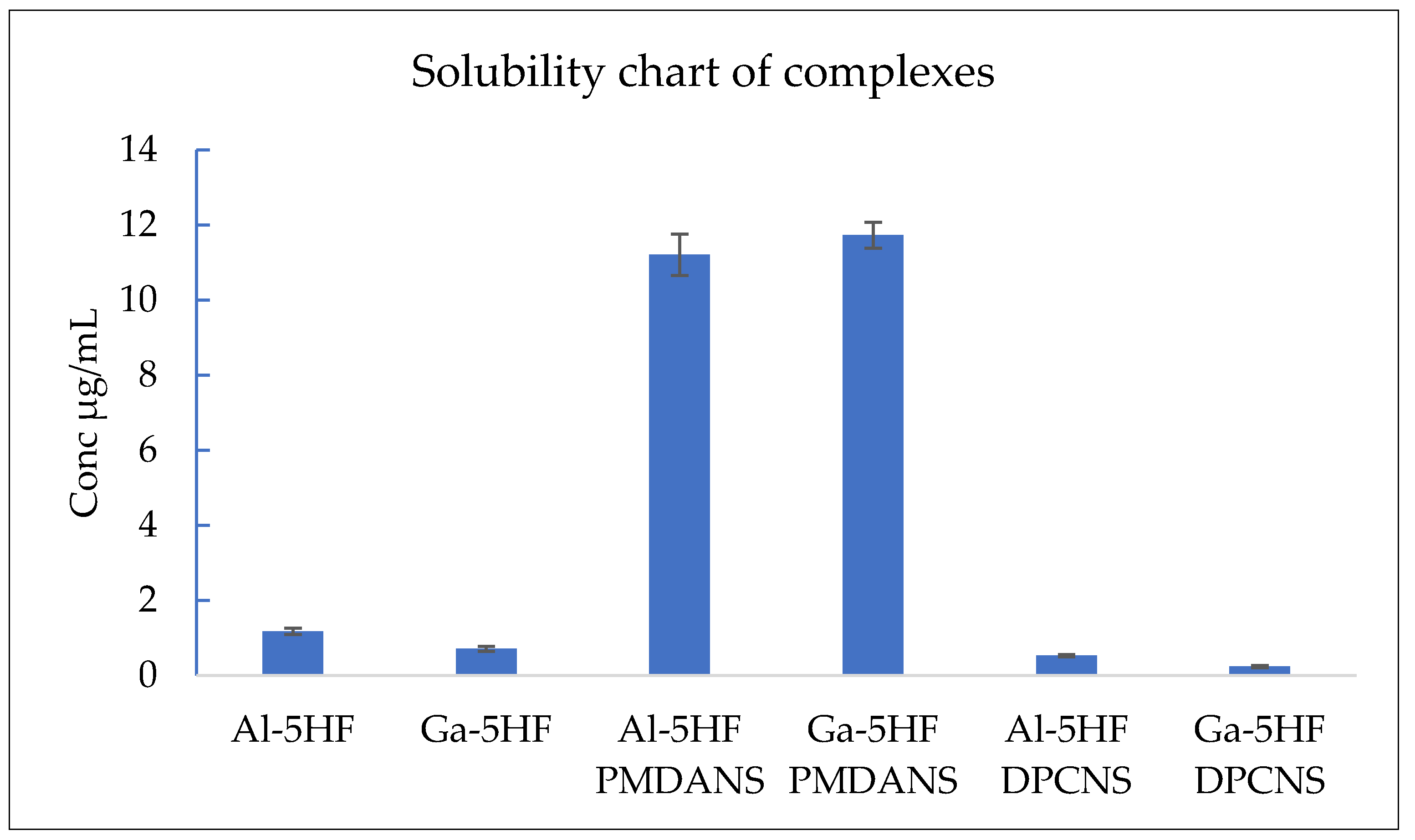
- Solubility studies
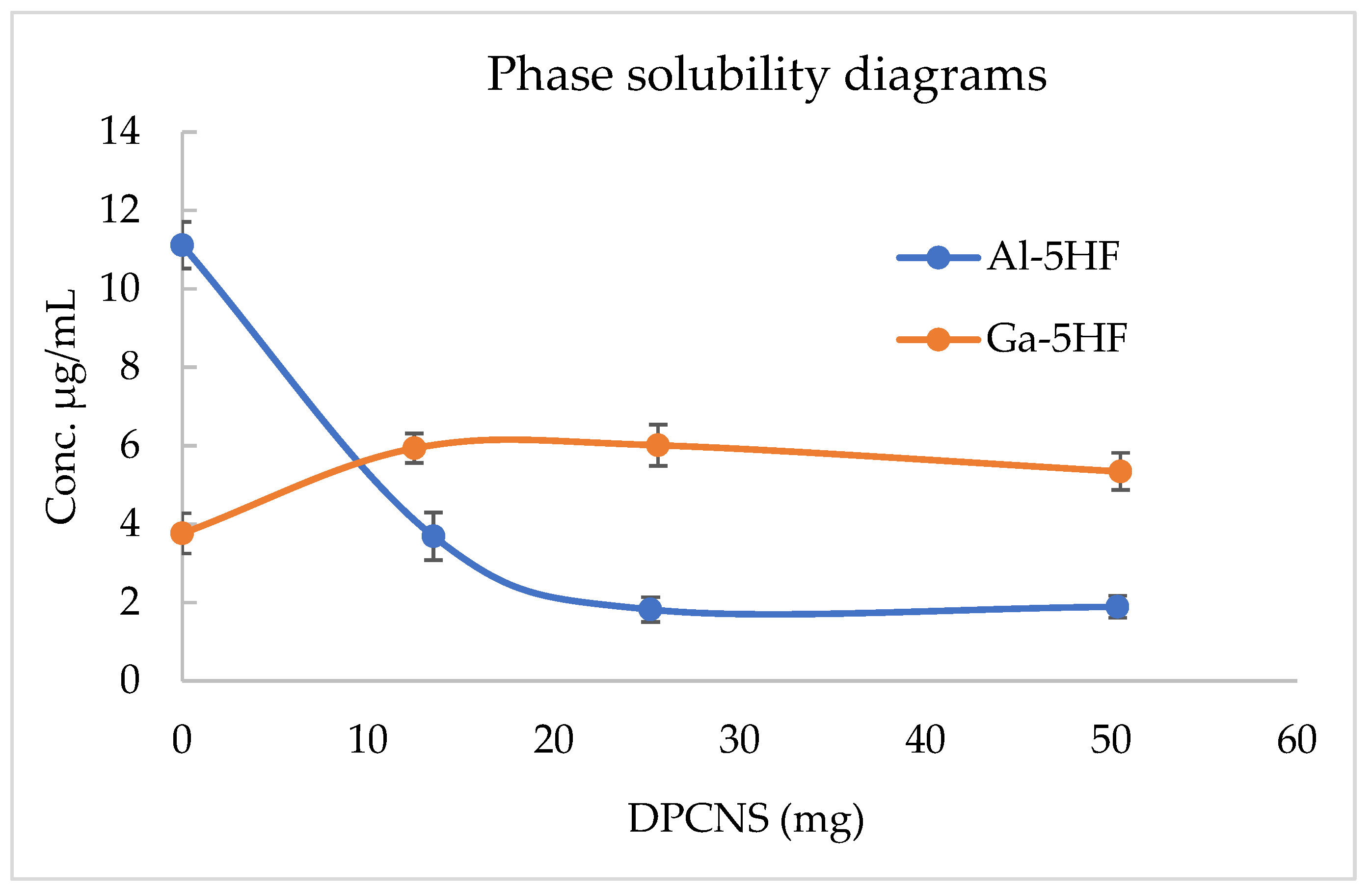
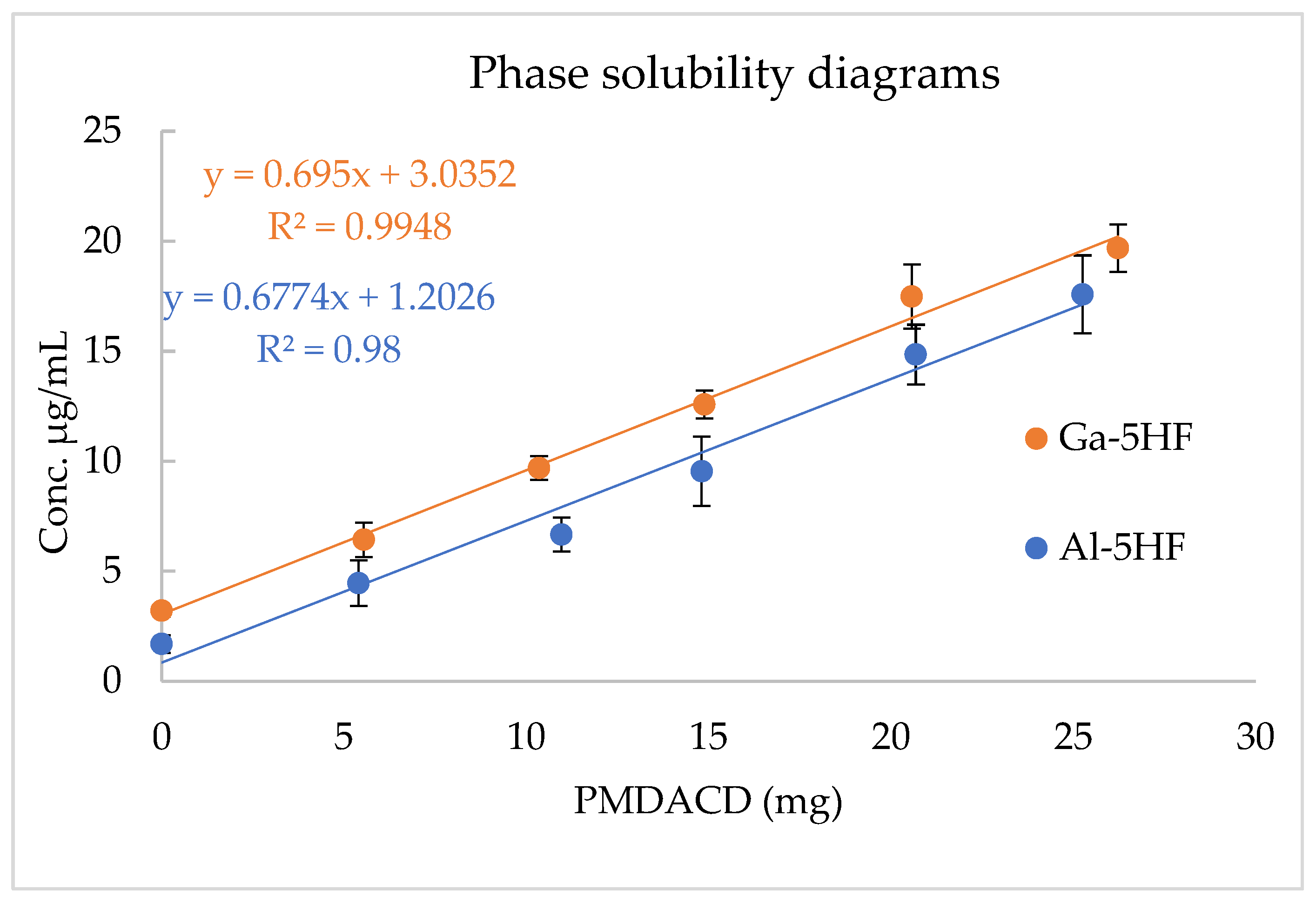
- Phase solubility diagrams
- Loading capacity and entrapment efficiency
- Cell cytotoxicity
- Cell culture and treatment
- Cytotoxicity assay
- Apoptosis analysis
3. Results and Discussion
- Physicochemical Characterization
- IR Spectra
- Thermal analysis
- Solubility studies
- Phase diagram studies
- The loading capacity and entrapment efficiency
- Cell cytotoxicity
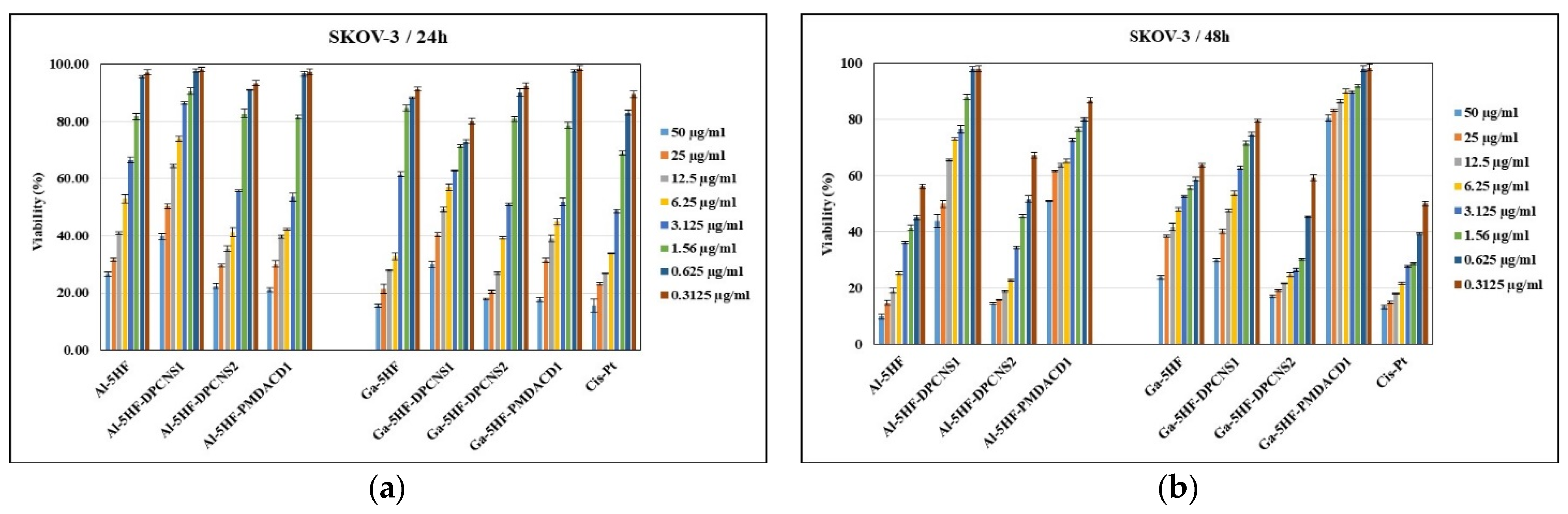
- The effect of the compounds on cell viability
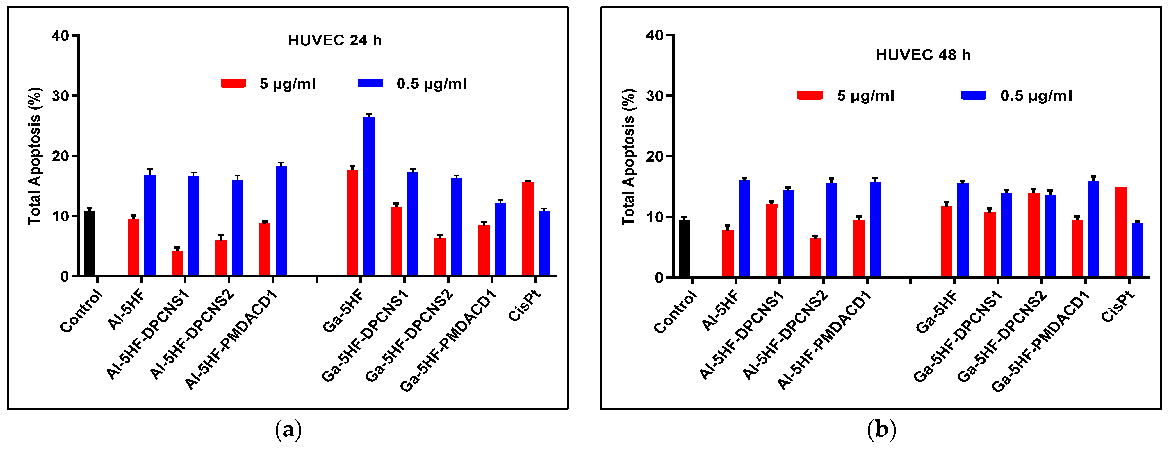
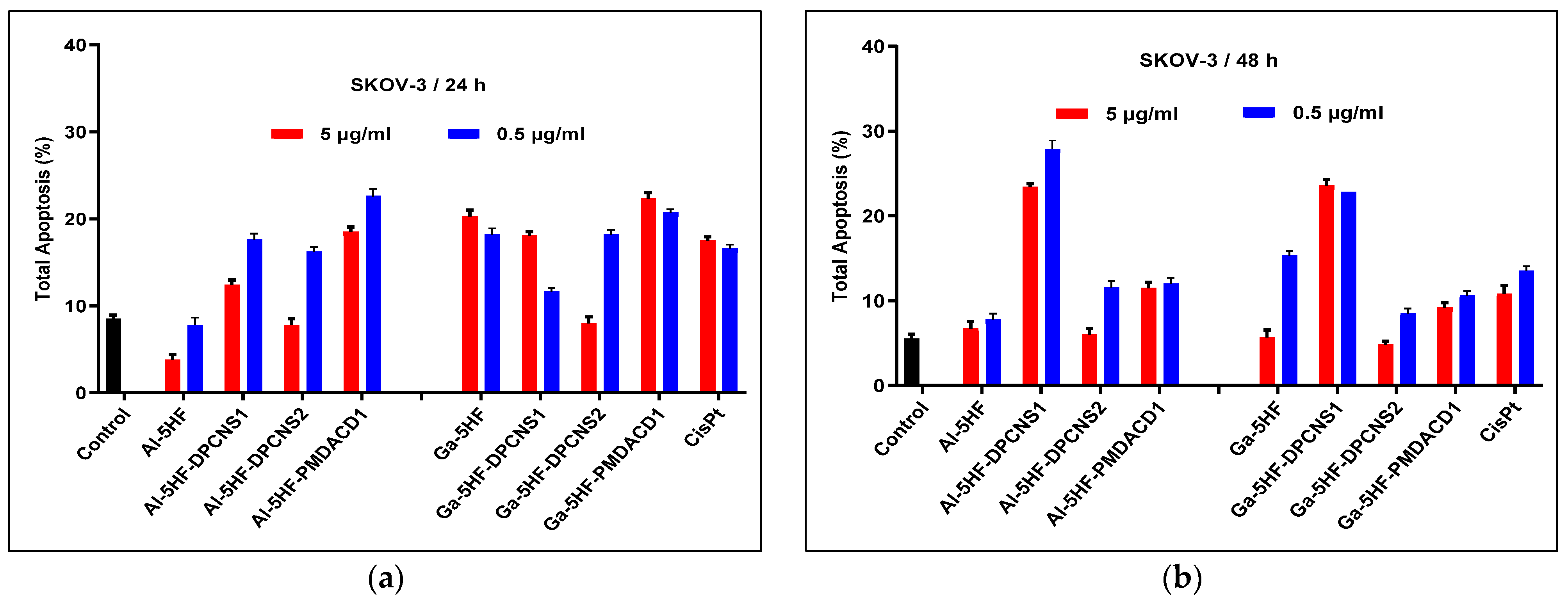
- The analyzed compounds were studied for their impact on the apoptotic process
4. Conclusions
Supplementary Materials
Author Contributions
Funding
Institutional Review Board Statement
Informed Consent Statement
Data Availability Statement
Acknowledgments
Conflicts of Interest
References
- Herath, W.; Mikell, J.R.; Hale, A.L.; Ferreira, D.; Khan, I.A. Microbial Metabolism Part 9. 1. Structure and Antioxidant Significance of the Metabolites of 5,7-Dihydroxyflavone (Chrysin), and 5-and 6-Hydroxyflavones. Chem. Pharm. Bull. 2008, 56, 418–422. [Google Scholar] [CrossRef] [PubMed]
- Winkel-Shirley, B. Biosynthesis of flavonoids and effects of stress. Curr. Opin. Plant. Biol. 2002, 5, 218–223. [Google Scholar] [CrossRef] [PubMed]
- Samsonowicz, M.; Regulska, E.; Kalinowska, M. Hydroxyflavone metal complexes—Molecular structure, antioxidant activity and biological effects. Chem. Biol. Interact. 2017, 273, 245–256. [Google Scholar] [CrossRef] [PubMed]
- Cotelle, N.; Bernier, J.-L.; Catteau, J.-P.; Pommery, J.; Wallet, J.-C.; Gaydou, E.M. Antioxidant Properties of Hydroxy-Flavones. Free Radic. Biol. Med. 1996, 20, 35–43. [Google Scholar] [CrossRef] [PubMed]
- Shamsudin, N.F.; Ahmed, Q.U.; Mahmood, S.; Ali Shah, S.A.; Khatib, A.; Mukhtar, S.; Alsharif, M.A.; Parveen, H.; Zakaria, Z.A. Antibacterial Effects of Flavonoids and Their Structure-Activity Relationship Study: A Comparative Interpretation. Molecules 2022, 27, 1149. [Google Scholar] [CrossRef] [PubMed]
- Badshah, S.L.; Faisal, S.; Muhammad, A.; Poulson, B.G.; Emwas, A.H.; Jaremko, M. Antiviral activities of flavonoids. Biomed. Pharmacother. 2021, 140, 111596. [Google Scholar] [CrossRef] [PubMed]
- Maleki, S.J.; Crespo, J.F.; Cabanillas, B. Anti-inflammatory effects of flavonoids. Food. Chem. 2019, 299, 125124. [Google Scholar] [CrossRef] [PubMed]
- Spagnuolo, C.; Moccia, S.; Russo, G.L. Anti-inflammatory effects of flavonoids in neurodegenerative disorders. Eur. J. Med. Chem. 2018, 153, 105–115. [Google Scholar] [CrossRef]
- Khan, A.U.; Dagur, H.S.; Khan, M.; Malik, N.; Alam, M.; Mushtaque, M. Therapeutic role of flavonoids and flavones in cancer prevention: Current trends and future perspectives. EJMCR 2021, 3, 100010. [Google Scholar] [CrossRef]
- Ren, W.; Qiao, Z.; Wang, H.; Zhu, L.; Zhang, L. Flavonoids: Promising anticancer agents. Med. Res. Rev. 2003, 23, 519–534. [Google Scholar] [CrossRef]
- Dajas, F.; Rivera-Megret, F.; Blasina, F.; Arredondo, F.; Abin-Carriquiry, J.A.; Costa, G.; Echeverry, C.; Lafon, L.; Heizen, H.; Ferreira, M.; et al. Neuroprotection by flavonoids. Braz. J. Med. Biol. Res. 2003, 36, 1613–1620. [Google Scholar] [CrossRef] [PubMed]
- Chen, J.; Li, X. Hypolipidemic effect of flavonoids from mulberry leaves in triton WR-1339 induced hyperlipidemic mice. Asia Pac. J. Clin. Nutr. 2007, 16, 290–294. [Google Scholar] [PubMed]
- Snijman, P.W.; Swanevelder, S.; Joubert, E.; Green, I.R.; Gelderblom, W.C.A. The antimutagenic activity of the major flavonoids of rooibos (Aspalathus linearis): Some dose-response effects on mutagen activation-flavonoid interactions. Mutat. Res. Genet. Toxicol. Environ. Mutagen. 2007, 631, 111–123. [Google Scholar] [CrossRef] [PubMed]
- Peluso, I.; Miglio, C.; Morabito, G.; Ioannone, F.; Serafini, M. Flavonoids and Immune Function in Human: A Systematic Review. Crit. Rev. Food. Sci. Nutr. 2015, 55, 383–395. [Google Scholar] [CrossRef] [PubMed]
- Shen, N.; Wang, T.; Gan, Q.; Liu, S.; Wang, L.; Jin, B. Plant flavonoids: Classification, distribution, biosynthesis, and antioxidant activity. Food Chem. 2022, 383, 132531. [Google Scholar] [CrossRef] [PubMed]
- Abou Baker, D.H. An ethnopharmacological review on the therapeutical properties of flavonoids and their mechanisms of actions: A comprehensive review based on up to date knowledge. Toxicol. Rep. 2022, 9, 445–469. [Google Scholar] [CrossRef] [PubMed]
- Li, N.; Shou, Z.; Yang, S.; Cheng, X.; Chen, C.; Zheng, S.; Shi, Y.; Tang, H. Subtle distinction in molecular structure of flavonoids leads to vastly different coating efficiency and mechanism of metal-polyphenol networks with excellent antioxidant activities. Colloids. Surf. B Biointerfaces 2023, 229, 113454. [Google Scholar] [CrossRef] [PubMed]
- Valant-Vetschera, K.M.; Doma Bhutia, T.; Wollenweber, E. Exudate Flavonoids of Primula spp: Structural and Biogenetic Chemodiversity. Nat. Prod. Commun. 2009, 4, 365–370. [Google Scholar]
- Valant-Vetschera, K.M.; Bhutia, T.D.; Wollenweber, E. Chemodiversity of exudate flavonoids in Dionysia (Primulaceae): A comparative study. Phytochemistry 2010, 71, 937–947. [Google Scholar] [CrossRef]
- Cornard, J.P.; Merlin, J.C. Structural and spectroscopic investigation of 5-hydroxyflavone and its complex with aluminium. J. Mol. Struct. 2001, 569, 129–138. [Google Scholar] [CrossRef]
- Munteanu, A.C.; Badea, M.; Olar, R.; Silvestro, L.; Dulea, C.; Negut, C.D.; Uivarosi, V. Synthesis and structural investigation of new bio-relevant complexes of lanthanides with 5-hydroxyflavone: DNA binding and protein interaction studies. Molecules 2016, 21, 1737. [Google Scholar] [CrossRef] [PubMed]
- Uivarosi, V.; Badea, M.; Olar, R.; Drǎghici, C.; Bǎrbuceanu, Ş.F. Synthesis and characterization of some new complexes of magnesium (II) and zinc (II) with the natural flavonoid primuletin. Molecules 2013, 18, 7631–7645. [Google Scholar] [CrossRef] [PubMed]
- Jabeen, E.; Janjua, N.K.; Ahmed, S.; Murtaza, I.; Ali, T.; Hameed, S. Radical scavenging propensity of Cu2+, Fe3+ complexes of flavonoids and in-vivo radical scavenging by Fe3+-primuletin. Spectrochim. Acta A Mol. Biomol. Spectrosc. 2017, 171, 432–438. [Google Scholar] [CrossRef] [PubMed]
- Vătavu, V.-N.; Badea, M.; Olar, R.; Hudiță, A.; Gălățeanu, B.; Munteanu, A.C.; Uivarosi, V. Synthesis and Characterization of a New Cr(III) Complex with 5-Hydroxyflavone as a Potential Antidiabetic Agent. Proceedings 2019, 29, 118. [Google Scholar] [CrossRef]
- Alexiou, A.D.P.; Decandio, C.C.; Almeida, S.D.N.; Ferreira, M.J.P.; Romoff, P.; Rocha, R.C. A trinuclear oxo-chromium(III) complex containing the natural flavonoid primuletin: Synthesis, characterization, and antiradical properties. Molecules 2015, 20, 6310–6318. [Google Scholar] [CrossRef] [PubMed]
- Munteanu, A.C.; Badea, M.; Olar, R.; Silvestro, L.; Mihaila, M.; Brasoveanu, L.I.; Musat, M.G.; Andries, A.; Uivarosi, V. Cytotoxicity studies, DNA interaction and protein binding of new Al (III), Ga (III) and In (III) complexes with 5-hydroxyflavone. Appl. Organomet. Chem. 2018, 32, e4579. [Google Scholar] [CrossRef]
- De Souza, R.F.V.; De Giovani, W.F. Antioxidant properties of complexes of flavonoids with metal ions. Redox Rep. 2004, 9, 97–104. [Google Scholar] [CrossRef] [PubMed]
- Wang, H.; Tan, M.; Zhu, J.; Pan, Y.; Chen, Z.; Liang, H.; Liu, H.; Wang, H. Synthesis, cytotoxic activity, and DNA binding properties of copper (II) complexes with hesperetin, naringenin, and apigenin. Bioinorg. Chem. Appl. 2009, 2009, 347872. [Google Scholar]
- Vajragupta, O.; Boonchoong, P.; Sumanont, Y.; Watanabe, H.; Wongkrajang, Y.; Kammasud, N. Manganese-based complexes of radical scavengers as neuroprotective agents. Bioorg. Med. Chem. 2003, 11, 2329–2337. [Google Scholar] [CrossRef]
- Bello, U.; Shallangwa, G.A.; Idris, S.O.; Musa, M.; Aisha, A.A. Synthesis, spectroscopic characterization and in vitro antibacterial activity studies of Cu(II) complexes derived from 6- hydroxyflavone Schiff bases. BAJOPAS 2019, 11, 136. [Google Scholar] [CrossRef]
- Joseph, J.; Nagashri, K. Novel copper-based therapeutic agent for anti-inflammatory: Synthesis, characterization, and biochemical activities of copper(II) complexes of hydroxyflavone schiff bases. Appl. Biochem. Biotechnol. 2012, 167, 1446–1458. [Google Scholar] [CrossRef] [PubMed]
- Dias, K.; Nikolaou, S.; De Giovani, W.F. The in vitro antioxidant properties of the Al-quercetin/βCD and Al-catechin/βCD inclusion compounds, rationalized in terms of their electrochemical behaviour. Med. Chem. Res. 2012, 21, 2920–2925. [Google Scholar] [CrossRef]
- Deng, S.P.; Yang, Y.L.; Cheng, X.X.; Li, W.R.; Cai, J.Y. Synthesis, spectroscopic study and radical scavenging activity of Kaempferol derivatives: Enhanced water solubility and antioxidant activity. Int. J. Mol. Sci. 2019, 20, 975. [Google Scholar] [CrossRef] [PubMed]
- Hoti, G.; Lucia Appleton, S.; Rubin Pedrazzo, A.; Cecone, C.; Matencio, A.; Trotta, F.; Caldera, F. Strategies to Develop Cyclodextrin-Based Nanosponges for Smart Drug Delivery. Smart Drug Delivery; IntechOpen: London, UK, 2022. [Google Scholar]
- Trotta, F.; Zanetti, M.; Cavalli, R. Cyclodextrin-based nanosponges as drug carriers. Beilstein J. Org. Chem. 2012, 8, 2091–2099. [Google Scholar] [CrossRef] [PubMed]
- Krabicová, I.; Appleton, S.L.; Tannous, M.; Hoti, G.; Caldera, F.; Rubin Pedrazzo, A.; Cecone, C.; Cavalli, R.; Trotta, F. History of cyclodextrin nanosponges. Polymers 2020, 12, 1122. [Google Scholar] [CrossRef] [PubMed]
- Trotta, F.; Mele, A. Cyclodextrin Nanosponges. In Nanosponges, 1st ed.; Trotta, F., Mele, A., Eds.; Wiley Professional, Wiley-VCH: Berlin/Heidelberg, Germany, 2019; Volume 1, pp. 27–57. [Google Scholar]
- Swaminathan, S.; Pastero, L.; Serpe, L.; Trotta, F.; Vavia, P.; Aquilano, D.; Trotta, M.; Zara, G.; Cavalli, R. Cyclodextrin-based nanosponges encapsulating camptothecin: Physicochemical characterization, stability and cytotoxicity. Eur. J. Pharm. Biopharm. 2010, 74, 193–201. [Google Scholar] [CrossRef] [PubMed]
- Gigliotti, C.L.; Ferrara, B.; Occhipinti, S.; Boggio, E.; Barrera, G.; Pizzimenti, S.; Giovarelli, M.; Fantozzi, R.; Chiocchetti, A.; Argenziano, M.; et al. Enhanced cytotoxic effect of camptothecin nanosponges in anaplastic thyroid cancer cells in vitro and in vivo on orthotopic xenograft tumors. Drug. Deliv. 2017, 24, 670–680. [Google Scholar] [CrossRef] [PubMed]
- Allahyari, S.; Zahednezhad, F.; Khatami, M.; Hashemzadeh, N.; Zakeri-Milani, P.; Trotta, F. Cyclodextrin nanosponges as potential anticancer drug delivery systems to be introduced into the market, compared with liposomes. J. Drug Deliv. Sci. Technol. 2022, 67, 102931. [Google Scholar] [CrossRef]
- Serrano-Martínez, A.; Victoria-Montesinos, D.; García-Muñoz, A.M.; Hernández-Sánchez, P.; Lucas-Abellán, C.; González-Louzao, R. A Systematic Review of Clinical Trials on the Efficacy and Safety of CRLX101 Cyclodextrin-Based Nanomedicine for Cancer Treatment. Pharmaceutics 2023, 15, 1824. [Google Scholar] [CrossRef]
- Adeoye, O.; Bártolo, I.; Conceição, J.; da Silva, A.B.; Duarte, N.; Francisco, A.P.; Taveira, N.; Cabral-Marques, H. Pyromellitic dianhydride crosslinked soluble cyclodextrin polymers: Synthesis, lopinavir release from sub-micron sized particles and anti-HIV-1 activity. Int. J. Pharm. 2020, 583, 119356. [Google Scholar] [CrossRef]
- Singh, V.; Xu, J.; Wu, L.; Liu, B.; Guo, T.; Guo, Z.; York, P.; Gref, R.; Zhang, J. Ordered and disordered cyclodextrin nanosponges with diverse physicochemical properties. RSC Adv. 2017, 7, 23759–23764. [Google Scholar] [CrossRef]
- Higuchi, T.; Connors, K.A. Phase-solubility techniques. Adv. Anal. Chem. Instr. 1965, 4, 117–122. [Google Scholar]
- Mihaila, M.; Bostan, M.; Hotnog, D.; Ferdes, M.; Brasoveanu, L.I. Real-time analysis of quercetin, resveratrol and/or doxorubicin effects in mcf-7 cells. Rom. Biotechnol. Lett. 2013, 18, 8106–8114. [Google Scholar]
- Ivan, B.-C.; Barbuceanu, S.-F.; Hotnog, C.M.; Anghel, A.I.; Ancuceanu, R.V.; Mihaila, M.A.; Brasoveanu, L.I.; Shova, S.; Draghici, C.; Olaru, O.T.; et al. New Pyrrole Derivatives as Promising Biological Agents: Design, Synthesis, Characterization, In Silico, and Cytotoxicity Evaluation. Int. J. Mol. Sci. 2022, 23, 8854. [Google Scholar] [CrossRef]
- Hotnog, C.M.; Mihaila, M.; Puiu, L.; Botezatu, A.; Roman, V.; Popescu, I.D.; Bostan, M.; Brasoveanu, L.I. Modulation of the interplay between p53, ICAM-1 and VEGF in drug-treated LoVo colon cancer cells. Rom. Biotechnol. Lett. 2019, 24, 261–270. [Google Scholar] [CrossRef]
- Iancu, I.V.; Botezatu, A.; Plesa, A.; Huica, I.; Fudulu, A.; Albulescu, A.; Bostan, M.; Mihaila, M.; Grancea, C.; Manda, D.A.; et al. Alterations of regulatory factors and DNA methylation pattern in thyroid cancer. Cancer Biomark. 2020, 28, 255–268. [Google Scholar] [CrossRef]
- Saokham, P.; Muankaew, C.; Jansook, P.; Loftsson, T. Solubility of Cyclodextrins and Drug/Cyclodextrin Complexes. Molecules 2018, 23, 1161. [Google Scholar] [CrossRef]
- Maciuca, A.-M.; Munteanu, A.-C.; Mihaila, M.; Badea, M.; Olar, R.; Nitulescu, G.M.; Munteanu, C.V.A.; Bostan, M.; Uivarosi, V. Rare-Earth Metal Complexes of the Antibacterial Drug Oxolinic Acid: Synthesis, Characterization, DNA/Protein Binding and Cytotoxicity Studies. Molecules 2020, 25, 5418. [Google Scholar] [CrossRef]
- Mihaila, M.; Hotnog, C.M.; Bostan, M.; Munteanu, A.C.; Vacaroiu, I.A.; Brasoveanu, L.I.; Uivarosi, V. Anticancer Activity of Some Ruthenium(III) Complexes with Quinolone Antibiotics: In Vitro Cytotoxicity, Cell Cycle Modulation, and Apoptosis-Inducing Properties in LoVo Colon Cancer Cell Line. Appl. Sci. 2021, 11, 8594. [Google Scholar] [CrossRef]
- Romani, A.M.P. Cisplatin in cancer treatment. Biochem. Pharmacol. 2022, 206, 115323. [Google Scholar] [CrossRef]
- Kregielewski, K.; Fraczek, W.; Grodzik, M. Graphene Oxide Enhanced Cisplatin Cytotoxic Effect in Glioblastoma and Cervical Cancer. Molecules 2023, 28, 6253. [Google Scholar] [CrossRef] [PubMed]


| Loaded Polymers | Entrapment Efficiency (%) | Loading Capacity (%) |
|---|---|---|
| Al-5HF-DPCNS1 | 69.63 ± 2.62 | 11.18 ± 0.42 |
| Ga-5HF-DPCNS1 | 75.28 ± 0.78 | 11.43 ± 0.12 |
| Al-5HF-DPCNS2 | 81.35 ± 1.11 | 13.07 ± 0.18 |
| Ga-5HF-DPCNS2 | 91.00 ± 1.73 | 13.82 ± 0.26 |
| Al-5HF-PMDACD1 | 64.96 ± 1.92 | 10.43 ± 0.31 |
| Ga-5HF-PMDACD1 | 84.77 ± 3.04 | 12.87 ± 0.46 |
| HUVEC | LoVo | SKOV-3 | ||||
|---|---|---|---|---|---|---|
| Compound | 24 h | 48 h | 24 h | 48 h | 24 h | 48 h |
| Al-5HF | 9.05 ± 0.79 | 8.74 ± 0.77 | 4.75 ± 0.99 | 4.53 ± 0.80 | 5.53 ± 0.86 | 1.63 ± 1.14 |
| Al-5HF-DPCNS1 | 9.03 ± 1.11 | 6.22 ± 0.67 | 5.61 ± 1.11 | 5.78 ± 1.04 | 7.38 ± 0.79 | 7.43 ± 0.85 |
| Al-5HF-DPCNS2 | 9.52 ± 0.65 | 9.54 ± 1.17 | 3.92 ± 1.07 | 4.84 ± 1.09 | 5.07 ± 1.06 | 2.39 ± 0.99 |
| Al-5HF-PMDACD1 | 9.13 ± 0.80 | 8.35 ± 1.05 | 3.38 ± 0.95 | 2.14 ± 0.94 | 5.16 ± 1.13 | 8.77 ± 1.12 |
| Ga-5HF | 5.41 ± 0.92 | 7.99 ± 0.84 | 2.21 ± 1.03 | 8.25 ± 0.83 | 4.73 ± 0.72 | 4.08 ± 0.82 |
| Ga-5HF-DPCNS1 | 7.72 ± 0.58 | 5.40 ± 1.09 | 3.48 ± 1.01 | 2.96 ± 1.06 | 5.65 ± 0.90 | 5.55 ± 1.10 |
| Ga-5HF-DPCNS2 | 6.47 ± 0.93 | 6.07 ± 1.02 | 2.88 ± 0.59 | 1.73 ± 1.16 | 4.69 ± 1.18 | 0.89 ± 0.87 |
| Ga-5HF-PMDACD1 | 5.34 ± 1.02 | 4.27 ± 0.81 | 2.71 ± 0.70 | 0.49 ± 0.95 | 5.12 ± 1.05 | 20.03 ± 1.17 |
| CisPt | 10.07 ± 0.83 | 8.42 ± 0.79 | 2.00 ± 1.21 | 0.97 ± 0.92 | 4.39 ± 0.97 | 2.41 ± 1.20 |
Disclaimer/Publisher’s Note: The statements, opinions and data contained in all publications are solely those of the individual author(s) and contributor(s) and not of MDPI and/or the editor(s). MDPI and/or the editor(s) disclaim responsibility for any injury to people or property resulting from any ideas, methods, instructions or products referred to in the content. |
© 2024 by the authors. Licensee MDPI, Basel, Switzerland. This article is an open access article distributed under the terms and conditions of the Creative Commons Attribution (CC BY) license (https://creativecommons.org/licenses/by/4.0/).
Share and Cite
Radu, C.; Olteanu, A.A.; Aramă, C.C.; Mihăilă, M.; Uivaroși, V. Investigating the Effect of Cyclodextrin Nanosponges and Cyclodextrin-Based Hydrophilic Polymers on the Chemical Pharmaceutical and Toxicological Profile of Al(III) and Ga(III) Complexes with 5-Hydroxyflavone. Appl. Sci. 2024, 14, 5441. https://doi.org/10.3390/app14135441
Radu C, Olteanu AA, Aramă CC, Mihăilă M, Uivaroși V. Investigating the Effect of Cyclodextrin Nanosponges and Cyclodextrin-Based Hydrophilic Polymers on the Chemical Pharmaceutical and Toxicological Profile of Al(III) and Ga(III) Complexes with 5-Hydroxyflavone. Applied Sciences. 2024; 14(13):5441. https://doi.org/10.3390/app14135441
Chicago/Turabian StyleRadu, Claudiu, Andreea Alexandra Olteanu, Corina Cristina Aramă, Mirela Mihăilă, and Valentina Uivaroși. 2024. "Investigating the Effect of Cyclodextrin Nanosponges and Cyclodextrin-Based Hydrophilic Polymers on the Chemical Pharmaceutical and Toxicological Profile of Al(III) and Ga(III) Complexes with 5-Hydroxyflavone" Applied Sciences 14, no. 13: 5441. https://doi.org/10.3390/app14135441