Article

Light and Nutrient Conditions Influence Fucoxanthin Production of the Microalgae Cyclotella meneghiniana

1
Guangdong Provincial Key Laboratory of Marine Disaster Prediction and Prevention, Shantou University, Shantou 515063, China
2
South China Sea Environmental Monitoring Center, State Oceanic Adiministration, Guangzhou 510300, China
*
Author to whom correspondence should be addressed.
These authors contributed equally to this work.
Appl. Sci. 2024, 14(13), 5504; https://doi.org/10.3390/app14135504
Submission received: 21 April 2024 / Revised: 4 June 2024 / Accepted: 20 June 2024 / Published: 25 June 2024
(This article belongs to the Special Issue Microalgae: Physiology, Biotechnology, and Industrial Applications)

Abstract

:
Fucoxanthin has attracted the attention of scholars because of its health benefits in terms of anticancer, weight loss, antidiabetic, hypolipidemic, and antioxidant functions. Researchers have found that the fucoxanthin content of microalgae was higher than that of macroalgae. Therefore, the microalgae Cyclotella meneghiniana was isolated and maintained under varying light and modified nutrient conditions. The results of this study showed that Cyclotella meneghiniana had better photosynthetic activity and higher biomass under low light. Both high trace elements and high nitrogen promoted the accumulation of fucoxanthin in Cyclotella meneghiniana. Low light levels and high trace metal contents enhanced the fucoxanthin production (7.76 ± 0.30 mg g−1 DW). The results of the current study will help to enhance fucoxanthin production for commercialization.

1. Introduction

The recent surge in attention from global researchers towards microalgae stems from their vast potential uses in industries such as renewable energy, biopharmaceuticals, and nutraceuticals [1,2,3]. Microalgae are of high commercial value and also produce proteins, lipids, vitamins, pigments, cosmetics, and food and feed additives [4]. Microalgae offer a renewable and sustainable way to obtain bioactive substances like essential amino acids, polyunsaturated fatty acids, and antioxidants, which have been documented to have beneficial effects on health and nutrition [5]. Prior studies have confirmed the effectiveness of microalgae in serving as a source of fucoxanthin, with diatoms exhibiting a golden-brown hue, attributed to their richness in pigments such as fucoxanthin [6,7]. Fucoxanthin, an essential xanthophyll carotenoid present in both macroalgae and microalgae, possesses significant market worth [8,9,10]. Earlier research indicates that fucoxanthin exhibits a range of pharmacological properties, encompassing antidiabetic, antioxidant, anti-inflammatory, anti-obesity, antimalarial, anticancer, and more [8,11,12]. For instance, fucoxanthin (10 mg kg−1 or 30 mg kg−1) attenuates inflammation and oxidative stress in inflammatory tracheal epithelial cells and improves the pathological changes related to asthma in mice [13]. Contemporary studies point to fucoxanthin serving as a potent remedy for anti-inflammatory disorders; it reduces inflammation internal and external to the body by diminishing NF-kB activation, and it also impedes the proliferation of cancer cells and aids in weight reduction [14,15]. Fucoxanthin could provide health advantages in preventing long-term diseases like malignant obesity, cancer, diabetes mellitus, and liver cirrhosis [16].
The trade-based production of fucoxanthin relies on brown macroalgae [17]. In industrial applications, seaweed has the disadvantages of scarcity, low growth rates, less fucoxanthin, and poor product quality, making it an expensive and unsustainable original material for practical production [18,19,20]. The fucoxanthin production in macroalgae is still reliant on collection from wild populations (68% of macroalgae production units) and is limited by fucoxanthin yields (less than 3.7 mg g−1 DW) and long incubation periods [8,21,22]. Microalgae are characterized by their high biomass productivity, fast growth rate, accessibility and cultivation, ease of scale-up, and continuous supply of stocks, which can help them to scale up industrial production, improve the controllability and sustainability of production, and utilize resources more efficiently [23,24]. Numerous studies have highlighted the variance in fucoxanthin levels between macroalgae and microalgae, noting that microalgae possess a notably greater fucoxanthin concentration compared to macroalgae (up to 100 times that of seaweed) [6,23,24,25,26,27]. These characteristics make microalgae more suitable than macroalgae as promising candidates as a reliable source of commercially produced fucoxanthin [6,24].
Previous studies have shown that the amassment of fucoxanthin in microalgae is critically influenced by light and nutrients, with the cumulative content varying among different species [28,29,30]. For instance, among the cells of Isochrysis galbana under different nitrogen concentrations, those in medium containing 4 mM nitrogen (N-NO3) had the largest fucoxanthin content, which was 18.8 mg g−1 [31]. Similarly, research conducted on Odontella aurita revealed that cells under initial high nitrogen plus supplementary nitrogen conditions maximized the fucoxanthin accumulation: a maximum productivity of 6.01 mg L−1 d−1 [32]. Another study obtained a maximum yield of 50.5 mg L−1 of fucoxanthin from Poterioochromonas malhamensis CMBB-1 by coupling light with high-cell-density fermentation [28]. Microalgae may act as a consistent and dependable supplier of natural goods, with their effectiveness amplified by the corresponding target compounds [33]. Consequently, exploring how various environmental elements influence the build-up of fucoxanthin in microalgae becomes crucial.
The feasibility of using microalgae to commercially produce fucoxanthin, a carotenoid, is questionable [34]. However, culturing microalgae at favorable conditions has great potential to increase the viability of fucoxanthin sources, and most of the microalgae that are currently used to study the content of fucoxanthin are marine microalgae due to their abundance of compounds and ease of cultivation [6]. Many scientists have conducted studies aimed at optimizing the culture conditions of microalgae with a view to obtaining higher production of fucoxanthin [6,35,36,37]. Within the realm of microalgae cultivation, the accumulation of fucoxanthin is primarily governed by two critical factors: illumination and the availability of nutrients [17,28].
Diatoms of microalgae possess fucoxanthin chlorophyll proteins (FCPs) that act as light-harvesting systems [38]. Two major FCP complexes can be isolated from Cyclotella meneghiniana as a central diatom [38,39]. It has been shown that the FCP complexes are sensitive to changes in environmental conditions and that the enhanced formation of FCP proteins has the potential to increase fucoxanthin biosynthesis [40,41]. Therefore, different environmental conditions may affect the production of fucoxanthin by Cyclotella meneghiniana. Hence, in this study, Cyclotella meneghiniana was isolated from Rongjiang River and cultured under different light and nutrient conditions to increase the possible sources of fucoxanthin for commercial production, thereby enhancing the practicality and sustainability of commercially producing fucoxanthin for its practical and enduring production.

2. Materials and Methods

2.1. Sample Collection and Culture Conditions

The centric diatom, Cyclotella meneghiniana (homotypic synonym: Stephanocyclus meneghinianus), was collected from the Rongjiang River (23°17′ N, 116°47′ E) on the southeast coast of China; it was isolated and cultured in F/2 medium at 15 °C [42]. Before the isolation, the medium was autoclaved at 121 °C for 20 min and cultivated in a 250 mL Erlenmeyer flask under illumination from the fluorescent lamp (40 µmol m−2 s−1) with light: dark cycles of 12 h:12 h until reaching the exponential stage. Subsequently, algal cells were introduced into 1000 mL conical containers filled with 400 mL of sterile medium, maintaining a salinity of 24.5‰, and cultivated at 24 °C, mirroring the conditions of axenic culture. The collection of C. meneghiniana cells occurred during their mid-exponential growth stage, involving a 20 min centrifugation at 2000× g (Eppendorf Centrifuge 5810R, Hamburg, HH, Germany), followed by a 48 h freeze-drying at −70 °C prior to experimental use.
Investigating improved growth conditions and nutrient environments for cell pigment generation, C. meneghiniana was grown at 24 °C for 12 days under three distinct light intensities and nutrient levels. A total of eight different nutrient therapies were sustained, with the specifics provided in Table S1. The levels of vitamins remained consistent across all experimental environments. The various light intensity treatments are categorized as LL (low light, 45 µmol photons m−2 s−1), ML (medium light, 110 µmol photons m−2 s−1), HL (high light, 170 µmol photons m−2 s−1), nutrient concentrations are denoted as C (Control, 882.457 mM NaNO3, 36.235 mM NaH2PO4, 105.559 mM Na2SiO3, 11.686 mM Na2EDTA, 11.654 mM FeCl3, 909.504 mM MnCl2, 76.506 mM ZnSO4, 42.029 mM CoCl2, 39.249 mM CuSO4, 26.038 mM Na2MoO4, 11.86 mM VB1, 4.09 mM VB7, 2.21 mM VB12), HN (High nitrate, 1588.422 mM NaNO3), MN (Mediumnitrate, 1176.609 mM NaNO3), HP (High phosphate, 253.642 mM NaH2PO4), MP (Mediumphosphate, 144.938 mM NaH2PO4), HS (High silicate, 316.678 mM Na2SiO3), HM (High trace metals, 80.593 mM Na2EDTA, 96.193 mM FeCl3, 1263.2 mM MnCl2, 208.652 mM ZnSO4, 126.088 mM CoCl2, 120.149 mM CuSO4, 103.327 mM Na2MoO4), LC (lower concentration than F/2 medium, 588.305 mM NaNO3, 21.741 mM NaH2PO4, 52.78 mM Na2SiO3, 5.722 mM Na2EDTA, 4.477 mM FeCl3, 429.48 mM MnCl2, 21.015 mM CoCl2, 17.622 mM CuSO4, and 13.226 mM Na2MoO4).

2.2. Species Identification

To conduct scanning electron microscopy (SEM), a 10 mL sample underwent centrifugation at 5000× g for 10 min, and utilized an electron microscopy (JEOLJSM6390LV, Pleasanton, CA, USA) to examine the structure of diatom frustules [43]. By applying a platinum coating on the edges using a fine auto coater (JEOLFC1600, Pleasanton, CA, USA), an electrical condition bridge was created within the stubs and samples. For DNA extraction, 1 mL of the cultured diatom was spun in a centrifuge at 10,000× g for a period of 3 min. The cellular pellet was subsequently rinsed with 1 mL distilled water, centrifuged at 10,000× g for 3 min, and then further processed according to Sambrook and Russell (2001) [44]. Internal Transcribed Spacer (ITS) sequences have risen to prominence as exceptional molecular instruments and an essential adjunct to conventional morphological identification [45]. Primers, ITS1-F (5′CCGTAGGTGAACGTGCGGAAGGATC3′), (5′CGTATCGCATTTCGCTGCGTTCTTC3′) ITS1-R, were designed in this study. The polymerase chain reaction was conducted using 25 µL reaction mixtures composed of 1.0 µL of the first PCR product, 0.5 µL of 10 µL MdNTPs, 0.5 µL of 10 µM of each primer, and 0.25 µL of Taq DNA polymerase (5 units/µL, Invitrogen, Waltham, MA, USA), 0.5 µL of 50 mM MgCl2, 2.5 µL of 10 × PCR buffer, and 19.25× of ultra-pure distilled water according to the protocol [46]. Subsequently, the PCR products underwent purification through a Qiagen PCR kit and were sequenced by BGI Biotechnology (BGI, Shenzhen, China).

2.3. Cell Densities and Determination of Photosystem II Photosynthetic Functions

Cell densities were calculated by Z2 Particle Coulter (Beckman Coulter, Brea, CA, USA) at a 20× dilution. Cultures of microalgae were gathered, and the optimal quantum yield (Fv/Fm) of photosystem II (PSII) was measured with a pulse amplitude modulation (PAM) fluorometer (Walz, Effeltrich, BAV, Germany). Each sample was dark adjusted for 15 min before measurement.

2.4. Determination of Photosynthetic Pigment Contents

2.4.1. Determination of Fucoxanthin and β-Carotene

Growth and fucoxanthin content of the centric diatom C. meneghiniana were determined in the crud sample and compared with standard fucoxanthin (Sigma Aldrich, Burlington, MA, USA) and the methods described by a previous study [47]. The measurement of fucoxanthin levels was conducted through a Waters2965 HPLC system equipped with a PDA detector and a C18 reverse-phase bar. The measurement of β-carotene was conducted using a YMC carotenoid column (5 µm particle size, 250 mm × 4.6 mm I.D; Waters, Milford, MA, USA), adhering to the methodology outlined according to previous research [48].

2.4.2. Determination of and Chlorophyll-a, Chlorophyll-c, and Carotenoids

Cells of each sample were filtered through GF/F glass microfiber filters (Whatman, 25 mm diameter) at day 12 to determine the contents of chlorophyll-a (Chl-a), chlorophyll-c (Chl-c), and carotenoids (Caro). Pigments underwent extraction using 90% acetone (5 mL) at 4 °C in the darkness for 24 h, then centrifuging for 5 min at 3000× g (Eppendorf AG, Hamburg, HH, Germany). The optical densities (OD) of supernatant were measured by a spectrophotometer (Shanghai sunny hengping, Shanghai, China). Calculations for Chl-a, Chl-c, and Caro were performed using these equations [49,50]:
Chl-a = (11.4902 × A664 − 0.4504 × A630) × V1/V2
Chl-c = Chl-(c1 + c2) = (22.6792 × A630 − 3.4041 × A664) × V1/V2
Caro = 7.6 × (A480 − 1.49 × A510) × V1/V2
In this context, A664, A630, A480, and A510 correspond sequentially to the optical densities at the wavelengths of 664, 630, 480, and 510 nm. V1 represents the quantity of 90% acetone utilized, while V2 denotes the volume of the algal liquid. When performing the final data processing, the units were converted to μg mL−1.

2.5. Statistical Analysis

All data were collected from triplicate samples, and the last values were expressed as mean values ± the standard deviation (SD). Statistical significance was evaluated using Origin (2018) (OriginLab, Northampton, MA, USA) software and SPSS 21.0 (SPSS, Inc., Chicago, IL, USA) for one-way analysis of variance (ANOVA). The significance level was set at p ≤ 0.05.

3. Results and Discussion

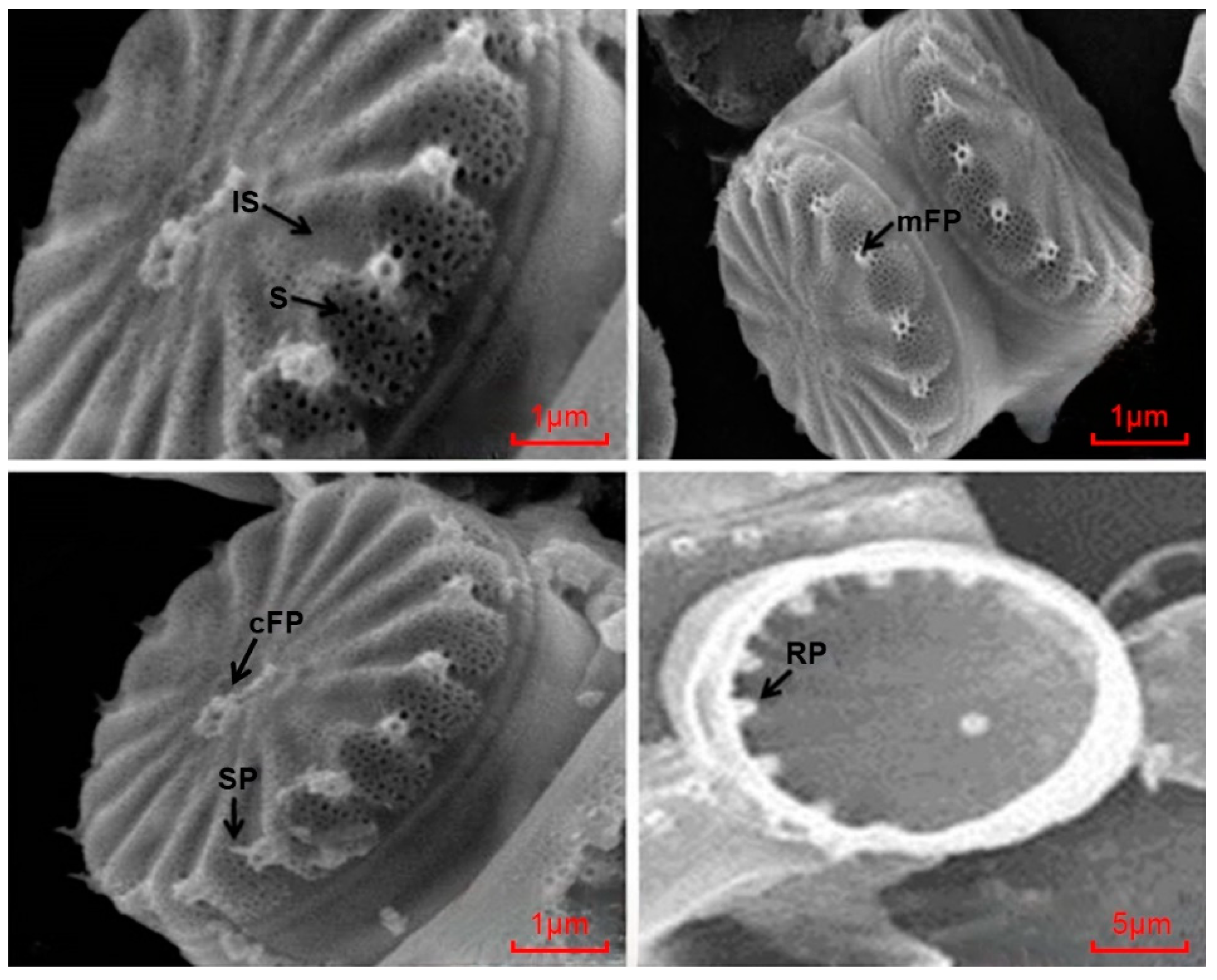
3.1. Molecular Identification of C. meneghiniana

As a typical representative of diatoms, Cyclotella meneghiniana Kützing 1844 (Basionym: Stephanocyclus meneghinianus Kützing Skabitschevsky 1975) is one of the most insightful explored diatoms with a wide distribution and is able to rapidly absorb nutrients from the water column because of its large cell surface area to volume ratio [51,52]. The algal species selected for this study (Cyclotella meneghiniana) were collected from the field at the mouth of the Rongjiang River, China, site: 23°17′5″ N, 116°47′11″ E (Figure 1).
The SEM image revealed that it was circular (centric), and the striae, marginal fultoportula almost on every costa, central fultoportula, rimoportula, spine, and interstice were clearly distinguished by scanning electron microscopy (SEM). The sub-microscopic structure pictures of the algal cells obtained by SEM were basically the same as the results of the study on the identification of the same algal species [53] (Figure 2). Morphological features of Cyclotella meneghiniana have been reported, but there have also been reports of the erroneous extrapolation of Cyclotella cryptic to Cyclotella meneghiniana under morphological observation, and the morphology by itself has proved insufficient to detect the species boundaries in this algal group [54,55]. The sole reliance on morphological techniques for establishing species limits has proven inadequate for precise species recognition; thus, employing molecular research approaches can enhance the detection and discernment of the nuanced variances in diatom species classification and demarcation studies [52,55]. The SEM images identified the diatom as C. meneghiniana (Figure 2), which was further confirmed by PCR using primers designed in this study targeting the ITS.
In conducting the phylogenetic study, we retrieved the Internal Transcribed Spacer regions of C. meneghiniana and related sequences from the NCBI database and discovered that the ITS gene sequences in this study were highly similar (100%) to Cyclotella meneghiniana BIMS-PP0014 [46]. A phylogram with closely related species constructed using ITS1 sequences of Cyclotella meneghiniana (Stephanocyclus meneghinianus) is shown in Figure S1. The sequences of the organism have been deposited in GenBank under accession number STUMBI KY200339.1, named Stephanocyclus meneghinianus STUMBI under the nomenclature Santhoshkumar Chinnappan (Figure 3).

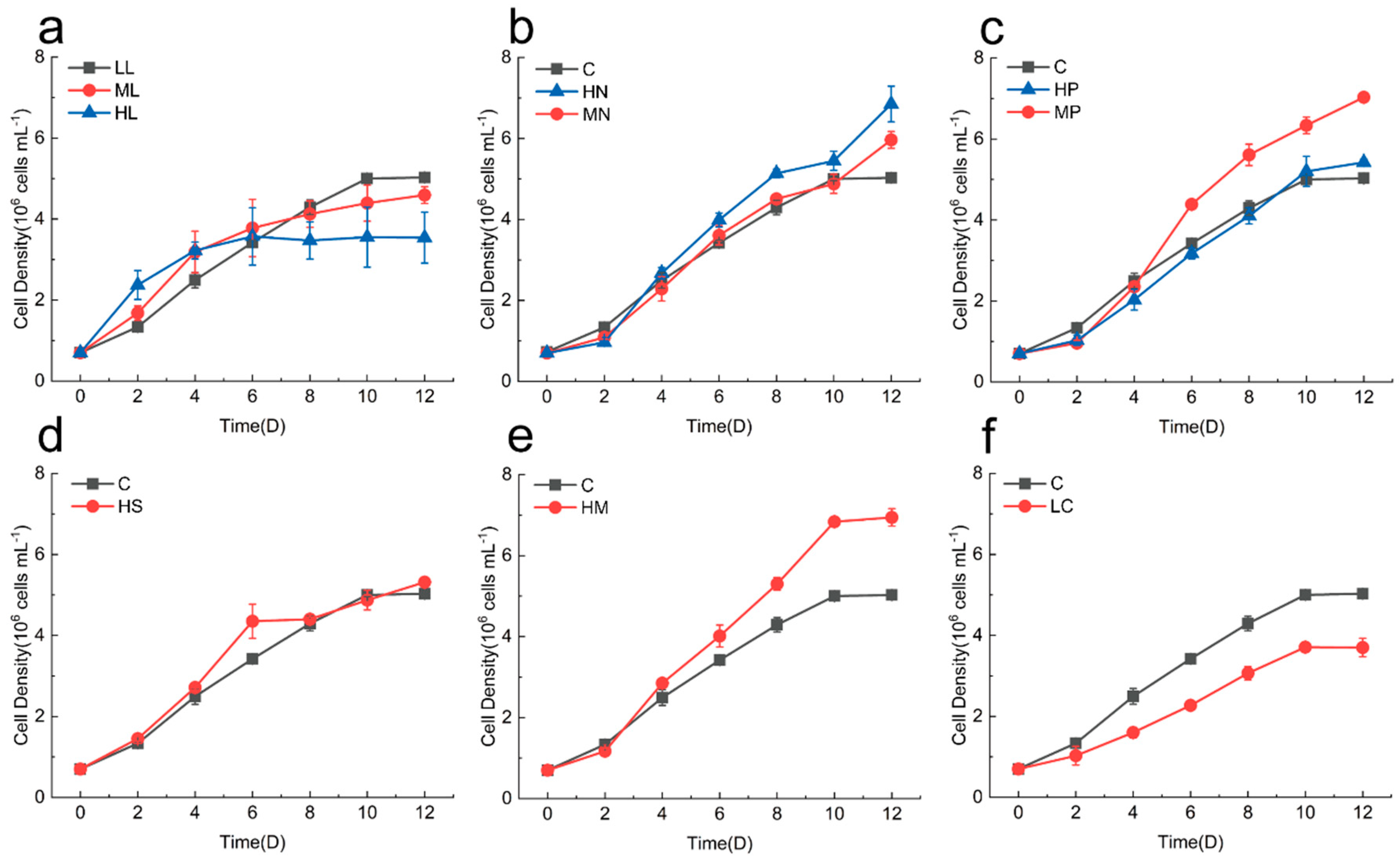
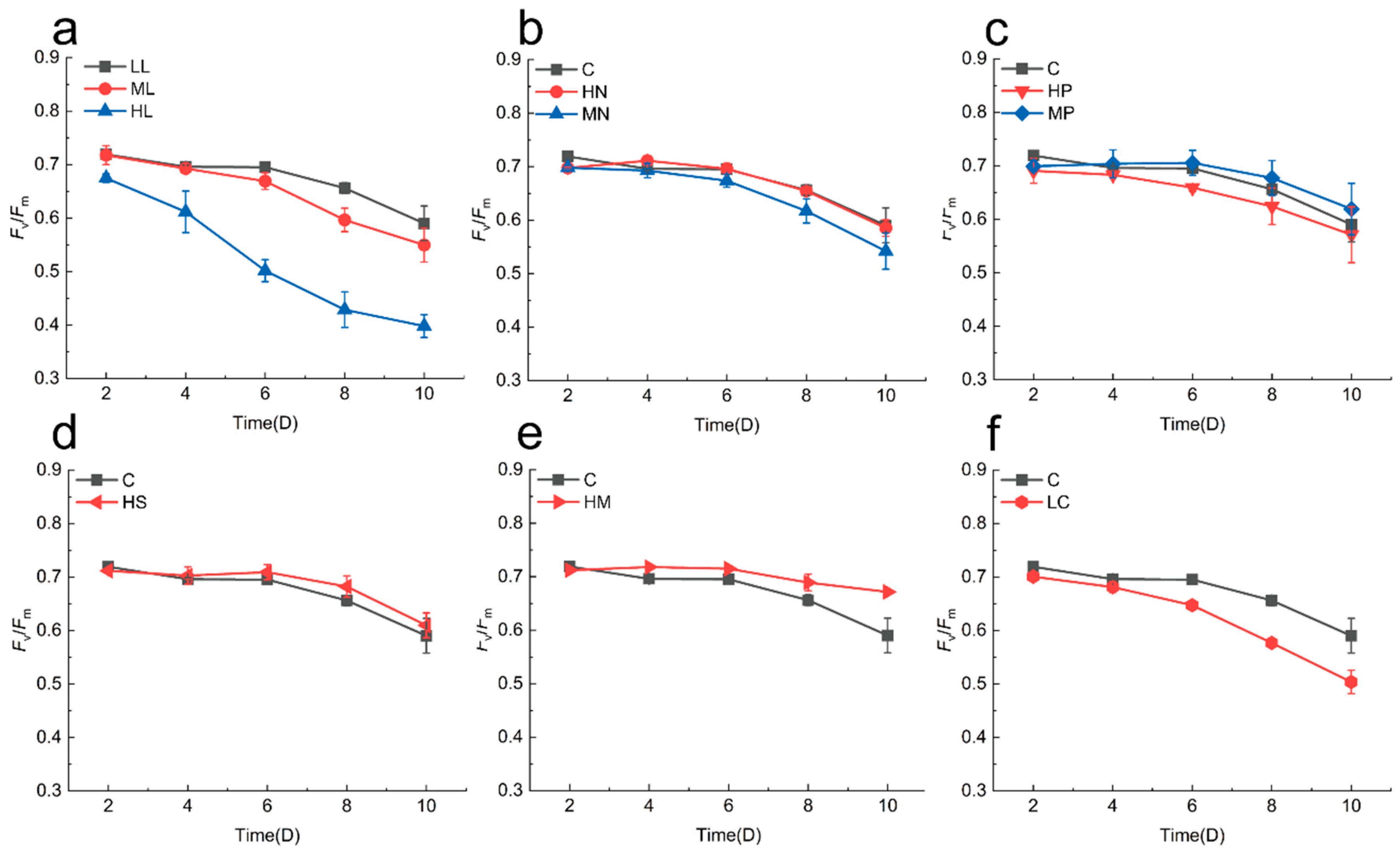
3.2. Physiological Effects on Cyclotella meneghiniana

The generation of fucoxanthin in macroalgae ranges from 0.001 to 0.356% DW, with microalgae yielding more advantageous amounts of fucoxanthin than macroalgae [56,57]. The output of fucoxanthin depends on the type of microalgae, its growth, the content of fucoxanthin, and the efficiency of extraction [6]. The autotrophic growth of microalgae is facilitated by photosynthesis, enriched with light, inorganic nutrients, and carbon dioxide inorganic nutrients present in the water [58]. The proliferation of algae is shaped by a mix of physical, chemical, and biological elements, predominantly light and nutrients [59]. Our research focused on examining microalgae biomass at three different light levels, revealing that Cyclotella meneghiniana in low-light conditions exhibited the greatest cell density after a 12-day incubation period (Figure 4a). The outcome mirrors that of Cyclotella cryptica CCMP333, where a minimal light intensity of 30 μmol photons m−2 s−1 was employed to achieve the peak biomass concentration (roughly 1.25 g L−1) [47]. One plausible reason for this occurrence could be the prolonged acclimatization of experimental microalgae to certain culture environments prior to conducting experiments with varying light intensities. Nutrients such as nitrogen and phosphorus play a fundamental role in the growth of phytoplankton [60]. Typically, the proliferation of microalgae is constrained by the levels and respective ratios of nitrate (N) and phosphorus (P) present in the aquatic environment [58]. We adjusted different nutrient condition variables under low-light conditions to compare with the control group (F/2 group) and found that the HN (Figure 4b) and MP (Figure 4c) groups had significantly higher biomass than the other groups. In general, diatoms are the dominant species in phytoplankton cultures with high nitrogen to phosphorus ratios [61]. This is similar to the findings that nitrate enrichment alone promoted algal growth [62]. Another study was similar to the conclusion of this paper that the biomass of diatoms increased with the increase in the nitrogen concentration when cultured under nitrogen-rich conditions [63]. Interestingly, medium concentrations of phosphorus (MP) in this experiment would promote diatom biomass production more than low (C) or high (HP) phosphorus concentrations (Figure 4c). Our single change in the phosphorus concentration affected the nitrogen–phosphorus ratio in the medium, which may have been the key reason for affecting the algal cell growth. It is shown that a moderate phosphorus concentration favors algal growth, while too much phosphorus may alter the effect on the algal cell biomass due to changes in the nitrogen–phosphorus ratio. Similar studies have shown that the highest concentration of nitrogen and phosphorus (40 mg L−1 NO3 + 20 mg L−1 PO4) did not have the greatest growth-promoting effect on Ulva rigida, while the appropriate concentration of nitrogen and phosphorus (30 mg L−1 NO3 + 15 mg L−1 PO4) was more suitable for the growth of Ulva rigida [64]. The biomass of the HM group (Figure 4e) exhibited a notable increase compared to the control group, reaching (6.94 ± 0.21) × 106 cells mL−1 following a period of growth lasting 12 days. Conversely, the cells within the LC group (Figure 4f) experienced a notable reduction in growth, recording a biomass of (3.70 ± 0.23) × 106 cells mL−1 on the 12th day. This is probably due to the fact that diatoms utilize fundamentally different metabolic responses and cellular regulation to cope with varying nutritional stresses and maintain cellular viability [65].
In addition to the high light culture group, the values of Fv/Fm in C. meneghiniana varied from 0.4 to 0.8 throughout the incubation period (Figure 5). This indicated that the photosynthetic efficiency of the microalgae remained within normal limits. Nonetheless, the HL group exhibited a notable reduction in Fv/Fm when contrasted with the control group (Figure 5a). This observation aligns with the finding on the diatom Thalassiosira pseudonana, which reported a significant decrease in the value of Fv/Fm in HL cultures [66]. The variation in the light adaptations among the diatoms from diverse aquatic environments could explain this phenomenon [67]. The diatoms acclimatized to reduced light intensity (45 μmol photons m−2 s−1) exhibited increased growth rates (Figure 4a) alongside the activity of photosynthesis (Figure 5a) in contrast to comparatively greater light intensities (110 and 170 μmol photons m−2 s−1) [67]. Oversaturated light intensities lead to photoinhibition, which inactivates the reaction centers of PSII, thereby damaging the cell and limiting its optimal physiology and primary productivity [68,69,70].

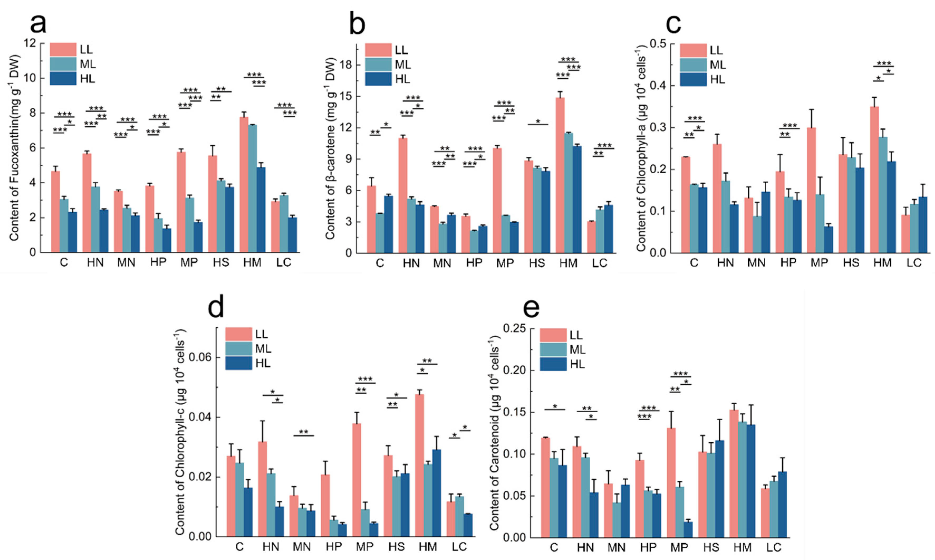
3.3. Influence of Different Conditions on the Content of Photosynthetic Pigments

3.3.1. Effect of Light on Photosynthetic Pigment Production

The growth of microalgae biomass and the build-up of valuable compounds are influenced by multiple elements, including illumination, nutrient levels, and cultivation techniques [71]. Considering the reliance of photoautotrophic microalgae on light for absorbing energy and chemically transforming, light acts as the primary energy source for their biochemical processes, greatly impacting the growth of algae [6,71]. A distinct trend was observed in the build-up of fucoxanthin within microalgae, where the highest concentration of fucoxanthin ranged from 0.52% to 4.28% DW, under varying light intensities from 10 μmol photons m−2 s−1 to 100 μmol photons m−2 s−1 [6]. Nevertheless, the ideal lighting conditions for fucoxanthin production differ across various species [63,72,73,74]. There was a notable disparity in the fucoxanthin production between the low-light and high-light intensity groups (Figure 6a), where every test group under low light exhibited conspicuous variances in fucoxanthin production relative to the control group (Figure S2). Comparable findings were observed in a different investigation on Isochrysis zhangjiangensis, where low light enhanced the accumulation of fucoxanthin, achieving 23.29 mg g−1 DW [72]. The increase in light intensity, along with the decrease in fucoxanthin levels, might be due to fucoxanthin acting as a light-harvesting pigment in PSII photosynthesis, balancing out the reduced light intensity [74]. In terms of addition, the diadinoxanthin cycle can synthesize fucoxanthin, and higher light intensities result in the conversion of diadinoxanthin to diatoxanthin, which diminishes the biosynthesis of fucoxanthin [75]. It was established that the maximum concentration of fucoxanthin reached 7.76 ± 0.30 mg g−1 DW in high trace metals (HMs) concentrations, and these HM levels also enhanced the biosynthesis of fucoxanthin to a cell yield of 0.98 µg 10−7 cells (Table S2). The findings of our research indicated that, under low-light culture conditions, high trace elements, high silicate, and high nitrate contributed to the growth of biomass and improved photosynthesis of C. meneghiniana to a certain extent (Figure 4) while also encouraging fucoxanthin accumulation (Figure 6).
Chlorophyll-a is vital as a pigment that absorbs light, crucial for absorbing and transferring the energy in algal cells throughout photosynthesis [76]. As a crucial carotenoid, β-carotene also plays a role in photosynthesis by absorbing light energy as a key concentrating pigment [77]. Our experimental findings revealed markedly higher levels of Chl-a and carotenoids in low-light environments as opposed to high-light settings. The cause of Chl-a accumulation may be related to the energy transmission routes of the fucoxanthin–chlorophyll protein (FCP) complex in C. meneghiniana [78]. The levels of β-carotene remained consistent across both high-light and low-light conditions, probably because low light is the optimal light intensity for C. meneghiniana growth, so the accumulated content was relatively high. Yet, an abrupt exposure to intense light changed the physiological state of C. meneghiniana, altering its activity in the photosynthetic function and impacting the creation and accumulation of β-carotene [77].

3.3.2. Effect of Nutrient Conditions on the Content of Photosynthetic Pigments

The nitrogen element serves an irreplaceable function in the enzyme that generates photosynthetic pigments, with its composition and quantity directly affecting the elements that foster algal proliferation and enzymatic metabolism, thereby impacting algal life cycles and nutrient renewal [79]. The findings revealed a notable diminution in all the pigment levels among all the MN groups (Figure S2) in LL conditions compared to the control group, with the HN group under LL exhibiting substantially increased levels of fucoxanthin (Figure S2a) and β-carotene (Figure S2d) than the control group. The maximum fucoxanthin yield was 5.66 ± 0.17 mg g−1 DW (under LL and HN) among the different levels of light intensities and N treatments (Figure 6a). This result is in accordance with other research indicating that the fucoxanthin content of Thalassiosira weissflogii at a nitrogen concentration of 300 mg L−1 was about seven times greater than in cultures without nitrogen [80]. Similarly, a study found that nitrogen-enriched conditions (150 mg L−1 NaNO3) were more favorable for diatoms to accumulate fucoxanthin under autotrophic conditions, which is consistent with the findings of this paper [63]. This suggests that photosystems are extremely susceptible to the nitrogen supply, and that the production of fucoxanthin, one of the most essential pigments in photosynthesis, is vulnerable to nitrogen limitation or enrichment [81]. The existing studies have reported that high concentrations of N (≥300 mg L−1), although not exceeding 9 g L−1, are favorable for microalgae to accumulate more fucoxanthin [6,32,82]. The chlorophyll-a content (0.35 ± 0.02 μg 104 cells−1) was measured in a culture containing both low light (LL) and high trace metals (HM), succeeded by the MP group (0.30 ± 0.04 µg 104 cells−1) (Figure 6). The scarcity of nitrogen resulted in reduced pigmentation, suggesting a decelerated photosynthesis process owing to nitrogen deficiency, consequently diminishing the Chl-a content [83]. Furthermore, under low-light conditions, suitable phosphorus enrichment (MP) benefits the production of fucoxanthin (Figure S2a) and β-carotene (Figure S2d) by algal cells, which may be related to the effect of the suitable nitrogen-to-phosphorus ratio on the physiological status of microalgae.
In addition to nitrogen, factors such as silicate and light intensity also play an important role in the biosynthesis of the microalgae fucoxanthin [17]. The growth of diatoms heavily relies on silicates as vital nutrients, which are crucial for the formation of the rigid outer shells of frustules. Due to the potential disruption of the cell growth and structure by a lack of silicate, this research employed nutrient-rich environments rich in silicate to cultivate cells, aiming for enhanced growth and increased fucoxanthin output [84]. As the concentration of silicate escalates, the number of cells (Figure 4d) and the fucoxanthin (Figure 6a) and β-carotene (Figure 6b) contents increased, and the fucoxanthin content of the cells cultured under high-silicate conditions was higher than that of the control group under the same light intensity (Figure S2a–c), in which the highest fucoxanthin content was found under low light (Figure 6a). The results of a study examining the cellular response of the marine diatom Navicula laevis to different concentrations of silicate showed that the biomass was highest on day four (2.41 g L−1) at a silicate level of 480 mg L−1. This result robustly indicates that high silicate concentrations (over 240 mg L−1) profoundly enhance the fucoxanthin content of diatoms [85].
The HM group incubated in low light exhibited the highest photosynthetic pigment content among all the nutrient conditions (Figure 6). We attribute this to the fact that the enrichment of trace metals can periodically have a stimulatory effect on phytoplankton. An analogous study conducted on trace metals showed that an ethylenediaminetetraacetic acid chelated mixture containing iron, copper, zinc, cobalt, manganese, and molybdenum did effectively promote the chlorophyll-a production in plankton across a span of three days [86]. These results can be strung together for the low light intensity and high trace metals condition, under which the Fv/Fm values of algal cells increase, indicating an increase in the photosynthetic efficiency, which in turn promotes an increase in the cellular biomass and thus enhances the accumulation of cell-associated photosynthetic pigments.
Furthermore, we collected the fucoxanthin content in microalgae in scholarly articles and found that the amount of fucoxanthin extracted from some algae was low, probably due to subpar extraction or culture conditions (Table 1). The findings of our study showed that the centric diatom C. meneghiniana measured 7.76 ± 0.30 mg g−1 DW, preserved in low light with high trace metals. In comparison, the cultivation environment was fine-tuned for fucoxanthin synthesis using C. meneghiniana, which was extracted in relatively high amounts. Going forward, the goal is to determine the ideal conditions for extracting fucoxanthin from C. meneghiniana through the compound study of multiple environmental elements, which will provide a favorable theoretical backing for its commercial manufacture.

4. Conclusions

In this study, we characterized Cyclotella meneghiniana at morphological and molecular levels and determined the physiological parameters affecting the production of fucoxanthin in Cyclotella meneghiniana under varying light levels and nutrient environments. The findings showed that low light (45 μmol photons m−2 s−1) and high trace elements yielded the maximum fucoxanthin contents of 7.76 ± 0.30 mg g−1 DW. Consequently, Cyclotella meneghiniana presents as a potentially fucoxanthin-rich resource for the production of durable and cost-effective fucoxanthin.

Supplementary Materials

The following supporting information can be downloaded at https://www.mdpi.com/article/10.3390/app14135504/s1, Figure S1: Phylogram with closely related species constructed using ITS1 sequences of Cyclotella meneghiniana (Stephanocyclus meneghinianus). Phylogram was constructed by the neighbor-joining method using the K2P distance protocol in MEGA ver.5.0. Numerical value at the nodes of the branches indicates bootstrap values and asterisks indicate species studied in this study; Figure S2: Fucoxanthin (a–c), β-carotene (d–f), Chlorophyll-a (g–i), Chlorophyll-c (j–l), and Carotenoid (m–o) contents of C. meneghiniana under three light conditions and different nutritional conditions. The vertical bars represented averages ± STDEV (n = 3); Table S1: Different nutritional concentrations treatments were used in this study. All other concentrations not indicated were the same as Control group; Table S2: Fucoxanthin content yields from cell density under three light conditions.

Author Contributions

S.C.: Investigation, Methodology. J.C.: Investigation, Methodology, Writing—original draft. Y.L.: Investigation, Methodology. Z.Y.: Conceptualization, Methodology, Supervision. Y.S.: Investigation, Methodology. K.C.: Investigation, Methodology. H.D.: Conceptualization, Methodology, Supervision. W.L.: Conceptualization, Methodology, Supervision. P.L.: Conceptualization, Methodology, Supervision, Writing—review and editing, Funding acquisition. All authors have read and agreed to the published version of the manuscript.

Funding

This research was funded by [Science and Technology Planning Project of Guangdong Province] and [Department of Education of Guangdong Province] grant number [2021B1212050025] and [2022zdzx4008].

Data Availability Statement

The data of this study are available from the corresponding author upon reasonable request.

Conflicts of Interest

The authors declare no conflicts of interest.

References

  1. Khan, M.I.; Shin, J.H.; Kim, J.D. The promising future of microalgae: Current status, challenges, and optimization of a sustainable and renewable industry for biofuels, feed, and other products. Microb. Cell Factories 2018, 17, 36. [Google Scholar] [CrossRef]
  2. Goshtasbi, H.; Okolodkov, Y.B.; Movafeghi, A.; Awale, S.; Safary, A.; Barar, J.; Omidi, Y. Harnessing microalgae as sustainable cellular factories for biopharmaceutical production. Algal Res. 2023, 74, 103237. [Google Scholar] [CrossRef]
  3. Udayan, A.; Pandey, A.K.; Sirohi, R.; Sreekumar, N.; Sang, B.-I.; Sim, S.J.; Kim, S.H.; Pandey, A. Production of microalgae with high lipid content and their potential as sources of nutraceuticals. Phytochem. Rev. 2023, 22, 833–860. [Google Scholar] [CrossRef]
  4. De Jesus Raposo, M.F.; De Morais, R.M.S.C.; De Morais, A.M.M.B. Health applications of bioactive compounds from marine microalgae. Life Sci. 2013, 93, 479–486. [Google Scholar] [CrossRef] [PubMed]
  5. Mavrommatis, A.; Tsiplakou, E.; Zerva, A.; Pantiora, P.D.; Georgakis, N.D.; Tsintzou, G.P.; Madesis, P.; Labrou, N.E. Microalgae as a Sustainable Source of Antioxidants in Animal Nutrition, Health and Livestock Development. Antioxidants 2023, 12, 1882. [Google Scholar] [CrossRef]
  6. Khaw, Y.S.; Yusoff, F.M.; Tan, H.T.; Noor Mazli, N.A.I.; Nazarudin, M.F.; Shaharuddin, N.A.; Omar, A.R.; Takahashi, K. Fucoxanthin Production of Microalgae under Different Culture Factors: A Systematic Review. Mar. Drugs 2022, 20, 592. [Google Scholar] [CrossRef]
  7. Hou, H.Y.; Xiang, W.P.; Zhang, J.R.; Cheng, P.F.; Zhou, C.X. Strain and light selection improved fucoxanthin content in the diatom. Acta Hydrobiol. Sin. 2020, 44, 912–919. [Google Scholar] [CrossRef]
  8. Peng, J.; Yuan, J.-P.; Wu, C.-F.; Wang, J.-H. Fucoxanthin, a Marine Carotenoid Present in Brown Seaweeds and Diatoms: Metabolism and Bioactivities Relevant to Human Health. Mar. Drugs 2011, 9, 1806–1828. [Google Scholar] [CrossRef]
  9. Pereira, H.; Sá, M.; Maia, I.; Rodrigues, A.; Teles, I.; Wijffels, R.H.; Navalho, J.; Barbosa, M. Fucoxanthin production from Tisochrysis lutea and Phaeodactylum tricornutum at industrial scale. Algal Res. 2021, 56, 102322. [Google Scholar] [CrossRef]
  10. Li, M.; Feng, H.; Ouyang, X.; Ling, J. Determination of Fucoxanthin in Bloom-Forming Macroalgae by HPLC-UV. J. Chromatogr. Sci. 2021, 59, 978–982. [Google Scholar] [CrossRef]
  11. Sarmiento-Padilla, A.L.; Moreira, S.; Oliveira Rocha, H.A.; Araujo, R.G.; Govea-Salas, M.; Pinales-Marquez, C.D.; Ruiz, H.A.; Rodriguez-Jasso, R.M. Circular bioeconomy in the production of fucoxanthin from aquatic biomass: Extraction and bioactivities. J. Chem. Technol. Biotechnol. 2022, 97, 1363–1378. [Google Scholar] [CrossRef]
  12. Heffernan, N.; Smyth, T.J.; FitzGerald, R.J.; Vila-Soler, A.; Mendiola, J.; Ibáñez, E.; Brunton, N.P. Comparison of extraction methods for selected carotenoids from macroalgae and the assessment of their seasonal/spatial variation. Innov. Food Sci. Emerg. Technol. 2016, 37, 221–228. [Google Scholar] [CrossRef]
  13. Wu, S.-J.; Liou, C.-J.; Chen, Y.-L.; Cheng, S.-C.; Huang, W.-C. Fucoxanthin Ameliorates Oxidative Stress and Airway Inflammation in Tracheal Epithelial Cells and Asthmatic Mice. Cells 2021, 10, 1311. [Google Scholar] [CrossRef]
  14. Zhao, D.; Kwon, S.-H.; Chun, Y.S.; Gu, M.-Y.; Yang, H.O. Anti-Neuroinflammatory Effects of Fucoxanthin via Inhibition of Akt/NF-κB and MAPKs/AP-1 Pathways and Activation of PKA/CREB Pathway in Lipopolysaccharide-Activated BV-2 Microglial Cells. Neurochem. Res. 2017, 42, 667–677. [Google Scholar] [CrossRef]
  15. Xiao, H.; Zhao, J.; Fang, C.; Cao, Q.; Xing, M.; Li, X.; Hou, J.; Ji, A.; Song, S. Advances in Studies on the Pharmacological Activities of Fucoxanthin. Mar. Drugs 2020, 18, 634. [Google Scholar] [CrossRef]
  16. Bae, M.; Kim, M.-B.; Park, Y.-K.; Lee, J.-Y. Health benefits of fucoxanthin in the prevention of chronic diseases. Biochim. Biophys. Acta (BBA) Mol. Cell Biol. Lipids 2020, 1865, 158618. [Google Scholar] [CrossRef]
  17. Leong, Y.K.; Chen, C.-Y.; Varjani, S.; Chang, J.-S. Producing fucoxanthin from algae-Recent advances in cultivation strategies and downstream processing. Bioresour. Technol. 2022, 344, 126170. [Google Scholar] [CrossRef]
  18. Zarekarizi, A.; Hoffmann, L.; Burritt, D. Approaches for the sustainable production of fucoxanthin, a xanthophyll with potential health benefits. J. Appl. Phycol. 2019, 31, 281–299. [Google Scholar] [CrossRef]
  19. Lu, X.; Sun, H.; Zhao, W.; Cheng, K.-W.; Chen, F.; Liu, B. A Hetero-Photoautotrophic Two-Stage Cultivation Process for Production of Fucoxanthin by the Marine Diatom Nitzschia laevis. Mar. Drugs 2018, 16, 219. [Google Scholar] [CrossRef]
  20. Pajot, A.; Huynh, G.H.; Picot, L.; Marchal, L.; Nicolau, E. Fucoxanthin from Algae to Human, an Extraordinary Bioresource: Insights and Advances in up and Downstream Processes. Mar. Drugs 2022, 20, 222. [Google Scholar] [CrossRef]
  21. Araújo, R.; Vázquez Calderón, F.; Sánchez López, J.; Azevedo, I.C.; Bruhn, A.; Fluch, S.; Garcia Tasende, M.; Ghaderiardakani, F.; Ilmjärv, T.; Laurans, M.; et al. Current Status of the Algae Production Industry in Europe: An Emerging Sector of the Blue Bioeconomy. Front. Mar. Sci. 2021, 7, 626389. [Google Scholar] [CrossRef]
  22. Terasaki, M.; Hirose, A.; Narayan, B.; Baba, Y.; Kawagoe, C.; Yasui, H.; Saga, N.; Hosokawa, M.; Miyashita, K. Evaluation of recoverable functional lipid components of several brown seaweeds (phaeophyta) from Japan with special reference to fucoxanthin and fucosterol contents. J. Phycol. 2009, 45, 974–980. [Google Scholar] [CrossRef] [PubMed]
  23. Mohamadnia, S.; Tavakoli, O.; Faramarzi, M.A.; Shamsollahi, Z. Production of fucoxanthin by the microalga Tisochrysis lutea: A review of recent developments. Aquaculture 2020, 516, 734637. [Google Scholar] [CrossRef]
  24. Wang, S.; Wu, S.; Yang, G.; Pan, K.; Wang, L.; Hu, Z. A review on the progress, challenges and prospects in commercializing microalgal fucoxanthin. Biotechnol. Adv. 2021, 53, 107865. [Google Scholar] [CrossRef] [PubMed]
  25. Yang, R.; Wei, D.; Xie, J. Diatoms as cell factories for high-value products: Chrysolaminarin, eicosapentaenoic acid, and fucoxanthin. Crit. Rev. Biotechnol. 2020, 40, 993–1009. [Google Scholar] [CrossRef] [PubMed]
  26. Foo, S.C.; Yusoff, F.M.; Ismail, M.; Basri, M.; Yau, S.K.; Khong, N.M.H.; Chan, K.W.; Ebrahimi, M. Antioxidant capacities of fucoxanthin-producing algae as influenced by their carotenoid and phenolic contents. J. Biotechnol. 2017, 241, 175–183. [Google Scholar] [CrossRef]
  27. Kim, S.M.; Jung, Y.-J.; Kwon, O.-N.; Cha, K.H.; Um, B.-H.; Chung, D.; Pan, C.-H. A potential commercial source of fucoxanthin extracted from the microalga Phaeodactylum tricornutum. Appl. Biochem. Biotechnol. 2012, 166, 1843–1855. [Google Scholar] [CrossRef] [PubMed]
  28. Jin, H.; Guo, Y.; Li, Y.; Chen, B.; Ma, H.; Wang, H.; Wang, L.; Yuan, D. Effective fucoxanthin production in the flagellate alga Poterioochromonas malhamensis by coupling heterotrophic high-cell-density fermentation with illumination. Front. Bioeng. Biotechnol. 2022, 10, 1074850. [Google Scholar] [CrossRef] [PubMed]
  29. Tachihana, S.; Nagao, N.; Katayama, T.; Yusoff, F.M.; Banerjee, S.; Shariff, M.; Yamada, Y.; Imaizumi, Y.; Toda, T.; Furuya, K. High productivity of fucoxanthin and eicosapentaenoic acid in a marine diatom Chaetoceros gracilis by perfusion culture under high irradiance. Algal Res. 2023, 72, 103123. [Google Scholar] [CrossRef]
  30. Zhang, X.; Zhao, J.; Zhang, J.; Su, S.; Huang, L.; Ye, J. Kinetic modelling of microalgal growth and fucoxanthin synthesis in photobioreactor. Int. J. Chem. React. Eng. 2022, 20, 723–734. [Google Scholar] [CrossRef]
  31. Mousavi Nadushan, R.; Hosseinzade, I. Optimization of production and antioxidant activity of fucoxanthin from marine haptophyte algae, Isochrysis galbana. Iran. J. Fish. Sci. 2020, 19, 2901–2908. [Google Scholar] [CrossRef]
  32. Xia, S.; Gao, B.; Fu, J.; Xiong, J.; Zhang, C. Production of fucoxanthin, chrysolaminarin, and eicosapentaenoic acid by Odontella aurita under different nitrogen supply regimes. J. Biosci. Bioeng. 2018, 126, 723–729. [Google Scholar] [CrossRef] [PubMed]
  33. Zittelli, G.C.; Lauceri, R.; Faraloni, C.; Benavides, A.M.S.; Torzillo, G. Valuable pigments from microalgae: Phycobiliproteins, primary carotenoids, and fucoxanthin. Photochem. Photobiol. Sci. 2023, 22, 1733–1789. [Google Scholar] [CrossRef] [PubMed]
  34. Novoveska, L.; Ross, M.E.; Stanley, M.S.; Pradelles, R.; Wasiolek, V.; Sassi, J.-F. Microalgal Carotenoids: A Review of Production, Current Markets, Regulations, and Future Direction. Mar. Drugs 2019, 17, 640. [Google Scholar] [CrossRef] [PubMed]
  35. Zhang, H.; Gong, P.; Cai, Q.; Zhang, C.; Gao, B. Maximizing fucoxanthin production in Odontella aurita by optimizing the ratio of red and blue light-emitting diodes in an auto-controlled internally illuminated photobioreactor. Bioresour. Technol. 2022, 344, 126260. [Google Scholar] [CrossRef] [PubMed]
  36. Chen, C.-Y.; Liu, P.-Y.; Chang, Y.-H.; Nagarajan, D.; Latagan, M.J.D.; de Luna, M.D.G.; Chen, J.-H.; Chang, J.-S. Optimizing cultivation strategies and scaling up for fucoxanthin production using Pavlova sp. Bioresour. Technol. 2024, 399, 130609. [Google Scholar] [CrossRef] [PubMed]
  37. Ishika, T.; Laird, D.W.; Bahri, P.A.; Moheimani, N.R. Co-cultivation and stepwise cultivation of Chaetoceros muelleri and Amphora sp. for fucoxanthin production under gradual salinity increase. J. Appl. Phycol. 2019, 31, 1535–1544. [Google Scholar] [CrossRef]
  38. Roeding, A.; Boekema, E.; Buechel, C. The structure of FCPb, a light-harvesting complex in the diatom Cyclotella meneghiniana. Photosynth. Res. 2018, 135, 203–211. [Google Scholar] [CrossRef] [PubMed]
  39. Gardian, Z.; Litvin, R.; Bina, D.; Vacha, F. Supramolecular organization of fucoxanthin-chlorophyll proteins in centric and pennate diatoms. Photosynth. Res. 2014, 121, 79–86. [Google Scholar] [CrossRef]
  40. Truong, T.Q.; Park, Y.J.; Winarto, J.; Huynh, P.K.; Moon, J.; Choi, Y.B.; Song, D.-G.; Koo, S.Y.; Kim, S.M. Understanding the Impact of Nitrogen Availability: A Limiting Factor for Enhancing Fucoxanthin Productivity in Microalgae Cultivation. Mar. Drugs 2024, 22, 93. [Google Scholar] [CrossRef]
  41. Gelzinis, A.; Augulis, R.; Buechel, C.; Robert, B.; Valkunas, L. Confronting FCP structure with ultrafast spectroscopy data: Evidence for structural variations. Phys. Chem. Chem. Phys. 2021, 23, 806–821. [Google Scholar] [CrossRef] [PubMed]
  42. Guillard, R.R.; Ryther, J.H. Studies of marine planktonic diatoms. I. Cyclotella nana Hustedt, and Detonula confervacea (cleve) Gran. Can. J. Microbiol. 1962, 8, 229–239. [Google Scholar] [CrossRef] [PubMed]
  43. Mazumder, N.; Gogoi, A.; Kalita, R.D.; Ahmed, G.A.; Buragohain, A.K.; Choudhury, A. Luminescence studies of fresh water diatom frustules. Indian J. Phys. 2010, 84, 665–669. [Google Scholar] [CrossRef]
  44. Sambrook, J.; Russell, D.W. Molecular Cloning: A Laboratory Manual, 3rd ed.; Cold Spring Harbor Laboratory Press: New York, NY, USA, 2001; Volume 1, pp. 131–142. ISBN 978-0-87969-577-4. [Google Scholar]
  45. Zhang, B.Y.; Wang, G.C.; Zhang, Y.; Han, X.T.; Lu, S.H.; Qi, Y.Z.; Zou, J.Z.; Zeng, C.K.; Tseng, C.K. Cloning and sequence analysis of 5.8s rDNA and its region from Prorocentrum donghaiense and P. micans APBM. Oceanol. Limnol. Sin. 2004, 35, 272–277. [Google Scholar]
  46. Senapin, S.; Phiwsaiya, K.; Withyachumnarnkul, K.B. Development of primers and a procedure for specific identification of the diatom Thalassiosira weissflogii. Aquac. Int. 2011, 19, 693–704. [Google Scholar] [CrossRef]
  47. Guo, B.; Liu, B.; Yang, B.; Sun, P.; Lu, X.; Liu, J.; Chen, F. Screening of Diatom Strains and Characterization of Cyclotella cryptica as A Potential Fucoxanthin Producer. Mar. Drugs 2016, 14, 125. [Google Scholar] [CrossRef] [PubMed]
  48. Kim, S.-H.; Cho, H.-K.; Shin, H.-S. Physicochemical properties and antioxidant activities of commercial vinegar drinks in Korea. Food Sci. Biotechnol. 2012, 21, 1729–1734. [Google Scholar] [CrossRef]
  49. Ritchie, R.J. Consistent Sets of Spectrophotometric Chlorophyll Equations for Acetone, Methanol and Ethanol Solvents. Photosynth. Res. 2006, 89, 27–41. [Google Scholar] [CrossRef] [PubMed]
  50. Strickland, J.D.H.; Parsons, T.R. A Practical Handbook of Seawater Analysis; Fisheries Research Board of Canada: Ottawa, ON, Canada, 1968. [Google Scholar]
  51. Rzodkiewicz, M.; Gąbka, M.; Szpikowska, G.; Woszczyk, M. Diatom assemblages as indicators of salinity gradients: A case study from a coastal lake. Oceanol. Hydrobiol. Stud. 2017, 46, 325–339. [Google Scholar] [CrossRef]
  52. Beszteri, B.; John, U.; Medlin, L.K. An assessment of cryptic genetic diversity within the Cyclotella meneghiniana species complex (Bacillariophyta) based on nuclear and plastid genes, and amplified fragment length polymorphisms. Eur. J. Phycol. 2007, 42, 47–60. [Google Scholar] [CrossRef]
  53. Goswami, B.; Choudhury, A.; Buragohain, A.K. Luminescence properties of a nanoporous freshwater diatom. Luminescence 2012, 27, 16–19. [Google Scholar] [CrossRef]
  54. McLachlan, J. The growth of unicellular algae in artificial and enriched sea water media. Can. J. Microbiol. 1959, 5, 9–15. [Google Scholar] [CrossRef] [PubMed]
  55. Hevia-Orube, J.; Orive, E.; David, H.; Díez, A.; Laza-Martínez, A.; Miguel, I.; Seoane, S. Molecular and morphological analyses of solitary forms of brackish Thalassiosiroid diatoms (Coscinodiscophyceae), with emphasis on their phenotypic plasticity. Eur. J. Phycol. 2016, 51, 11–30. [Google Scholar] [CrossRef]
  56. Xia, S.; Wang, K.; Wan, L.; Li, A.; Hu, Q.; Zhang, C. Production, characterization, and antioxidant activity of fucoxanthin from the marine diatom Odontella aurita. Mar. Drugs 2013, 11, 2667–2681. [Google Scholar] [CrossRef] [PubMed]
  57. Kanamoto, A.; Kato, Y.; Yoshida, E.; Hasunuma, T.; Kondo, A. Development of a Method for Fucoxanthin Production Using the Haptophyte Marine Microalga Pavlova sp. OPMS 30543. Mar. Biotechnol. 2021, 23, 331–341. [Google Scholar] [CrossRef]
  58. Ahn, C.-Y.; Lee, J.-Y.; Oh, H.-M. Control of Microalgal Growth and Competition by N: P Ratio Manipulation. Korean J. Environ. Biol. 2013, 31, 61–68. [Google Scholar] [CrossRef]
  59. Rashid, N.; Rehman, M.S.U.; Han, J. Enhanced growth rate and lipid production of freshwater microalgae by adopting two-stage cultivation system under diverse light and nutrients conditions. Water Environ. J. 2015, 29, 533–540. [Google Scholar] [CrossRef]
  60. Kraus, C.N.; Bonnet, M.-P.; de Souza Nogueira, I.; Pereira Souza Lobo, M.T.M.; da Motta Marques, D.; Garnier, J.; Galli Vieira, L.C. Unraveling Flooding Dynamics and Nutrients’ Controls upon Phytoplankton Functional Dynamics in Amazonian Floodplain Lakes. Water 2019, 11, 154. [Google Scholar] [CrossRef]
  61. Mikaelyan, A.S.; Pautova, L.A.; Chasovnikov, V.K.; Mosharov, S.A.; Silkin, V.A. Alternation of diatoms and coccolithophores in the north-eastern Black Sea: A response to nutrient changes. Hydrobiologia 2015, 755, 89–105. [Google Scholar] [CrossRef]
  62. Zhang, X.; Mei, X.; Gulati, R.D.; Liu, Z. Effects of N and P enrichment on competition between phytoplankton and benthic algae in shallow lakes: A mesocosm study. Environ. Sci. Pollut. Res. 2015, 22, 4418–4424. [Google Scholar] [CrossRef]
  63. Wang, H.; Zhang, Y.; Chen, L.; Cheng, W.; Liu, T. Combined production of fucoxanthin and EPA from two diatom strains Phaeodactylum tricornutum and Cylindrotheca fusiformis cultures. Bioprocess Biosyst. Eng. 2018, 41, 1061–1071. [Google Scholar] [CrossRef]
  64. Shakouri, A.; Balouch, G.M. The effects of nitrate and phosphate on growth of algae, Ulva rigida. Iran. J. Fish. Sci. 2020, 19, 59–66. [Google Scholar] [CrossRef]
  65. Lin, Q.; Liang, J.-R.; Huang, Q.-Q.; Luo, C.-S.; Anderson, D.M.; Bowler, C.; Chen, C.-P.; Li, X.-S.; Gao, Y.-H. Differential cellular responses associated with oxidative stress and cell fate decision under nitrate and phosphate limitations in Thalassiosira pseudonana: Comparative proteomics. PLoS ONE 2017, 12, e0184849. [Google Scholar] [CrossRef] [PubMed]
  66. Zhang, R.; Kong, Z.; Chen, S.; Ran, Z.; Ye, M.; Xu, J.; Zhou, C.; Liao, K.; Cao, J.; Yan, X. The comparative study for physiological and biochemical mechanisms of Thalassiosira pseudonana and Chaetoceros calcitrans in response to different light intensities. Algal Res. 2017, 27, 89–98. [Google Scholar] [CrossRef]
  67. Shi, P.; Shen, H.; Wang, W.; Yang, Q.; Xie, P. Habitat-specific differences in adaptation to light in freshwater diatoms. J. Appl. Phycol. 2016, 28, 227–239. [Google Scholar] [CrossRef]
  68. Heiden, J.P.; Bischof, K.; Trimborn, S. Light Intensity Modulates the Response of Two Antarctic Diatom Species to Ocean Acidification. Front. Mar. Sci. 2016, 3, 260. [Google Scholar] [CrossRef]
  69. Raven, J.A. The cost of photoinhibition. Physiol. Plant. 2011, 142, 87–104. [Google Scholar] [CrossRef]
  70. Carvalho, A.P.; Silva, S.O.; Baptista, J.M.; Malcata, F.X. Light requirements in microalgal photobioreactors: An overview of biophotonic aspects. Appl. Microbiol. Biotechnol. 2011, 89, 1275–1288. [Google Scholar] [CrossRef] [PubMed]
  71. Parveen, A.; Bhatnagar, P.; Gautam, P.; Bisht, B.; Nanda, M.; Kumar, S.; Vlaskin, M.S.; Kumar, V. Enhancing the bio-prospective of microalgae by different light systems and photoperiods. Photochem. Photobiol. Sci. 2023, 22, 2687–2698. [Google Scholar] [CrossRef]
  72. Li, Y.; Sun, H.; Wu, T.; Fu, Y.; He, Y.; Mao, X.; Chen, F. Storage carbon metabolism of Isochrysis zhangjiangensis under different light intensities and its application for co-production of fucoxanthin and stearidonic acid. Bioresour. Technol. 2019, 282, 94–102. [Google Scholar] [CrossRef]
  73. Gao, F.; Teles Cabanelas Itd, I.; Wijffels, R.H.; Barbosa, M.J. Process optimization of fucoxanthin production with Tisochrysis lutea. Bioresour. Technol. 2020, 315, 123894. [Google Scholar] [CrossRef] [PubMed]
  74. McClure, D.D.; Luiz, A.; Gerber, B.; Barton, G.W.; Kavanagh, J.M. An investigation into the effect of culture conditions on fucoxanthin production using the marine microalgae Phaeodactylum tricornutum. Algal Res. 2018, 29, 41–48. [Google Scholar] [CrossRef]
  75. Sun, Z.; Wang, X.; Liu, J. Screening of Isochrysis strains for simultaneous production of docosahexaenoic acid and fucoxanthin. Algal Res. 2019, 41, 101545. [Google Scholar] [CrossRef]
  76. Zhang, J.; Kong, L.; Zhao, Y.; Lin, Q.; Huang, S.; Jin, Y.; Ma, Z.; Guan, W. Antagonistic and synergistic effects of warming and microplastics on microalgae: Case study of the red tide species Prorocentrum donghaiense. Environ. Pollut. 2022, 307, 119515. [Google Scholar] [CrossRef]
  77. Zarandi-Miandoab, L.; Hejazi, M.-A.; Bagherieh-Najjar, M.-B.; Chaparzadeh, N. Optimization of the Four Most Effective Factors on β-Carotene Production by Dunaliella salina Using Response Surface Methodology. Iran. J. Pharm. Res. 2019, 18, 1566–1579. [Google Scholar] [CrossRef] [PubMed]
  78. Papagiannakis, E.; Van Stokkum, I.H.M.; Fey, H.; Büchel, C.; Van Grondelle, R. Spectroscopic Characterization of the Excitation Energy Transfer in the Fucoxanthin–Chlorophyll Protein of Diatoms. Photosynth. Res. 2005, 86, 241–250. [Google Scholar] [CrossRef] [PubMed]
  79. Shah, J.M.; Bukhari, S.A.H.; Zeng, J.; QuAN, X.; Ali, E.; Muhammad, N.; Zhang, G. Nitrogen (N) metabolism related enzyme activities, cell ultrastructure and nutrient contents as affected by N level and barley genotype. J. Integr. Agric. 2017, 16, 190–198. [Google Scholar] [CrossRef]
  80. Marella, T.K.; Tiwari, A. Marine diatom Thalassiosira weissflogii based biorefinery for co-production of eicosapentaenoic acid and fucoxanthin. Bioresour. Technol. 2020, 307, 123245. [Google Scholar] [CrossRef]
  81. Mohamadnia, S.; Tavakoli, O.; Faramarzi, M.A. Production of fucoxanthin from the microalga Tisochrysis lutea in the bubble column photobioreactor applying mass transfer coefficient. J. Biotechnol. 2022, 348, 47–54. [Google Scholar] [CrossRef]
  82. Mohamadnia, S.; Tavakoli, O.; Faramarzi, M.A. Enhancing production of fucoxanthin by the optimization of culture media of the microalga Tisochrysis lutea. Aquaculture 2021, 533, 736074. [Google Scholar] [CrossRef]
  83. Kim, S.M.; Kang, S.-W.; Kwon, O.-N.; Chung, D.; Pan, C.-H. Fucoxanthin as a major carotenoid in Isochrysis aff. galbana: Characterization of extraction for commercial application. J. Korean Soc. Appl. Biol. Chem. 2012, 55, 477–483. [Google Scholar] [CrossRef]
  84. Jiang, Y.; Laverty, K.S.; Brown, J.; Brown, L.; Chagoya, J.; Burow, M.; Quigg, A. Effect of silicate limitation on growth, cell composition, and lipid production of three native diatoms to Southwest Texas desert. J. Appl. Phycol. 2015, 27, 1433–1442. [Google Scholar] [CrossRef]
  85. Mao, X.; Chen, S.H.Y.; Lu, X.; Yu, J.; Liu, B. High silicate concentration facilitates fucoxanthin and eicosapentaenoic acid (EPA) production under heterotrophic condition in the marine diatom Nitzschia laevis. Algal Res. 2020, 52, 102086. [Google Scholar] [CrossRef]
  86. Twiss, M.R.; Gouvêa, S.P.; Bourbonniere, R.A.; McKay, R.M.L.; Wilhelm, S.W. Field Investigations of Trace Metal Effects on Lake Erie Phytoplankton Productivity. J. Great Lakes Res. 2005, 31, 168–179. [Google Scholar] [CrossRef]
  87. Sun, P.; Wong, C.-C.; Li, Y.; He, Y.; Mao, X.; Wu, T.; Ren, Y.; Chen, F. A novel strategy for isolation and purification of fucoxanthinol and fucoxanthin from the diatom Nitzschia laevis. Food Chem. 2019, 277, 566–572. [Google Scholar] [CrossRef] [PubMed]
  88. Goiris, K.; Muylaert, K.; Fraeye, I.; Foubert, I.; De Brabanter, J.; De Cooman, L. Antioxidant potential of microalgae in relation to their phenolic and carotenoid content. J. Appl. Phycol. 2012, 24, 1477–1486. [Google Scholar] [CrossRef]
  89. Pasquet, V.; Chérouvrier, J.-R.; Farhat, F.; Thiéry, V.; Piot, J.-M.; Bérard, J.-B.; Kaas, R.; Serive, B.; Patrice, T.; Cadoret, J.-P.; et al. Study on the microalgal pigments extraction process: Performance of microwave assisted extraction. Process Biochem. 2011, 46, 59–67. [Google Scholar] [CrossRef]
Figure 1. Collecting site of C. meneghiniana. The red dot on the map represents study area of this research. The purple dot on the map represents sample site of Cyclotella meneghiniana.
Applsci 14 05504 g001
Figure 2. SEM pictures of C. meneghiniana. Arrows indicate important morphological features, such as striae (S), marginal fultoportula (mFP), central fultoportula (cFP), rimoportula (RP), spine (SP), and interstriae (IS).
Applsci 14 05504 g002
Figure 3. Phylogram constructed using ITS1 sequences of Cyclotella meneghiniana (Stephanocyclus meneghinianus). Phylogram was constructed by the neighbor-joining method using the K2P distance protocol in MEGA ver.5.0. Numerical values at the nodes of the branches indicate bootstrap values and asterisks indicate species studied in this study.
Applsci 14 05504 g003
Figure 4. Growth of C. meneghiniana under three light conditions and different nutrient conditions: three light conditions (a), different nitrate conditions (b), different phosphate conditions (c), high silicate (d), high trace metals (e), and lower concentration than F/2 medium (f). The vertical bars represent averages ± STDEV (n = 3).
Applsci 14 05504 g004
Figure 5. Changes in the maximum quantum efficiency of PSII (Fv/Fm) of C. meneghiniana under three light conditions and different nutrient conditions: three light conditions (a), different nitrate conditions (b), different phosphate conditions (c), high silicate (d), high trace metals (e), and lower concentration than F/2 medium (f). The vertical bars represent averages ± STDEV (n = 3).
Applsci 14 05504 g005
Figure 6. Fucoxanthin (a), β-carotene (b), chlorophyll-a (c), chlorophyll-c (d), and carotenoid (e) contents of C. meneghiniana under three light conditions and different nutritional conditions. The vertical bars represent averages ± STDEV (n = 3). * p < 0.05, ** p < 0.01, *** p < 0.001.
Applsci 14 05504 g006
Table 1. Comparison with fucoxanthin contents in other microalgae.
MicroalgaeFucoxanthin Contents (mg g−1 DW)Sample ConditionReferences
Cyclotella meneghiniana7.76DriedIn this study
Saccharina japonica0.03DriedFoo et al., 2017 [26]
Skeletonema costatum0.36DriedFoo et al., 2017 [26]
Odontella sinensis1.18DriedFoo et al., 2017 [26]
Nitzschia laevis1.68DriedSun et al., 2019 [87]
Isochrysis galbana2.19DriedFoo et al., 2017 [26]
Chaetoceros gracilis2.24DriedKim et al., 2012 [83]
Chaetoceros calcitrans2.33DriedGoiris et al., 2012 [88]
Nitzschia sp.4.92DriedKim et al., 2012 [83]
Cylindrotheca closterium5.23DriedPasquet et al., 2011 [89]
Isochrysis galbana6.04DriedKim et al., 2012 [83]
Phaeodactylum tricornutum8.55DriedKim et al., 2012 [83]
Isochrysis aff. galbana18.23DriedKim et al., 2012 [83]
Odontella aurita21.67DriedSong et al., 2013 [56]
Isochrysis zhangjiangensis23.29DriedLi et al., 2019 [72]
Disclaimer/Publisher’s Note: The statements, opinions and data contained in all publications are solely those of the individual author(s) and contributor(s) and not of MDPI and/or the editor(s). MDPI and/or the editor(s) disclaim responsibility for any injury to people or property resulting from any ideas, methods, instructions or products referred to in the content.

