Review

Applications of Raman Microscopy/Spectroscopy-Based Techniques to Plant Disease Diagnosis

by
Ioannis Vagelas
1,
Ioannis Manthos
2 and
Thomas Sotiropoulos
3,*
1
Laboratory of Plant Pathology, Department of Agriculture Crop Production and Rural Environment, School of Agricultural Sciences, University of Thessaly, 38446 Volos, Greece
2
Department of Nut Trees, Institute of Plant Breeding & Genetic Resources, Hellenic Agricultural Organization (ELGO)-DIMITRA, Neo Krikello, 35100 Lamia, Greece
3
Department of Deciduous Fruit Trees, Institute of Plant Breeding & Genetic Resources, Hellenic Agricultural Organization (ELGO)-DIMITRA, 59200 Naoussa, Greece
*
Author to whom correspondence should be addressed.
Appl. Sci. 2024, 14(13), 5926; https://doi.org/10.3390/app14135926
Submission received: 6 June 2024 / Revised: 27 June 2024 / Accepted: 3 July 2024 / Published: 7 July 2024
(This article belongs to the Special Issue Raman Spectroscopy: Novel Advances and Applications: 2nd Edition)

Abstract

:
Plant diseases pose a significant threat to plant and crop health, leading to reduced yields and economic losses. The traditional methods for diagnosing plant diseases are often invasive and time-consuming and may not always provide accurate results. In recent years, there has been growing interest in utilizing Raman microscopy as a non-invasive and label-free technique for plant disease diagnosis. Raman microscopy is a powerful analytical tool that can provide detailed molecular information about samples by analyzing the scattered light from a laser beam. This technique has the potential to revolutionize plant disease diagnosis by offering rapid and accurate detection of various plant pathogens, including bacteria and fungi. One of the key advantages of Raman microscopy/spectroscopy is its ability to provide real-time and in situ analyses of plant samples. By analyzing the unique spectral fingerprints of different pathogens, researchers can quickly identify the presence of specific diseases without the need for complex sample preparation or invasive procedures. This article discusses the development of a Raman microspectroscopy system for disease diagnosis that can accurately detect and identify various plant pathogens, such as bacteria and fungi.

1. Introduction

Plant pathogenic microorganisms cause significant yield losses in many economically important crops, leading to social and economic change. The spread of these plant pathogens and the emergence of new diseases are facilitated by human practices such as monoculture farming and global trade. Therefore, it is crucial to detect and identify these pathogens early to minimize agricultural losses [1].
New strategies and new knowledge on plant pathogen life cycles show that the study of pre-symptomatic disease detection in the latent period is crucial for crop production and for food security [2]. Literature studies have highlighted the importance of understanding latent infections of soybean and seeds by fungi [3] and the importance of understanding Phomopsis durionis leaf spot and latent infection to develop effective strategies for management and control, respectively [4].
Recent studies such as whole-genome sequencing of plant pathogens can improve diagnostic accuracy [5,6,7], but this kind of diagnosis (using new-generation sequencing technology in the diagnosis of fungal plant pathogens) usually generates big data that need sophisticated bioinformatic tools (machine learning, artificial intelligence) for their management [8,9,10,11,12]. Furthermore, early detection of plant diseases using spectral data is a rapidly growing field that combines the power of spectroscopy with machine learning and artificial intelligence [13,14].
Existing literature on plant diseases may not always provide accurate results due to various factors, such as methodology limitations (PCR or serology may produce varying results) or variability in disease expression, so combining innovative techniques with traditional–modern methods to improve our understanding of plant diseases and develop more accurate diagnostic tools is required. To address these limitations, in this article, we explore new techniques and technologies, such as advanced microscopy techniques like Raman microscopy and Raman spectroscopy, in order to develop more accurate diagnostic tools for plant pathology.

2. Plant Disease Diagnosis an Overview—Why Early Detection Matters—The Problem

As mentioned above, plant disease diagnosis is a crucial aspect of plant pathology, essential for mitigating economic losses and ensuring food security. In general, plant diseases are caused by many pathogenic agents, including viruses, bacteria, mycoplasmas, rickettsias, fungi, nematodes and parasitic higher plants [15,16]. These agents can be transmitted naturally from plant to plant by various means, such as the wind, rain or physical disturbance of plant tissues by other animals. Plant pathologists have attempted to characterize and classify each disease based on the host or the associated host–pathogen relationship, the environmental patterns of disease frequency and the mode of the pathogen’s survival, dispersal and infection [15,16,17,18]. Once diseases are characterized, agronomists can focus on important methods for managing them. The first step in disease management is the accurate diagnosis of each disease. Some common methods used for plant disease diagnosis are visual observations (identifying characteristic symptoms such as lesions on plants to identify the plant pathogen), microscopy (involving examining tissue samples under a microscope to identify pathogenic microorganisms), serological tests (involving antibodies to detect the presence of specific pathogens in plant tissue) and molecular diagnostics (involving techniques such as PCR or DNA sequencing to identify plant pathogens) [16].
Besides the new tools and technologies, e.g., PCR, that have been created for plant disease diagnosis, recent advances in computer technology offer tools that can be used to develop systems for accurate and timely plant disease diagnostics [19,20,21]. These systems have introduced computer applications in disease diagnostics focused on providing fast information access, from databases of disease symptoms and their causes to computerized practical aids and expert systems for plant disease diagnostics [22].
The above-mentioned disease diagnostic systems enable the rapid and accurate identification of plant diseases, which can help farmers and plant breeders take timely action to control the spread of diseases. However, they have limitations in terms of understanding the interactions between plants and pathogens, including the host’s immune system, the pathogen’s virulence factors and environmental factors [1]. Furthermore, while we have a general understanding of plant defense mechanisms, there is still much to be learned about the specific mechanisms involved in plant–pathogen interactions. For example, walnut blight pathogen (Xanthomonas arboricola pv. juglandis) causes one of the most destructive diseases of the walnut (Juglans regia L.), so specific and reliable diagnosis tools for this nut based on either conventional PCR or real-time PCR have been developed [23]. However, these tools can become somewhat inappropriate (due to different genomic profiles when it comes to in-field diagnosis, as demonstrated by symptomatic plants [24]). Moreover, due to heavy rainfall and high relative humidity and temperatures in the main walnut-producing areas, walnut disease occurs frequently, with severe pathogenesis. By addressing these limitations, prompt diagnosis (in-field diagnosis) of the occurrence of this disease provides the potential to undertake effective management responses to reduce damage to walnut trees, minimize economic losses and protect the environment [25,26].
Many more plant–pathogen interactions are complex and influenced by many factors, making it challenging to develop accurate diagnostic methods. Furthermore, multiple virulence factors being produced by pathogens, such as toxins, enzymes and signaling molecules, which can interact with each other, increases the risk of false diagnosis and makes it challenging to develop effective diagnostic methods [1,27].
Why does early detection matter? An answer could be that early detection allows for prompt action to prevent the spread of the disease to other plants, reducing the risk of epidemic outbreaks (reduces crop losses and minimizes economic impact). To address the challenges mentioned above and focus on early detection, it becomes essential to join the modern methods of detection with computer-aided automated studies such as machine learning and deep understanding detection models or field-based disease detection and monitoring [28,29]. But these techniques require specialized expertise and equipment, which can be costly and may not be feasible for researchers and agronomists.
Overall, to overcome these above-mentioned challenges, researchers are employing innovative approaches, such as the following:
  • Advanced genomics and transcriptomics: Analyzing gene expression and genomic data to better understand pathogen–host interactions and disease development [30].
  • Machine learning algorithms: Using machine learning algorithms to analyze large datasets and identify patterns in plant disease latency [31,32,33,34].
  • Experimental design and simulation modeling: Designing experiments to study disease latency under controlled conditions and using simulation models to predict plant disease spread and management [35,36].
We believe that these innovative approaches have limited access to researchers and can be expensive. Based on these problems, we propose in this novel review a system (Raman spectroscopy/microscopy) which (i) can detect disease-related biomarkers on the surface of plant leaves without causing physical damage, (ii) can analyze the chemical composition of plant samples, providing information on disease-related changes, (iii) can be combined with other techniques, such as hyperspectral imaging or machine learning, to enhance the detection accuracy and (iv) can detect very low concentrations of biomarkers, making it potentially suitable for early detection of disease.

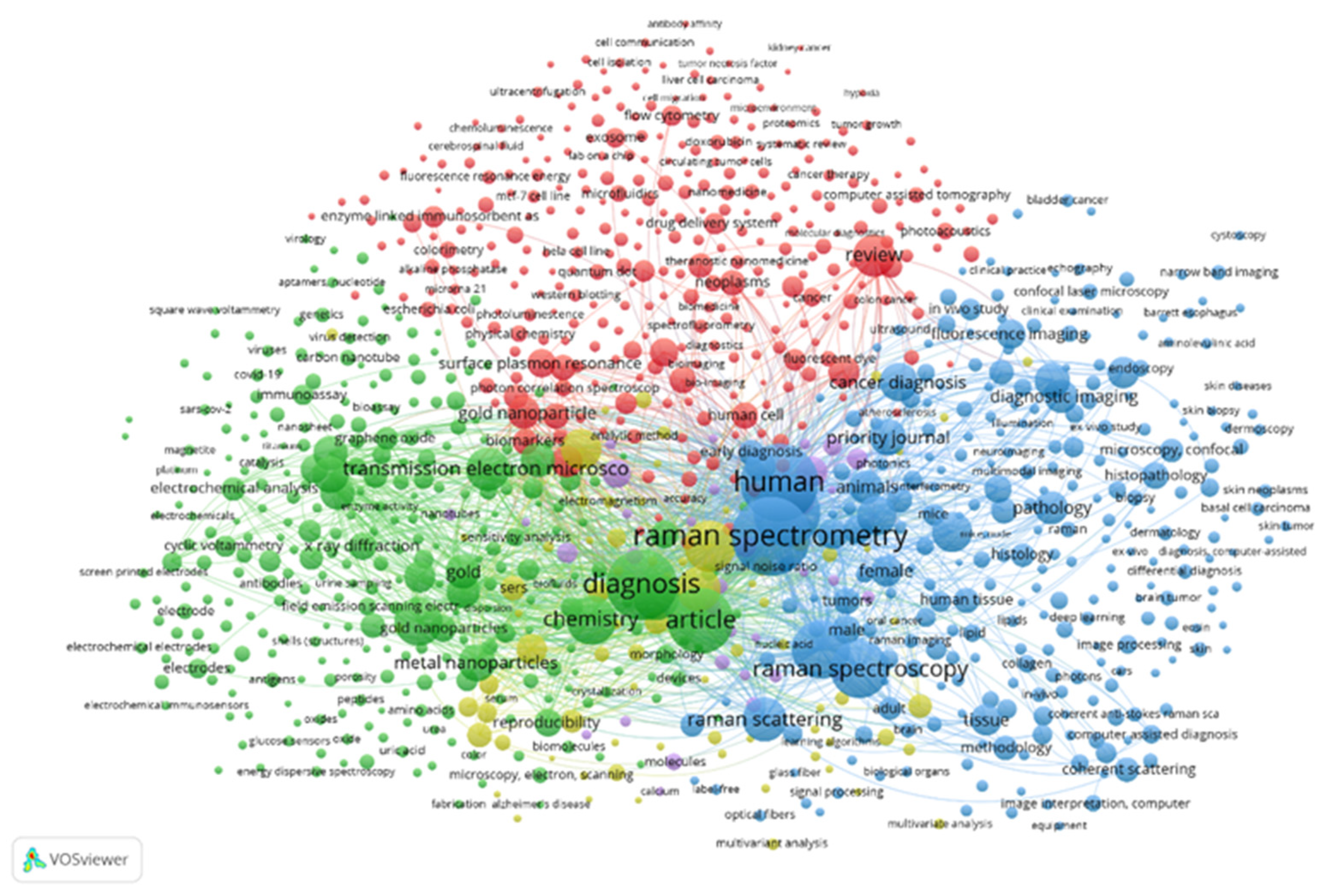
3. Raman Microscopy—A New Method Used to Study Cell Biology

Microscopic Raman imaging and confocal Raman microscopy (CRM) are current and advanced analytical tools for biomedicine; they offer spectral fingerprint recognition, molecular presence and biochemical mapping. CRM was first developed almost 20 years ago, though its applications have mainly been restricted to surface investigations of cellular components and drugs at the micrometer scale. Its development in the past few years has been particularly advanced with respect to biological specimens, particularly in the case of soft tissues [37].
A new method used to study cell biology is Raman microspectroscopy, which has become common due to the ability to provide analytical data with little prior sample preparation and to enable the visualization and biochemical characterization of live cells, subcellular structures and specific organelles [38,39].
Moreover, the Raman spectra of microorganisms are superpositions of spectra due to variations in various organelles and biochemical components inside the cell, such as peptidoglycan (the predominant polymer in the cell wall of Gram-positive bacteria), chitin (a fungal cell wall component), ergosterol (the steroid molecule in fungal cell membranes) and polymers (F-actin, microtubules and septins—fungal cytoskeleton polymers) [39]. Furthermore, since fungal hyphal growth is closely related to the ability to secrete large amounts of enzymes, polysaccharides, lipids and proteins, including cytochromes, Raman spectroscopic imaging provides more accurate spatially resolved information on their relative amounts and chemical states compared with other methods [40].
The properties of Raman spectroscopy make it an interesting technique for non-invasive characterization and differentiation of certain cell types and tissues. Studies applying Raman scattering to living eukaryotic cells appeared in the late 1990s, with pioneering works in this decade mainly investigating the effect of Raman scattering in single cells and the spatial distribution of cell structures and their conformational status. Raman scattering characteristics have been used by biologists to identify cancerous cells and to differentiate malignancy levels in a noninvasive way, without the need for chemical markers [41,42]. With the use of a lower-intensity wavelength, molecular damage can mostly be minimized, which provides a successful approach to studying in vivo soft tissues, which are sensitive to optical damage. Such a technique can be invaluable to cancer researchers for studying cell dynamics and metabolisms [41,42,43].

4. Raman Spectroscopy—Principles of Raman Spectroscopy

Light scattering occurs in a molecule when a photon interacting with a coherent molecular vibration of the medium collides with a vibrationally hot state. If this process occurs without energy loss, the act of scattering changes the direction of the photon and the polarizability of the coherent mode of the medium. The degree of the polarizability change is related to the eternally scattered photon, originating from an electronic excited state and a molecular vibrational state [44]. The inelastic scattering rules for the final state imply that the energy of the scattered photon is directly correlated with the energy of the accessible vibrational states in the medium. Hence, the scattered Raman photon has an energy that deviates as a Raman shift from the energy of the incident photon [44,45].
Raman spectroscopy is a powerful tool for probing levels of biological organization from molecules to whole cells without extrinsic staining. The molecular specificity of Raman spectra facilitates the quantification and spatial identification of functional groups, such as proteins and lipids or carotenoids [37]. For example, tomato carotenoids exhibit two strong Raman peaks in spectral regions of 1100–1200 and 1400–1600 cm−1 due to the stretching vibrations of C-C bonds [39]. Quantitative and label-free spatially resolved measurements of lipid and protein contents are particularly important for understanding cell biology because of their physiological demands. Additionally, Raman microspectroscopy is compatible with a variety of live-cell time-resolved measurements. Compatibility with life and depth-resolved imaging are particularly advantageous for studying the cellular dynamics and functions underlying complex (dys)regulation mechanisms and the spatial distribution of molecular species in the environment of living cells [37,46].

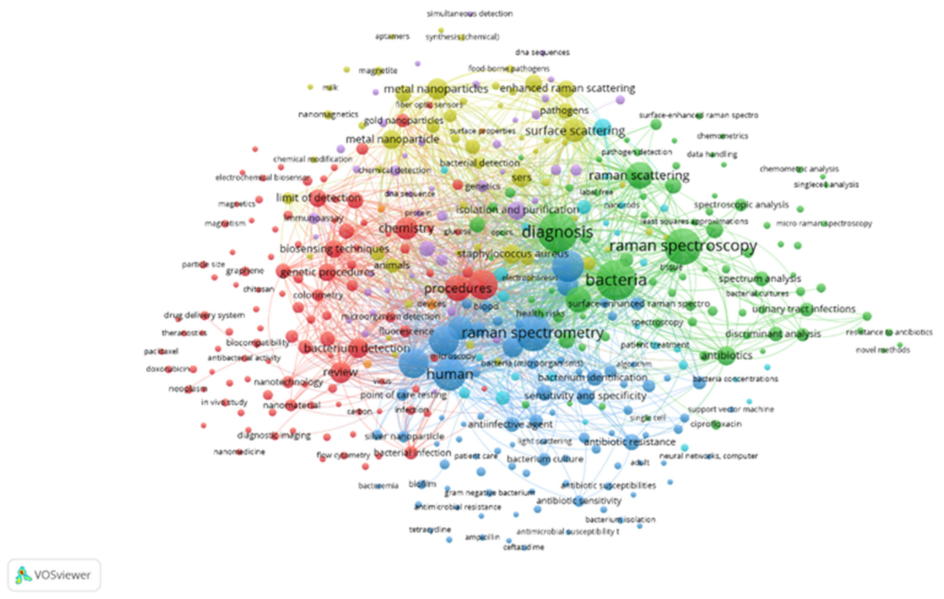
5. Raman Microscopy in Single Cells—Single Microbial Cells

Cells, whether individually or in groups, can be observed using various techniques. The most common method is light microscopy, which is frequently used in cell culture laboratories. It allows for quick assessment of the shape and coverage of cultured cells. However, this visual examination does not provide specific information about relevant molecules and detailed cellular structures. Other techniques, such as electron microscopy and fluorescence microscopy, are utilized to observe cells. Currently, confocal fluorescence microscopy is considered the most advanced technique for cell visualization. This method involves the use of fluorescent marker molecules that selectively bind to the structures of interest, enabling the specific visualization of different cell structures [47,48,49].
The need for techniques that allow for chemically selective analysis to visualize and understand uptake is important in addition to visualization. Therefore, IR absorption spectroscopy and Raman spectroscopy are attractive methods for non-destructive and label-free imaging while maintaining chemical selectivity. Furthermore, both techniques can be combined with optical microscopy to enable powerful approaches to non-invasive chemical imaging. Due to the superior resolution of Raman microscopy, which makes it possible to analyze individual particles with dimensions of <1 μm compared to IR microscopy (~10 μm) and its applicability in aqueous environments, confocal Raman microscopy is now the predominant technique for cellular visualization [50,51,52].
Therefore, the utilization of Raman microscopy in cell biology has definitively added to the visualization of cell biology for a series of biochemical species involved in cell division. Moreover, nowadays, this method contributes to cellular phenotyping. This is mainly because Raman spectroscopy can supply reliable information about the concentration of various amino acids and nucleotides, besides other components. Raman spectroscopy can highlight the concentration of both proteins and nuclear acids, separated from lipids and other cellular components [53].
Raman spectroscopy can distinguish between different cell types and states (e.g., healthy vs. diseased cells) based on their unique spectral signatures. For example, Raman spectroscopy is used to detect cancerous cells and understand their biochemical changes, aiding in early diagnosis and treatment monitoring [54,55].
Raman spectroscopy is increasingly being used to study single microbial cells within populations, offering insights into their biochemical composition, metabolic states and interactions. This technique is particularly valuable for microbial ecology, biotechnology and medical microbiology [56,57]. Raman spectroscopy can differentiate between microbial species and strains based on their unique spectral fingerprints. This is valuable for microbial taxonomy and diagnosing infections. This technique can analyze the biochemical composition of microbes at the single-cell level, providing insights into their formation, structure and biochemical composition. Therefore, Raman spectroscopy can be used to study microbial communities in their natural environments, such as soil or water, providing information on their metabolic activities and interactions [58,59].
Raman spectra can provide insight into the molecular composition of specific locations, allowing for the differentiation of components within a sample, such as organelles and cells in tissues. In addition to Raman peak mapping, advanced analysis methods like multiple curve resolution and independent component analysis (multivariate analysis techniques) can be used to visualize nuclei, lipid droplets and cell bodies individually. This offers valuable morphological information about samples without the need for labeling [46].

6. Raman Microscopy/Spectroscopy and Chemical Information

Based on the above, Raman microscopy is widely used in modern analytical laboratories because it is a powerful and fast non-destructive technique that can be applied to many sample types. The chemical compounds found in a mixture can be analyzed and identified, and quantitative data can be obtained. Raman microscopy equipment is robust, reliable and easy to use. The technique is sensitive, allowing for analysis of numerous compounds in trace amounts in a single measurement [46].
So, Raman microscopy can identify specific molecules and their structures, including biomolecules, polymers and small molecules. It can provide information about chemical bonding patterns, including functional groups and molecular interactions, and can measure the concentration of specific molecules in a sample, allowing for quantitative analysis. Raman microscopy is non-invasive, it can detect very small concentrations of molecules, making it suitable for analyzing complex samples, and it is able to analyze multiple components in a sample simultaneously, making it suitable for analyzing complex mixtures [37,53,60].
Due to these advantages, we believe that Raman microspectroscopy is a significant technique, particularly in the fields of biology and plant pathology for early disease detection, as it retains the spatial resolution inherent in microscopy and benefits from the highly specific chemical analysis provided by Raman spectroscopy.

7. “Raman Shifts” in Cell Biology

“Raman shifts” are shifts between the energy of the incident photon of laser light and the energy of the photon which has interacted with the molecule under analysis, allowing for the exact assessment of vibrations in the normal functional groups of biochemical compounds, such as proteins, lipids or nucleic acids [46,61]. This makes it possible to study their conformation and changes in a non-invasive way. The distinctive features of the method include its chemical selectivity, biological molecule sensitivity, noninvasive manner and applicability to conducting in situ analyses of living single cells [53,60]. So, the energy of the scattered photon is shifted by the energy of the vibrational mode, resulting in a characteristic Raman shift, which is a fundamental concept in Raman spectroscopy, a technique used to analyze the molecular structure of materials. In summary, the “Raman shift” (i) is a unique “fingerprint” for each molecule; (ii) provides information about the molecular structure, including the arrangement of atoms, bonds and functional groups; (iii) helps assign specific vibrational modes to specific molecular structures, allowing for a deeper understanding of the molecular dynamics; (iv) is sensitive to subtle changes in the molecular structure, making it an effective tool for detecting small changes in molecular conformation or interactions; (v) can be used to monitor changes in molecular structure in real time, making it useful for applications such as process control and monitoring; (vi) can be used to quantify the concentration of specific molecules or species, allowing for accurate chemical analysis; (vii) can been used to study biological processes, such as protein–ligand interactions and enzyme activity [37].

8. Raman Spectroscopy in Cancer Research

Raman spectroscopy can be used for non-invasive diagnosis of cancer, allowing for early detection and treatment. The method provides molecular-level information on the chemical composition of cancer cells, enabling researchers to identify biomarkers of cancer. Applications of Raman spectroscopy in cancer diagnosis show that Raman spectroscopy can be used to diagnose cancer in tissues, understand the evolution of disease and improve prognostic evaluation [42,62].
In cancer diagnosis, one of the most important aspects of Raman spectroscopy is its sensitivity and specificity for identifying breast cancer at the asymptomatic or very early stages [42]. Moreover, at the early stage of cancer, in cancerous tissues compared to healthy tissues, (i) when cancerous lesions arise and their degree increases, the intensity of β-carotene decreases sharply and even almost disappears, and (ii) during cancer progression, the amounts of tryptophan, nucleic acids and proteins increase accordingly. Raman microspectroscopy was conducted in this context using an Alpha 500R confocal Raman microscopy system (WITec GmbH, Ulm, Germany) coupled with a continuous 532 nm semiconductor laser [63]. All the acquired spectral data were pre-processed by using the NWU-Spectral-Analysis (NWUSA) (Northwest University, Xi’an, China) Toolbox, a graphical user interface for Raman spectral processing [64]. Further, for Raman imaging processing, K-means cluster analysis (KCA) was adopted to identify and visualize the chemical components [63].
Overall, we believe that Raman spectroscopy in plant disease diagnosis is a key tool, especially in early pre-symptomatic, middle pre-symptomatic and symptomatic plant disease for testing and studying indicators.

9. Why Raman Techniques Are Important in Diagnosis

To provide a scientific possible answer to “why are Raman techniques important in diagnosis?”, we accessed the Scopus database on 30 May 2024. The Scopus bibliographic database yielded a total of 398,574, 1442 and 384 documents using the criteria terms “Raman”, “Raman AND microscopy AND diagnosis” and “Raman AND diagnosis AND bacteria”, respectively.
The results are presented as graph-based maps by creating maps based on bibliographic data and distance-based maps to reflect the strength of the relation between the items [65,66]. All the distance-based and graph-based maps were analyzed using the following methods of analysis: (i) the type of analysis: co-occurrence; (ii) the unit of analysis: all keywords and (iii) the counting method: full counting.
Figure 1 and Figure 2 show the co-keyword networks of the keywords visualized using the bibliometric analysis software VOSviewer (1.6.20). In detail, the co-keyword network visualizations were “Raman” AND “microscopy” AND “diagnosis” (Figure 1) and “Raman” AND “diagnosis” AND “bacteria” (Figure 2). The size of a keyword node represents the keyword’s frequency of occurrence. A link between two nodes represents a co-occurrence relationship, with the thickness indicating the length strength.
The keywords “Raman” AND “microscopy” AND “diagnosis” were presented as five clusters, defined by 999 keywords (items), which contributed a Total Link Strength (TLS) of 465.805, or 100%, as presented in Figure 1.
Cluster 1 (red circles) is defined by 314 keywords, with keywords including “review”, which contributed 6.496 TLS, or 1.39%; “fluorescence microscopy”, which contributed 3.557 TLS, or 0.76%; “human cell”, contributing 3.178 TLS, or 0.68%, and “nanomaterial”, which contributed 3.022 TLS, or 0.65%.
Cluster 2 (green circles) is defined by 305 keywords, with keywords including “diagnosis”, contributing 15.300 TLS, or 3.28%; “chemistry”, contributing 10.185 TLS, or 2.18%; “scanning electron microscopy”, contributing 8.679 TLS, or 1.34%; “transmission electron microscope”, contributing 7.910 TLS, or 1.86%, and “biosensing techniques”, contributing 5.755 TLS, or 1.23%
Cluster 3 (blue circles) is defined by 272 keywords, with keywords including “Raman spectroscopy” contributing 21.950 TLS, or 6.94%; “human” contributing 21.734 TLS, or 1.92%; “humans” contributed 16.555 TLS, or 1.78%; “spectrum analysis, Raman” contributing 11.889 TLS, or 0.92%, and “Raman spectroscopy” contributed 8.339, or 0.84%.
Cluster 4 (mustard color circles) is defined by 272 keywords, with keywords including “procedure” contributing 10.400 TLS, or 2.32%; “surface enhanced Raman spectroscopy” contributing 6.891 TLS, or 1.48; “surface scattering” contributing 3.889 TLS, or 0.83%; “proteins” contributing 1.980 TLS, or 0.81%, and “early cancer diagnosis” contributing 1.250 TLS, or 0.43%.
Cluster 5 (purple) is defined by 75 keywords, with keywords including “metabolism” contributing 3.522 TLS, or 0.76%; “cells” contributing 2.186 TLS, or 0.47%; “cell line, tumor” contributing 1.798 TLS, or 0.39%; “tumor cell line” contributing 1.504 TLS, or 0.32%, and “breast neoplasms” contributing 928 TLS, or 0.199%.
From the above, Raman spectroscopy is an advanced analytical technique used for the identification of the molecular composition and structure of microbes. The technique is based on the inelastic scattering of photons, known as Raman scattering, and it has shown promise in pathology, with an emphasis on the early diagnosis of cancer.
The keywords for “Raman” AND “diagnosis” AND “bacteria” were presented as seven clusters, defined by 414 keywords (items), which contributed a Total Link Strength (TLS) of 75.838, or 100%, as presented in Figure 2.
Cluster 1 (red circles) is defined by 109 keywords, with keywords including “surface enhanced Raman spectroscopy”, which contributed 2.693 TLS, or 3.55%; “procedures”, which contributed 2.150 TLS, or 2.83%; “chemistry”, which contributed 2.273 TLS, or 0.68%, and “bacterium detection”, which contributed 1.462 TLS, or 1.93%.
Cluster 2 (green circles) is defined by 89 keywords, with keywords including “bacteria”, which contributed 3.716 TLS, or 4.90%; “diagnosis”, which contributed 3.595 TLS, or 4.74%; “Raman spectroscopy”, which contributed 2.667 TLS, or 3.52%; “Escherichia coli”, which contributed 1.772 TLS, or 2.34%, and “Raman scattering”, which contributed 1.742 TLS, or 2.30%
Cluster 3 (blue circles) is defined by 88 keywords, with keywords including “Raman spectrometry” contributing 3.679 TLS, or 4.85%; “human” contributing 3.245 TLS, or 4.28%; “nonhuman” contributing 2.882 TLS, or 3.80%; “humans” contributing 2.703 TLS, or 3.56%, and “bacterium”, which contributed 1.815, or 2.39%.
Cluster 4 (mustard color circles) is defined by 55 keywords, with keywords including “surface scattering” contributing 1.426 TLS, or 1.88%; “metal nanoparticles” contributing 1.399 TLS, or 1.84%; “staphylococcus aureus” contributing 1156 TLS, or 1.52%; “enhanced Raman scattering” contributing 802 TLS, or 1.06%, and “pathogens” contributing 646 TLS, or 0.85%.
Cluster 5 (purple) is defined by 43 keywords, with keywords including “isolation and purification” contributing 1.041 TLS, or 1.37%, “polymerase chain reaction” contributing 698 TLS, or 0.92%, “genetics” contributing 492 TLS, or 0.65%, “sensitivity analysis” contributing 318 TLS, or 0.42%, and “rapid detection” contributing 285 TLS, or 0.38%.
Cluster 6 (sky blue) is defined by 26 keywords, with keywords including “microfluidics” contributing 587 TLS or 0.77%, “biological marker” contributing 470 TLS or 0.62%, “metabolism” contributing 416 TLS or 0.55%, “clinical diagnosis” contributing 290 TLS or 0.38%, and “pathogenicity” contributing 263 TLS or 0.35%.
Cluster 7 (orange) is defined by four keywords, with keywords including “devices” contributing 399 TLS, or 0.53%, and “surface properties” contributing 123 TLS, or 0.16%.
In Figure 2, Raman spectroscopy was utilized to identify pathogenic bacteria such as Staphylococcus aureus. Despite the limitations of Raman spectroscopy as a diagnostic tool, such as the lack of commercial databases for identifying bacterial spectra, application of Raman spectroscopy to various clinical Staphylococcus species enables successful identification. Overall, the use of Raman spectroscopy is a reliable tool for Staphylococci identification, making it an important diagnostic method in microbiological research for successful identification in a single measurement [59].
In addition, Figure 2 includes Raman spectroscopy and the polymerase chain reaction as important methods for bacterial diagnosis. The significance of Raman spectroscopy in diagnosing Huanglongbing (yellow dragon disease), previously known as citrus greening, a severe citrus tree disease was reported [67]. Raman spectroscopy was reported to be more sensitive in diagnosing Huanglongbing on both orange and grapefruit trees compared to qPCR, the “golden standard” in pathogen diagnostics according to Sánchez et al. (2020). Furthermore, Raman spectroscopy was described as an important diagnostic tool for both biotic and abiotic plant disease stresses [68].

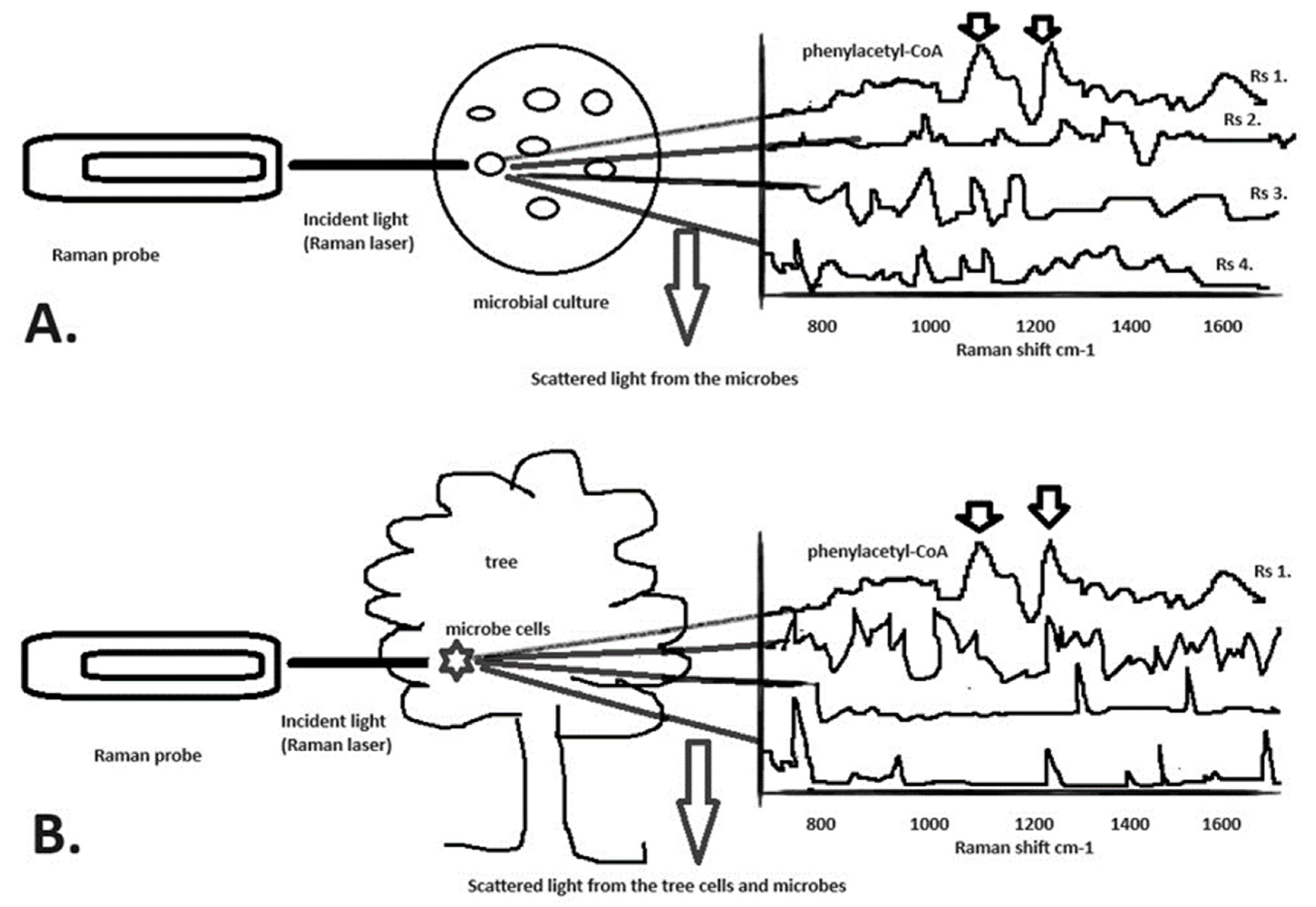
10. Raman Microspectroscopy in Plant Disease Diagnosis

Raman microspectroscopy has emerged as a powerful tool in the field of plant disease diagnosis due to its ability to provide detailed molecular information without the need for extensive sample preparation (Figure 3). By utilizing this technique, researchers can identify specific biomarkers associated with various diseases in plants (Figure 3b–d). For instance, changes in the spectral profile of plant tissue can reveal alterations in cell composition, allowing for early detection of pathogens or physiological disorders. Additionally, Raman spectroscopy can distinguish between different plant species and specific strains of diseases, providing valuable information for targeted treatment strategies.
Raman spectra indicated the presence of lycopene in tomato fruit at the ripe stage with bands, as a peak, at 1156 and 1510 cm−1 and the presence of β-carotene as a strong Raman peak in the spectral regions at 1100 and 1200 cm−1, correlated with the C-C bonds [39]. When tomato fruits which were infected with Alernaria alternata and its toxins, such as tenuazonic acid, alternariol and alternariol monomethyl, the lycopene, β-carotene, ascorbic acid and phenolic contents were found to be lowest in the pathogen-infected fruits, concluding that A. alternata affects the nutritional value of tomato fruits the most, alone or due to a combined effect with the toxins [69,70]. Based on the above-mentioned results, we can conclude that the study of the presence of lycopene and β-carotene in tomato fruit using Raman spectra exhibits strong evidence of tomato health or infection being affected by the pathogen A. alternata.
In Figure 3, we present plant disease progress flow graphs that illustrate the progression of plant infection and the establishment of fungus in plant tissue (Figure 3a). Among various plant pathology techniques (Figure 3b), Raman spectroscopy can be utilized (Figure 3c) for fast and reliable identification of the infection of the fungus A. alternata (Figure 3d).
It is well known that in plants, chlorophyll a exhibits strong fluorescence, particularly under visible light excitation, which can interfere with Raman signal detection. Researchers [71] have demonstrated that the chlorophyll a molecule contains an electron donor group (carbonyl at the g-position, abbreviated as “C9=O”) with electronic absorption at 660–665 nm. Additionally, the same authors [71] found that when measuring the Raman spectra of typical chlorophyll forms, the Raman lines in the low-frequency region (below 300 cm−1) were sensitive to these forms of chlorophyll. This is an important consideration for our review article when using Raman spectroscopy.
Furthermore, many microorganisms contain carotenoids that fluoresce, giving them their bright colors. These pigments can produce strong fluorescence when they are exposed to Raman laser excitation. Raman spectroscopy has been successfully used to detect β-carotene in a wide range of microorganisms, as well as bacterioruberin as a primary carotenoid in halophilic archaea. Another article presented data from Raman spectroscopy for carotenoid detection in microorganisms, with implications for astrobiology researchers, demonstrating that the Raman spectra of various carotenoids from representative species of archaea, bacteria, cyanobacteria and algae, when compared with HPLC extracts, showed that carotenoids can be easily identified using the Raman method. The researchers concluded that using 514 and 532 nm lasers provided excellent results due to the advantage of Raman resonance enhancement.
Furthermore, the accumulation of phenolic compounds was significantly increased in all walnut genotypes after inoculation with the bacterium Xanthomonas arboricola pv. juglandis, the causal pathogen of walnut blight, suggesting that these phenolic compounds contribute to early defense against walnut blight [63]. We believe that further estimations can be made related to these crucial points using Raman microspectroscopy: (i) the health of plant tissue and (ii) identifying through particular molecular characterization early walnut blight disease as caused by X. arboricola pv. juglandis.
We propose using Raman detection to identify specific plant molecules such as carotenoids or antioxidant enzymes like catalase or peroxidase that are linked to plant responses to pathogens, such as X. arboricola pv. juglandis [63]. By using Raman microspectroscopy to observe these biochemical reactions, we believe researchers can assess the early stages of the disease by detecting the accumulation of these compounds in the infected plant tissue. Our goal is to identify the early stages of walnut blight disease using Raman microspectroscopy, even in particularly damaging wet years or in early-leafing walnut cultivars. Further, by using Raman spectra, it is possible to analyze plant tissues in real time to detect early plant diseases.

11. An Example Based on the Raman Microscopy Techniques Proposed in this Article

Phenylacetyl-coenzyme A (CoA) is a key intermediate in the aerobic catabolism of phenylacetate in microbes such as Pseudomonas when cultured in minimal media using phenylacetate as the sole carbon source. Using Raman spectroscopy, as Figure 4 shows, the spectrum of phenylacetyl-CoA was detected at 1108 cm−1 and 1245 cm−1 and further was detected in the tree before the microbe tree pathogen (Pseudomonas) produced any visible symptoms.

12. Conclusions

Raman microscopy-based systems have emerged as powerful tools for plant disease diagnosis in recent years [72,73,74]. This technique utilizes the scattering of laser light to provide detailed molecular information about a sample, allowing for the identification of subtle biochemical changes associated with diseases in plants. By analyzing the vibrational modes of the molecules in a sample, Raman microscopy-based systems can detect alterations in cell structure, composition and metabolism that are indicative of disease presence. This non-invasive and label-free approach offers significant advantages over traditional methods, such as histological staining or genetic markers, by providing rapid and accurate results without the need for complex sample preparation. Furthermore, Raman microscopy-based systems offer the potential for early disease detection [67], aiding in the timely implementation of disease management strategies to minimize crop losses and enhance agricultural productivity.
In the context of Raman microspectroscopy for plant disease diagnosis, understanding the principle of this technique is paramount to its successful application. Raman microspectroscopy leverages the phenomenon of the inelastic scattering of photons to provide valuable molecular information about the sample under study. This technique allows for the identification and characterization of the chemical composition within the plant tissues with high spatial resolution and specificity. Moreover, Raman microscopy-based systems can enhance the precision and efficiency of plant disease diagnosis, ultimately contributing to significant advancements in agricultural sciences.
Raman microspectroscopy emerges as a powerful tool for the early and accurate diagnosis of plant diseases [67,68,71,72,73,74,75]. Our article has demonstrated the ability of this technique to distinguish between healthy and infected plant tissues with high sensitivity and specificity. By analyzing the spectral signatures of various plant pathogens, Raman microscopy-based systems offer a non-destructive and rapid method for identifying diseases such as those caused by fungi and bacteria. Moreover, the technique’s ability to provide information on the biochemical changes occurring in plants during infection enables researchers to gain a deeper understanding of the mechanisms underlying pathogenesis. Raman microscopy-based systems hold great potential for revolutionizing the field of plant pathology by facilitating timely and targeted disease management strategies. Future research efforts should focus on expanding the application of this technique to a broader range of plant species and diseases, ultimately enhancing crop productivity and food security.
In conclusion, Raman microscopy shows great promise as a cutting-edge tool for plant disease diagnosis. Its non-invasive nature, rapid analysis capabilities and high sensitivity make it an asset for ensuring the health and productivity of plants and crops. As research in this field continues to advance, we can expect to see further developments in Raman microscopy-based systems for plant disease diagnosis, ultimately leading to more effective disease management strategies in agriculture.

Author Contributions

Conceptualization, I.V., I.M. and T.S.; methodology, I.V.; software, I.V. and T.S.; validation, I.M. and I.V.; formal analysis, I.V.; investigation, I.M., I.V. and T.S.; resources, I.V.; data curation, T.S. and I.V.; writing—original draft preparation, I.V. and T.S.; writing—review and editing, I.V., T.S. and I.M.; visualization, I.V.; supervision, I.M., I.V. and T.S.; project administration, I.V.; funding acquisition, I.V. All authors have read and agreed to the published version of the manuscript.

Funding

This work received no external funding.

Institutional Review Board Statement

Not applicable.

Informed Consent Statement

Not applicable.

Data Availability Statement

Not applicable.

Conflicts of Interest

The authors declare no conflicts of interest.

References

  1. Venbrux, M.; Crauwels, S.; Rediers, H. Current and emerging trends in techniques for plant pathogen detection. Front. Plant Sci. 2023, 14, 1120968. [Google Scholar] [CrossRef]
  2. Vagelas, I.; Papadimos, A.; Lykas, C. Pre-symptomatic disease detection in the vine, chrysanthemum, and rose leaves with a low-cost infrared sensor. Agronomy 2021, 11, 1682. [Google Scholar] [CrossRef]
  3. Sinclair, J.B. Latent infection of soybean plants and seeds by fungi. Plant Dis. 1991, 75, 220–224. [Google Scholar] [CrossRef]
  4. Tongsri, V.; Songkumarn, P.; Sangchote, S. Leaf spot characteristics of Phomopsis durionis on durian (Durio zibethinus Murray) and latent infection of the pathogen. Acta Univ. Agric. Silvic. Mendel. Brun. 2016, 64, 185–193. [Google Scholar] [CrossRef]
  5. Aylward, J.; Steenkamp, E.T.; Dreyer, L.L.; Roets, F.; Wingfield, B.D.; Wingfield, M.J. A plant pathology perspective of fungal genome sequencing. IMA Fungus 2017, 8, 1–15. [Google Scholar] [CrossRef] [PubMed]
  6. Baltrus, D.A.; McCann, H.C.; Guttman, D.S. Evolution, genomics and epidemiology of Pseudomonas syringae: Challenges in bacterial molecular plant pathology. Mol. Plant Pathol. 2017, 18, 152–168. [Google Scholar] [CrossRef] [PubMed]
  7. Moreno-Pérez, A.; Pintado, A.; Murillo, J.; Caballo-Ponce, E.; Tegli, S.; Moretti, C.; Rodríguez-Palenzuela, P.; Ramos, C. Host range determinants of Pseudomonas savastanoi pathovars of woody hosts revealed by comparative genomics and cross-pathogenicity tests. Front. Plant Sci. 2020, 11, 973. [Google Scholar] [CrossRef] [PubMed]
  8. Aragona, M.; Haegi, A.; Valente, M.T.; Riccioni, L.; Orzali, L.; Vitale, S.; Luongo, L.; Infantino, A. New-generation sequencing technology in diagnosis of fungal plant pathogens: A dream comes true? J. Fungi 2022, 8, 737. [Google Scholar] [CrossRef] [PubMed]
  9. Panchal, P.; Raman, V.C.; Mantri, S. Plant diseases detection and classification using machine learning models. In Proceedings of the 4th International Conference on Computational Systems and Information Technology for Sustainable Solution (CSITSS), Bengaluru, India, 20–21 December 2019; Volume 4, pp. 1–6. [Google Scholar] [CrossRef]
  10. Nagaraju, M.; Chawla, P. Systematic review of deep learning techniques in plant disease detection. Int. J. Syst. Assur. Eng. Manag. 2020, 11, 547–560. [Google Scholar] [CrossRef]
  11. Sarkar, C.; Gupta, D.; Gupta, U.; Hazarika, B.B. Leaf disease detection using machine learning and deep learning: Review and challenges. Appl. Soft Comput. 2023, 145, 110534. [Google Scholar] [CrossRef]
  12. Shah, S.K.; Kumbhar, V.; Singh, T.P. A Systematic Review on Crop Leaf Disease Identification Using Machine Learning and Deep Learning Techniques. In Proceedings of the 7th International Conference On Computing, Communication, Control and Automation (ICCUBEA), Pune, India, 18–19 August 2023; pp. 1–7. [Google Scholar] [CrossRef]
  13. Omran, E.E. Early sensing of peanut leaf spot using spectroscopy and thermal imaging. Arch. Agron. Soil Sci. 2017, 63, 883–896. [Google Scholar] [CrossRef]
  14. Conrad, A.O.; Li, W.; Lee, D.; Wang, G.; Rodriguez-Saona, L.E.; Bonello, P. Machine learning-based presymptomatic detection of rice sheath blight using spectral profiles. Plant Phenom. 2020, 2020, 8954085. [Google Scholar] [CrossRef] [PubMed]
  15. Lucas, J.A. Plant Pathology and Plant Pathogens, 4th ed.; Willey-Blackwell: Chichester, UK, 2020; p. 432. [Google Scholar]
  16. Agrios, G.N. Plant Pathology, 5th ed.; Elsevier Academic Press: Amsterdam, The Netherlands, 2005; p. 952. [Google Scholar]
  17. Pathak, R.K.; Kumar Singh, S.; Tak, A.; Gehlot, P.S. Impact of climate change on host, pathogen and plant disease adaptation regime: A review. Biosci. Biotechnol. Res. Asia 2018, 15, 529–540. [Google Scholar] [CrossRef]
  18. Liaqat, W.; Barutçular, C.; Farooq, M.U.; Ahmad, H.; Jan, M.F.; Ahmad, Z.; Nawaz, H.; Li, M. Climate change in relation to agriculture: A review. Span. J. Agric. Res. 2022, 20, e03R01. [Google Scholar] [CrossRef]
  19. Henson, J.M.; French, R.C. The polymerase chain reaction and plant disease diagnosis. Annu. Rev. Phytopathol. 1993, 31, 81–109. [Google Scholar] [CrossRef] [PubMed]
  20. Balodi, R.; Bisht, S.; Ghatak, A.; Rao, K.H. Plant disease diagnosis: Technological advancements and challenges. Indian Phytopathol. 2017, 70, 275–281. [Google Scholar] [CrossRef]
  21. Yang, Y.; Zhou, Q.; Zahr, K.; Harding, M.W.; Feindel, D.; Feng, J. Impact of DNA extraction efficiency on the sensitivity of PCR-based plant disease diagnosis and pathogen quantification. Europ. J. Plant Pathol. 2021, 159, 583–591. [Google Scholar] [CrossRef]
  22. Buja, I.; Sabella, E.; Monteduro, A.G.; Chiriacò, M.S.; Bellis, L.D.; Luvisi, A.; Maruccio, G. Advances in plant disease detection and monitoring: From traditional assays to in-field diagnostics. Sensors 2021, 21, 2129. [Google Scholar] [CrossRef]
  23. Martins, L.; Fernandes, C.; Albuquerque, P.M.; Tavares, F. Assessment of Xanthomonas arboricola pv. juglandis bacterial load in infected walnut fruits by quantitative PCR. Plant Dis. 2019, 103, 2577–2586. [Google Scholar] [CrossRef]
  24. Scortichini, M.; Marchesi, U.; Prospero, P.D. Genetic diversity of Xanthomonas arboricola pv. juglandis (synonyms: X. campestris pv. juglandis; X. juglandis pv. juglandis) strains from different geographical areas shown by repetitive polymerase chain reaction genomic fingerprinting. J. Phytopathol. 2001, 149, 325–332. [Google Scholar] [CrossRef]
  25. Moragrega, C.; Llorente, I. Effects of leaf wetness duration, temperature, and host phenological stage on infection of walnut by Xanthomonas arboricola pv. juglandis. Plants 2023, 12, 2800. [Google Scholar] [CrossRef] [PubMed]
  26. Manthos, I.; Sotiropoulos, T.; Vagelas, I. Is the artificial pollination of walnut trees with drones able to minimize the presence of Xanthomonas arboricola pv. juglandis? A Review. Appl. Sci. 2024, 14, 2732. [Google Scholar] [CrossRef]
  27. John, M.A.; Bankole, I.; Ajayi-Moses, O.; Ijila, T.; Jeje, O.; Lalit, P. Relevance of advanced plant disease detection techniques in disease and pest management for ensuring food security and their implication: A Review. Am. J. Plant Sci. 2023, 14, 1260–1295. [Google Scholar] [CrossRef]
  28. Jafar, A.; Bibi, N.; Naqvi, R.A.; Sadeghi-Niaraki, A.; Jeong, D. Revolutionizing agriculture with artificial intelligence: Plant disease detection methods, applications, and their limitations. Front. Plant Sci. 2024, 15, 1356260. [Google Scholar] [CrossRef] [PubMed]
  29. Sahoo, L.; Mohapatra, D.; Raghuvanshi, H.R.; Kumar, S.; Kaur, R.; Chawla, R.; Afreen, N. Transforming agriculture through artificial intelligence: Advancements in plant disease detection, applications and challenges. J. Adv. Biol. Biotechnol. 2024, 27, 381–388. [Google Scholar] [CrossRef]
  30. Mena, E.; Garaycochea, S.; Stewart, S.; Montesano, M.; Ponce de León, I. Comparative genomics of plant pathogenic Diaporthe species and transcriptomics of Diaporthe caulivora during host infection reveal insights into pathogenic strategies of the genus. BMC Genom. 2022, 23, 175. [Google Scholar] [CrossRef]
  31. Das, S.; Pattanayak, S.; Behera, P.R. Application of machine learning: A recent advancement in plant diseases detection. J. Plant Prot. Res. 2023, 62, 122–135. [Google Scholar] [CrossRef]
  32. Précigout, P.; Claessen, D.; Makowski, D.; Robert, C. Does the latent period of leaf fungal pathogens reflect their trophic type? A meta-analysis of biotrophs, hemibiotrophs and necrotrophs. Phytopathology 2020, 110, 345–361. [Google Scholar] [CrossRef]
  33. Routis, G.; Michailidis, M.; Roussaki, I. Plant disease identification using machine learning algorithms on single-board computers in IoT environments. Electronics 2024, 13, 1010. [Google Scholar] [CrossRef]
  34. Takahashi, H.; Fukuhara, T.; Kitazawa, H.; Kormelink, R. Virus latency and the impact on plants. Front. Microbiol. 2019, 10, 2764. [Google Scholar] [CrossRef]
  35. Jeger, M.J.; Pautasso, M.; Holdenrieder, O.; Shaw, M.W. Modelling disease spread and control in networks: Implications for plant sciences. New Phytol. 2007, 174, 279–297. [Google Scholar] [CrossRef] [PubMed]
  36. González-Domínguez, E.; Caffi, T.; Rossi, V.; Salotti, I.; Fedele, G. Plant disease models and forecasting: Changes in principles and applications over the last 50 years. Phytopathology 2023, 113, 678–693. [Google Scholar] [CrossRef] [PubMed]
  37. Pezzotti, G. Raman spectroscopy in cell biology and microbiology. J. Raman Spectrosc. 2021, 52, 2348–2443. [Google Scholar] [CrossRef]
  38. Matthäus, C.; Bird, B.; Miljković, M.; Chernenko, T.; Romeo, M.J.; Diem, M. Chapter 10: Infrared and Raman microscopy in cell biology. Methods Cell Biol. 2008, 89, 275–308. [Google Scholar] [CrossRef] [PubMed]
  39. Radić, D. Characterization of Microorganisms Using Raman Microscopy. In Application of Molecular Methods and Raman Microscopy/Spectroscopy in Agricultural Sciences and Food Technology; Vucelić Radović, B., Lazić, D., Nikšić, M., Eds.; Ubiquity Press: London, UK, 2019; pp. 161–165. [Google Scholar] [CrossRef]
  40. Shigeto, S.; Takeshita, N. Raman microspectroscopy and imaging of filamentous fungi. Microbes Environ. 2022, 37, ME22006. [Google Scholar] [CrossRef] [PubMed]
  41. Cui, S.; Zhang, S.; Yue, S. Raman spectroscopy and imaging for cancer diagnosis. J. Healthc. Eng. 2018, 2018, 8619342. [Google Scholar] [CrossRef]
  42. Hanna, K.; Krzoska, E.; Shaaban, A.M.; Muirhead, D.K.; Abu-Eid, R.; Speirs, V. Raman spectroscopy: Current applications in breast cancer diagnosis, challenges and future prospects. Br. J. Cancer 2022, 126, 1125–1139. [Google Scholar] [CrossRef] [PubMed]
  43. Zhang, B.; Zhang, Z.; Gao, B.; Zhang, F.; Tian, L.; Zeng, H.; Wang, S. Raman microspectroscopy based TNM staging and grading of breast cancer. Spectrochim. Acta A Mol. Biomol. Spectrosc. 2023, 285, 121937. [Google Scholar] [CrossRef]
  44. Vriens, L. Raman scattering cross sections for In and T1 atoms and multiphoton processes in Sr. Opt. Commun. 1974, 11, 396–401. [Google Scholar] [CrossRef]
  45. Langer, J.; Aberasturi, D.J.; Aizpurua, J.; Álvarez-Puebla, R.A.; Auguié, B.; Auguié, B.; Baumberg, J.J.; Bazan, G.C.; Bell, S.E.; Boisen, A.; et al. Present and future of surface-enhanced Raman scattering. ACS Nano 2020, 14, 28–117. [Google Scholar] [CrossRef]
  46. Dodo, K.; Fujita, K.; Sodeoka, M. Raman spectroscopy for chemical biology research. J. Am. Chem. Soc. 2022, 144, 19651–19667. [Google Scholar] [CrossRef]
  47. Alberts, B.; Johnson, A.; Lewis, J.; Raff, M.; Roberts, K.; Walter, P. Visualizing Cells. In Molecular Biology of the Cell, 4th ed.; Garland Science: New York, NY, USA, 2002. Available online: https://www.ncbi.nlm.nih.gov/books/NBK21048/ (accessed on 1 June 2024).
  48. Cuny, A.P.; Schlottmann, F.P.; Ewald, J.C.; Pelet, S.; Schmoller, K.M. Live cell microscopy: From image to insight. Biophys. Rev. 2022, 3, 021302. [Google Scholar] [CrossRef] [PubMed]
  49. Balasubramanian, H.; Hobson, C.M.; Chew, T.; Aaron, J.S. Imagining the future of optical microscopy: Everything, everywhere, all at once. Commun. Biol. 2023, 6, 1096. [Google Scholar] [CrossRef] [PubMed]
  50. Gierlinger, N.; Keplinger, T.; Harrington, M. Imaging of plant cell walls by confocal Raman microscopy. Nat. Protoc. 2012, 7, 1694–1708. [Google Scholar] [CrossRef] [PubMed]
  51. Lević, S. Materials Characterization by Raman Microscopy. In Application of Molecular Methods and Raman Microscopy/Spectroscopy in Agricultural Sciences and Food Technology; Vucelić Radović, B., Lazić, D., Nikšić, M., Eds.; Ubiquity Press: London, UK, 2019. [Google Scholar] [CrossRef]
  52. Mateu, B.P.; Bock, P.; Gierlinger, N. Raman imaging of plant cell walls. Methods Mol. Biol. 2020, 2149, 251–295. [Google Scholar] [CrossRef] [PubMed]
  53. Nitta, N.; Iino, T.; Isozaki, A.; Yamagishi, M.; Kitahama, Y.; Sakuma, S.; Suzuki, Y.; Tezuka, H.; Oikawa, M.; Arai, F.; et al. Raman image-activated cell sorting. Nat. Commun. 2020, 11, 3452. [Google Scholar] [CrossRef] [PubMed]
  54. Derely, L.; Collart Dutilleul, P.; Michotte de Welle, S.; Szabo, V.; Gergely, C.; Cuisinier, F. Raman confocal microscopy and AFM combined studies of cancerous cells treated with Paclitaxel. In Proceedings of the Nanoscale Imaging, Sensing, and Actuation for Biomedical Applications, Event: SPIE BiOS, San Francisco, CA, USA, 22–27 January 2011; Volume 7908. [Google Scholar] [CrossRef]
  55. Yan, S.; Cui, S.; Ke, K.; Zhao, B.; Liu, X.; Yue, S.; Wang, P. Hyperspectral stimulated Raman scattering microscopy unravels aberrant accumulation of saturated fat in human liver cancer. Anal. Chem. 2018, 90, 6362–6366. [Google Scholar] [CrossRef]
  56. Ramesh, G.; Paul, W.; Valparambil Puthanveetil, V.; Raja, K.; Embekkat Kaviyil, J. Raman spectroscopy as a novel technique for the identification of pathogens in a clinical microbiology laboratory. Spectrosc. Lett. 2022, 55, 546–551. [Google Scholar] [CrossRef]
  57. Dinçtürk, E. Determination of Raman spectrum under different culture conditions: Preliminary research on bacterial fish pathogens. Anim. Biotechnol. 2024, 35, 2299733. [Google Scholar] [CrossRef]
  58. Wang, L.; Liu, W.W.; Tang, J.; Wang, J.; Liu, Q.; Wen, P.; Wang, M.; Pan, Y.; Gu, B.; Zhang, X. Applications of Raman spectroscopy in bacterial infections: Principles, advantages, and shortcomings. Front. Microbiol. 2021, 12, 683580. [Google Scholar] [CrossRef]
  59. Rebrošová, K.; Samek, O.; Kizovský, M.; Bernatová, S.; Holá, V.; Růžička, F. Raman spectroscopy—A novel method for identification and characterization of microbes on a single-cell level in clinical settings. Front. Cell. Infect. Microbiol. 2022, 12, 866463. [Google Scholar] [CrossRef]
  60. Wang, Y.; Song, Y.; Zhu, D.; Ji, Y.; Wang, T.; McIlvenna, D.; Yin, H.; Xu, J.; Huang, W.E. Probing and sorting single cells—The application of a Raman-activated cell sorter. Spectrosc. Eur. 2013, 25, 16–20. [Google Scholar]
  61. Yuan, Y.; Wang, D.; Zhang, J.; Liu, J.; Chen, J.; Zhang, X. Raman spectra of the GFP-like fluorescent proteins. Biophys. Rep. 2018, 4, 265–272. [Google Scholar] [CrossRef]
  62. Auner, G.W.; Koya, S.K.; Huang, C.; Broadbent, B.; Trexler, M.; Auner, Z.; Elias, A.; Mehne, K.C.; Brusatori, M.A. Applications of Raman spectroscopy in cancer diagnosis. Cancer Metastasis Rev. 2018, 37, 691–717. [Google Scholar] [CrossRef] [PubMed]
  63. Zhang, Q.; Li, M.; Yang, G.; Liu, X.; Yu, Z.; Peng, S. Protocatechuic acid, ferulic acid and relevant defense enzymes correlate closely with walnut resistance to Xanthomonas arboricola pv. juglandis. BMC Plant Biol. 2022, 22, 598. [Google Scholar] [CrossRef] [PubMed]
  64. Song, D.; Chen, Y.; Li, J.; Wang, H.; Ning, T.; Wang, S. A graphical user interface (NWUSA) for Raman spectral processing, analysis and feature recognition. J. Biophotonisc. 2021, 14, e202000456. [Google Scholar] [CrossRef]
  65. Vagelas, I.; Leontopoulos, S. A bibliometric analysis and a citation mapping process for the role of soil recycled organic matter and microbe interaction due to climate change using Scopus database. AgriEngineering 2023, 5, 581–610. [Google Scholar] [CrossRef]
  66. Lykas, C.; Vagelas, I. Innovations in agriculture for sustainable agro-systems. Agronomy 2023, 13, 2309. [Google Scholar] [CrossRef]
  67. Sánchez, L.R.; Pant, S.R.; Mandadi, K.K.; Kurouski, D. Raman spectroscopy vs quantitative polymerase chain reaction in early stage Huanglongbing diagnostics. Sci. Rep. 2020, 10, 10101. [Google Scholar] [CrossRef]
  68. Sánchez, L.R.; Pant, S.R.; Xing, Z.; Mandadi, K.K.; Kurouski, D. Rapid and noninvasive diagnostics of Huanglongbing and nutrient deficits on citrus trees with a handheld Raman spectrometer. Anal. Bioanal. Chem. 2019, 411, 3125–3133. [Google Scholar] [CrossRef] [PubMed]
  69. Meena, M.K.; Zehra, A.; Swapnil, P.; Dubey, M.K.; Patel, C.B.; Upadhyay, R.S. Effect on lycopene, β-carotene, ascorbic acid and phenolic content in tomato fruits infected by Alternaria alternata and its toxins (TeA, AOH and AME). Arch. Phytopathol. Plant Protect. 2017, 50, 317–329. [Google Scholar] [CrossRef]
  70. Jiang, S.; Han, S.; He, D.; Cao, G.; Fang, K.; Xiao, X.; Yi, J.; Wan, X. The accumulation of phenolic compounds and increased activities of related enzymes contribute to early defense against walnut blight. Physiol. Mol. Plant Pathol. 2019, 108, 101433. [Google Scholar] [CrossRef]
  71. Koyama, Y.; Umemoto, Y.; Akamatsu, A.; Uehara, K.; Tanaka, M. Raman spectra of chlorophyll forms. J. Mol. Struct. 1986, 146, 273–287. [Google Scholar] [CrossRef]
  72. Lin, Y.; Lin, H.; Lin, Y. Construction of Raman spectroscopic fingerprints for the detection of Fusarium wilt of banana in Taiwan. PLoS ONE 2020, 15, e0230330. [Google Scholar] [CrossRef] [PubMed]
  73. Payne, W.Z.; Kurouski, D. Raman-Based Diagnostics of Biotic and Abiotic Stresses in Plants. A Review. Front. Plant Sci. 2021, 11, 616672. [Google Scholar] [CrossRef] [PubMed]
  74. Parlamas, S.; Goetze, P.K.; Humpal, D.M.; Kurouski, D.; Jo, Y. Raman Spectroscopy Enables Confirmatory Diagnostics of Fusarium Wilt in Asymptomatic Banana. Front. Plant Sci. 2022, 13, 922254. [Google Scholar] [CrossRef]
  75. Jehlička, J.; Edwards, H.G.; Osterrothová, K.; Novotná, J.; Nedbalová, L.; Kopecký, J.; Němec, I.; Oren, A. Potential and limits of Raman spectroscopy for carotenoid detection in microorganisms: Implications for astrobiology. Philos. Trans. R. Soc. A Math. Phys. Eng. Sci. 2014, 372, 20140199. [Google Scholar] [CrossRef]
Figure 1. Co-keyword network visualization based on “Raman” AND “microscopy” AND “diagnosis”.
Applsci 14 05926 g001
Figure 2. Co-keyword network visualization based on “Raman” AND “diagnosis” AND “bacteria”.
Applsci 14 05926 g002
Figure 3. Flow graphs showing the plant infection and the establishment of fungi (plant disease progress). (a) Methods of plant disease identification; (b) Raman spectroscopy for plant disease diagnosis; (c) Raman spectroscopy for plant disease diagnosis; (d) understanding mechanisms of pathogenesis. (a) aims to explain the plant disease progress as follows: 1. Fungus conidia germination; 2. First step of plant tissue infection; 3. Conidia hyphae invading the plant tissue and multiplying; 4. Initial infection is limited in the plant without visual symptoms; 5. Fungus mycelial network growing between plant cells; 6. Visual disease symptoms observed on leaf tissue; 7. Fungus sporulation occurring across the damaged leaf tissue. (b) aims to explain reliable diagnostic tools for the detection of plant diseases: microscope (A1), PCR (B1), ELISA (B2), fluorescence microscopy (B3), Raman spectroscopy (C1). (c) aims to explain Raman (micro)spectroscopy as a tool for identifying microbes with a Raman spectrum. Moreover, (d) shows a Raman chemical fingerprint that identifies molecules of the invading fungus. Figure 3d displays the benefits of Raman (micro)spectroscopy, using a chemical Raman fingerprint to identify molecules of the invading fungus, such as the species Alternaria alternata. All scientific figures and icons were created using BioRender scientific software (Version 04) (https://www.biorender.com/).
Applsci 14 05926 g003
Figure 4. Illustration of microbe detection with Raman spectra. In laboratory tests (A), a probing laser is focused on the Petri dish sample (microbes), and a small amount of light, which transports the chemical structure of analyzed microbes, is reflected as individual Raman shift lines at 800 to 1600 cm−1. The final graph shows four Raman spectra (Rs 1., Rs 2., Rs 3. and Rs 4). The Raman spectra show information about the molecular bond vibrations of a given microbe. The Raman spectrum Rs 1. shows the phenylacetyl-CoA spectrum (arrows) with a peak at 1108 cm−1 and 1245 cm−1. This spectrum Rs 1. at 1108 cm−1 and 1245 cm−1 was detected when the Raman probing laser was focused on the tree sample (B). Microbe identification was detected at the single-cell (-s) level before the microbe caused visible symptoms in plant tissue, concluding with early microbe detection.
Applsci 14 05926 g004
Disclaimer/Publisher’s Note: The statements, opinions and data contained in all publications are solely those of the individual author(s) and contributor(s) and not of MDPI and/or the editor(s). MDPI and/or the editor(s) disclaim responsibility for any injury to people or property resulting from any ideas, methods, instructions or products referred to in the content.

