Review

From the Environment to Molecular Interactions of Nanoplastics: Unraveling the Neurotoxic Impacts and the Implications in Neurodegenerative Processes

by
Chiara Urani
1,*,†,
Raffaella Barbieri
2,
Susanna Alloisio
2,3,† and
Marina Tesauro
4,‡
1
Department of Earth and Environmental Sciences, University of Milano-Bicocca, Piazza della Scienza, 1, 20126 Milan, Italy
2
Institute of Biophysics, National Research Council, Via De Marini 6, 16149 Genoa, Italy
3
ETT S.p.A., Via Albareto 21, 16154 Genova, Italy
4
Department of Biomedical, Surgical and Dental Sciences, University of Milan, Via Carlo Pascal 36, 20133 Milan, Italy
*
Author to whom correspondence should be addressed.
Integrated Models for Prevention and Protection in Environmental and Occupational Health, (MISTRAL) Interuniversity Research Center, 25121 Brescia, Italy.
Coordinated Research Center “Epidemiology and Molecular surveillance of Infections (EpiSoMI)”, University of Milan, 20122 Milano, Italy.
Appl. Sci. 2024, 14(16), 7280; https://doi.org/10.3390/app14167280
Submission received: 20 July 2024 / Revised: 7 August 2024 / Accepted: 9 August 2024 / Published: 19 August 2024

Abstract

:
Nanoplastics (NPs) represent an escalating hazard to both humans and the ecosystem due to their pervasive presence. This review delves into (i) the widespread occurrence of NPs across the different environmental matrices, including food; (ii) routes and estimates for human exposure; (iii) the mechanisms of blood–brain barrier (BBB) crossing; and (iv) implications for human health, with a specific focus on molecular features associated with neurotoxicity and neurodegenerative processes. The impact of NPs on the central nervous system, their ability to cross the BBB and the underpinning mechanisms, the potential to initiate neurotoxicity by fostering β-amyloid aggregation, and their interactions with metallo-enzymes (such as superoxide dismutase) are elucidated. The analysis of transcriptomics and epigenomic results, including microRNA dysregulation, unveil how NPs could contribute to neurological disorders. The need for considering overlaps among diverse pathogenetic mechanisms when probing the effects of NPs is discussed. Additional urgent needs are the development of reliable in vitro models for neurotoxicity studies able to mimic the complexity of the nervous system and the exposure of such models to more environmentally relevant NPs. Finally, the development of extremely sensitive detection and analysis methodologies to quantify NPs in environmental and biological matrices is a pressing priority.

1. Introduction and Focus of the Review: From the Environment to Molecular Interactions of Plastic Particles

Plastic has been a fundamental and omnipresent material, providing numerous socio-economic benefits due to its multifaceted attributes. However, worldwide plastic production has surged by nearly 200-fold, from 2 million tons in 1950 to nearly 390 million tons in 2021, enforced by economic and population growth, particularly in emerging regions such as China, the Middle East, and Africa (PLASTICS—THE FACTS, 2022). Consequently, plastic pollution is of central concern on the global environmental agenda, prompting European and international agreements and regulations aimed at curbing plastic usage and bolstering recycling efforts [1].
For instance, the European Chemical Agency (ECHA) recently unveiled a timetable for restricting intentionally added microplastics, effective from September 2023, aimed at curtailing intentional release [2]. However, while these regulatory interventions are imperative, they fail to address the issue of existing plastic particles permeating all environmental matrices. Noteworthy, plastic particles in the environment undergo mechanical and chemical–physical degradation, and for certain types of polymers, biodegradation. These phenomena cause increasing fragmentation, leading to the formation of smaller plastic particles, from macro to micro and nano sizes [3]. In this review, recent knowledge on the diffusion of nanoplastic particles in different matrices is firstly critically examined, although literature papers and reviews are mainly available on microplastics (MPs) rather than on nanoplastics (NPs). This mainly appears to be due to instrumental and technological limitations for detection and analyses of NPs in different environmental and biological matrices.
The aim is to give an in-depth and critical analysis on NPs’ neurotoxic effects and implications for neurodegenerative disorders at the molecular level, merging different fields of research to achieve a global comprehension. The review’s starting point is from a macroscopic perspective encompassing the occurrence of NPs and, unavoidably, of MPs into various environmental matrices, progressing to the exposure pathways of NPs and consequent molecular effects impacting human health.
In particular, the primary focus lies on an overlooked aspect: the capability to cross barriers, mostly the blood–brain barrier; the accumulation into brain target cells; and the interaction at the molecular level with biological structures and functions leading to impairment of neuronal features.
Major gaps are identified regarding the mechanisms of blood–brain barrier (BBB) crossing; the molecular mechanisms and interactions of NPs with key enzymes and proteins involved in neurodegenerative processes; as well as the dysregulation of pathways, genes, and epigenetic factors by these smaller plastic particles.

2. Definition and Characterization of Plastics

Color, flexibility, lightness, and durability are among the primary characteristics that drive the utilization of plastic polymers. Various categories of plastics, as classified by the Society of the Plastics Industry, e.g., polyethylene terephthalate (PET), high-density polyethylene (HDPE), polyvinyl chloride (PVC), low-density polyethylene (LDPE), polypropylene (PP), polystyrene (PS), and polycarbonate (PC), along with polylactide and nylon, are available. These particles comprise solid plastic composed of polymer blends and functional additives, potentially containing residual impurities [4].
As per the International Organization for Standardization, nanomaterials are defined as substances with dimensions in the nanoscale or possessing internal or surface structures within the nanoscale range [5]. The term “nanoscale” refers to sizes ranging approximately from 1 nm to 100 nm. Recently, a revised definition has been proposed, describing NPs as particles ranging from 1 to 1000 nm and originating from mechanical–physical–chemical fragmentation of industrial plastic items [6]. In this review, we regard MPs as plastic particles smaller than 5 mm and NPs as even smaller particles ranging from 1 to 100 nanometers, as defined by the European Food Safety Authority [7]. However, the precise definition of these plastic materials remains subject to debate. Therefore, a formal reassessment by competent authorities, such as the International Organization for Standardization, is advisable in the near future, considering that the varied sizes of plastic particles and their consequent area-to-mass ratios confer varying chemical and physical properties which can have divergent impacts on biological contact and chemical adsorption. In addition, another relevant feature of NPs to be considered in view of their potential toxicity is represented by their physical and chemical properties and the role of the corona [8]. High surface area and surface energies along with the propensity of NPs to interact with molecules, proteins, and biological probes could, in fact, invalidate laboratory analyses and give artefactual results [9]. In addition, the analysis of NPs in complex mixtures such as sea spray is even more complex because of the ionization and speciation of the aerosol matrix [10].

3. Presence of Micro- and Nanoplastics in Environmental Matrices and Food

In recent years, research has evidenced that plastic particles are omnipresent, and consequently, they are discovered in nearly all environmental matrices upon investigation. The abundance of plastic materials is so significant that the scientific community has coined the term “Plasticene” to define the current historical era [11].
Recent reports estimate that, worldwide, 13 million tons of plastic waste (fossil-based, recycled, and bio-based) enter aquatic ecosystems, such as rivers and oceans [12].
Water, air, soil, and food are environmental determinants of human health to which an individual is exposed in daily life. The negative qualities of these determinants, such as the pollution of these matrices by plastic particles, adversely affect human health [13].
The ingestion of water and food; inhalation of air; and dermal contact with contaminated water, textiles, and cosmetics represent major exposure routes to MPs and NPs (Figure 1).
From the literature, it emerges that available data are more frequently related to both plastic particle sizes (MPs and NPs) and to a mix of these two forms and dimensions sometimes identified in the literature as MNPs (micro–nanoplastics), and not specifically to the smaller size range, even if, in the bibliographic research in databases (PubMed, Scopus, WoS), the keywords are focused on NPs. Thus, the subsequent sections consider the three primary environmental matrices and food through which humans encounter NPs and, inevitably, MPs.

3.1. Water

Water serves as a major route for the ingestion of plastic pollutants, particularly in drinking water, including tap water, commercial bottled water, and beverages made with tap water.
MPs and NPs can occur at various stages as contaminants of drinking water, but the isolation and analysis of nano-sized particles are complex, and data regarding their occurrence are often unavailable [14,15,16]. In drinking water treatment plants, plastic particles are mostly detected as fibers [14,16,17,18] or as spherical and fragmented particles of 1–50 µm in treated water [19] and bottled water, with mean concentrations of 4.23 MPs/L and 94.37 MPs/L, respectively [20]. Note that the abundance of plastic particles increases as their size decreases [21]. Huang et al. [22] reported a relatively high volume of spherical nanoparticles derived from PET bottled water ranging from 60 nm to 5300 µm, but little is known about the occurrence of nano-scale plastics in drinking water systems, mainly due to the methodological difficulties mentioned above.
MPs and NPs have also been extensively documented in various aquatic environments [23], including marine waters [24], surface water bodies [25,26,27], and wastewaters [28,29]. Plastic debris in the Sargasso Sea was first reported in 1972 [30], and since then, numerous studies have emphasized how human activities have transformed marine waters into the ultimate repositories for plastic waste and how plastic particles of different dimensions accumulate throughout various trophic levels and transport through the entire food web, not only in the marine environment [31]. NPs were quantified in lakes and streams in Siberia (mean 51 µg/L) and Sweden (mean 563 µg/L) [32], and particularly, NPs < 200 nm were found in river waters with average concentrations ranging from 5 to 336 µg/L in Austria and Thailand, respectively. Moreover, NPs were detected in marine environments from 4.2 to 9.25 µg/L in the Netherlands and China [23], and, intriguingly, in the Greenland ice core and in Antarctic Sea ice, where the average concentrations of the NPs were 13.2 ng/mL or ranged from 37.7 to 67 ng/mL, respectively [33].
Municipal wastewater treatment plants act as primary receptors of MPs and NPs, originating from landfills, industrial effluents, civil discharge (especially fibers from washing machines), and stormwater before discharge into open waterways [34,35,36]. Water stirring, mixing, and pumping generate shear forces, potentially causing MP fragmentation and NP generation. Mechanical impellers, for example, can fragment MPs from 75 to 300 µm into smaller particles averaging 0.74–1.88 µm, to the point that NPs released post-wastewater treatment are at least 40 times higher than the initial NP/MP levels [37]. Over 95% of MPs > 300 µm are removed through primary and secondary treatments, but to reach a 98% removal efficacy, tertiary treatment is required [34,38], which many plants lack. Despite this, it is estimated that around 65 million MPs < 300 µm could still be discharged into receiving water bodies daily, even with the maximum removal efficacy, due to the vast volume of wastewater treated globally each day [39], raising concerns about the quality of open water environments.

3.2. Soil

Plastic particles pose a global concern in agricultural soil ecosystems, impacting soil biota biodiversity and food security. Agricultural plastic materials, primarily mulch films, along with irrigation water, street runoff, flooding, sewage sludge, agricultural compost, soil amendments, and atmospheric deposition, constitute significant sources of MPs and NPs in soil [40,41,42,43,44]. Modern agricultural techniques, often referred to as “plasticulture”, rely on plastic to enhance crop productivity, quality, and sustainability, inadvertently leading to the production, dispersion, and retention of plastic fragments, particularly MPs and NPs, in rural ecosystems [45].
However, the current database on soil pollution with plastic remains limited due to the absence of a validated method for plastic detection in soil, stemming from methodological, instrumental, and applicability constraints. It is estimated that from 63,000 to 430,000 tons and from 44,000 to 300,000 tons of MPs enter agroecosystems in Europe and in North America, respectively [46]. Plastic concentrations ranging from 2.38 to 1200 mg/kg have been reported in compost, while sewage sludge can contain between 1000 and 24,000 plastic items/kg. Additionally, irrigation with untreated and treated wastewaters, as well as surface freshwaters, serves as a pathway for MPs and NPs to enter the soil, with varying estimated concentrations [47].
Other significant sources of MPs and NPs include roadside waste, illegal garbage disposal, road runoff, and atmospheric input. Most plastics, especially those larger than 1 µm, are likely to become trapped in the soil, where they can persist for an unpredictable period, influencing groundwater quality; soil geochemistry; and biota comprising plants, earthworms, and microorganisms [48]. The fall of airborne MPs near roadsides from tire wear due to abrasion between the road and rubber further alters the biogeochemical properties of the soil [49].
To address this emerging problem regarding soil quality, the European Commission has proposed a mission to ensure that 75% of soils are healthy by 2030, aiming to safeguard food quality, human health, ecosystems, and climate [50].

3.3. Air

The deposition of plastic particles in the atmosphere encompasses both dry and wet deposition processes.
Critical sources of plastic particles comprise polymer-modified bitumen utilized in road pavement, road-marking paint, and emissions from road traffic. Kole and colleagues [49] estimate the total mean emissions of tire wear particles to be 0.81 kg/year per capita, with particle sizes from 10 nm to 100 µm. In another study, airborne particle sizes near a road ranged from 6 to 562 nm and from 30 to 60 nm when cars were braking, while no increase in particle concentration was observed during normal driving [51].
It is also worth considering the indoor pollution by NPs that can result from the use of daily equipment, such kitchen blenders (<500 nm–2 µm) [52], or equipment for professional purposes, such as 3D printers, which release particles of irregular shapes ranging from 3 to 250 nm [53].

3.4. Food

Food and food contact materials represent significant sources of plastic particles for human ingestion.
NPs, along with plastic particles of higher dimensions, depending on their composition, have the capacity to infiltrate various parts of vegetables, such as seeds, roots, leaves, and fruits. For instance, studies have detected plastic particles in the root cap mucilage (200 nm) and leaves (100 nm) of lettuce; in the roots (1219.7 nm) and leaves (607.2 nm) of carrots; in radish roots (300 nm-2 µm); and in seeds and fruits such as peas (20 nm), apples (2.17 µm), pears (1.99 µm), and cabbage (2.10 µm) [54,55,56,57].
Studies have shown the presence of microplastics (MPs) in milk, with concentrations ranging from 6.5 ± 2.3 to 40 particles per liter (p/L) and dimensions measuring <11–6700 µm. In the case of honey, there is significant variation in microplastic concentrations, with ranges from 10 to 9000 µm and from 22–76 to 36–114 p/kg for craft or industrial origin, respectively. Similarly, sea salt demonstrates considerable variability in contamination levels, ranging from a few particles to 31,680 p/kg and dimensions varying from 1 to 9360 µm [15].
Mortensen and co-authors reviewed the presence and concentrations (as p/g) of MPs and NPs in oysters and bivalves and report that oysters and other bivalves contain MPs of <1–250 µm and 0.2 to 5.8 p/g wet weight. In particular, mussels have been found to contain MPs from 0.12 to 2.3 p/g wet weight [14]. Data on wild-caught fishes indicate varying contamination levels, with values ranging from 0.43 ± 0.69 p/individual in 24 different species from the East China Sea to 14–94 p/individual in Taiwan. It is worth noting that these concentrations are primarily derived from analyses of the gastrointestinal tract and gill tissues rather than edible tissues [58].
Moreover, food containers have been studied, and research conducted in China revealed that PP packages hold particles from 3 ± 1.13 to 38 ± 5.29 mg/pack in disposable plastic bags, and in PS packaged meat from 4 to 18.7 p/kg [59,60]; particles of 113 nm–31.7 µm in PE, PS, and PP food containers were also observed [61]. The heat from hot food and beverages stored in plastic containers accelerates the release of MPs and NPs. For example, Ranjan et al. [62] reported the release of 25,000 MPs into 100 mL of hot water within 15 min from a paper cup, along with 102 ± 21 × 106 sub-micron particles (105 nm–4.2 µm). Containers made of PP, PS, PE, and PET present MPs ranging from 3 to 29 items per container [63]. Even PP feeding bottles can harbor over 16 million p/L [64], while plastic teabags release roughly 11.6 billion MPs and 3.1 billion NPs into a cup at brewing temperature [65,66].
Plastic degradation from packaging and cross-contamination are the primary pathways through which MPs and NPs infiltrate food sources. Once ingested, these particles can undergo fermentation by gut microbiota, particularly within the upper gastrointestinal tract [67]. Understanding the extent to which MPs and NPs permeate both the environment and the food chain is crucial for assessing their potential risks to human health.

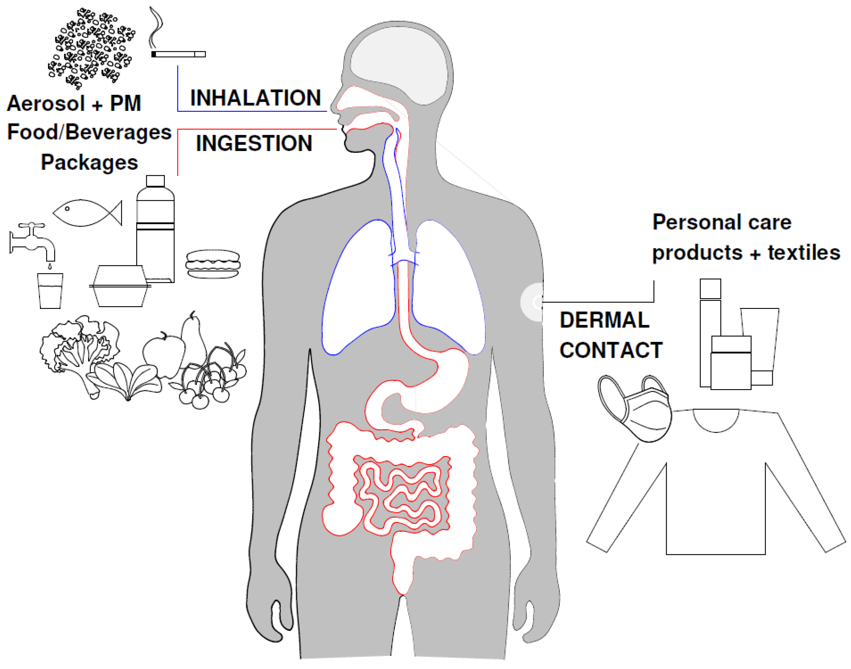
4. Human Exposure through Different Routes

Human exposure to plastic particles is based on generalized estimations and pooled data derived from literature mainly on MPs, which unavoidably suffers from crude assumptions and simplifications, since exposure is highly subject- and lifestyle-dependent or based on human standard food intake. Nevertheless, estimations on exposure to MPs are useful to represent indirectly the exposure to NPs, which most likely could be even higher and undetermined because of the biased methods of analyses [65].
Domenech and Marcos [68] demonstrated that ingestion is the most studied pathway, but other means of exposure to MPs and NPs, such as inhalation and dermal contact, should not be neglected. Fruits, vegetables, meat, fish, grains, legumes, and water constitute the fundamental human diet. It is estimated that the intake of particles through the consumption of vegetables and fruits amounts to an average of 132,740 particles per gram (p/g) of MPs and NPs in five frequently consumed fruits and vegetables (apples, pears, broccoli, lettuce, and carrots). Considering these food categories and complying with the WHO recommendation of a daily intake of at least 400 g of fruit and vegetables, an average of around 53 million particles/day (p/d) are ingested [68,69]. Evaluating approximately 15% of Americans’ caloric intake, Cox et al. [20] estimate that MP consumption ranges from 39,000 to 52,000 p/y depending on age and sex, increasing to 74,000 and 121,000 p/y when inhalation is considered. Additionally, individuals who follow recommended water intake through only bottled sources may be ingesting an additional 90,000 p/y instead of 4000 p/y in case of only tap water consumption. Other authors report high variability in the numbers of MPs and NPs in both bottled and tap water. Regarding the extent of exposure to plastic particles, the following data on mean concentrations obtained by various methodologies are reported, clearly showing different orders of magnitude. An average of 325 p/L was detected in 259 samples from 11 different brands of bottled water purchased globally [26], while Oßmann et al. [70] and Zuccarello et al. [71] detected 2649 ± 2857 p/L and 5.42 × 10⁷ ± 1.95 × 10⁷ p/L in bottled water, respectively. Kosuth et al. [72] detected 3.57 ± 1.79 p/L in tap water. Considering these estimates, humans ingest around 13.5 million p/L in single used bottled water and a total of around 27 million p/d, assuming a daily intake of 2 L of bottled water [73]. The lack of standardized methods for identifying and quantifying MPs and NPs clearly is the cause of this significant variability in exposure data [68]. The actual numbers of MPs and NPs in drinking water have yet to be fully evaluated, but according to Zhang et al. [74], a human intake of almost 5 thousand p/y per person could be estimated for direct consumption. Additionally, food processing should be considered, as drinking water could contribute to the MPs and NPs content in processed food items. A range from 9.77 p/kg to 506 p/kg of MPs and NPs in table salt is reported [75,76], and humans would ingest 0.714 p/d with a consumption of 5 g per day (g/d) for healthy adults. In sugar, Liebezeit and Liebezeit [77] measured 217 fibers/kg and 32 fragments/kg, suggesting that humans could ingest around 2150 fibers/year and around 320 fragments/year with a consumption of 27 g/d per capita [78]. A general exposure of over 50 to billions of fibers/day, depending on dietary habits, is estimated [79].
Considering an airborne concentration of MPs and NPs of about 0.7 particles per cubic meter (p/m3), a respiratory frequency of 12 breaths per minute, and a tidal volume of 0.5 L, the inhalation rate is roughly 8 cubic meters per day, leading humans to inhale approximately 5900 p/d. However, this estimation of airborne MPs and NPs depends on various factors, such as sampling methodologies, air renewal rates, human and cleaning habits, furniture, and even the use of masks for extended periods, as occurred during the COVID-19 pandemic [68]. Morgana et al. [80] evidenced the release of spherical submicrometric particles from discarded face mask fabrics, mostly comprising nano-sized ones (size classes 0.1–0.5 µm and <0.1 µm), at values (2.1 ± 1.4 × 10¹⁰ items/mask) notably higher than those found for MPs (>100 µm) by microscopy (1.2 ± 1.07 × 10⁴ items/mask).
Additionally, in some work environments, the likelihood of exposure to MPs and NPs produced by both mechanical and environmental deterioration of plastic goods, as well as by MPs and NPs present in printer inks, spray paints, injection moldings, and abrasives, is increased [81]. Dris et al. [82] observed a higher indoor concentration of MPs or NPs (1.0 and 60.0 fibers/m3) in private residences and public offices compared with the outdoor concentrations (0.3 and 1.5 fibers/m3).
Besides oral intake and air inhalation, dermal exposure to NPs can occur through taking a shower with water or using personal care products, but the variability of the analyzed conditions makes it difficult to establish the real situation. Nanoparticles, comprising NPs < 40 nm, may enter the body through the epidermal barrier. In care products, concentrations of MPs and NPs range from 2.15 up to 3.11 million p/g [83,84].
Assuming a deposition ratio ranging from 36 to 1008 p/m2 per day, with an average value of almost 370 p/m2 per day, and taking into account the continuous release of fibers from clothing, dermal contact with MPs and NPs is evident, even if not well quantified [68].
Data on human exposure to MPs and NPs are controversial because they refer to specific conditions, whereas plastic particles can be identified in many more items and processes. Therefore, current data on human exposure should be considered as indicative and could serve as a guideline for studies that aim to assess human toxicity.

5. Neurodegenerative Diseases and Environmental Factors

Neurodegenerative diseases pose a significant public health challenge, affecting millions of individuals globally. This group of disorders, which includes Alzheimer’s disease (AD), Amyotrophic Lateral Sclerosis (ALS), Huntington’s disease (HD), and Parkinson’s disease (PD), among others, presents symptoms such as memory loss, impaired motor function, and cognitive decline. The hallmark of these diseases is the progressive loss of neurons in the central and/or peripheral nervous system [85].
While genetic factors have long been associated with these conditions, research underscores the crucial role of environmental influences in their onset and progression. For instance, while the causes of ALS are largely unknown, approximately 5 to 20% of cases (familial cases) have been linked to mutations in identified genes. However, for the remaining 95–80% of cases (sporadic cases), a combination of genetic and environmental factors likely contributes to disease development [86,87,88].
Potential environmental determinants include, but are not limited to, exposure to cyanotoxins, electromagnetic fields, and various chemicals such as pesticides, solvents, heavy metals, and selenium. Studies have investigated the association between traffic-related air pollution and increased ALS risk in the US, as well as the impact of long-term exposure to air pollutants like PM2.5, NOx, and NO2 on ALS risk [89]. Emerging evidence also suggests that exposure to air pollution, which contains components like ultrafine particulate matter (UFPM), could contribute to neurodegenerative diseases such as AD, PD, Multiple Sclerosis (MS), and Motor Neuron Diseases (MND), although the evidence remains inconsistent and limited for MS and MND [90]. While the exact mechanisms are still under study, both animal and epidemiological research indicate that air pollution exposure may induce oxidative stress, leading to neurotoxicity and neuroinflammation [91,92]. In addition to animal and epidemiological studies on air pollutants and brain impairment, data from in vitro models of target tissues, such as cells of the olfactory mucosa, support the implications of air pollutants in neurodegeneration. Indeed, a key route for particles entry into the brain is through the olfactory epithelium [93].
Recent research has examined the metal content in various environmental compartments and the genetic contribution to ALS among patients living in specific areas [94]. In these areas, metal contaminants have been found to exceed the legal limits in surface water, wastewater, and soil, supporting the hypothesis that increased ALS incidence may be linked to environmental metal(loids) contamination along with other environmental factors. In addition, and further supporting these findings, biomonitoring studies describe how heavy metals are significantly found in the neurons of patients affected by motor neuron disease in comparison to controls [95,96].
Interestingly, MPs in the environment are rapidly colonized by bacteria, cyanobacteria, and microalgae, forming biofilms that alter their surface chemistry and attract environmental metals. Exposure to metal-MPs biofilms can disrupt cell homeostasis, leading to toxicities. Consequently, imbalances in metal concentrations can result in neuronal network dysfunction; reactive oxygen species (ROS) production; and mitochondrial damage in diseases such as PD, AD, and Prion disorder [97].

6. Mechanisms of Plastic Particles’ Transfer to the Central Nervous System (CNS) and Effects on the Blood–Brain Barrier

Exposure of humans to plastic particles has been estimated to account for over hundreds of thousands particle/year and to accumulate at various concentrations in different tissues and organs such as the skin, heart, blood, testis, and intestine, among others, exhibiting the ability to accumulate into internal organs/tissues thanks to their ability to pass through epithelial barriers, as was recently reviewed by De Boever et al. [65]. Studies on organisms, including mammals, have demonstrated that ingested plastic particles can diffuse throughout the organism, exerting harmful effects on numerous organs at the macroscopic, histological, and metabolic levels [98,99]. These particles tend to accumulate in tissues, particularly in the liver and intestine, leading to cytotoxicity, inflammation, alteration of genetic expression profiles, and metabolic changes [100]. The digestive tract, renowned for its absorptive function, offers an efficient route for orally ingested particles to enter the body, much akin to the mechanisms observed in many oral delivery formulations based on polymeric nanoparticles designed to withstand degradation by enzymes and acids during transit [101]. To gain entry into the body, both MPs and NPs acquire a biomolecular corona composed of proteins and other biomolecules that accumulate on their surface [102]. Consequently, factors such as the size, negative charge, hydrophilicity, and Van del Waals’ forces of MPs and NPs facilitate their passage through the mucus layer of the gastrointestinal epithelium and trans-intestinal transport, primarily reliant on endocytosis, wherein the particles traverse through the cell and are released at the basolateral membrane. In vitro experiments have underscored the ability of PS-plastic particles to traverse biological plasma membranes, with various sizes (50–500 nm) internalized through diverse mechanisms such as passive diffusion, endocytosis, and macropinocytosis, as evidenced by studies on cultured cells [103]. PS-NPs (50 nm) follow a water–phospholipid partition system and are internalized through endocytosis via the clatrin-and caveolin-mediated pathways and through micropinocytosis. The lysosomes represent the main accumulation site, where the plastic particles are transported through an energy-dependent mechanism. Interestingly, the same authors analyzed the mechanism of NPs release for the first time, which relies on both lysosomal exocytosis by energy consumption and an energy-free penetration mechanism [103].
MPs and NPs induce an imbalance in the gut microbiome, characterized by an increase in harmful bacteria and a reduction in beneficial bacteria, which could compromise the intestinal barrier. Consequently, certain bacterial products or species may breach into the bloodstream, potentially causing damage to other tissues and organs [104]. The gut microbiota may significantly influence the effects of foreign environmental pollutants on the nervous system. A recent study hypothesizes that NPs ingested through the digestive tract could alter the composition of the gut microbiota, subsequently impacting the host’s rhythmic function and causing damage to the nervous system [105]. Additionally, enteric neurons, situated in the gut wall, are likely the initial neurons exposed to MPs and NPs, given their location and involvement in regulating gastrointestinal functions. It is suggested that central neurodegeneration might originate peripherally, with growing evidence implicating the enteric nervous system in various neurodegenerative disorders such as Alzheimer’s, Parkinson’s, and prion diseases, possibly mediated by interactions with the gut microbiome [106,107,108,109].
The intestinal absorption pathway serves as a conduit for plastic particle uptake, potentially leading to their dissemination into non-target organs and tissues via the bloodstream and systemic circulation, including the CNS. Thus, the involuntary intake of plastic particles may be a crucial aspect triggering the initial development of neurodegenerative diseases or accelerating the illness’ progression. The effects of MNPs on enteric neurons’ morphology and functionality have been hypothesized [110,111]. However, even though these features have not been extensively studied yet, an important advancement in this field has been made by studying bigger sizes of plastic particles (namely, PET-MPs), and the effects on the enteric nervous system in animal models have now been demonstrated [112].
The type of corona formed significantly influences the ability of NPs to breach the blood–brain barrier (BBB) and their overall toxicity. Molecular dynamics simulations using lipids 1,2-Dioleoyl-sn-glycero-3-phosphocholine as a model of the membrane and a representative of the BBB demonstrate that polystyrene (PS) plastic particles (5 nm) can traverse both the gastrointestinal barrier and the BBB swiftly, contingent on their specific surface coronas [8]. These findings are corroborated by in vivo mouse models, which indicate the presence of nanometric-sized PS particles in brain tissues as early as 2 h post-exposure. Size is another critical determinant influencing the capacity of plastic particles to penetrate the BBB, with only particles measuring 293 nm being absorbed by the gastrointestinal tract and capable of breaching the BBB in mice models exposed to a mixture of different sizes of MP and NP particles (9.55 µm,1.14 µm, 293 nm) [8]. Synthesized fluorescent PS-NPs (30–50 nm) can reach brain tissues of orally exposed mice [113], suggesting that the gut–brain connection is pivotal in oral exposure to plastic particles and brain damage caused by NPs. Recent advances [114,115] using in vivo (mouse) and in vitro models (i.e., bEnd.3 endothelial cells from mouse brain tissue or hCMEC/D3 cells, recognized as a cell line for human BBB model) confirm the ability of PS-NPs to cross through the BBB and clarify the underlying mechanisms [115]. PS-NPs 50 nm in size, characterized by transmission electron microscopy and scanning electron microscopy, reduce the transendothelial electrical resistance by altering the expression of the scaffold protein zona occludens-1 (ZO-1), responsible for the actin cytoskeleton’s connection to the transmembrane protein occludin, which preserves the integrity of the BBB. The molecular analyses further suggest that pathways of ferroptosis, iron-mediated cell death which adversely affects brain function (see, e.g., [116]), is related to BBB disruption [114]. The internalization of PS-NPs (100 nm) was evidenced in a neuronal model (mouse hippocampal neuronal cells, HT22), leading to decreased cell viability [117]. In addition, studies both in mice and in vitro demonstrate the ability of PS-NPs to accumulate and activate microglia cells, which in turn leads to neuronal damage [115].
Given the BBB’s pivotal role in shielding the brain from harmful substances, its compromise can precipitate various neurological issues. Therefore, the short-term health ramifications of NPs warrant consideration, as their presence in the brain may precipitate cognitive impairment, neurological disorders, and neurotoxicity, ultimately impeding brain function [118].
Furthermore, the intestinal tract, beyond its role in digestion and nutrient absorption, serves as a crucial interface between the organism and the environment. It also facilitates communication with the CNS through the so called “gut–brain axis”, a complex network where neuroendocrine and immunological signaling pathways, along with bidirectional neural mechanisms, interact, influenced by the gut microbiome, potentially impacting brain damage and neuroinflammatory processes. These factors modulate various processes, including neuroinflammation, aberrant BBB permeability, microglial activation, immune system responses, and mitochondrial dysfunction, all of which are critical to the progression of brain-related pathologies [119,120,121].
Neurodegenerative processes may be initiated by the enteric nervous system, the vagus nerve, and the bloodstream through neuroactive compounds such as serotonin or dopamine, as well as cytokines [122]. These pathways’ activation, demonstrated for ambient ultrafine particulate matter (UFPM) and the transfer of iron from UFPM into the brain, and for several metals transiting via olfactory receptor neurons from the nasal lumen to the olfactory bulb, could similarly be exploited by plastic particles [123,124].
In addition to BBB crossing, using in vivo pregnant rat models exposed to PS-NPs (30–50 nm), the ability of NPs to cross placental barriers and transgenerationally accumulate into the brains of fetuses has just been demonstrated [125].
Thus, as summarized in Table 1, the recent advancements with in silico, in vitro (mammalian and human cells), and animal models (mouse and rat) demonstrate the ability of NPs, even those of bigger dimension (293 nm), to cross the BBB and accumulate in the brain. When translating these findings from the biological models to humans, a great concern emerges regarding the possible neurotoxic effect and neurodegenerative process of NPs.

7. Molecular Effects of Nanoplastics in Processes of Neurotoxicity and Neurodegeneration

Environmental and food data are largely available on MPs and MNPs rather than NPs. In fact, the detection in environmental samples and even more in human tissues and organs of smaller-sized plastic particles (NPs) is currently limited by technology, which should be improved for a better understanding of NPs’ burdens in terms of quantities and polymer type. However, due to recent data that demonstrate the ability of NPs to pass barriers and exert (neuro)toxicological effects, the following sections are specifically dedicated to the neurotoxic and possible neurodegenerative effects of NPs. Therefore, the focus is on NPs which are able to pass the BBB, as described in Section 6 and summarized in Table 1, and to affect brain and neuron functions.

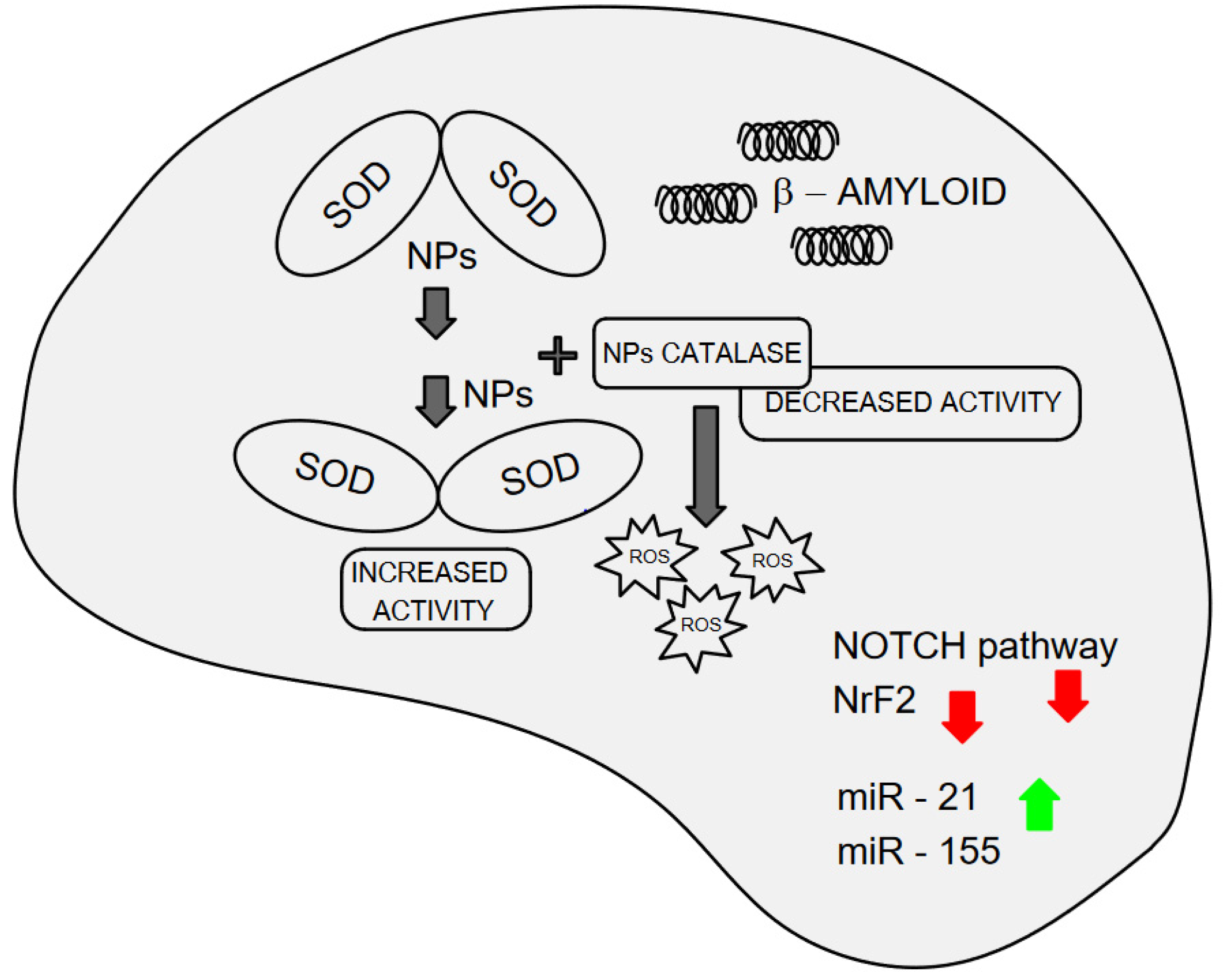
7.1. In Vitro and In Silico Interaction of Nanoplastics with Superoxide Dismutase and Amyloid Protein Formation: Implications for Oxidative Stress and Neurodegeneration

The central nervous system (CNS) is particularly susceptible to oxidative imbalance owing to its abundance of polyunsaturated fatty acids, high metabolic oxidative rate, and elevated content of transient metals and ascorbate levels, collectively acting as pro-oxidants. Nonetheless, the CNS possesses relatively fewer antioxidant systems compared to other organs [126]. Substantial evidence underscores the roles of inflammation and oxidative stress as prominent features in AD, PD, and ALS, linking oxidative stress to neuronal death and neural dysfunction. Notably, mitochondrial dysfunction also features prominently in these diseases, likely playing a critical role in generating and amplifying reactive oxygen species (ROS), thereby contributing to the pathophysiology of these diseases [127,128].
Numerous genes have been implicated in the onset of neurodegenerative diseases, with mutations in superoxide dismutase (SOD) genes identified in ALS patients, present in nearly 20% of familial cases. The SOD family proteins represent one of the primary lines of defense against oxidative stress in organisms, with most oxygen-reliant organisms expressing at least one SOD isoform. This family of metalloenzymes acts as antioxidants by scavenging superoxides (e.g., O2), and deleterious mutations have been shown to alter SOD1 activity, resulting in the accumulation of highly toxic hydroxyl radicals. Mammals possess three isoforms of SOD: cytoplasmic Cu/ZnSOD (SOD1), mitochondrial MnSOD (SOD2), and extracellular Cu/ZnSOD (SOD3), all of which necessitate catalytic metals (Cu or Mn) for activation [129]. The consequences of NPs’ interactions with proteins have been investigated and described, including protein corona formation, NPs-induced coalescence, and alterations in secondary protein structure [112]. Of particular relevance to neurotoxic and neurodegenerative processes is the interaction of plastics with the key enzyme superoxide dismutase (SOD), with recently described structural and functional consequences [130]. These authors employed PS microspheres of 70 nm (PS-NPs) and conducted binding studies on purified SOD1, the Cu/Zn isoform, employing multispectroscopic approaches (UV-vis, circular dichroism, fluorescence emission spectra). Addition of PS-NPs induces peptide bond alterations in the backbone of SOD1, loosening and unfolding its structure. PS-NPs alter the secondary structure of SOD1, affecting the enzyme’s percentage of α-helix, β-sheet, β-turn, and random coil. Moreover, PS-NPs and SOD form new complexes, leading to larger aggregate sizes. At varying concentrations of NPs, structural alterations contribute to increased SOD1 activity [130]. The underlying hypothesis suggests that, with increased PS-NPs concentrations, aggregate sizes enlarge, loosening the skeletal structure of SOD1 and facilitating substrate entry into the enzyme’s active center. However, elevated SOD activity, without a concomitant rise in catalase levels, may escalate the Fenton reaction, generating hydroxyl radicals from hydrogen peroxide, thereby heightening oxidative stress [131]. Catalase serves as a crucial enzyme in the pathway for ROS removal, converting hydrogen peroxide into water and molecular oxygen. The interaction of catalase with NPs and ensuing enzymatic activity assumes particular significance. Multispectral approaches and enzyme activation assays reveal alterations in catalase’s internal structure and a corresponding decrease in enzyme activity upon exposure to polystyrene NPs [132]. These findings complement the depiction of an ROS-enriched environment devoid of key enzymes crucial for their removal, conditions intimately linked to neurodegeneration. Both NP size (20–1000 nm) and different concentrations (2–16 mg/L) can differentially impact SOD activity and structure [133]. Smaller NPs are more prone to inducing SOD coalescence and a looser SOD skeleton. With increasing NP concentrations, enhanced binding with the enzyme’s metal active site is observed, resulting in reduced SOD activity. The observed effects can be summarized as NPs binding to SOD, inducing changes in the enzyme’s conformational structure, ultimately affecting the activity of superoxides’ removal of this pivotal enzyme. However, it is important to note that Wang and collaborators [133] included and classified plastic particles of 500 and 1000 nm into the NPs, but what is relevant is that they observed major effects on SOD skeleton due to the smaller size.
NPs not only interact with the three-dimensional structure of antioxidant enzymes, but also alter the polarity around tyrosine residues in SOD. In 2009, the role of a highly conserved tyrosine residue (Tyr34) on human SOD2 (MnSOD) structure and catalysis was elucidated [134]. Intriguingly, mutational studies substituting Tyr with residues possessing varying polarity (e.g., Asp, Phe, Val) substantially diminished the catalytic rate constant for superoxide reduction. Disruption of SOD’s catalytic site affects enzyme activity in a loss-of-function manner, as was recently demonstrated in vitro. In a human neuronal cell model, despite the normal expression of SOD1, the protein failed to eliminate ROS [135].
All changes in the catalytic site(s), backbone structure, and polarity of key residues in SOD suggest a role of NPs in the biotoxicity of these plastic particles concerning key enzymes associated with neurotoxicity and neurodegenerative mechanisms.
Other common cellular and molecular mechanisms in neurodegenerative diseases such as AD, PD, and ALS, which have long been recognized, include inclusion body formation and protein aggregation. These aggregates are typically characterized by fibers containing misfolded proteins with a β-sheet conformation, termed amyloids [136]. Amyloidosis primarily manifests in the CNS, although protein aggregates can form and deposit in various organs or systematically. In silico studies using molecular dynamics simulations with plastic nanoparticles of different polymer origins have demonstrated interactions with proteins for different types of plastics abundant in nature as both MPs and NPs, including PE, PET, PP, and nylon-6,6 [137,138]. Molecular dynamics simulations were conducted either on two peptides, representing the two most prevalent kinds of secondary structures in proteins, or on β-amyloid fibril models. The first peptide was a tryptophan zipper, characterized by a β-hairpin structure resembling β-sheets in proteins, and the second was an α-helix polypeptide of 12 alanine amino acids, which intrinsically stabilizes α-helices in proteins. Observations of changes in secondary structure and peptide conformational rearrangements upon exposure to NPs suggest that PE and, particularly, PP-NPs may delay fibrillation processes and augment the content of more cytotoxic oligomers, akin to aggregates implicated in amyloidosis. Furthermore, in addition to in silico models, exposure of human neuroblastoma cells (SH-SY5Y) to 70 nm PS-NPs at very low concentrations (100 pM) accelerates the nucleation rate of β-amyloid oligomer formation [139], raising significant concerns regarding NPs’ associations with neurodegenerative diseases.
The protein corona may significantly influence NPs’ behavior and biological interactions, as mentioned before. Consequently, one of the principal challenges in this field is understanding the composition and structure of the protein corona and its impact on the biological effects of NPs.
The ability of NPs to cross the blood–brain barrier and accumulate into the brain (see Section 6), as well as their interactions with protein folding, strongly suggest their plausible ability to induce the aggregation of amyloid proteins, thereby creating a conducive environment for neurodegenerative diseases.
Observations of these significant changes at the molecular level in key proteins and their consequent altered functions underscore the urgent need to expand investigations into the effects of these materials through further modeling and molecular biological methods, focusing on additional key enzymes.

7.2. Nanoplastics Affect the Transcriptomics and Epigenomic in Neuronal Models

Transcriptomic analysis serves as a powerful tool for unraveling the intricate regulatory networks within cells and identifying specific altered pathways and deregulated genes. This method enables the simultaneous detection of gene regulation on a large scale, offering mechanistic insights. Such an approach has been instrumental in understanding the deregulatory effects of NPs at the gene expression level. From this perspective, the alterations of gene regulation by NPs in biological in vivo and in vitro models suitable for neurological studies are described to achieve a deeper insight into the molecular mechanisms involved in neurotoxicity processes, which could lead to or could interplay with other factors in neurodegenerative processes caused by these environmental contaminants.
Nearly a decade ago, Hoelting et al. [140] published one of the pioneering papers describing the effects of PE-NPs with an average particle size of 33 nm on transcripts in a neural model characterized by a 3D neurosphere system. PE-NPs were found to reduce the transcript levels of NOTCH pathway genes, including NOTCH1 and HES5, as well as downstream neuronal precursor genes (NEUROD1, ASCL1, FOXG1). The dysfunction of these genes results in severe nervous system impairments in mice, particularly affecting neurodevelopmental functions. More recently, Cho et al. [141] demonstrated that an imbalance in NOTCH1 signaling might be implicated in AD, where NOTCH1 plays a role in regulating the proteolytic processing of amyloid precursor protein (APP).
In studies focusing on gene expression and exposure to NPs in animal models, Liang et al. [142] provided transcriptomics data on isolated nuclei from frozen brains of adult male C57BL/6 J mice, commonly used in neurobiology studies, which were exposed to 50 nm pristine PS-NPs. The analysis revealed evidence of Parkinson’s disease-associated genes among differentially expressed genes (DEGs). Astrocytes exhibited the highest number of DEGs (597 genes), followed by oligodendrocytes, neurons, and endotheliocytes, with 486, 326, and 280 DEGs, respectively. PS-NPs were found to inhibit multiple biological processes in a cell-specific manner, including mitochondrial function, proteostasis, ATP metabolism, and synaptic function regulation. Notably, ATP metabolism was the most enriched biological process in neurons, while mitochondrial and proteostasis functions were most enriched in neurons, oligodendrocytes, astrocytes, and endotheliocytes. Furthermore, synaptic function regulation was enriched in astrocytes and endotheliocytes. Overall, the genes and pathways analyzed by these authors provide a comprehensive understanding that PS-NPs induce PD-like neurodegeneration through cell-specific pathways in mouse brains. In the same mouse model, exposure to NPs through the diet revealed neurotoxic effects by reprogramming circadian rhythm-related genes involving the gut–brain axis [105]. Key genes involved in PS-NPs neurotoxicity include Camk2g, which encodes a calcium/calmodulin-dependent protein; Adcyap1, encoding key mediators of neuroendocrine stress responses; and Per1, a critical gene in circadian rhythms. Thus, the ingestion of PS-NPs could affect the expression of genes related to circadian rhythms in the hippocampus through dietary intake, affecting the gut–brain axis, reducing neuroplasticity, and further causing nerve damage in mice.
These findings in mice offer a distinct perspective on the potential effects of exposure to NPs through ingestion via contaminated food and water consumption, and further support the role of the “gut–brain axis” described in Section 6.
Recent studies have examined the gene expression profiles following exposure to NPs using in vitro neuronal cell models. In one study, different metabolic pathways were identified in human neural stem cells (hNS1) exposed to 0.5, 2.5, and 10 µg/mL of PS-NPs for 4 days. The study revealed alterations in stress response genes (hsp27/hspB1, hsp70/hspA5, and hsp90α), with a notable increase in mRNA expression of hsp27/hspB1, which plays crucial roles in neurodegenerative diseases such as AD and MS. Additionally, 30 nm PS-NPs induced altered expression of Cu/ZnSOD1 and catalase, along with inflammatory and mitochondrial responses in the same in vitro neural model [143]. In another study, PS-NPs beads (25 nm) were exposed for 1 day to a modified human neuronal cell model (SH-SY5Y cell line) overexpressing α-synuclein, a protein associated with the formation of pathological inclusions (Lewy bodies) in the brains of individuals with Parkinson’s disease. The penetration of PS-NPs into the cells further increased α-synuclein aggregation, suggesting potential implications for human neurodegeneration [144].
Brain organoids are complex 3D cell models differentiated from human embryonic stem cells which have emerged as innovative in vitro models for neurodevelopmental studies and neurological diseases (see, e.g., [145] for a comprehensive review). The transcriptomic analysis of DEGs in this 3D in vitro model exposed to 100 nm PS-NPs reveals the molecular mechanisms in which Wnt, PI3K-Akt, and TGF-beta signaling pathways are the most enriched [146]. The Wnt and PI3K-Akt are recognized signaling pathway in Alzheimer’s disease [147,148], and TGF-beta is a common feature of neurodegenerative disorders and particularly in ALS [149].
Important cues as to the comprehension of neurotoxicological effects of NPs derive from animal models, which can be used to transfer systemic data to humans. Among these, zebrafish (Danio rerio) is a National Institute of Health (NIH)-validated model organism for studies in (neuro)developmental toxicology due to its high genomic conservation with humans, and it represents an emerging model in toxicity studies of MPs and NPs [150].
Exposure of larval zebrafish to PS-NPs (50–200 nm) alters transcriptome analysis, affecting gene functions related to neurodegeneration and motor dysfunction. DEGs related to nervous system sub-pathways, including movement disorders, neuromuscular diseases, morphology of the nervous system, and development of neurons, are affected by NPs [151]. In the adult model, the same authors observe alterations in movement disorders and neuromuscular disease pathways, including basal ganglia, dyskinesia, and cerebellar hypoplasia. Upregulation of the membrane protein trafficking gene ap1s2, involved in basal ganglia disease in humans, and downregulation of ahi1, associated with cerebellar development in mice and humans, are induced in adult zebrafish upon exposure to NPs. Additionally, multiple neuromuscular genes and cytoskeletal genes are downregulated, while genes related to synaptic signaling and pumps are upregulated. In addition, multiple neuromuscular genes (e.g., skeletal muscle subunit myhb and axonal transport genes bcd2 and kif1ab, encoding for axonal transport proteins in the opposite directions, retrograde vs. anterograde, respectively) and cytoskeletal gene prph are downregulated. PS-NPs (25–134 nm) at concentrations which can be environmentally encountered (0.1 and 0.5 mg/L) were exposed to zebrafish to perform a battery of behavioral and gene expression assays. PS-NPs affected genes of the hypothalamic–pituitary–thyroid axis and of the dopamine metabolism [152].
These transcriptomics findings in this animal model, with structures and functions homologous to those of mammals and humans, provide crucial insights into the mechanisms of NPs implicated in promoting neurological and neuromuscular disorders.
Epigenetic evaluations provide important insights into transcriptomics studies, with epigenetics encompassing a wide range of heritable changes in gene expression in response to various environmental factors, such as pollutants, diet, smoke, and lifestyle factors, without DNA sequence modifications. Key factors controlling gene expression at the translational and post-translational levels include DNA methylation or hydroxymethylation, phosphorylation, histone acetylation, and deacetylation, as well as the actions of histone variants, microRNAs (miRNAs), and long non-coding RNAs (lncRNAs). The emerging field of neuroepigenetics explores how epigenetic dysregulation contributes to neuronal death associated with neurodegenerative diseases [153,154].
PS-NPs (44 nm mean size) have been shown to alter the transcriptional profile and DNA methylation of fibroblasts and human-induced pluripotent stem cells (hiPSCs). Exposure to PS-NPs resulted in the downregulation of the dopaminergic synapse signaling pathway, which is known to be related to amino acid metabolism and Parkinson’s disease [155]. Additionally, enrichment analysis of transcriptome data from hiPSCs exposed to PS-NPs revealed the downregulation of genes associated with the alteration of molecular functions of metal ion transmembrane transporter activity and voltage-gated cation channel activity, both of which are key activities related to neuronal functions.
As previously described (Section 4), one of the routes of NP exposure to humans is through epidermal exposure, with systemic transport allowing for deposition into various organs and tissues, including the nervous system. Thus, there is an urgent need to develop robust human cell models to investigate the potentially harmful effects of NPs on human health that consider the possible different routes of exposure, such as dermal contact. The use of fibroblasts and hiPSCs as cell models to identify epigenomic signatures following NPs exposure has been proposed to identify pathways involved in human diseases. Transcriptomic and epigenomic signatures can be revealed through RNA-seq and whole-genome methyl-seq [155]. Fibroblasts and hiPSCs exposed even to low concentrations of PS-NPs exhibited cellular responses that might trigger diseases such as cancer and neurodegeneration, with the ATG7 gene identified among them. Autophagy-related (ATG) proteins, including ATG7, play crucial roles in protecting against complex diseases like neurodegeneration, although the precise mechanism by which dysfunctional ATG7 contributes to neurodegenerative diseases in humans remains to be fully elucidated [156].
In both fibroblasts and hiPSCs, differentially methylated regions (DMRs) have been identified, with hypermethylated or hypomethylated regions. Gene ontology (GO) analysis for methylation data has revealed genes with the most prominent differentially methylated regions associated with differential gene expression. Fibroblasts exposed to NPs are affected in various GO terms related to axon guidance, cell adhesion, synapse assembly, and nervous system development, indicating potential impacts on neural development and function.
Expanding our understanding to common processes and molecular markers in seemingly different diseases, like carcinogenesis and neurodegeneration, reveals intriguing insights and shared features. Techniques such as gene expression evaluations and the identification of different families of microRNAs have unveiled the carcinogenic potential of pristine PS-NPs [157]. Prolonged in vitro exposure (6 months) of mouse embryonic fibroblasts, which are sensitive to oxidative stress and prone to transformation progress (PTP), to 25 µg/mL of PS-NPs leads to stress-related gene expression and deregulation. Significant downregulation of genes crucial for neurodegenerative processes, such as Sod1, Sod2, and Nrf2, is observed. The Nrf2 gene product, a transcriptional factor, plays essential roles in regulating antioxidant defense mechanisms against oxidative stress and is implicated in neurodegenerative disorders [158]. The downregulation of these gene groups by PS-NPs supports their involvement in neurodegenerative processes. Keap1, another gene in the pathway activated by pro-oxidant conditions, is also downregulated by PS-NP exposure, regulating Nrf2 activity and sensing oxidative and electrophilic stresses [158].
MicroRNAs are now recognized as universal regulators of differentiation, activation, and polarization of microglia and macrophages in both normal and pathological conditions of the CNS. Dysregulation of these small non-coding RNAs, which are involved in post-transcriptional regulation of gene expression, has been described in various neurodegenerative diseases [159]. Among the most prevalent miRNAs found in neurodegenerative diseases and their animal models are miR-21-5p (miR-21) and miR-155-5p (miR-155). Interestingly, these miRNAs are upregulated in PTP cells exposed to PS-NPs, according to Barguilla and colleagues [157]. MiR-21 downregulation in animal models has shown a neuroprotective role by reducing the inflammatory response. However, the dysregulation of miR-21 suggests its complex involvement in neurodegeneration and necessitates further research. Conversely, miR-155, predominantly upregulated across different neurodegenerative diseases, plays roles in promoting neuroinflammation and may exacerbate neurodegenerative diseases through inflammatory pathways [159]. In Figure 2, representative features of NPs’ effects and interactions at the molecular levels are illustrated.
A noteworthy comment and suggestion arising from the analysis of miR dysregulation upon exposure to NPs is to consider processes sharing overlapping dysfunctions and triggering mechanisms, such as cancer and neurodegeneration. A broader perspective on the results obtained could aid in identifying subsequent diseases and potential therapeutic interventions.

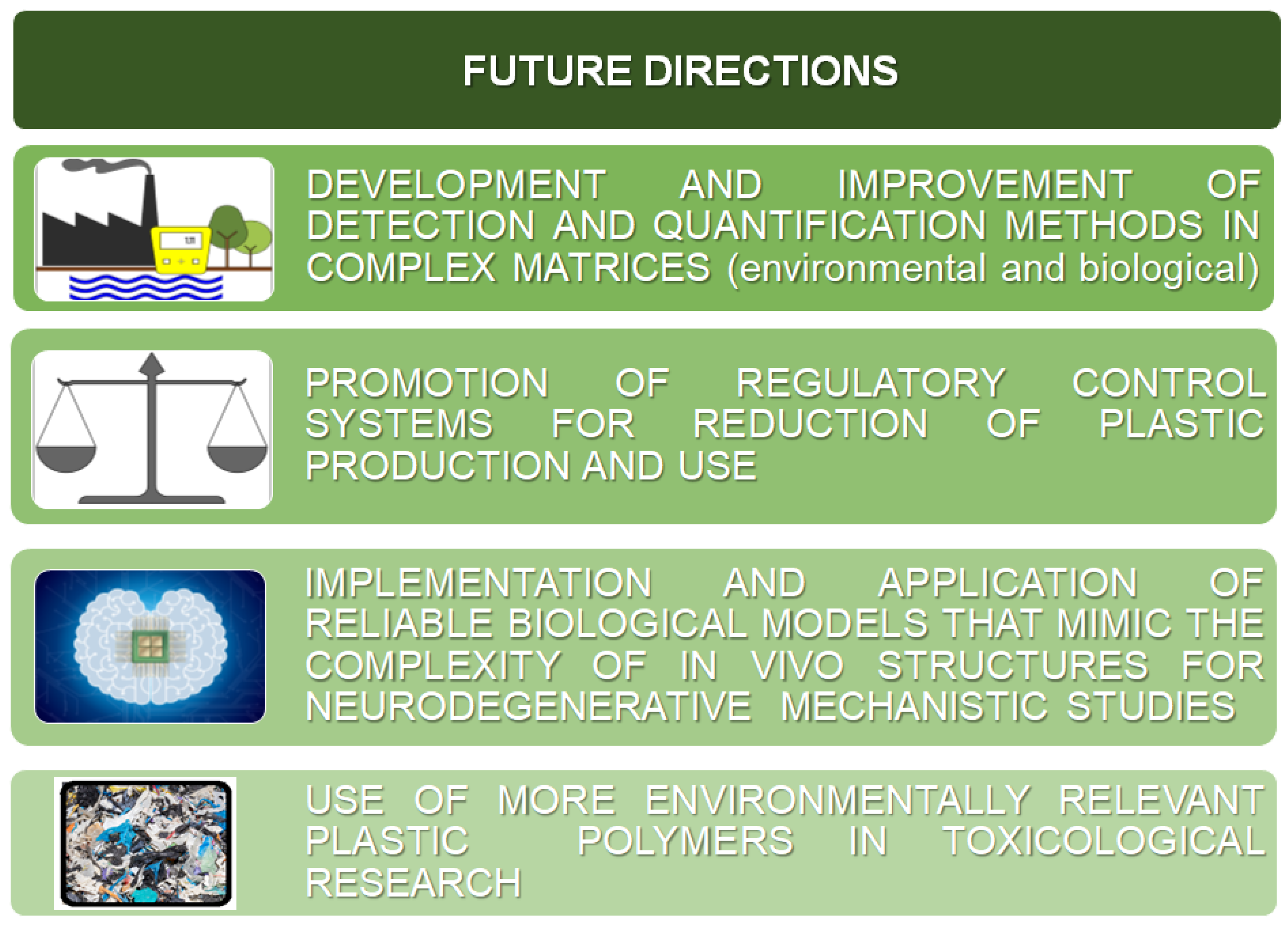
8. Conclusions and Future Directions

The pervasive presence of plastic particles, particularly NPs, across all environmental matrices, including food, has sparked significant apprehension regarding human health. Their ubiquitous distribution and ability to permeate organisms and humans through various exposure pathways have heightened these concerns. Moreover, the unpredictable accumulation of NPs within organisms adds another layer of complexity. Compounding the issue is the challenge of accurately quantifying NPs in complex matrices, especially in biological samples rich in organic matter. Addressing this necessitates the development of improved collection and detection methods alongside the advancement of highly sensitive instrumentation for direct analysis of NPs in environmental matrices, human tissues, or biological models. While there have been advancements in extracting and quantifying NPs from certain biological samples [160], there remains substantial room for technological progress. For these advances, more precise detection of NP levels will follow, providing an improvement to human risk assessment.
While we acknowledge the newly implemented regulations aimed at curtailing plastic production and waste, it is important to recognize that these measures do not offer a definitive solution to the widespread presence of plastic materials to which organisms are already exposed. At present, no barrier systems are available to definitively block or reduce plastic’s entry into organisms.
Regarding human health, and particularly neurotoxicity and neurodegenerative implications, great attention should be paid to this central issue, as demonstrated by the literature [161]. Experimental data from in silico, in vitro, and in vivo models (see Section 6) have evidenced the ability of NPs to accumulate in the brain by destroying the BBB’s molecular structure and permeability. Very interestingly, PS particles of bigger dimensions (293 nm) have been found in brain tissues of experimental animals, suggesting the relevance of in-depth studies in this area of concern. Considering the various pathways through which particulate matter carrying NPs can access the brain, including the olfactory and trigeminal nerves, the vagal nerve, and the bloodstream, it is conceivable and expected that plastic particles can also breach the BBB directly through these routes. Similarly, the routes established for different environmental contaminants present in particulate matter can be extrapolated to NPs. Therefore, future research should prioritize understanding the precise mechanisms underlying the accumulation of plastic particles in the brain and their interactions with various metallo-enzymes involved in other pathogenic processes, as well as all potential epigenetic mechanisms. This research is pivotal for advancing our understanding of the neurotoxic and neurodegenerative effects of NPs themselves. Furthermore, the access of particulate matter to the brain, characterized by multiple organic and inorganic chemicals, and the environmental interaction of NPs with different pollutants and metals, pose another critical issue to be explored: the co-presence and co-transport of multiple chemicals with NPs and the subsequent interplay at the biological level. In addition, a further level of complexity involves considering the possible synergistic effects of NPs and the different contaminants to which humans are exposed.
Another important final consideration is to take into account the overlapping of biological processes leading to different chronic pathologies, such as cancer and neurodegeneration. These considerations open new perspectives on the knowledge of common pathways and mechanisms, and, therefore, to possible therapeutic solutions.
Lastly, due to the intricate architecture of the nervous system, it is advisable to develop and implement in vitro models that better reflect the biological complexity to address neurotoxicity and neurodegenerative mechanistic studies by applying models at different scales of organization, such as co-cultures, organoids, or microfluidic systems. The use of animal models is a powerful tool, since it allows for the investigation of aspects such as behavioral and physiological mechanisms of diseases, but limitations involve the possible translation of the effects to humans due to inter-species differences [162]. On the other hand, embryonic stem cells and induced pluripotent stem cells, in vitro models frequently used in neurodegenerative disease studies, pose concerns due to genetic instability, absence of epigenetic markers, and ethical issues [163].
Indeed, in addition to epidemiological and animal research, the use of in vitro mammalian and even more human cell cultures represent relevant biological models providing numerous advantages, such as standardized conditions, rapid screening of substances’ toxicity, and understanding the cell and molecular mechanisms. Furthermore, these models are in accordance with high-throughput testing strategies which provide a large amount of data useful even for regulatory purposes, and are in compliance with the 3Rs Principles of Refining, Reducing, and Replacing the use of animal models. In more recent terms, the NAMs (any non-animal-based approaches), including not only biological models, but also in silico simulations and any approaches and methodologies that can provide information on chemical hazards, should be integrated within the Integrated Approaches to Testing and Assessment (IATA) protocols [164].
Finally, although the studies described herein illustrate directions and suggest clear mechanistic interactions at a molecular level, it is advisable that future studies utilize more environmentally relevant polymers, since animal and in vitro studies are mainly based on PS-NPs. In Figure 3, all advisable future directions are summarized.

Author Contributions

All authors contributed to the study’s conceptualization and literature search. The first draft of the manuscript was written by C.U., R.B. and M.T., and all authors (C.U., R.B., S.A. and M.T.) commented on previous versions of the manuscript. All authors have read and agreed to the published version of the manuscript.

Funding

This research received no external funding.

Data Availability Statement

Not applicable.

Acknowledgments

The authors gratefully acknowledge Marco Maggiora for graphical assistance and Mechall Abrahams for English proof-reading.

Conflicts of Interest

Author Susanna Alloisio was employed by the company ETT S.p.A. The remaining authors declare that the research was conducted in the absence of any commercial or financial relationships that could be construed as a potential conflict of interest.

References

  1. Liu, C.; Liu, C. Exploring Plastic-Management Policy in China: Status, Challenges and Policy Insights. Sustainability 2023, 15, 9087. [Google Scholar] [CrossRef]
  2. European Chemical Agency. Available online: https://echa.europa.eu/hot-topics/microplastics (accessed on 18 July 2024).
  3. Ng, E.-L.; Huerta Lwanga, E.; Eldridge, S.M.; Johnston, P.; Hu, H.-W.; Geissen, V.; Chen, D. An Overview of Microplastic and Nanoplastic Pollution in Agroecosystems. Sci. Total Environ. 2018, 627, 1377–1388. [Google Scholar] [CrossRef]
  4. Stapleton, P.A. Microplastic and Nanoplastic Transfer, Accumulation, and Toxicity in Humans. Curr. Opin. Toxicol. 2021, 28, 62–69. [Google Scholar] [CrossRef]
  5. European Commission. Commission Recommendation of 18 October 2011 on the Definition of Nanomaterial (2011/696/EU). Official Journal of the European Union L 275/38- L 275/40. 2011. Available online: https://eur-lex.europa.eu/LexUriServ/LexUriServ.do?uri=OJ:L:2011:275:0038:0040:EN:PDF (accessed on 18 July 2024).
  6. Gigault, J.; Halle, A.T.; Baudrimont, M.; Pascal, P.-Y.; Gauffre, F.; Phi, T.-L.; El Hadri, H.; Grassl, B.; Reynaud, S. Current Opinion: What Is a Nanoplastic? Environ. Pollut. 2018, 235, 1030–1034. [Google Scholar] [CrossRef]
  7. European Food Safety Authority (EFSA). 2021. Available online: https://www.efsa.europa.eu/en/events/event/update-scientific-colloquium-25-coordinated-approach-assess-human-health (accessed on 18 July 2024).
  8. Kopatz, V.; Wen, K.; Kovács, T.; Keimowitz, A.S.; Pichler, V.; Widder, J.; Vethaak, A.D.; Hollóczki, O.; Kenner, L. Micro- and Nanoplastics Breach the Blood–Brain Barrier (BBB): Biomolecular Corona’s Role Revealed. Nanomaterials 2023, 13, 1404. [Google Scholar] [CrossRef] [PubMed]
  9. Martin, L.M.A.; Gan, N.; Wang, E.; Merrill, M.; Xu, W. Materials, Surfaces, and Interfacial Phenomena in Nanoplastics Toxicology Research. Environ. Pollut. 2022, 292, 118442. [Google Scholar] [CrossRef] [PubMed]
  10. Petters, S.S.; Kjærgaard, E.R.; Hasager, F.; Massling, A.; Glasius, M.; Bilde, M. Morphology and Hygroscopicity of Nanoplastics in Sea Spray. Phys. Chem. Chem. Phys. 2023, 25, 32430–32442. [Google Scholar] [CrossRef] [PubMed]
  11. Muthulakshmi, L.; Mohan, S.; Tatarchuk, T. Microplastics in Water: Types, Detection, and Removal Strategies. Environ. Sci Pollut Res 2023, 30, 84933–84948. [Google Scholar] [CrossRef]
  12. Plastic—The Facts. 2022. Available online: https://plasticseurope.org/knowledge-hub/plastics-the-facts-2022/ (accessed on 19 July 2024).
  13. European Parliament. Environmental Determinants of Health, Including Those Caused by Climate Change. 2024. Available online: https://www.europarl.europa.eu/RegData/etudes/STUD/2024/754209/IPOL_STU(2024)754209_EN.pdf (accessed on 6 August 2024).
  14. Mortensen, N.P.; Fennell, T.R.; Johnson, L.M. Unintended Human Ingestion of Nanoplastics and Small Microplastics through Drinking Water, Beverages, and Food Sources. NanoImpact 2021, 21, 100302. [Google Scholar] [CrossRef]
  15. Sewwandi, M.; Wijesekara, H.; Rajapaksha, A.U.; Soysa, S.; Vithanage, M. Microplastics and Plastics-Associated Contaminants in Food and Beverages; Global Trends, Concentrations, and Human Exposure. Environ. Pollut. 2023, 317, 120747. [Google Scholar] [CrossRef]
  16. Yang, L.; Kang, S.; Luo, X.; Wang, Z. Microplastics in Drinking Water: A Review on Methods, Occurrence, Sources, and Potential Risks Assessment. Environ. Pollut. 2024, 348, 123857. [Google Scholar] [CrossRef] [PubMed]
  17. Negrete Velasco, A.; Ramseier Gentile, S.; Zimmermann, S.; Le Coustumer, P.; Stoll, S. Contamination and Removal Efficiency of Microplastics and Synthetic Fibres in a Conventional Drinking Water Treatment Plant in Geneva, Switzerland. Sci. Total Environ. 2023, 880, 163270. [Google Scholar] [CrossRef] [PubMed]
  18. Sarkar, D.J.; Das Sarkar, S.; Das, B.K.; Praharaj, J.K.; Mahajan, D.K.; Purokait, B.; Mohanty, T.R.; Mohanty, D.; Gogoi, P.; Kumar, V.S.; et al. Microplastics Removal Efficiency of Drinking Water Treatment Plant with Pulse Clarifier. J. Hazard. Mater. 2021, 413, 125347. [Google Scholar] [CrossRef] [PubMed]
  19. Pivokonsky, M.; Cermakova, L.; Novotna, K.; Peer, P.; Cajthaml, T.; Janda, V. Occurrence of Microplastics in Raw and Treated Drinking Water. Sci. Total Environ. 2018, 643, 1644–1651. [Google Scholar] [CrossRef] [PubMed]
  20. Cox, K.D.; Covernton, G.A.; Davies, H.L.; Dower, J.F.; Juanes, F.; Dudas, S.E. Human Consumption of Microplastics. Environ. Sci. Technol. 2019, 53, 7068–7074. [Google Scholar] [CrossRef] [PubMed]
  21. Gambino, I.; Bagordo, F.; Grassi, T.; Panico, A.; De Donno, A. Occurrence of Microplastics in Tap and Bottled Water: Current Knowledge. Int. J. Environ. Res. Public Health 2022, 19, 5283. [Google Scholar] [CrossRef] [PubMed]
  22. Huang, Y.; Wong, K.K.; Li, W.; Zhao, H.; Wang, T.; Stanescu, S.; Boult, S.; Van Dongen, B.; Mativenga, P.; Li, L. Characteristics of Nano-Plastics in Bottled Drinking Water. J. Hazard. Mater. 2022, 424, 127404. [Google Scholar] [CrossRef] [PubMed]
  23. Shi, C.; Liu, Z.; Yu, B.; Zhang, Y.; Yang, H.; Han, Y.; Wang, B.; Liu, Z.; Zhang, H. Emergence of Nanoplastics in the Aquatic Environment and Possible Impacts on Aquatic Organisms. Sci. Total Environ. 2024, 906, 167404. [Google Scholar] [CrossRef]
  24. Ter Halle, A.; Jeanneau, L.; Martignac, M.; Jardé, E.; Pedrono, B.; Brach, L.; Gigault, J. Nanoplastic in the North Atlantic Subtropical Gyre. Environ. Sci. Technol. 2017, 51, 13689–13697. [Google Scholar] [CrossRef]
  25. Ding, R.; Tong, L.; Zhang, W. Microplastics in Freshwater Environments: Sources, Fates and Toxicity. Water Air Soil Pollut. 2021, 232, 181. [Google Scholar] [CrossRef]
  26. Mason, S.A.; Welch, V.G.; Neratko, J. Synthetic Polymer Contamination in Bottled Water. Front. Chem. 2018, 6, 407. [Google Scholar] [CrossRef] [PubMed]
  27. Sighicelli, M.; Pietrelli, L.; Lecce, F.; Iannilli, V.; Falconieri, M.; Coscia, L.; Di Vito, S.; Nuglio, S.; Zampetti, G. Microplastic Pollution in the Surface Waters of Italian Subalpine Lakes. Environ. Pollut. 2018, 236, 645–651. [Google Scholar] [CrossRef] [PubMed]
  28. Simon, M.; van Alst, N.; Vollertsen, J. Quantification of Microplastic Mass and Removal Rates at Wastewater Treatment Plants Applying Focal Plane Array (FPA)-Based Fourier Transform Infrared (FT-IR) Imaging. Water Res. 2018, 142, 1–9. [Google Scholar] [CrossRef] [PubMed]
  29. Ziajahromi, S.; Neale, P.A.; Telles Silveira, I.; Chua, A.; Leusch, F.D.L. An Audit of Microplastic Abundance throughout Three Australian Wastewater Treatment Plants. Chemosphere 2021, 263, 128294. [Google Scholar] [CrossRef] [PubMed]
  30. Carpenter, E.J.; Smith, K.L. Plastics on the Sargasso Sea Surface. Science 1972, 175, 1240–1241. [Google Scholar] [CrossRef] [PubMed]
  31. Alimba, C.G.; Faggio, C. Microplastics in the Marine Environment: Current Trends in Environmental Pollution and Mechanisms of Toxicological Profile. Environ. Toxicol. Pharmacol. 2019, 68, 61–74. [Google Scholar] [CrossRef] [PubMed]
  32. Allen, S.; Allen, D.; Karbalaei, S.; Maselli, V.; Walker, T.R. Micro(Nano)Plastics Sources, Fate, and Effects: What We Know after Ten Years of Research. J. Hazard. Mater. Adv. 2022, 6, 100057. [Google Scholar] [CrossRef]
  33. Materić, D.; Kjær, H.A.; Vallelonga, P.; Tison, J.-L.; Röckmann, T.; Holzinger, R. Nanoplastics Measurements in Northern and Southern Polar Ice. Environ. Res. 2022, 208, 112741. [Google Scholar] [CrossRef] [PubMed]
  34. Carr, S.A.; Liu, J.; Tesoro, A.G. Transport and Fate of Microplastic Particles in Wastewater Treatment Plants. Water Res. 2016, 91, 174–182. [Google Scholar] [CrossRef]
  35. Monira, S.; Roychand, R.; Hai, F.I.; Bhuiyan, M.; Dhar, B.R.; Pramanik, B.K. Nano and Microplastics Occurrence in Wastewater Treatment Plants: A Comprehensive Understanding of Microplastics Fragmentation and Their Removal. Chemosphere 2023, 334, 139011. [Google Scholar] [CrossRef]
  36. Ngo, P.L.; Pramanik, B.K.; Shah, K.; Roychand, R. Pathway, Classification and Removal Efficiency of Microplastics in Wastewater Treatment Plants. Environ. Pollut. 2019, 255, 113326. [Google Scholar] [CrossRef]
  37. Pramanik, B.K.; Pramanik, S.K.; Monira, S. Understanding the Fragmentation of Microplastics into Nano-Plastics and Removal of Nano/Microplastics from Wastewater Using Membrane, Air Flotation and Nano-Ferrofluid Processes. Chemosphere 2021, 282, 131053. [Google Scholar] [CrossRef] [PubMed]
  38. Talvitie, J.; Heinonen, M.; Pääkkönen, J.-P.; Vahtera, E.; Mikola, A.; Setälä, O.; Vahala, R. Do Wastewater Treatment Plants Act as a Potential Point Source of Microplastics? Preliminary Study in the Coastal Gulf of Finland, Baltic Sea. Water Sci. Technol. 2015, 72, 1495–1504. [Google Scholar] [CrossRef]
  39. Murphy, F.; Ewins, C.; Carbonnier, F.; Quinn, B. Wastewater Treatment Works (WwTW) as a Source of Microplastics in the Aquatic Environment. Environ. Sci. Technol. 2016, 50, 5800–5808. [Google Scholar] [CrossRef] [PubMed]
  40. de Souza Machado, A.A.; Lau, C.W.; Till, J.; Kloas, W.; Lehmann, A.; Becker, R.; Rillig, M.C. Impacts of Microplastics on the Soil Biophysical Environment. Environ. Sci. Technol. 2018, 52, 9656–9665. [Google Scholar] [CrossRef]
  41. Pérez-Reverón, R.; González-Sálamo, J.; Hernández-Sánchez, C.; González-Pleiter, M.; Hernández-Borges, J.; Díaz-Peña, F.J. Recycled Wastewater as a Potential Source of Microplastics in Irrigated Soils from an Arid-Insular Territory (Fuerteventura, Spain). Sci. Total Environ. 2022, 817, 152830. [Google Scholar] [CrossRef]
  42. Rolf, M.; Laermanns, H.; Kienzler, L.; Pohl, C.; Möller, J.N.; Laforsch, C.; Löder, M.G.J.; Bogner, C. Flooding Frequency and Floodplain Topography Determine Abundance of Microplastics in an Alluvial Rhine Soil. Sci. Total Environ. 2022, 836, 155141. [Google Scholar] [CrossRef] [PubMed]
  43. Salehi, Z.; Hashemi, S.H.; Flury, M. Micro- and Mesoplastics in Farmlands with Different Irrigation Water Sources. Water Air Soil Pollut. 2023, 234, 267. [Google Scholar] [CrossRef]
  44. Schell, T.; Hurley, R.; Buenaventura, N.T.; Mauri, P.V.; Nizzetto, L.; Rico, A.; Vighi, M. Fate of Microplastics in Agricultural Soils Amended with Sewage Sludge: Is Surface Water Runoff a Relevant Environmental Pathway? Environ. Pollut. 2022, 293, 118520. [Google Scholar] [CrossRef] [PubMed]
  45. Astner, A.F.; Gillmore, A.B.; Yu, Y.; Flury, M.; DeBruyn, J.M.; Schaeffer, S.M.; Hayes, D.G. Formation, Behavior, Properties and Impact of Micro- and Nanoplastics on Agricultural Soil Ecosystems (A Review). NanoImpact 2023, 31, 100474. [Google Scholar] [CrossRef]
  46. Nizzetto, L.; Futter, M.; Langaas, S. Are Agricultural Soils Dumps for Microplastics of Urban Origin? Environ. Sci. Technol. 2016, 50, 10777–10779. [Google Scholar] [CrossRef] [PubMed]
  47. Bläsing, M.; Amelung, W. Plastics in Soil: Analytical Methods and Possible Sources. Sci. Total Environ. 2018, 612, 422–435. [Google Scholar] [CrossRef]
  48. Maddela, N.R.; Reddy, K.V.; Ranjit, P. (Eds.) Micro and Nanoplastics in Soil: Threats to Plant-Based Food; Springer International Publishing: Cham, Switzerland, 2023; ISBN 978-3-031-21194-2. [Google Scholar]
  49. Kole, P.J.; Löhr, A.J.; Van Belleghem, F.; Ragas, A. Wear and Tear of Tyres: A Stealthy Source of Microplastics in the Environment. Int. J. Environ. Res. Public Health 2017, 14, 1265. [Google Scholar] [CrossRef]
  50. European Commission. Caring for Soil Is Caring for Life—Ensure 75% of Soils Are Healthy by 2030 for Food, People, Nature and Climate. Caring for Soil is Caring for Life—Publications Office of the EU (europa.eu). 2020. Available online: https://op.europa.eu/en/publication-detail/-/publication/4ebd2586-fc85-11ea-b44f-01aa75ed71a1/ (accessed on 8 August 2024).
  51. Mathissen, M.; Scheer, V.; Vogt, R.; Benter, T. Investigation on the Potential Generation of Ultrafine Particles from the Tire–Road Interface. Atmos. Environ. 2011, 45, 6172–6179. [Google Scholar] [CrossRef]
  52. Luo, Y.; Awoyemi, O.S.; Naidu, R.; Fang, C. Detection of Microplastics and Nanoplastics Released from a Kitchen Blender Using Raman Imaging. J. Hazard. Mater. 2023, 453, 131403. [Google Scholar] [CrossRef]
  53. Byrley, P.; Boyes, W.K.; Rogers, K.; Jarabek, A.M. 3D Printer Particle Emissions: Translation to Internal Dose in Adults and Children. J. Aerosol Sci. 2021, 154, 105765. [Google Scholar] [CrossRef]
  54. Kim, J.; Pham, D.T.; Park, H.-J.; Chae, M.; Lee, S.-H.; Hong, S.; Kim, J.-Y.; Jung, J.; Lee, B.-T.; Kwon, J.-H. Development and Validation of Analytical Methods for Detecting and Identifying Microplastics in Salts, Soy Sauce, and Salted Pollock Roe. J. Food Compos. Anal. 2022, 114, 104856. [Google Scholar] [CrossRef]
  55. Lazăr, N.-N.; Călmuc, M.; Milea, Ș.-A.; Georgescu, P.-L.; Iticescu, C. Micro and Nano Plastics in Fruits and Vegetables: A Review. Heliyon 2024, 10, e28291. [Google Scholar] [CrossRef]
  56. Sohail, M.; Urooj, Z.; Noreen, S.; Baig, M.M.F.A.; Zhang, X.; Li, B. Micro- and Nanoplastics: Contamination Routes of Food Products and Critical Interpretation of Detection Strategies. Sci. Total Environ. 2023, 891, 164596. [Google Scholar] [CrossRef] [PubMed]
  57. Tympa, L.-E.; Katsara, K.; Moschou, P.N.; Kenanakis, G.; Papadakis, V.M. Do Microplastics Enter Our Food Chain Via Root Vegetables? A Raman Based Spectroscopic Study on Raphanus Sativus. Materials 2021, 14, 2329. [Google Scholar] [CrossRef]
  58. Liu, Q.; Chen, Z.; Chen, Y.; Yang, F.; Yao, W.; Xie, Y. Microplastics and Nanoplastics: Emerging Contaminants in Food. J. Agric. Food Chem. 2021, 69, 10450–10468. [Google Scholar] [CrossRef] [PubMed]
  59. Fadare, O.O.; Wan, B.; Guo, L.-H.; Zhao, L. Microplastics from Consumer Plastic Food Containers: Are We Consuming It? Chemosphere 2020, 253, 126787. [Google Scholar] [CrossRef] [PubMed]
  60. Kedzierski, M.; Lechat, B.; Sire, O.; Le Maguer, G.; Le Tilly, V.; Bruzaud, S. Microplastic Contamination of Packaged Meat: Occurrence and Associated Risks. Food Packag. Shelf Life 2020, 24, 100489. [Google Scholar] [CrossRef]
  61. Liu, G.; Wang, J.; Wang, M.; Ying, R.; Li, X.; Hu, Z.; Zhang, Y. Disposable Plastic Materials Release Microplastics and Harmful Substances in Hot Water. Sci. Total Environ. 2022, 818, 151685. [Google Scholar] [CrossRef]
  62. Ranjan, V.P.; Joseph, A.; Goel, S. Microplastics and Other Harmful Substances Released from Disposable Paper Cups into Hot Water. J. Hazard. Mater. 2021, 404, 124118. [Google Scholar] [CrossRef]
  63. Du, F.; Cai, H.; Zhang, Q.; Chen, Q.; Shi, H. Microplastics in Take-out Food Containers. J. Hazard. Mater. 2020, 399, 122969. [Google Scholar] [CrossRef] [PubMed]
  64. Li, D.; Yang, L.; Kavanagh, R.; Xiao, L.; Shi, Y.; Kehoe, D.K.; Sheerin, E.D.; Gun’ko, Y.K.; Boland, J.J.; Wang, J.J. Sampling, Identification and Characterization of Microplastics Release from Polypropylene Baby Feeding Bottle during Daily Use. JoVE 2021, 173, e62545. [Google Scholar] [CrossRef]
  65. De Boever, S.; Devisscher, L.; Vinken, M. Unraveling the Micro- and Nanoplastic Predicament: A Human-Centric Insight. Sci. Total Environ. 2024, 916, 170262. [Google Scholar] [CrossRef] [PubMed]
  66. Hernandez, L.M.; Xu, E.G.; Larsson, H.C.E.; Tahara, R.; Maisuria, V.B.; Tufenkji, N. Plastic Teabags Release Billions of Microparticles and Nanoparticles into Tea. Environ. Sci. Technol. 2019, 53, 12300–12310. [Google Scholar] [CrossRef]
  67. Tamargo, A.; Molinero, N.; Reinosa, J.J.; Alcolea-Rodriguez, V.; Portela, R.; Bañares, M.A.; Fernández, J.F.; Moreno-Arribas, M.V. PET Microplastics Affect Human Gut Microbiota Communities during Simulated Gastrointestinal Digestion, First Evidence of Plausible Polymer Biodegradation during Human Digestion. Sci. Rep. 2022, 12, 528. [Google Scholar] [CrossRef]
  68. Domenech, J.; Marcos, R. Pathways of Human Exposure to Microplastics, and Estimation of the Total Burden. Curr. Opin. Food Sci. 2021, 39, 144–151. [Google Scholar] [CrossRef]
  69. Oliveri Conti, G.; Ferrante, M.; Banni, M.; Favara, C.; Nicolosi, I.; Cristaldi, A.; Fiore, M.; Zuccarello, P. Micro- and Nano-Plastics in Edible Fruit and Vegetables. The First Diet Risks Assessment for the General Population. Environ. Res. 2020, 187, 109677. [Google Scholar] [CrossRef] [PubMed]
  70. Oßmann, B.E.; Sarau, G.; Holtmannspötter, H.; Pischetsrieder, M.; Christiansen, S.H.; Dicke, W. Small-Sized Microplastics and Pigmented Particles in Bottled Mineral Water. Water Res. 2018, 141, 307–316. [Google Scholar] [CrossRef] [PubMed]
  71. Zuccarello, P.; Ferrante, M.; Cristaldi, A.; Copat, C.; Grasso, A.; Sangregorio, D.; Fiore, M.; Oliveri Conti, G. Exposure to Microplastics (<10 Μm) Associated to Plastic Bottles Mineral Water Consumption: The First Quantitative Study. Water Res. 2019, 157, 365–371. [Google Scholar] [CrossRef] [PubMed]
  72. Kosuth, M.; Mason, S.A.; Wattenberg, E.V. Anthropogenic Contamination of Tap Water, Beer, and Sea Salt. PLoS ONE 2018, 13, e0194970. [Google Scholar] [CrossRef] [PubMed]
  73. European Food Safety Authority (EFSA). Panel on Dietetic Products, Nutrition, and Allergies: Scientific Opinion on Dietary Reference Values for Water. EFSA Journal 8:1459. Scientific Opinion on Dietary Reference Values for Water—2010–EFSA Journal—Wiley Online Library. 2010. Available online: https://www.efsa.europa.eu/it/efsajournal/pub/1459 (accessed on 8 August 2024).
  74. Zhang, Q.; Xu, E.G.; Li, J.; Chen, Q.; Ma, L.; Zeng, E.Y.; Shi, H. A Review of Microplastics in Table Salt, Drinking Water, and Air: Direct Human Exposure. Environ. Sci. Technol. 2020, 54, 3740–3751. [Google Scholar] [CrossRef] [PubMed]
  75. Kim, J.-S.; Lee, H.-J.; Kim, S.-K.; Kim, H.-J. Global Pattern of Microplastics (MPs) in Commercial Food-Grade Salts: Sea Salt as an Indicator of Seawater MP Pollution. Environ. Sci. Technol. 2018, 52, 12819–12828. [Google Scholar] [CrossRef] [PubMed]
  76. Lee, H.; Kunz, A.; Shim, W.J.; Walther, B.A. Microplastic Contamination of Table Salts from Taiwan, Including a Global Review. Sci. Rep. 2019, 9, 10145. [Google Scholar] [CrossRef] [PubMed]
  77. Liebezeit, G.; Liebezeit, E. Non-Pollen Particulates in Honey and Sugar. Food Addit. Contam. Part A 2013, 30, 2136–2140. [Google Scholar] [CrossRef]
  78. World Health Organization (WHO) 2015 Guideline: Sugars Intake for Adults and Children. Geneva. Available online: https://www.who.int/publications/i/item/9789241549028 (accessed on 19 July 2024).
  79. Zarus, G.M.; Muianga, C.; Hunter, C.M.; Pappas, R.S. A Review of Data for Quantifying Human Exposures to Micro and Nanoplastics and Potential Health Risks. Sci. Total Environ. 2021, 756, 144010. [Google Scholar] [CrossRef]
  80. Morgana, S.; Casentini, B.; Amalfitano, S. Uncovering the Release of Micro/Nanoplastics from Disposable Face Masks at Times of COVID-19. J. Hazard. Mater. 2021, 419, 126507. [Google Scholar] [CrossRef] [PubMed]
  81. Ramsperger, A.F.R.M.; Bergamaschi, E.; Panizzolo, M.; Fenoglio, I.; Barbero, F.; Peters, R.; Undas, A.; Purker, S.; Giese, B.; Lalyer, C.R.; et al. Nano- and Microplastics: A Comprehensive Review on Their Exposure Routes, Translocation, and Fate in Humans. NanoImpact 2023, 29, 100441. [Google Scholar] [CrossRef] [PubMed]
  82. Dris, R.; Gasperi, J.; Mirande, C.; Mandin, C.; Guerrouache, M.; Langlois, V.; Tassin, B. A First Overview of Textile Fibers, Including Microplastics, in Indoor and Outdoor Environments. Environ. Pollut. 2017, 221, 453–458. [Google Scholar] [CrossRef] [PubMed]
  83. Hernandez, L.M.; Yousefi, N.; Tufenkji, N. Are There Nanoplastics in Your Personal Care Products? Environ. Sci. Technol. Lett. 2017, 4, 280–285. [Google Scholar] [CrossRef]
  84. Sun, Q.; Ren, S.-Y.; Ni, H.-G. Incidence of Microplastics in Personal Care Products: An Appreciable Part of Plastic Pollution. Sci. Total Environ. 2020, 742, 140218. [Google Scholar] [CrossRef] [PubMed]
  85. Dugger, B.N.; Dickson, D.W. Pathology of Neurodegenerative Diseases. Cold Spring Harb. Perspect. Biol. 2017, 9, a028035. [Google Scholar] [CrossRef]
  86. Filippini, T.; Hatch, E.E.; Vinceti, M. Residential Exposure to Electromagnetic Fields and Risk of Amyotrophic Lateral Sclerosis: A Dose–Response Meta-Analysis. Sci. Rep. 2021, 11, 11939. [Google Scholar] [CrossRef]
  87. Tesauro, M.; Consonni, M.; Filippini, T.; Mazzini, L.; Pisano, F.; Chiò, A.; Esposito, A.; Vinceti, M. Incidence of Amyotrophic Lateral Sclerosis in the Province of Novara, Italy, and Possible Role of Environmental Pollution. Amyotroph. Lateral Scler. Front. Degener. 2017, 18, 284–290. [Google Scholar] [CrossRef]
  88. Vasta, R.; Callegaro, S.; Sgambetterra, S.; Cabras, S.; Di Pede, F.; De Mattei, F.; Matteoni, E.; Grassano, M.; Bombaci, A.; De Marco, G.; et al. Presymptomatic Geographical Distribution of ALS Patients Suggests the Involvement of Environmental Factors in the Disease Pathogenesis. J. Neurol. 2023, 270, 5475–5482. [Google Scholar] [CrossRef]
  89. Filippini, T.; Mandrioli, J.; Malagoli, C.; Costanzini, S.; Cherubini, A.; Maffeis, G.; Vinceti, M. Risk of Amyotrophic Lateral Sclerosis and Exposure to Particulate Matter from Vehicular Traffic: A Case-Control Study. Int. J. Environ. Res. Public Health 2021, 18, 973. [Google Scholar] [CrossRef]
  90. Van Wijngaarden, E.; Rich, D.Q.; Zhang, W.; Thurston, S.W.; Lin, S.; Croft, D.P.; Squizzato, S.; Masiol, M.; Hopke, P.K. Neurodegenerative Hospital Admissions and Long-Term Exposure to Ambient Fine Particle Air Pollution. Ann. Epidemiol. 2021, 54, 79–86.e4. [Google Scholar] [CrossRef] [PubMed]
  91. Costa, L.G.; Cole, T.B.; Coburn, J.; Chang, Y.-C.; Dao, K.; Roque, P. Neurotoxicants Are in the Air: Convergence of Human, Animal, and In Vitro Studies on the Effects of Air Pollution on the Brain. BioMed. Res. Int. 2014, 2014, 736385. [Google Scholar] [CrossRef] [PubMed]
  92. Jayaraj, R.L.; Rodriguez, E.A.; Wang, Y.; Block, M.L. Outdoor Ambient Air Pollution and Neurodegenerative Diseases: The Neuroinflammation Hypothesis. Curr. Environ. Health Rep. 2017, 4, 166–179. [Google Scholar] [CrossRef] [PubMed]
  93. Kanninen, K.M.; Lampinen, R.; Rantanen, L.M.; Odendaal, L.; Jalava, P.; Chew, S.; White, A.R. Olfactory Cell Cultures to Investigate Health Effects of Air Pollution Exposure: Implications for Neurodegeneration. Neurochem. Int. 2020, 136, 104729. [Google Scholar] [CrossRef] [PubMed]
  94. Tesauro, M.; Bruschi, M.; Filippini, T.; D’Alfonso, S.; Mazzini, L.; Corrado, L.; Consonni, M.; Vinceti, M.; Fusi, P.; Urani, C. Metal(Loid)s Role in the Pathogenesis of Amyotrophic Lateral Sclerosis: Environmental, Epidemiological, and Genetic Data. Environ. Res. 2021, 192, 110292. [Google Scholar] [CrossRef] [PubMed]
  95. Pamphlett, R.; Bishop, D.P.; Kum Jew, S.; Doble, P.A. Age-Related Accumulation of Toxic Metals in the Human Locus Ceruleus. PLoS ONE 2018, 13, e0203627. [Google Scholar] [CrossRef]
  96. Pamphlett, R.; Kum Jew, S. Heavy Metals in Locus Ceruleus and Motor Neurons in Motor Neuron Disease. Acta Neuropathol. Commun. 2013, 1, 81. [Google Scholar] [CrossRef] [PubMed]
  97. Pan, I.; Umapathy, S.; Issac, P.K.; Rahman, M.M.; Guru, A.; Arockiaraj, J. The Bioaccessibility of Adsorped Heavy Metals on Biofilm-Coated Microplastics and Their Implication for the Progression of Neurodegenerative Diseases. Environ. Monit. Assess. 2023, 195, 1264. [Google Scholar] [CrossRef] [PubMed]
  98. Deng, Y.; Zhang, Y.; Lemos, B.; Ren, H. Tissue Accumulation of Microplastics in Mice and Biomarker Responses Suggest Widespread Health Risks of Exposure. Sci. Rep. 2017, 7, 46687. [Google Scholar] [CrossRef]
  99. Yong, C.; Valiyaveettil, S.; Tang, B. Toxicity of Microplastics and Nanoplastics in Mammalian Systems. Int. J. Environ. Res. Public Health 2020, 17, 1509. [Google Scholar] [CrossRef]
  100. Wang, Y.-L.; Lee, Y.-H.; Chiu, I.-J.; Lin, Y.-F.; Chiu, H.-W. Potent Impact of Plastic Nanomaterials and Micromaterials on the Food Chain and Human Health. Int. J. Mol. Sci. 2020, 21, 1727. [Google Scholar] [CrossRef] [PubMed]
  101. Babadi, D.; Dadashzadeh, S.; Osouli, M.; Daryabari, M.S.; Haeri, A. Nanoformulation Strategies for Improving Intestinal Permeability of Drugs: A More Precise Look at Permeability Assessment Methods and Pharmacokinetic Properties Changes. J. Control Release 2020, 321, 669–709. [Google Scholar] [CrossRef] [PubMed]
  102. Vethaak, A.D.; Legler, J. Microplastics and Human Health. Science 2021, 371, 672–674. [Google Scholar] [CrossRef] [PubMed]
  103. Liu, L.; Xu, K.; Zhang, B.; Ye, Y.; Zhang, Q.; Jiang, W. Cellular Internalization and Release of Polystyrene Microplastics and Nanoplastics. Sci. Total Environ. 2021, 779, 146523. [Google Scholar] [CrossRef] [PubMed]
  104. Yin, K.; Wang, Y.; Zhao, H.; Wang, D.; Guo, M.; Mu, M.; Liu, Y.; Nie, X.; Li, B.; Li, J.; et al. A Comparative Review of Microplastics and Nanoplastics: Toxicity Hazards on Digestive, Reproductive and Nervous System. Sci. Total Environ. 2021, 774, 145758. [Google Scholar] [CrossRef]
  105. Kang, H.; Zhang, W.; Jing, J.; Huang, D.; Zhang, L.; Wang, J.; Han, L.; Liu, Z.; Wang, Z.; Gao, A. The Gut-Brain Axis Involved in Polystyrene Nanoplastics-Induced Neurotoxicity via Reprogramming the Circadian Rhythm-Related Pathways. J. Hazard. Mater. 2023, 458, 131949. [Google Scholar] [CrossRef] [PubMed]
  106. Barrenschee, M.; Zorenkov, D.; Böttner, M.; Lange, C.; Cossais, F.; Scharf, A.B.; Deuschl, G.; Schneider, S.A.; Ellrichmann, M.; Fritscher-Ravens, A.; et al. Distinct Pattern of Enteric Phospho-Alpha-Synuclein Aggregates and Gene Expression Profiles in Patients with Parkinson’s Disease. Acta Neuropathol. Commun. 2017, 5, 1. [Google Scholar] [CrossRef] [PubMed]
  107. Baumuratov, A.S.; Antony, P.M.A.; Ostaszewski, M.; He, F.; Salamanca, L.; Antunes, L.; Weber, J.; Longhino, L.; Derkinderen, P.; Koopman, W.J.H.; et al. Enteric Neurons from Parkinson’s Disease Patients Display Ex Vivo Aberrations in Mitochondrial Structure. Sci. Rep. 2016, 6, 33117. [Google Scholar] [CrossRef]
  108. Pellegrini, C.; Daniele, S.; Antonioli, L.; Benvenuti, L.; D’Antongiovanni, V.; Piccarducci, R.; Pietrobono, D.; Citi, V.; Piragine, E.; Flori, L.; et al. Prodromal Intestinal Events in Alzheimer’s Disease (AD): Colonic Dysmotility and Inflammation Are Associated with Enteric AD-Related Protein Deposition. Int. J. Mol. Sci. 2020, 21, 3523. [Google Scholar] [CrossRef]
  109. Rao, M.; Gershon, M.D. The Bowel and beyond: The Enteric Nervous System in Neurological Disorders. Nat. Rev. Gastroenterol. Hepatol. 2016, 13, 517–528. [Google Scholar] [CrossRef]
  110. Tang, B.L. Commentary: Tissue Accumulation of Microplastics in Mice and Biomarker Responses Suggest Widespread Health Risks of Exposure. Front. Environ. Sci. 2017, 5, 63. [Google Scholar] [CrossRef]
  111. Windheim, J.; Colombo, L.; Battajni, N.C.; Russo, L.; Cagnotto, A.; Diomede, L.; Bigini, P.; Vismara, E.; Fiumara, F.; Gabbrielli, S.; et al. Micro- and Nanoplastics’ Effects on Protein Folding and Amyloidosis. Int. J. Mol. Sci. 2022, 23, 10329. [Google Scholar] [CrossRef] [PubMed]
  112. Gałęcka, I.; Szyryńska, N.; Całka, J. Influence of Polyethylene Terephthalate (PET) Microplastic on Selected Active Substances in the Intramural Neurons of the Porcine Duodenum. Part. Fibre Toxicol. 2024, 21, 5. [Google Scholar] [CrossRef] [PubMed]
  113. Paing, Y.M.M.; Eom, Y.; Song, G.B.; Kim, B.; Choi, M.G.; Hong, S.; Lee, S.H. Neurotoxic Effects of Polystyrene Nanoplastics on Memory and Microglial Activation: Insights from in Vivo and in Vitro Studies. Sci. Total Environ. 2024, 924, 171681. [Google Scholar] [CrossRef] [PubMed]
  114. Li, C.; Chen, X.; Du, Z.; Geng, X.; Li, M.; Yang, X.; Bo, C.; Jia, Q.; Yu, G.; Shi, L. Inhibiting Ferroptosis in Brain Microvascular Endothelial Cells: A Potential Strategy to Mitigate Polystyrene Nanoplastics–induced Blood–brain Barrier Dysfunction. Environ. Res. 2024, 250, 118506. [Google Scholar] [CrossRef]
  115. Shan, S.; Zhang, Y.; Zhao, H.; Zeng, T.; Zhao, X. Polystyrene Nanoplastics Penetrate across the Blood-Brain Barrier and Induce Activation of Microglia in the Brain of Mice. Chemosphere 2022, 298, 134261. [Google Scholar] [CrossRef]
  116. Hare, D.; Ayton, S.; Bush, A.; Lei, P. A Delicate Balance: Iron Metabolism and Diseases of the Brain. Front. Aging Neurosci. 2013, 5, 34. [Google Scholar] [CrossRef]
  117. Liu, S.; Li, Y.; Shang, L.; Yin, J.; Qian, Z.; Chen, C.; Yang, Y. Size-Dependent Neurotoxicity of Micro- and Nanoplastics in Flowing Condition Based on an in Vitro Microfluidic Study. Chemosphere 2022, 303, 135280. [Google Scholar] [CrossRef]
  118. González-Acedo, A.; García-Recio, E.; Illescas-Montes, R.; Ramos-Torrecillas, J.; Melguizo-Rodríguez, L.; Costela-Ruiz, V.J. Evidence from in Vitro and in Vivo Studies on the Potential Health Repercussions of Micro- and Nanoplastics. Chemosphere 2021, 280, 130826. [Google Scholar] [CrossRef]
  119. Busch, M.; Bredeck, G.; Kämpfer, A.A.M.; Schins, R.P.F. Investigations of Acute Effects of Polystyrene and Polyvinyl Chloride Micro- and Nanoplastics in an Advanced in Vitro Triple Culture Model of the Healthy and Inflamed Intestine. Environ. Res. 2021, 193, 110536. [Google Scholar] [CrossRef]
  120. Domenech, J.; Hernández, A.; Rubio, L.; Marcos, R.; Cortés, C. Interactions of Polystyrene Nanoplastics with in Vitro Models of the Human Intestinal Barrier. Arch. Toxicol. 2020, 94, 2997–3012. [Google Scholar] [CrossRef] [PubMed]
  121. Grodzicki, W.; Dziendzikowska, K.; Gromadzka-Ostrowska, J.; Kruszewski, M. Nanoplastic Impact on the Gut-Brain Axis: Current Knowledge and Future Directions. Int. J. Mol. Sci. 2021, 22, 12795. [Google Scholar] [CrossRef] [PubMed]
  122. Dabrowski, W.; Siwicka-Gieroba, D.; Kotfis, K.; Zaid, S.; Terpilowska, S.; Robba, C.; Siwicki, A.K. The Brain-Gut Axis-Where Are We Now and How Can We Modulate These Connections? Curr. Neuropharmacol. 2021, 19, 1164–1177. [Google Scholar] [CrossRef] [PubMed]
  123. Cory-Slechta, D.A.; Sobolewski, M.; Marvin, E.; Conrad, K.; Merrill, A.; Anderson, T.; Jackson, B.P.; Oberdorster, G. The Impact of Inhaled Ambient Ultrafine Particulate Matter on Developing Brain: Potential Importance of Elemental Contaminants. Toxicol. Pathol. 2019, 47, 976–992. [Google Scholar] [CrossRef] [PubMed]
  124. Sunderman, F.W. Nasal Toxicity, Carcinogenicity, and Olfactory Uptake of Metals. Ann. Clin. Lab. Sci. 2001, 31, 3–24. [Google Scholar]
  125. Zhang, Y.; Tian, L.; Chen, J.; Liu, X.; Li, K.; Liu, H.; Lai, W.; Shi, Y.; Lin, B.; Xi, Z. Selective Bioaccumulation of Polystyrene Nanoplastics in Fetal Rat Brain and Damage to Myelin Development. Ecotoxicol. Environ. Saf. 2024, 278, 116393. [Google Scholar] [CrossRef]
  126. Buizza, L.; Cenini, G.; Lanni, C.; Ferrari-Toninelli, G.; Prandelli, C.; Govoni, S.; Buoso, E.; Racchi, M.; Barcikowska, M.; Styczynska, M.; et al. Conformational Altered P53 as an Early Marker of Oxidative Stress in Alzheimer’s Disease. PLoS ONE 2012, 7, e29789. [Google Scholar] [CrossRef] [PubMed]
  127. Nakagawa, Y.; Yamada, S. A Novel Hypothesis on Metal Dyshomeostasis and Mitochondrial Dysfunction in Amyotrophic Lateral Sclerosis: Potential Pathogenetic Mechanism and Therapeutic Implications. Eur. J. Pharmacol. 2021, 892, 173737. [Google Scholar] [CrossRef] [PubMed]
  128. Yan, M.H.; Wang, X.; Zhu, X. Mitochondrial Defects and Oxidative Stress in Alzheimer Disease and Parkinson Disease. Free Radic. Biol. Med. 2013, 62, 90–101. [Google Scholar] [CrossRef]
  129. Mondola, P.; Damiano, S.; Sasso, A.; Santillo, M. The Cu, Zn Superoxide Dismutase: Not Only a Dismutase Enzyme. Front. Physiol. 2016, 7, 594. [Google Scholar] [CrossRef]
  130. Hu, S.; Xu, M.; Cui, Z.; Xiao, Y.; Liu, C.; Liu, R.; Li, X. Study on the Binding of Polystyrene Microplastics with Superoxide Dismutase at the Molecular Level by Multi-Spectroscopy Methods. Spectrochim. Acta Part A Mol. Biomol. Spectrosc. 2023, 294, 122511. [Google Scholar] [CrossRef] [PubMed]
  131. Peter, Y.; Rotman, G.; Lotem, J.; Elson, A.; Shiloh, Y.; Groner, Y. Elevated Cu/Zn-SOD Exacerbates Radiation Sensitivity and Hematopoietic Abnormalities of Atm-Deficient Mice. EMBO J. 2001, 20, 1538–1546. [Google Scholar] [CrossRef] [PubMed]
  132. Hu, S.; Xu, M.; Cui, Z.; Xiao, Y.; Liu, C.; Liu, R.; Zhang, G. Probing the Molecular Mechanism of Interaction between Polystyrene Nanoplastics and Catalase by Multispectroscopic Techniques. Chem.-Biol. Interact. 2023, 382, 110648. [Google Scholar] [CrossRef] [PubMed]
  133. Wang, Y.; Shi, H.; Li, T.; Yu, L.; Qi, Y.; Tian, G.; He, F.; Li, X.; Sun, N.; Liu, R. Size-Dependent Effects of Nanoplastics on Structure and Function of Superoxide Dismutase. Chemosphere 2022, 309, 136768. [Google Scholar] [CrossRef] [PubMed]
  134. Perry, J.J.P.; Hearn, A.S.; Cabelli, D.E.; Nick, H.S.; Tainer, J.A.; Silverman, D.N. Contribution of Human Manganese Superoxide Dismutase Tyrosine 34 to Structure and Catalysis. Biochemistry 2009, 48, 3417–3424. [Google Scholar] [CrossRef]
  135. Bovio, F.; Melchioretto, P.; Forcella, M.; Fusi, P.; Urani, C. Cadmium Promotes Glycolysis Upregulation and Glutamine Dependency in Human Neuronal Cells. Neurochem. Int. 2021, 149, 105144. [Google Scholar] [CrossRef] [PubMed]
  136. Ross, C.A.; Poirier, M.A. Protein Aggregation and Neurodegenerative Disease. Nat. Med. 2004, 10, S10–S17. [Google Scholar] [CrossRef] [PubMed]
  137. Gabbrielli, S.; Colnaghi, L.; Mazzuoli-Weber, G.; Redaelli, A.C.L.; Gautieri, A. In Silico Analysis of Nanoplastics’ and β-Amyloid Fibrils’ Interactions. Molecules 2023, 28, 388. [Google Scholar] [CrossRef] [PubMed]
  138. Hollóczki, O.; Gehrke, S. Nanoplastics Can Change the Secondary Structure of Proteins. Sci. Rep. 2019, 9, 16013. [Google Scholar] [CrossRef]
  139. Gou, X.; Fu, Y.; Li, J.; Xiang, J.; Yang, M.; Zhang, Y. Impact of Nanoplastics on Alzheimer ’s Disease: Enhanced Amyloid-β Peptide Aggregation and Augmented Neurotoxicity. J. Hazard. Mater. 2024, 465, 133518. [Google Scholar] [CrossRef]
  140. Hoelting, L.; Scheinhardt, B.; Bondarenko, O.; Schildknecht, S.; Kapitza, M.; Tanavde, V.; Tan, B.; Lee, Q.Y.; Mecking, S.; Leist, M.; et al. A 3-Dimensional Human Embryonic Stem Cell (hESC)-Derived Model to Detect Developmental Neurotoxicity of Nanoparticles. Arch. Toxicol. 2013, 87, 721–733. [Google Scholar] [CrossRef] [PubMed]
  141. Cho, S.-J.; Yun, S.-M.; Jo, C.; Jeong, J.; Park, M.H.; Han, C.; Koh, Y.H. Altered Expression of Notch1 in Alzheimer’s Disease. PLoS ONE 2019, 14, e0224941. [Google Scholar] [CrossRef] [PubMed]
  142. Liang, B.; Huang, Y.; Zhong, Y.; Li, Z.; Ye, R.; Wang, B.; Zhang, B.; Meng, H.; Lin, X.; Du, J.; et al. Brain Single-Nucleus Transcriptomics Highlights That Polystyrene Nanoplastics Potentially Induce Parkinson’s Disease-like Neurodegeneration by Causing Energy Metabolism Disorders in Mice. J. Hazard. Mater. 2022, 430, 128459. [Google Scholar] [CrossRef] [PubMed]
  143. Martin-Folgar, R.; González-Caballero, M.C.; Torres-Ruiz, M.; Cañas-Portilla, A.I.; De Alba González, M.; Liste, I.; Morales, M. Molecular Effects of Polystyrene Nanoplastics on Human Neural Stem Cells. PLoS ONE 2024, 19, e0295816. [Google Scholar] [CrossRef] [PubMed]
  144. Jeong, A.; Park, S.J.; Lee, E.J.; Kim, K.W. Nanoplastics Exacerbate Parkinson’s Disease Symptoms in C. Elegans and Human Cells. J. Hazard. Mater. 2024, 465, 133289. [Google Scholar] [CrossRef] [PubMed]
  145. Hartung, T.; Morales Pantoja, I.E.; Smirnova, L. Brain Organoids and Organoid Intelligence from Ethical, Legal, and Social Points of View. Front. Artif. Intell. 2024, 6, 1307613. [Google Scholar] [CrossRef] [PubMed]
  146. Chen, S.; Chen, Y.; Gao, Y.; Han, B.; Wang, T.; Dong, H.; Chen, L. Toxic Effects and Mechanisms of Nanoplastics on Embryonic Brain Development Using Brain Organoids Model. Sci. Total Environ. 2023, 904, 166913. [Google Scholar] [CrossRef] [PubMed]
  147. Inestrosa, N.C.; Montecinos-Oliva, C.; Fuenzalida, M. Wnt Signaling: Role in Alzheimer Disease and Schizophrenia. J. Neuroimmune Pharmacol. 2012, 7, 788–807. [Google Scholar] [CrossRef] [PubMed]
  148. Martín, D.; Salinas, M.; López-Valdaliso, R.; Serrano, E.; Recuero, M.; Cuadrado, A. Effect of the Alzheimer Amyloid Fragment Aβ(25–35) on Akt/PKB Kinase and Survival of PC12 Cells. J. Neurochem. 2001, 78, 1000–1008. [Google Scholar] [CrossRef]
  149. Peters, S.; Zitzelsperger, E.; Kuespert, S.; Iberl, S.; Heydn, R.; Johannesen, S.; Petri, S.; Aigner, L.; Thal, D.R.; Hermann, A.; et al. The TGF-β System As a Potential Pathogenic Player in Disease Modulation of Amyotrophic Lateral Sclerosis. Front. Neurol. 2017, 8, 669. [Google Scholar] [CrossRef]
  150. Bhagat, J.; Zang, L.; Nishimura, N.; Shimada, Y. Zebrafish: An Emerging Model to Study Microplastic and Nanoplastic Toxicity. Sci. Total Environ. 2020, 728, 138707. [Google Scholar] [CrossRef] [PubMed]
  151. Pedersen, A.F.; Meyer, D.N.; Petriv, A.-M.V.; Soto, A.L.; Shields, J.N.; Akemann, C.; Baker, B.B.; Tsou, W.-L.; Zhang, Y.; Baker, T.R. Nanoplastics Impact the Zebrafish (Danio rerio) Transcriptome: Associated Developmental and Neurobehavioral Consequences. Environ. Pollut. 2020, 266, 115090. [Google Scholar] [CrossRef] [PubMed]
  152. Torres-Ruiz, M.; De Alba González, M.; Morales, M.; Martin-Folgar, R.; González, M.C.; Cañas-Portilla, A.I.; De La Vieja, A. Neurotoxicity and Endocrine Disruption Caused by Polystyrene Nanoparticles in Zebrafish Embryo. Sci. Total Environ. 2023, 874, 162406. [Google Scholar] [CrossRef]
  153. Hwang, J.-Y.; Aromolaran, K.A.; Zukin, R.S. The Emerging Field of Epigenetics in Neurodegeneration and Neuroprotection. Nat. Rev. Neurosci. 2017, 18, 347–361. [Google Scholar] [CrossRef]
  154. Kwon, M.J.; Kim, S.; Han, M.H.; Lee, S.B. Epigenetic Changes in Neurodegenerative Diseases. Mol. Cells 2016, 39, 783–789. [Google Scholar] [CrossRef] [PubMed]
  155. Stojkovic, M.; Ortuño Guzmán, F.M.; Han, D.; Stojkovic, P.; Dopazo, J.; Stankovic, K.M. Polystyrene Nanoplastics Affect Transcriptomic and Epigenomic Signatures of Human Fibroblasts and Derived Induced Pluripotent Stem Cells: Implications for Human Health. Environ. Pollut. 2023, 320, 120849. [Google Scholar] [CrossRef] [PubMed]
  156. Collier, J.J.; Suomi, F.; Oláhová, M.; McWilliams, T.G.; Taylor, R.W. Emerging Roles of ATG7 in Human Health and Disease. EMBO Mol. Med. 2021, 13, e14824. [Google Scholar] [CrossRef]
  157. Barguilla, I.; Domenech, J.; Ballesteros, S.; Rubio, L.; Marcos, R.; Hernández, A. Long-Term Exposure to Nanoplastics Alters Molecular and Functional Traits Related to the Carcinogenic Process. J. Hazard. Mater. 2022, 438, 129470. [Google Scholar] [CrossRef] [PubMed]
  158. Suzen, S.; Tucci, P.; Profumo, E.; Buttari, B.; Saso, L. A Pivotal Role of Nrf2 in Neurodegenerative Disorders: A New Way for Therapeutic Strategies. Pharmaceuticals 2022, 15, 692. [Google Scholar] [CrossRef]
  159. Juźwik, C.A.; Drake, S.S.; Zhang, Y.; Paradis-Isler, N.; Sylvester, A.; Amar-Zifkin, A.; Douglas, C.; Morquette, B.; Moore, C.S.; Fournier, A.E. microRNA Dysregulation in Neurodegenerative Diseases: A Systematic Review. Prog. Neurobiol. 2019, 182, 101664. [Google Scholar] [CrossRef]
  160. Li, Y.; Lin, X.; Wang, J.; Xu, G.; Yu, Y. Quantification of Nanoplastics Uptake and Transport in Lettuce by Pyrolysis Gas Chromatography-Mass Spectrometry. Talanta 2023, 265, 124837. [Google Scholar] [CrossRef] [PubMed]
  161. Liu, S.; He, Y.; Yin, J.; Zhu, Q.; Liao, C.; Jiang, G. Neurotoxicities Induced by Micro/Nanoplastics: A Review Focusing on the Risks of Neurological Diseases. J. Hazard. Mater. 2024, 469, 134054. [Google Scholar] [CrossRef] [PubMed]
  162. McGonigle, P. Animal Models of CNS Disorders. Biochem. Pharmacol. 2014, 87, 140–149. [Google Scholar] [CrossRef] [PubMed]
  163. Mackay-Sim, A. Concise Review: Patient-Derived Olfactory Stem Cells: New Models for Brain Diseases. Stem Cells 2012, 30, 2361–2365. [Google Scholar] [CrossRef]
  164. Paparella, M.; Bennekou, S.H.; Bal-Price, A. An Analysis of the Limitations and Uncertainties of in Vivo Developmental Neurotoxicity Testing and Assessment to Identify the Potential for Alternative Approaches. Reprod. Toxicol. 2020, 96, 327–336. [Google Scholar] [CrossRef]
Figure 1. Possible routes for human exposure to plastic particles.
Applsci 14 07280 g001
Figure 2. Neurotoxicity and neurodegenerative effects of NPs at molecular level: Interaction of NPs with superoxide dismutase (SOD) and catalase affect the structure and function of these enzymes, leading to a high increase in reactive oxygen species (ROS); the NOTCH pathway, involved in regulating the proteolytic processes of amyloid precursors, is downregulated (red arrow) by NPs; Nrf2 gene activity is downregulated (red arrow), and the protein coded by this gene is a transcriptional factor that regulates antioxidant defense mechanisms and has key roles in neurodegenerative diseases; miR-21 and miR-155, related to neurodegeneration, are upregulated (green arrow); NPs’ interactions with protein folding can trigger the aggregation of amyloid proteins (β-amyloid).
Applsci 14 07280 g002
Figure 3. Future directions for the development and improvement of technical methods in regulatory context and in biomedical and toxicological research.
Applsci 14 07280 g003
Table 1. Overview of nanoplastic particles’ effects on the blood–brain barrier (BBB) and on brain accumulation in computational and mammalian in vivo and in vitro models.
Plastic Particle SizePlastic TypeModel UsedTarget and EffectReference
5 nm PSComputational model Gastrointestinal barrier and BBB crossing[8]
30–50 nmPSMouseBrain accumulation (hippocampus)[113]
30–50 nmPSRat primary culture of microgliaInternalization, microglia activation, and neuroinflammation[113]
25 and 50 nmPSPregnant ratTransgenerational accumulation in the brains of fetuses[125]
50 nmPSRBL-2H3 cells (rat basophilic leukemia cells)Cell membrane crossing and delivery to lysosomes[103]
50 nmPSMouseBrain accumulation[115]
50 nmPShCMEC/D3 human cerebral microvascular endothelial cells (BBB model)Internalization, tight junction disturbance, decreased occludin expression, necroptosis[115]
50 nmPSMouseIncreased permeability and BBB disruption[114]
50 nm PSMouse b.End.3 endothelial cells (BBB model)BBB disruption by ferroptosis contribution[114]
100 nmPSMouse hippocampal neuronal HT22 cellsInternalization and decreased viability[117]
293 nm *PSMouse Presence of particles in brain tissues[8]
* Referred to by the authors as NPs.
Disclaimer/Publisher’s Note: The statements, opinions and data contained in all publications are solely those of the individual author(s) and contributor(s) and not of MDPI and/or the editor(s). MDPI and/or the editor(s) disclaim responsibility for any injury to people or property resulting from any ideas, methods, instructions or products referred to in the content.

