Article

In Vitro, Ex Vivo, and In Vivo Evaluation of Silver Nanoparticles Synthesized Using Green Tomato Extract: Perspectives on Topical Application

1
NOVA School of Science & Technology, Chemistry Department, LAQV REQUIMTE, 2829-516 Caparica, Portugal
2
Research Institute for Medicines (iMed. ULisboa), Faculty of Pharmacy, Universidade de Lisboa, Avenida Professor Gama Pinto, 1649-003 Lisboa, Portugal
3
Instituto de Medicina Molecular, Faculdade de Medicina, Universidade de Lisboa, Avenida Professor Egas Moniz, 1649-028 Lisboa, Portugal
4
Centro de Estudos do Ambiente e do Mar (CESAM Lisboa), Faculdade de Ciências, Universidade de Lisboa, Campo Grande, 1749-016 Lisboa, Portugal
*
Author to whom correspondence should be addressed.
These authors contributed equally to this work.
Appl. Sci. 2024, 14(16), 7309; https://doi.org/10.3390/app14167309
Submission received: 26 July 2024 / Revised: 13 August 2024 / Accepted: 14 August 2024 / Published: 19 August 2024
(This article belongs to the Special Issue Advances in Bioactive Compounds from Plants and Their Applications)

Abstract

:
Biogenic silver nanoparticles (AgNP) are among the fastest-growing nanomaterials due to the simplicity, efficiency, and sustainability of their biosynthesis using phytochemicals as reducing and coating agents. The agro-food industry generates large quantities of organic waste, a renewable source of biomolecules for AgNP biosynthesis. The main objective of this work was to prepare and characterize biogenic AgNP using a green tomato waste extract (TE) obtained by subcritical water extraction. To the best of our knowledge, this is the first report on the use of such an extract in the synthesis of AgNP. The effects of the TE and AgNO3 concentrations, reaction time, pH, and temperature on AgNP physico-chemical characteristics and on in vitro cytotoxicity against HaCaT and THP-1 cells were assessed. Antimicrobial activity was determined in vitro and ex vivo. The wound-healing capability of AgNP was evaluated in vivo in an incisional wound mouse model. The developed AgNP have a Surface Plasmon Resonance (SPR) band between 402 and 406 nm and a size of ±60 nm, and they are negatively charged (−42 mV) and spherical. In vitro and ex vivo studies prove that AgNP do not compromise skin cells and can decrease cutaneous irritation. The AgNP formulated in a gel revealed similar wound-healing properties to a commercial silver-containing topical ointment. Overall, the biogenic synthesis of AgNP employing an extract of agricultural waste obtained by an eco-friendly method is simple and cost-effective and presents the potential for application in skin disease management.

1. Introduction

Silver salts have been used to prevent the growth of microorganisms. They are still used in burns, cuts, wounds, and catheters to suppress infections [1,2]. Silver nanoparticles (AgNP), normally ranging between 1 and 100 nm in size, are a nanomaterial produced using nanotechnology that contains silver atoms. AgNP have a widespread presence in different consumer products, mainly due to their strong bactericidal properties and broad-spectrum antimicrobial activity [3]. The biological effects of AgNP are enhanced due to their high surface-to-volume ratio. Their small size also optimizes electrical conductivity, as well as their thermal and optical activities [4]. There is no scientific consensus on how these nanoparticles work. However, it is likely that silver nanoparticles release Ag+ ions more efficiently due to their greater surface area.
The production of silver nanoparticles can be carried out through a bottom-up or top-down method. In the top-down method, a bulk material is fractioned by milling or cutting to manufacture nanoparticles [5]. The synthesis is fast and does not employ radiation or dangerous solvents as reducing agents. The use of energy/heat is expensive and does not comply with the sixth rule of green chemistry. An uneven distribution, a low yield, and solvent contamination can also occur [4,6]. In cases of chemical or biological synthesis, nanoparticles are manufactured by the bottom-up method. In this procedure, molecules or atoms are mobilized to form nanoparticles. In chemical methods, silver nitrate is reduced by different inorganic groups. The silver ions are reduced to metallic silver, causing their accumulation. Reducing solvents, such as polyethylene glycol-block copolymers, ascorbate, or sodium citrate, are used for this purpose [5]. To stabilize the newly formed nanoparticles, more solvents, such as trisodium citrate and sodium borohydride, are applied [7]. The disadvantages of these methods are the control of the nanoparticle size and agglomeration, which requires more procedures. The harmful and excessive use of solvents breaks the third and fifth rules of green chemistry [4].
In recent years, the biogenic synthesis of AgNP mediated by biological systems, including bacteria, fungi, and plants, has emerged as an environmentally friendly alternative [8,9,10]. In particular, plant extracts can be used to reduce Ag+ and form nanoparticles [9]. During this process, ions are reduced, followed by growth/nucleation and stabilization. In the case of bioreduction, plant functional groups, such as hydroxyl, carboxyl, and amino groups, act as reducing agents [11]. There is no need for a coating procedure, as nanoparticles are coated with organic groups from plants, like amino acids or peptides [12]. Extracts derived from agricultural waste are also being used to form nanoparticles [13,14]. On the other hand, green-synthesized AgNP using plant extracts as reducing and capping agents were tested in vitro on the germination and growth of tomato plants, showing an increase in the germination rate of tomato seeds and a decrease in the mean germination time [15].
Green tomatoes are subproducts of the agro-food industry that have no economic value, despite the pharmacological, nutritional, and health properties already reported [16]. Green tomatoes are also rich in flavonoids, flavanones, and flavones, relating to antioxidant, anticancer, anti-inflammatory, and anti-viral properties. Green tomatoes, which are discarded in large amounts by the food processing industry, are rich in several compounds that can act as silver-reducing agents. The use of environmentally friendly extraction methods to obtain phytocompounds has created new opportunities for the sustainable valorization of food wastes [17]. The starting point of this work was the production of a green tomato extract obtained by a subcritical water extraction procedure from green tomatoes discarded by the food industry. Extracts from vegetables can be obtained from various parts of the plant—including leaves, fruit, skin or peels, seeds, and other by-products—and, according to the literature, the extraction process typically involves simple steps: harvesting the plant material, thoroughly washing it with water, cutting it into small pieces, or drying it into a fine powder. The final extract is then prepared by adding water to this material and heating the mixture to release the phytocompounds [13,14,15]. Subcritical water, also described as pressurized hot water, corresponding to liquid water at temperatures above the normal boiling point and pressures higher than the respective vapor pressure, has been used as an effective solvent for both polar and nonpolar organic solutes [18,19]. Thus, the biogenic synthesis of AgNP proposed in this work allows compliance with various rules of green chemistry, such as a reduction in the use of solvents and toxic solvents, the minimization of energy used in the reaction, the use of renewable raw materials, safer synthesis, and the prevention of waste generation. Aqueous extracts obtained from tomato by-products have been successfully used to mediate the green synthesis of AgNP [20]. However, the originality of our work lies in the use of a less obvious industrial tomato waste: the unripe fruits left in the field after the tomato harvest. The developed nanoparticles were characterized in terms of their physico-chemical and morphological characteristics, and their biological properties were assessed in vitro, ex vivo, and in vivo.

2. Materials and Methods

2.1. Materials

The green tomatoes were kindly supplied by Italagro-Indústria de Transformação de Produtos Alimentares, S.A (Castanheira do Ribatejo, Portugal). Green tomatoes (average fruit weight of 50 g) from 5 different varieties were collected from Lezíria (Castanheira do Ribatejo, Portugal) in June 2022. The fruits were frozen at −20 °C and thawed immediately before extraction. Invitrogen Life Technologies (Waltham, MA, USA) was the supplier of both Dulbecco’s modified Eagle’s medium (DMEM) and RPMI (Roswell Park Memorial Institute Medium) 1640 medium. Sigma–Aldrich (Salamanca, Spain) supplied silver nitrate (AgNO3), dimethyl sulfoxide (DMSO), and 3-(4,5-dimethylthiazol-2-yl)-2,5-diphenyl tetrazolium bromide (MTT). LecigelTM (sodium acrylates copolymer and lecithin) and Dermosoft® OMP (methylpropanediol, caprylyl glycol, and phenylpropanol) were provided by Dr. Straetmans (Hamburg, Germany). All other chemical materials were of analytical grade and were used without any further purification. All solutions were freshly prepared using ultrapure water obtained in a MILLI-Q Elix3® System from Millipore® by the reverse osmosis process.

2.2. Green Tomato Extract Preparation and Characterization

Green tomatoes were cut into small pieces and blended in a mixer. Subcritical water treatment was performed using 600 g of tomatoes and around 60 mL of water at 190 °C with a 15 min residence time and at a pressure of 50 bar in a high-pressure batch reactor with a 1.2 L capacity from Parr (model 4547, Moline, IL, USA). The extract obtained was filtered through Whatman No. 1 filter paper and centrifuged at 1500× g for 10 min at room temperature (Beckman GPR, USA; rotor GH-3.8; Beckman, Brea, CA, USA). Extracts were stored in a refrigerator at 4 °C until use.
The total carbohydrate content of the green tomato extract was determined by the phenol–sulfuric acid colorimetric method, using D(+)-glucose monohydrate as the standard and water as blank. Absorbance was measured at 490 nm, and the results are expressed as mg of glucose equivalents/mL [21]. Measurements were made in three independently obtained extracts.
The total phenolic content of the green tomato extract was determined by the Folin–Ciocalteau method [22], using gallic acid (GA) as the standard. Before phenolic measurement, a protein precipitation step was performed using trichloroacetic acid. The absorbance was measured at 750 nm, and the results are expressed as mg of GA equivalents/mL [21]. Measurements were made in three independently obtained extracts, which were the same extracts used to quantify total carbohydrates.

2.3. Silver Nanoparticle Synthesis

Silver nanoparticles were synthesized via the bioreduction of silver ions, mediated by biomolecules present in the tomato extract that acted as both reducing and stabilizing agents. In the present work, a solution of silver nitrate salt (AgNO3) in ultrapure water was used as the silver source.
The unique chemical composition of the extract affects the characteristics and properties of the AgNP. The parameters examined included pH, AgNO3 concentration, extract volume as a percentage of total volume, reaction time, and temperature. To optimize the synthesis and obtain AgNP with the intended characteristics, the impacts of the selected variables were evaluated separately, with one variable changed and the others kept constant (Figure 1). All reactions occurred in 10 mL flasks under stirring. The formation of AgNP was confirmed by UV-Vis spectra, mean particle size, and zeta potential (described in Section 2.4).
The biosynthesis started with the addition of 5 mL of TE, drop by drop, to a glass beaker with 245 mL of a AgNO3 solution. The pH was adjusted under magnetic stirring. The reaction time was set to 2.5 h in the absence of light at room temperature. At the end, the mixture was divided into 15 mL aliquots and moved to 250 mL Schott flasks. Then, these flasks were frozen at −80 °C and freeze-dried overnight (Edwards Modulyo Freeze Drier, Crawley, UK). The mixture was suspended in 15 mL of ultrapure water and centrifuged at 4000 rpm (193× g) in a high-speed centrifuge (Beckman J2-MC, USA; Beckman, Brea, CA, USA) for 10 min at 4 °C. The product was centrifuged three times. The pellets in each cycle of centrifugation were collected and kept at 4 °C in the dark.

2.4. Silver Nanoparticle Characterization

2.4.1. UV-Vis Spectrophotometry

In a quartz cuvette, AgNP were diluted in ultrapure water at a 1:43 ratio. The wavelength of the absorption spectrum was set between 380 and 600 nm in a UV-Vis spectrophotometer (Shimadzu UV-160, Tokyo, Japan).

2.4.2. Mean Size and Zeta Potential

The mean size and polydispersity index (PdI) values of AgNP were acquired using a Zetasizer NanoS (Malvern Instruments, Malvern, UK) device at 20 °C by dynamic light scattering. The zeta potential was acquired by laser-Doppler anemometry in a Zetasizer NanoZ (Malvern Instruments®, Malvern, UK).

2.4.3. Quantification of AgNP

An aqueous suspension of purified AgNP was dried in an oven until it became a powder. A stock solution of 6 mg/mL AgNP was then prepared, allowing for the preparation of concentrations ranging from 0.01 to 0.4 mg/mL. The absorbance was acquired at 450 nm using water as blank [23].

2.4.4. Morphological Analysis

The morphology of AgNP was characterized by atomic force microscopy (AFM) and transmission electron microscopy (TEM). For AFM, the samples were diluted 1/10,000 with ultrapure water. The mixture was deposited onto a glass slide treated with poly-L-lysine for 20 min. Afterward, the slide was rinsed with water and dried at room temperature. The quantification was carried out using an Axiovert 200 inverted microscope (Carl Zeiss, Jena, Germany) equipped with a NanoWizard IV system (JPK Instruments, Berlin, Germany). The instrument has a piezoelectric scanner with a linear z-range of 15 µm and an infrared laser. The AFM images were taken in intermittent tapping mode. In this procedure, the surface of the sample is touched by the AFM cantilever with a sharpened tip, and the image is acquired. The AFM tip was made of silicon nitride with a 6 nm radius, a resonant frequency of 60 kHz, and a spring constant of 3 N/m. The image scanning speed was optimized to 0.7 Hz, and acquisition points were 512 × 512. Data were evaluated using the JPK v3 image processing software version 6.055 (JPK Instruments, Berlin, Germany).
For TEM analyses, aliquots (10 µL) of AgNP suspensions were placed on formvar/carbon-coated grids and left to adsorb for a few minutes. After, the excess solution was removed with filter paper, and the material was negatively stained with 1% uranyl acetate and left in room conditions for air-drying. Observations were carried out using a 1200EX transmission electron microscope (JEOL Ltd., Tokyo, Japan) operating at 80 kV, and images were recorded digitally.

2.4.5. Chemical Characterization of AgNP

AgNP were characterized in terms of total carbohydrates and phenolic contents, as described in Section 2.

2.5. In Vitro Cell Viability in Human Cell Lines

The cytotoxicity potential of the AgNP was evaluated in THP-1 cells (human acute monocytic leukemia cell line, ATCC, Barcelona, Spain) and in HaCaT cells (non-tumorigenic adherent immortalized human keratinocyte cell line, CLS, Eppelheim, Germany). THP-1 cells were cultured in RPMI-1640 medium, supplemented with 10% (v/v) inactivated fetal bovine serum (iFBS) and 1% (v/v) penicillin–streptomycin (10,000 U/mL) and incubated at 37 °C in a humidified incubator with 5% CO2 atmosphere. In a 96-well microplate, THP-1 cells were seeded at a concentration of 3 × 10⁵ cells/well and incubated for 48 h in RPMI medium with 50 ng/mL of PMA [phorbol 12-myristate 13-acetate] to promote differentiation into macrophages. Different concentrations of AgNP in PBS were tested (ranging from 2.83 to 181 µg/mL) and incubated again for 24 h under the previous conditions. For the determination of viable cells, an MTT solution at 0.5 mg/mL (50 µL/well) was used, and plates were incubated for 4 h. The crystals were dissolved with 200 µL/well of DMSO, and absorbances were measured at 570 nm using a Microplate Reader (BioTek® Instruments, Winooski, VT, USA). HaCaT cells were cultured in Dulbecco’s modified Eagle’s medium (DMEM) plus GlutaMAX™ dipeptide (L-glutamine and L-alanine), rich in glucose, supplemented with 10% (v/v) iFBS and 1% (v/v) penicillin–streptomycin (10,000 U/mL), and incubated at 37 °C in a humidified incubator with a 5% CO2 atmosphere. Cells were seeded at a density of 3 × 10⁴ cells/well in a 96-well microplate and allowed to grow until they reached 60–70% confluence. After 24 h, the cells were exposed to the same concentrations of AgNP as those used for THP-1 cells. Cells were incubated for another period of 24 h, and then the wells were washed. Cell viability was assessed using the MTS assay kit (Promega®). A solution of MTS + PMS (20 µL) was added to each well and incubated at 37 °C for 2 h. Absorbance was recorded at 490 and 630 nm using a Microplate Reader (BioTek® Instruments, Winooski, VT, USA).

2.6. MIC Assessment by Broth Microdilution

An in vitro antimicrobial study was carried out using the Gram (+) bacteria Staphylococcus aureus (ATCC 25923) and the Gram (-) bacteria Pseudomonas aeruginosa (ATCC PAO1); both strains were obtained from ATCC (Barcelona, Spain). S. aureus was grown in Mueller–Hinton broth (MHB) and P. aeruginosa in Lysogeny broth (LB) overnight at 37 °C. For each bacterium, a 96-well microplate was inoculated using a suspension of 5 × 10⁵ CFU/mL. Ten serial 2-fold dilutions were performed using concentrations between 0.0035 mg/mL and 3.62 mg/mL. The bacteria plates were incubated at 37 °C for 18 h. Controls were included in each plate: AgNP-free (positive control) and non-inoculated wells (negative control). The minimum inhibitory concentration (MIC), defined as the lowest AgNP concentration that inhibited growth, was calculated for each bacteria strain.

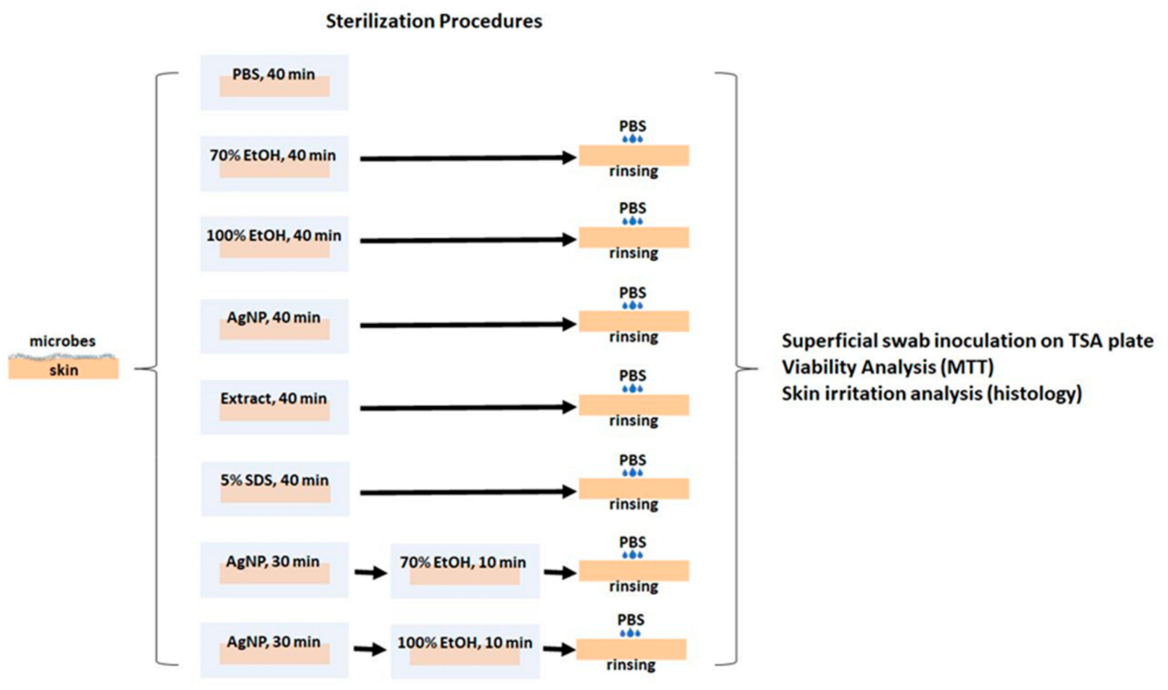
2.7. Antimicrobial Activity of AgNP Assessed in an Ex Vivo Model

To evaluate the antimicrobial activity, the sterilizing action of AgNP was tested through an ex vivo porcine skin model [24]. The sterilization protocol was performed as described in Figure 2. MTT and histology were used to determine skin viability and irritation. Photographic images of skin were collected. AgNP-treated skin (3.62 mg/mL) was compared to untreated skin soaked in PBS, in the TE used to produce the AgNP, in 70% or 100% ethanol (EtOH), and in 5% (w/v) SDS. Two-step treatments were also explored. Skin specimens were superficially swabbed with a cotton swab and plated on tryptic soy agar (TSA).
Porcine skin was acquired from a supermarket. Eight portions of skin were cut, placed in Petri dishes, and submerged in different solvents. PBS, 70% EtOH, 100% EtOH, AgNP, TE, and 5% SDS were used to submerge different skin portions for 40 min. Two other skin portions were submerged in AgNP for 30 min. After that, both skin portions were dried with a sterile gauze, and one was immersed in 70% EtOH and the other in 100% EtOH for 10 min (Figure 2). All skin samples were washed with PBS, dried with a sterile gauze, and incubated at 30 °C. The growth of microorganisms was evaluated for 14 days. Photographic images of the skin were taken on days 0, 7, and 14 after the sterilization procedure. Swabs of the skin surface were performed on days 0, 7, and 14, inoculated on TSA plates, and incubated at 37 °C. Photos of the plates were taken on days 2, 9, and 17 for inoculations made on days 0, 7, and 14, respectively.
The skin viability and cutaneous irritation were assessed for histological analysis. On day 0, six biopsies were performed on each skin fraction after the sterilization procedure. One biopsy of each group was stored in Eppendorfs with 500 µL of formalin and kept at 4 °C until histological analysis. The remaining 5 biopsies were incubated in a 96-well microplate with 100 µL of MTT at 37 °C for 3 h. Then, 100 µL of 0.1 M HCl in 2-propanol was placed in the wells and incubated overnight at 4 °C. The skin was taken, and the plates were read at 570 nm.

2.8. Animal Model of Excisional Wound

AgNP were tested in vivo in the form of a hydrogel. For this, an aqueous AgNP suspension was mixed with DermosoftTM (2%, w/w), and under vigorous stirring, Lecigel® (2%, w/v) was added to obtain a hydrogel with 1.5 mg/mL of AgNP. The placebo was made with the described method, using water instead of AgNP. Characteristics of the hydrogel, such as color and odor, were tested, and the pH was measured at 20 °C using a 713 pH meter from Metrohm (Filderstadt, Germany). Mice were anesthetized with isoflurane by inhalation. The dorsal skin was shaved and cleansed with PBS at pH 7.4, and a 1 cm line was made using a scalpel. The animals were randomly assigned to three different groups: the first group (n = 3), the negative control, was administered 100 µL of the placebo gel; the second group (n = 3) was administered 100 µL (1.5 mg/mL AgNP) of the AgNP gel; and the third group (n = 3), the positive group, was administered 100 µL of Silvederma®, a commercial cream of silver sulfadiazine (10 mg/g). These treatments were administered daily from day 0 (after incision) until day 6. Mice were accommodated individually with access to food and water ad libitum. Body weight was recorded from the beginning until the end of the assay. During wounding and each following day, macroscopic wound photographs were taken (D4S model, Nikon, Tokyo, Japan), and the area was measured (ImageJ 1.29xsoftware (National Institutes of Health, Bethesda, MD, USA)) to obtain a chronological healing profile. At the end of the assay on day 7, the mice were sacrificed. The wounded area was excised and handled for histological analysis. Hematoxylin and Eosin (H&E) staining was performed on a 4 µm longitudinal tissue area. The assessment was carried out by an independent pathologist.

2.9. Statistical Analysis

For in vitro assays, the results are expressed as mean ± standard deviation (SD) of three representative experiments. Statistical analysis was performed by one-way ANOVA with all pairwise multiple comparison procedures. For in vivo experiments, the results are expressed as mean ± standard error of the mean (SEM). One-way ANOVA followed by Tukey’s post hoc test was used.

3. Results and Discussion

Tomato by-products [20,25], ripened tomato fruits [26,27], or tomato leaves [28] have been considered for the green synthesis of AgNP. Green tomato waste has been recycled for human food products or animal feed [29]; however, this occurs on a very low scale and has only recently been considered for the extraction of natural bioactives to be incorporated into products in other industries, such as food supplements, cosmetics, and pharmaceuticals [30]. In this work, we obtained an aqueous extract from whole green tomato fruits through a green extraction process. Practically all studies using plant extracts in the synthesis of AgNP have evaluated the effect of the percentage of the extract on AgNP formation. This effect could be strongly determined by the extraction procedure and the concentration of bioactive molecules present in the extract. The results presented here describe the first report on the use of TE obtained by subcritical water extraction.

3.1. Characterization of the Tomato Extract

Total phenolic and carbohydrate compounds are important components of tomato extracts involved in the reduction and stabilization steps of AgNP biosynthesis. The aqueous tomato extract prepared by subcritical water presents a dark brown color. The carbohydrate and phenolic contents were 22.8 ± 2.9 mg GE/mL and 2.1 ± 0.4 mg GAE/mL, respectively. It is known that an increase in the temperature causes an increase in the ionic product of water [21], making it more reactive and able to catalyze the hydrolysis of the carbohydrate fraction of green tomatoes. Furthermore, the dielectric constant of water decreases with an increase in the temperature, thus allowing water to extract phenolic acids with a wide range of polarities. However, no comparison with other green tomato extracts can be made, as this is the first study of such an extract. Most of the studies regarding biogenic AgNP synthesis focus on nanoparticle characteristics and not on extract characteristics. Moreover, other biomolecules, such as proteins, amino acids, alkaloids, enzymes, saponins, tannins, terpenoids, and vitamins, present in the green tomato extract could also contribute to the formation and stabilization of the AgNP [31].

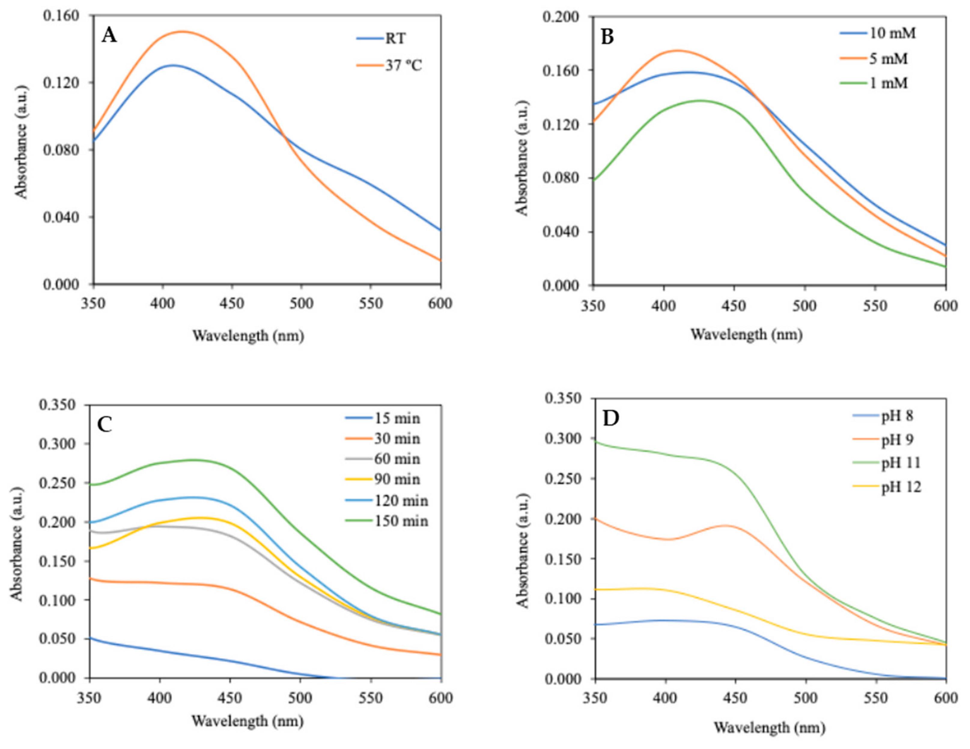
3.2. Biosynthesis of AgNP

In the current study, the biosynthesis of AgNP using a green tomato extract obtained by subcritical water treatment as a reducing agent is reported. A solution of silver nitrate salt (AgNO3) in deionized water was applied as the silver source (Ag). The Surface Plasmon Resonance (SPR) peak of AgNP ranges from 300 to 600 nm and depends on the environmental conditions, such as pH, size, shape, and the concentration of the extract or silver nitrate [32]. It has already been demonstrated that by varying key conditions/parameters in the biosynthesis of AgNP, it is possible to dramatically change the characteristics of the particles obtained [33]. In pre-formulation assays, it was possible to establish an optimal value for TE, and with this fixed concentration, different experimental conditions were tested, namely, the effects of the temperature, pH, reaction time, and concentration of AgNO3 (Figure 1). This procedure allowed the selection of the optimal conditions for the biosynthesis of AgNP using this TE.
The effect of the temperature on the SPR peak using 1.70% (v/v) TE and 1.67 mM AgNO3 is presented in Figure 3A. The reaction was carried out for 24 h in the dark at pH 11. As indicated in Table 1, the AgNP produced at 37 °C were smaller in size and had a lower PdI than those produced at room temperature. The zeta potential of AgNP produced at both temperatures was negative (<−20 mV), indicating that they can be considered stable. The intensity of the SPR peak was higher at 37 °C, which is in accordance with what has been found in other studies that correlated higher temperatures with an increased production rate of monodisperse and smaller nanoparticles [34].
Figure 3B depicts the SPR peaks resulting from different concentrations of silver nitrate tested in reaction mixtures with 0.5% (v/v) TE at room temperature. The reaction time was 2.5 h and carried out in the dark at pH 11. The SPR peak observed for the 1 mM silver nitrate concentration is narrower. The size difference in AgNP produced with 1 mM and 5 mM silver nitrate is minimal (Table 1). When the AgNO3 concentration is higher (10 mM), a reduction in the SPR peak may indicate the depletion of biomolecules in the TE, which cannot maintain AgNP formation [35].
The effect of the reaction time was evaluated using 1.67% (v/v) TE and 1.67 mM AgNO3 at 65 °C in the absence of light. The pH of the solution was 11 (Figure 3C). As seen in Table 1, all of the parameters evaluated (Z-average, PdI, and zeta potential) remained almost constant throughout the reaction time course. However, the intensity of the SPR band increased over time, indicating the continued production of AgNP. These results are in accordance with the literature and show that the reaction time is not the most critical parameter in AgNP optimization since it can be adjusted to obtain the best yield [33]. A higher temperature increases the rate of the reaction, but time is also crucial in developing stable AgNP [36]. Different pH values were evaluated using 10% (v/v) TE poured into 5 mM AgNO3, as shown in Figure 3D. The reaction time was 2 h 30 in the absence of light at room temperature. The presence of the SPR peak was only visible when the reaction was performed at pH 11. The results of the previous experiments demonstrated that the biosynthesis of silver nanoparticles is favored by alkaline pH, which is in accordance with published data [37]. Given the above-reported optimization studies, for further characterization, AgNP were biosynthesized using 2% (v/v) TE in a 2 mM AgNO3 solution at room temperature for a 150 min reaction time at pH 11 under constant stirring. The results obtained in a study using a tomato extract obtained from tomato fruits harvested at the light-red stage of ripeness are difficult to compare with our results, as the study employed different characterization techniques [26]. However, the reported average size of synthesized AgNP was between 9.6 and 72.7 nm, with the maximum size being similar to that obtained in our study.

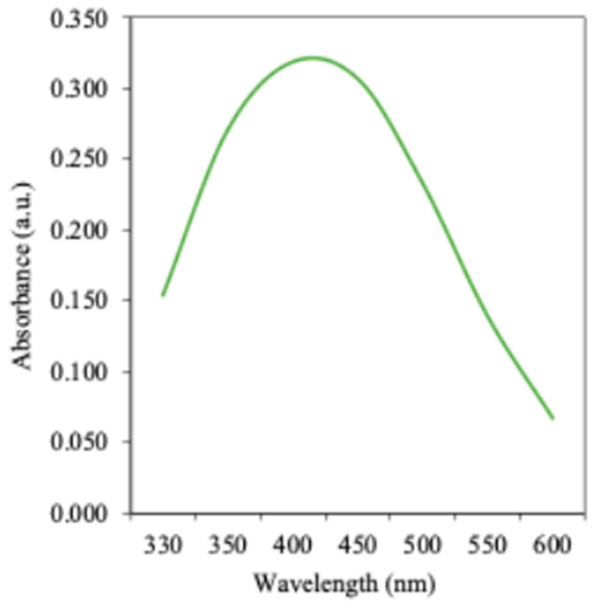
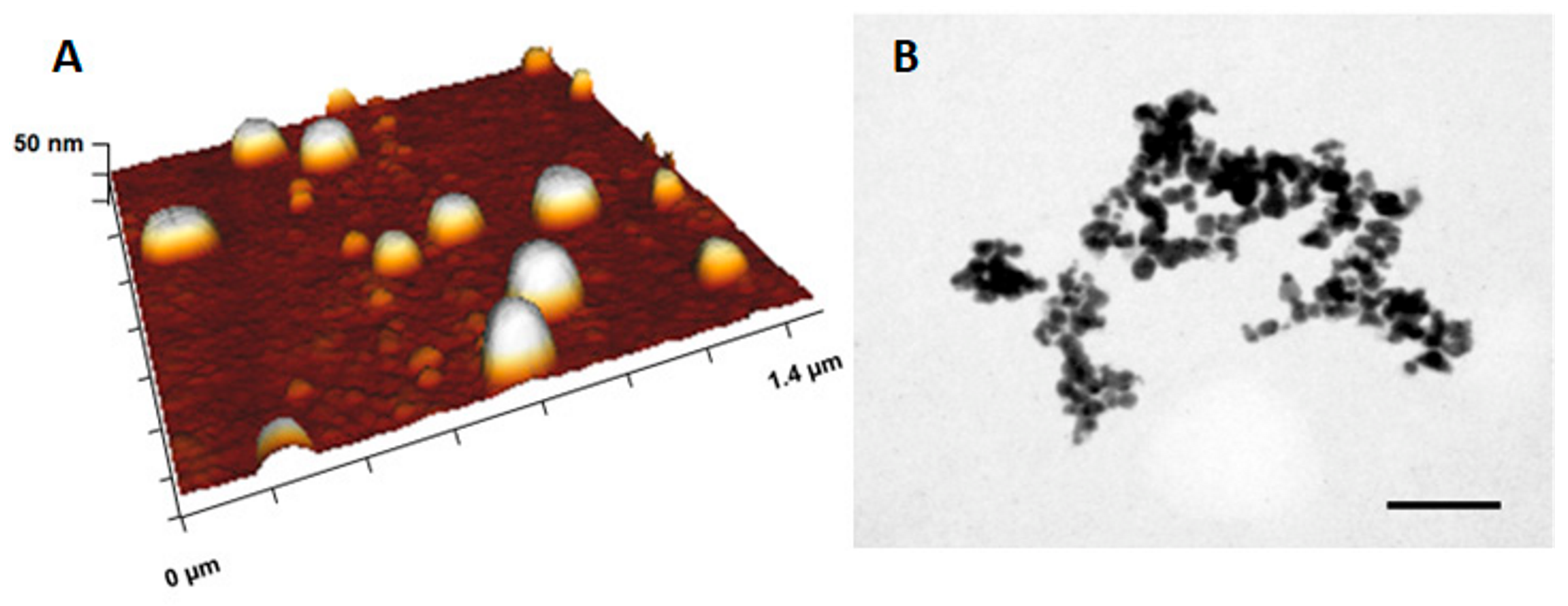
3.3. Physico-Chemical and Morphological Characterization of Optimized AgNP

As stated above, AgNP formation was monitored by measuring the SPR in the UV-Vis region. Nanoparticles were characterized in terms of their size distribution by DLS, and their morphology and state of aggregation were evaluated by AFM and TEM analyses. The stability of AgNP solutions was also evaluated through zeta potential analysis. As shown in Figure 4, the biosynthesized AgNP have an SPR peak located between 402 and 406 nm. This wavelength range suggests that the average size of AgNP produced is homogeneous since the peak is narrow. The SPR peak of small nanoparticles appears at lower wavelengths and vice versa [38]. The nanoparticles’ shape is influenced by their size and agglomeration. Some AgNP produced have a larger size, scattering more light and producing a broader SPR peak [39]. A calibration curve using different concentrations of dried AgNP was plotted to determine the concentration of the AgNP suspension. Using the equation obtained from linear regression, the mean concentration of the prepared AgNP was determined, and a value of 3.62 mg/mL was found. The carbohydrate and phenolic contents of AgNP were 0.12 ± 0.04 mg glucose equivalents/mL and 0.18 ± 0.03 mg gallic acid equivalents/mL, respectively. These values are lower compared to the values obtained for the TE used in their synthesis. However, the presence of these biomolecules in the AgNP suspension indicates that they contribute to the biosynthesis as reducing and stabilization agents for the nanoparticles [40,41,42].
DLS was used to determine the mean particle size and PdI of the AgNP suspension, which were found to be 60 ± 17 nm and 0.522 ± 0.052, respectively. This technique is a simple and fast method used to determine nanoparticle size. It can also be applied to determine the thickness of the capping or stabilizing layer surrounding metallic particles [43]. The small size of these nanoparticles is ideal for penetrating the skin, and their PdI of around 0.5 is reasonable within the criteria defined and indicates their relatively uniform dimensions. The zeta potential value is a measure of the surface charge of the synthesized AgNP, which promotes their stability in the suspension. The negative zeta potential exhibited (−42 ± 4 mV) provides repulsive electrostatic stabilization for the AgNP.
The AFM and TEM analyses used to characterize the shape, size, and distribution of the AgNP confirm that the nanoparticles have, in general, a spherical-like shape (Figure 5). Based on TEM, the estimated nanoparticle size is around 80 nm, while, according to AFM, the median size is 83 nm. According to the literature, the difference in size obtained between DLS and TEM/AFM is common and could be due to a non-homogeneous dispersion of the samples [44]. Also, for AFM and TEM measurements, the AgNP are adhered to a surface, and in DLS, the nanoparticle size is measured in solution. Spherical and oval AgNP (<87 nm) were obtained from biogenic synthesis using a tomato leaf extract [28]. The previously used tomato leaf extract differs from the one used in this work; however, similar size and polydispersity index values were reported.

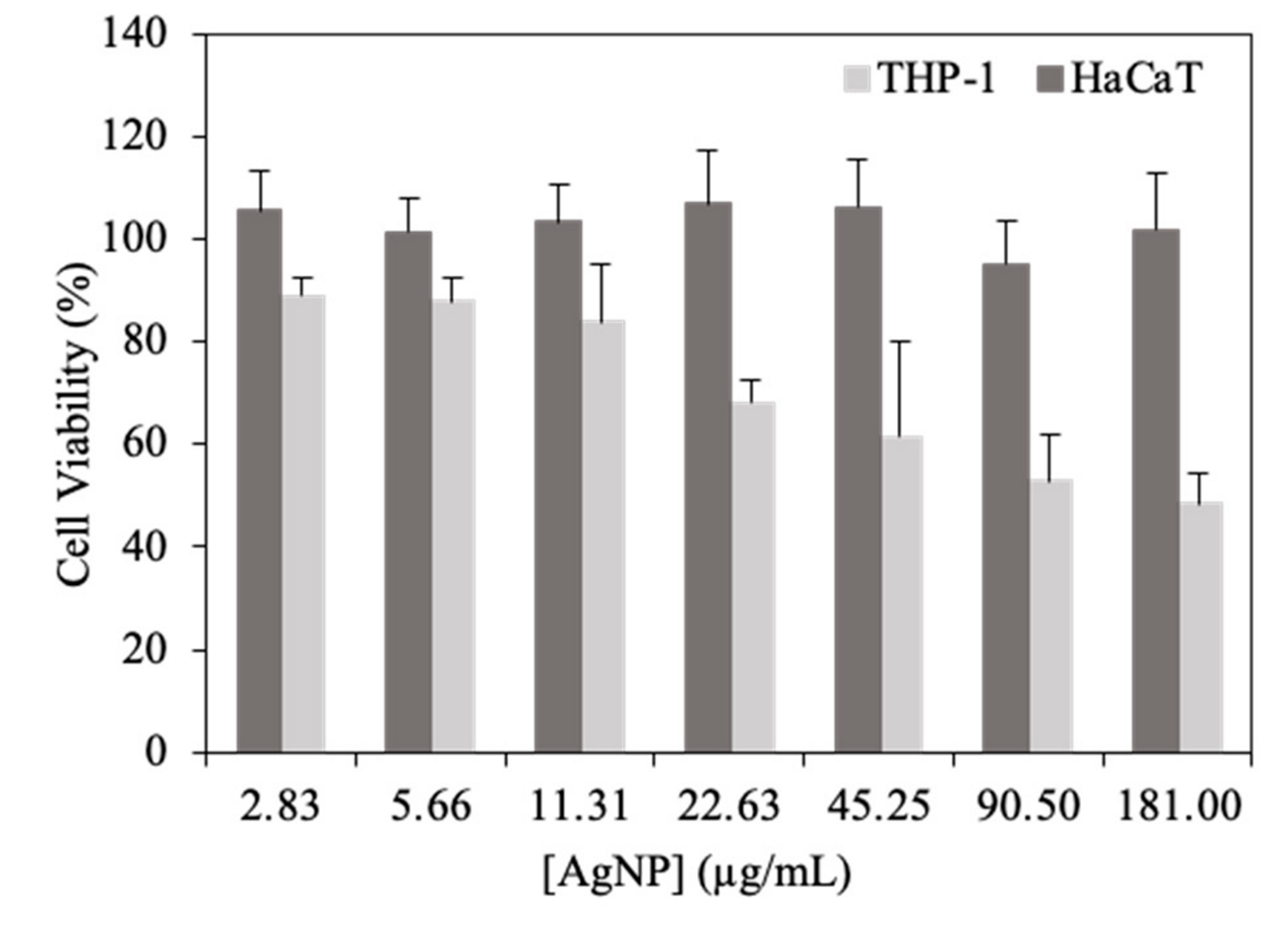
3.4. In Vitro Biocompatibility Assays

The cytotoxicity of AgNP was investigated in vitro against two human cell lines. A keratinocyte cell line (HaCaT) was used since the biosynthesized AgNP are intended for topical application; thus, it is critical that they are safe for skin cells. The human leukemia monocyte cell line THP-1 was used as host cells for microbes that infect the skin [45]. These assays were performed using the MTT colorimetric test for THP-1 cells and the MTS colorimetric test in the case of HaCaT cells. Cellular viability data for HaCaT and THP-1 macrophage-like cells exposed to AgNP are shown in Figure 6. AgNP did not show toxic effects in HaCaT cells in the range of tested concentrations. As for THP-1 cells, a dose-dependent reduction in cell viability was observed, with around 100% viability at the lower concentration decreasing to around 50% at the highest AgNP concentration tested. Overall, these findings indicate that the AgNP are not cytotoxic to either macrophages or skin cells. In vitro CC50 (50% cytotoxic concentration) values were > 181 µg/mL for HaCaT cells and 146.03 µg/mL for THP-1 cells.

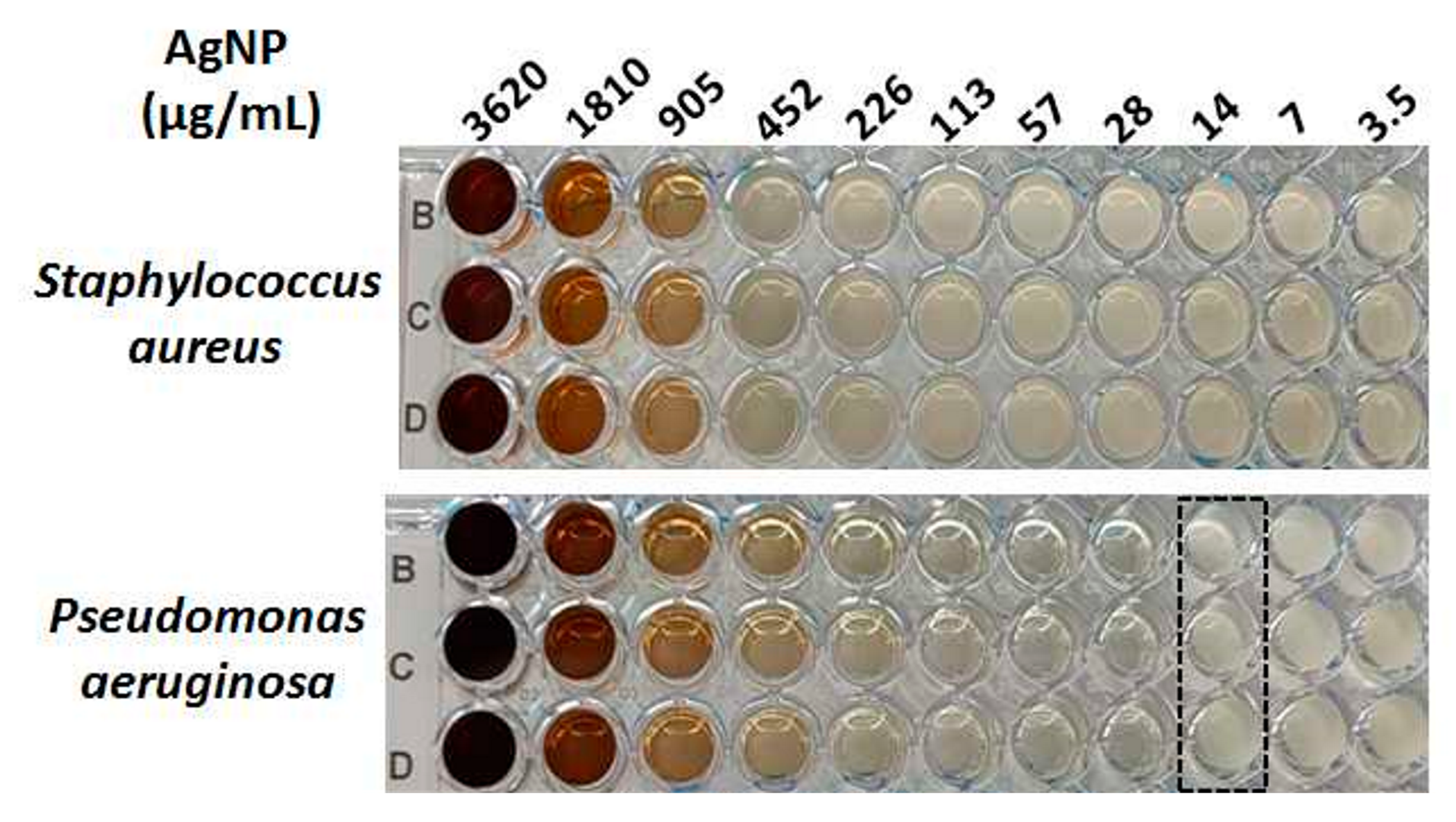
3.5. In Vitro Antimicrobial Activity

The results of the susceptibility of Staphylococcus aureus and Pseudomonas aeruginosa to the developed AgNP are shown in Figure 7. AgNP did not inhibit the growth of S. aureus at the tested concentrations. However, the MIC was able to be determined when P. aeruginosa was exposed to 14 µg/mL AgNP. These results are in accordance with reports found in the literature [46,47,48], where AgNP are described to be more efficient in Gram(−) bacteria than Gram(+). This can be justified by differences in the wall thickness of bacteria. The Gram(+) bacterial wall is almost 8 times thicker than that of Gram(−) [49]. These results are quite promising considering possible treatments for skin infections caused by Gram(−) bacteria.

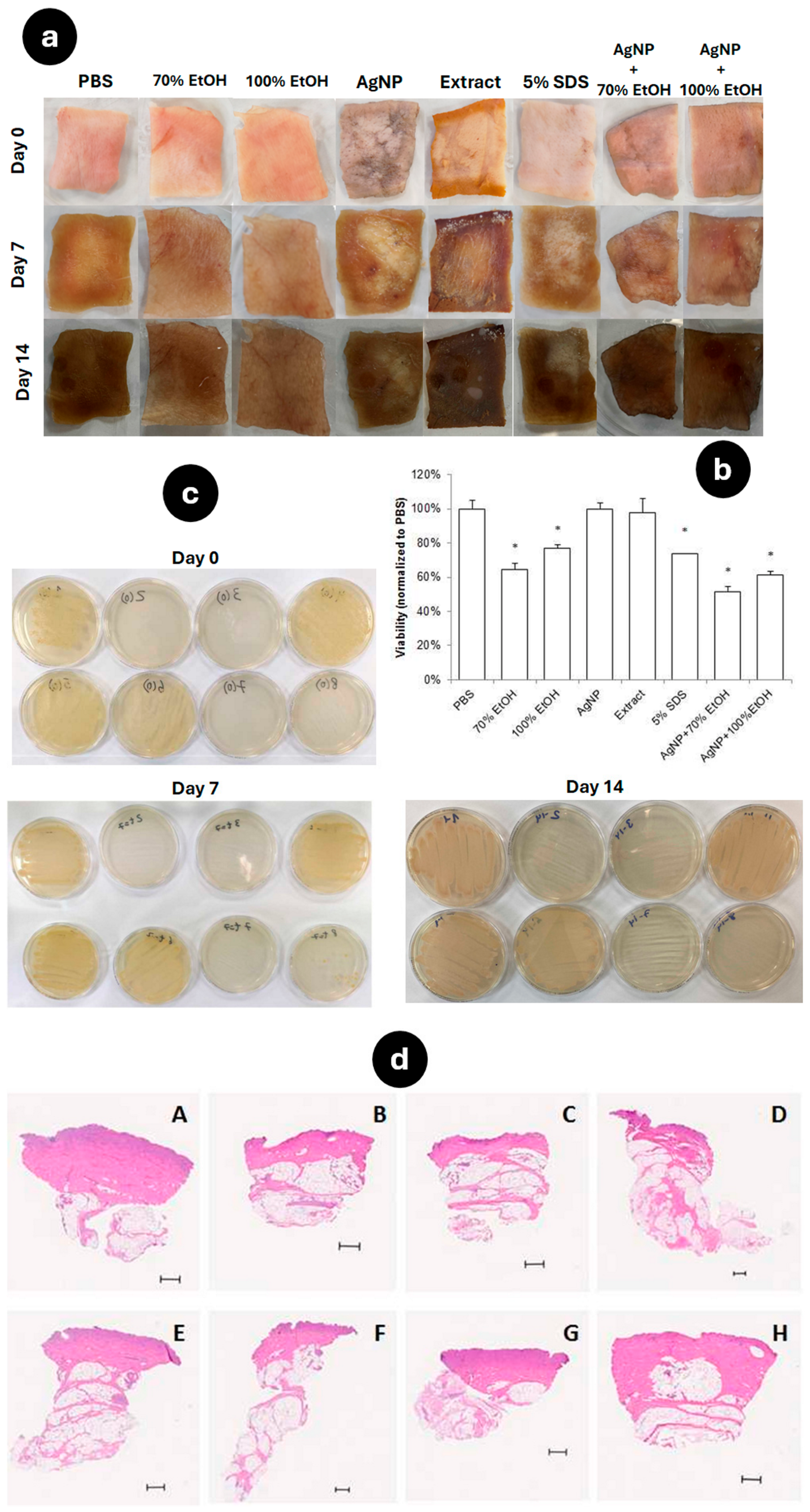
3.6. Antimicrobial Activity of AgNP Evaluated in an Ex Vivo Skin Model

AgNP have been proposed to be an excellent antimicrobial agent against bacteria both in vitro and in vivo, and scientific achievements in this area have been extensively reviewed [50]. However, studies on the effects of AgNP on the skin and their penetration have only been conducted in vitro, mainly using skin cell lines [51]. In the present work, the antimicrobial effect of the developed AgNP after topical application was compared with that of other solutions used in the disinfection of tissues and surfaces and with skin irritant solutions in an ex vivo model using full-thickness pig skin [24] initially developed to study skin bacterial translocation. PBS was used as a control in the experiments. A schematic representation of the procedure is presented in Figure 2 (Methods Section). Figure 8 shows the aspect of the skin incubated at 30 °C for 14 days from which, at defined time points, swabs were cultured on TSA plates.
In Figure 8a, the images from day 0, taken just after the soaking procedures, show a black color on the skin specimens treated with the AgNP solution, and this is more pronounced on the sample treated only with AgNP. On day 7, the skin treated with the extract already had colonies that could be seen with the naked eye. This rapid microorganism growth is explained by the high concentration of TE phytochemicals. All skin samples were darker, but ethanol-treated samples present a more preserved aspect. Over the experimental period, skin decay is more apparent. The skin portions submerged in 70% EtOH and 100% EtOH had less decay. The skin portion soaked in AgNP looks similar to the one with 5% SDS. The skin portions submerged in the extract, AgNP + 70% EtOH, and AgNP + 100% EtOH look like the control skin.
Chemical disinfectants can alter the skin structure and affect the viability of skin cells. Using the MTT assay, the viability of the cells was evaluated to understand which condition had the least impact on the skin cells (Figure 8b). Although the skin fractions treated with only EtOH looked more preserved, their overall skin viability was much lower. The viability of the skin cells was almost 80% with 100% EtOH and 5% SDS. The lower skin cell viability (<60%) was obtained with AgNP + 70% EtOH. In contrast, AgNP and extract provide a skin viability similar to PBS, almost 100%. This result shows that AgNP (non-diluted) do not compromise the skin cell viability as measured in an ex vivo model.
The swab tests (Figure 8c) had the purpose of assessing the sterilization that each solution provided during the time course of the assay. On day 0, skin samples treated with 70% EtOH, 100% EtOH, AgNP + 70% EtOH, and AgNP + 100% EtOH solutions show no growth of microorganisms. The skin slices treated with the TE or 5% SDS presented similar results, showing some microbial growth. PBS and AgNP presented comparable results, showing more colonies than the previous solutions. On days 7 and 14, plates with PBS, TE, 5% SDS, and AgNP show similar aspects, and the microbial growth is equivalent.
The histological results were crucial in determining the cutaneous irritation caused by the solvents used (Figure 8d). It is recognized that the source of porcine skin can have a substantial impact on contamination, epidermis structure, and usable time. Compared to PBS (Figure 8d-A), after treatment with 70% EtOH, 100% EtOH, AgNP, and 5% SDS, the skin’s superficial layer seems to be contracted. The extract, AgNP + 70% EtOH, and AgNP + 100% EtOH did not cause changes in the thickness of the skin layers when compared to PBS. These results indicate that the presence of AgNP with EtOH can decrease ethanol’s cutaneous irritation potential.

3.7. Wound-Healing Effect of AgNP in a Murine Incisional Acute Wound Model

In vivo preclinical wound-healing research commonly uses mouse models, either incisional or excisional [52]. In order to formulate AgNP in a semisolid pharmaceutical form, a hydrogel containing AgNP and the respective placebo were prepared. Semisolid formulations are convenient forms for topical applications since they increase the drug residence time on the skin [53]. The gel composition was selected from a previous study, as it showed compatibility with human skin without any irritation signs [54]. A gel containing 1.5 mg/mL AgNP was used to assess the healing performance of AgNP. The topical administration of the AgNP gel produced, of Silvederma®, a commercial cream of silver sulfadiazine (10 mg/g), and of the placebo started on day 0, and wounds were treated in the following 7 days. In Figure 9A, representative photos of the skin incision treatments at different time points are shown. During the assay, the body weight variation was less than 10% and not statistically different between the groups. As can be seen in the graphic (Figure 9B), mice treated with the placebo had a larger wound area, which was different from that present on mice treated with Silvederma® or AgNP (p ˂ 0.05). Statistically, there is no difference between Silvederma® and AgNP. Non-treated wounds increased in area in the initial 2–3 days, an indicator of the variable expansion of wound margins [52]. This phenomenon was not observed in the groups treated with Silvederma® or gel containing AgNP. The placebo wounds did not complete closure during the 7 days of the trial. Silvederma® has a higher silver concentration than the AgNP gel, both ensuring rapid wound closure. These results are very encouraging and highlight the potential of AgNP in wound healing.
The histological results presented in Figure 10 confirm that wound closure was successful in the groups treated with AgNP and Silvederma®. Figure 10A shows that the wound treated with the placebo did not close. The healing process looks more advanced with the AgNP formulation compared with silver sulfadiazine. The histological analysis corroborates the results obtained in the wound area evaluation (Figure 9).

4. Conclusions

The demand for the sustainable production of therapeutics is increasing. The biosynthesis of nanoparticles is an opportunity to use upcycled industrial waste since plants are rich in terpenoids, alkaloids, and phenolics, and any of these molecules can be used for the synthesis. This type of synthesis can also overcome the toxicity associated with chemical synthesis. Biogenic silver nanoparticles have interesting pharmacological features that look promising for the topical treatment of skin diseases. The unique chemical composition of the plant extract affects the characteristics and properties of the AgNP. To the best of our knowledge, this is the first report on the use of a green-tomato-derived extract obtained by subcritical water treatment for the biosynthesis of metallic nanoparticles. This subcritical water extraction process preserved the phenolic content of the green tomato extract. The pre-formulation studies elucidated the role of the main parameters involved in the biosynthesis of AgNP, namely, pH, silver nitrate concentration, time, and temperature. AgNP formation can be confirmed by a color change and by the SPR band in the UV-vis absorption spectrum. The AgNP produced had the proper size, a reasonable polydispersity index, and good stability, confirmed by the value of its zeta potential. AFM and TEM confirmed that AgNP had a spherical-like shape, and the size distribution was homogeneous. The size obtained by these methods can be considered suitable for topical administration [55]. In vitro assays confirmed the cytocompatible nature of the AgNP toward human cells. Further studies are needed to assess the internalization and elimination profiles of AgNP to accurately estimate the potential adverse effects associated with their interaction with human cells. In vitro antimicrobial activity showed the susceptibility of Gram(−) bacteria to biogenic AgNP. The ex vivo antimicrobial assay reinforces the safety of AgNP after topical application. The in vivo assay demonstrated good wound-healing properties, comparable to commercial silver-based topical formulations. Considering all of the results, this study demonstrated the potential of industrial waste (green tomatoes) to be used in a green approach to the biosynthesis of AgNP under simple and eco-friendly conditions. The developed AgNP were revealed to have antimicrobial and wound-healing properties with an adequate profile for future biomedical applications.

Author Contributions

Conceptualization, M.C. and S.S.; methodology, D.C.; software, C.F.-S.; validation, L.A., F.A.C. and P.S.; formal analysis, D.C.; investigation, M.C. and S.S.; resources, M.C. and S.S.; writing—original draft preparation, D.C.; writing—review and editing, D.C., C.F.-S., F.A.C., L.A., P.S., M.C. and S.S.; supervision, M.C. and S.S. All authors have read and agreed to the published version of the manuscript.

Funding

The authors acknowledge Fundação para a Ciência e a Tecnologia (FCT) through projects UIDB/04138/2020, UIDP/04138/2020, LA/P/0008/2020, UIDP/50006/2020, and UIDB/50006/2020. They also acknowledge financial support to CESAM by FCT/MCTES (UIDP/50017/2020 + UIDB/50017/2020 + LA/P/0094/2020).

Data Availability Statement

The raw data supporting the conclusions of this article will be made available by the authors on request.

Conflicts of Interest

The authors declare no conflicts of interest.

References

  1. Catauro, M.; Raucci, M.G.; de Gaetano, F.; Marotta, A. Antibacterial and bioactive silver-containing Na2O·CaO·2SiO2 glass prepared by sol–gel method. J. Mater. Sci. Mater. Med. 2004, 15, 831–837. [Google Scholar] [CrossRef] [PubMed]
  2. Crabtree, J.H.; Burchette, R.J.; Siddiqi, R.A.; Huen, I.T.; Hadnott, L.L.; Fishman, A. The Efficacy of Silver-Ion Implanted Catheters in Reducing Peritoneal Dialysis-Related Infections. Perit. Dial. Int. 2003, 23, 368–374. [Google Scholar] [CrossRef] [PubMed]
  3. Maillard, J.-Y.; Hartemann, P. Silver as an antimicrobial: Facts and gaps in knowledge. Crit. Rev. Microbiol. 2013, 39, 373–383. [Google Scholar] [CrossRef] [PubMed]
  4. Zhang, X.-F.; Liu, Z.-G.; Shen, W.; Gurunathan, S. Silver Nanoparticles: Synthesis, Characterization, Properties, Applications, and Therapeutic Approaches. Int. J. Mol. Sci. 2016, 17, 1534. [Google Scholar] [CrossRef] [PubMed]
  5. Iravani, S. Green synthesis of metal nanoparticles using plants. Green. Chem. 2011, 13, 2638. [Google Scholar] [CrossRef]
  6. Jamkhande, P.G.; Ghule, N.W.; Bamer, A.H.; Kalaskar, M.G. Metal nanoparticles synthesis: An overview on methods of preparation, advantages and disadvantages, and applications. J. Drug Deliv. Sci. Technol. 2019, 53, 101174. [Google Scholar] [CrossRef]
  7. Agnihotri, S.; Mukherji, S.; Mukherji, S. Size-controlled silver nanoparticles synthesized over the range 5–100 nm using the same protocol and their antibacterial efficacy. RSC Adv. 2014, 4, 3974–3983. [Google Scholar] [CrossRef]
  8. Garg, D.; Sarkar, A.; Chand, P.; Bansal, P.; Gola, D.; Sharma, S.; Khantwal, S.; Surabhi; Mehrotra, R.; Chauhan, N.; et al. Synthesis of silver nanoparticles utilizing various biological systems: Mechanisms and applications-A review. Prog. Biomater. 2020, 9, 81–95. [Google Scholar] [CrossRef] [PubMed]
  9. Dhir, R.; Chauhan, S.; Subham, P.; Kumar, S.; Sharma, P.; Shidiki, A.; Kumar, G. Plant-mediated synthesis of silver nanoparticles: Unlocking their pharmacological potential-a comprehensive review. Front. Bioeng. Biotechnol. 2024, 11, 1324805. [Google Scholar] [CrossRef]
  10. Iravani, S.; Korbekandi, H.; Mirmohammadi, S.V.; Zolfaghari, B. Synthesis of silver nanoparticles: Chemical, physical and biological methods. Res. Pharm. Sci. 2014, 9, 385–406. [Google Scholar] [PubMed]
  11. Aramwit, P.; Bang, N.; Ratanavaraporn, J.; Ekgasit, S. Green synthesis of silk sericin-capped silver nanoparticles and their potent anti-bacterial activity. Nanoscale Res. Lett. 2014, 9, 79. [Google Scholar] [CrossRef]
  12. Siddiqi, K.S.; Husen, A.; Rao, R.A.K. A review on biosynthesis of silver nanoparticles and their biocidal properties. J. Nanobiotechnol. 2018, 16, 14. [Google Scholar] [CrossRef]
  13. Kumar, R.; Roopan, S.M.; Prabhakarn, A.; Khanna, V.G.; Chakroborty, S. Agricultural waste Annona squamosa peel extract: Biosynthesis of silver nanoparticles. Spectrochim. Acta A Mol. Biomol. Spectrosc. 2012, 90, 173–176. [Google Scholar] [CrossRef]
  14. Roopan, S.M.; Bharathi, A.; Prabhakarn, A.; Abdul Rahuman, A.; Velayutham, K.; Rajakumar, G.; Padmaja, R.D.; Lekshmi, M.; Madhumitha, G. Efficient phyto-synthesis and structural characterization of rutile TiO2 nanoparticles using Annona squamosa peel extract. Spectrochim. Acta A Mol. Biomol. Spectrosc. 2012, 98, 86–90. [Google Scholar] [CrossRef]
  15. Ansari, M.; Ahmed, S.; Abbasi, A.; Khan, M.T.; Subhan, M.; Bukhari, N.A.; Hatamleh, A.A.; Abdelsalam, N.R. Plant mediated fabrication of silver nanoparticles, process optimization, and impact on tomato plant. Sci. Rep. 2023, 13, 18048. [Google Scholar] [CrossRef] [PubMed]
  16. Faria-Silva, C.; de Sousa, M.; Carvalheiro, M.C.; Simões, P.; Simões, S. Alpha-tomatine and the two sides of the same coin: An anti-nutritional glycoalkaloid with potential in human health. Food Chem. 2022, 391, 133261. [Google Scholar] [CrossRef]
  17. Costa, A.; Marques, M.; Congiu, F.; Paiva, A.; Simões, P.; Ferreira, A.; Bronze, M.R.; Marto, J.; Ribeiro, H.M.; Simões, S. Evaluating the Presence of Lycopene-Enriched Extracts from Tomato on Topical Emulsions: Physico-Chemical Characterization and Sensory Analysis. Appl. Sci. 2021, 11, 5120. [Google Scholar] [CrossRef]
  18. Faria-Silva, C.; Eleutério, C.; Simões, P.; Carvalheiro, M.; Simões, S. A new method for quantification of tomatine-enriched extracts. J. Sci. Food Agric. 2024. [Google Scholar] [CrossRef]
  19. Galamba, N.; Paiva, A.; Barreiros, S.; Simões, P. Solubility of Polar and Nonpolar Aromatic Molecules in Subcritical Water: The Role of the Dielectric Constant. J. Chem. Theory Comput. 2019, 15, 6277–6293. [Google Scholar] [CrossRef] [PubMed]
  20. Balčiūnaitienė, A.; Viškelis, J.; Urbonavičienė, D.; Viškelis, P. Tomatoes By-Products Extracts Mediated Green Synthesis of Silver Nanoparticles and Their Application as Antimicrobial Agent. In Tomato—From Cultivation to Processing Technology; Viškelis, P., Urbonavičienė, D., Viškelis, J., Eds.; IntechOpen Limited: London, UK, 2022. [Google Scholar] [CrossRef]
  21. Pedras, B.; Salema-Oom, M.; Sá-Nogueira, I.; Simões, P.; Paiva, A.; Barreiros, S. Valorization of white wine grape pomace through application of subcritical water: Analysis of extraction, hydrolysis, and biological activity of the extracts obtained. J. Supercrit. Fluids 2017, 128, 138–144. [Google Scholar] [CrossRef]
  22. Singleton, V.L.; Rossi, J.A. Colorimetry of Total Phenolics with Phosphomolybdic-Phosphotungstic Acid Reagents. Am. J. Enol. Vitic. 1965, 16, 144–158. [Google Scholar] [CrossRef]
  23. Jayachandran, P.; Ilango, S.; Suseela, V.; Nirmaladevi, R.; Shaik, M.R.; Khan, M.; Khan, M.; Shaik, B. Green Synthesized Silver Nanoparticle-Loaded Liposome-Based Nanoarchitectonics for Cancer Management: In Vitro Drug Release Analysis. Biomedicines 2023, 11, 217. [Google Scholar] [CrossRef]
  24. Wang, H.; Agrawal, A.; Wang, Y.; Crawford, D.W.; Siler, Z.D.; Peterson, M.L.; Woofter, R.T.; Labib, M.; Shin, H.Y.; Baumann, A.P.; et al. An ex vivo model of medical device-mediated bacterial skin translocation. Sci. Rep. 2021, 11, 5746. [Google Scholar] [CrossRef] [PubMed]
  25. Carbone, K.; de Angelis, A.; Mazzuca, C.; Santangelo, E.; Macchioni, V.; Cacciotti, I.; Petrella, G.; Cicero, D.; Micheli, L. Microwave-assisted synthesis of catalytic silver nanoparticles by hyperpigmented tomato skins: A green approach. LWT 2020, 133, 110088. [Google Scholar] [CrossRef]
  26. Mohamed, M.; Faraj, K.M.; Al-Jobori, K. Green Synthesis of Silver Nanoparticles Using Tomato (Lycopersicon esculentum) Extract and Evaluation of their Antifungal Activities. Plant Arch. 2020, 20, 5777–5786. [Google Scholar]
  27. Zia, M.; Gul, S.; Akhtar, J.; Haq, I.U.; Abbasi, B.H.; Hussain, A.; Naz, S.; Chaudhary, M.F. Green synthesis of silver nanoparticles from grape and tomato juices and evaluation of biological activities. IET Nanobiotechnol. 2017, 11, 193–199. [Google Scholar] [CrossRef]
  28. Santiago, T.R.; Bonatto, C.C.; Rossato, M.; Lopes, C.A.P.; Lopes, C.A.; G Mizubuti, E.S.; Silva, L.P. Green synthesis of silver nanoparticles using tomato leaf extract and their entrapment in chitosan nanoparticles to control bacterial wilt. J. Sci. Food Agric. 2019, 99, 4248–4259. [Google Scholar] [CrossRef] [PubMed]
  29. Piccolo, V.; Pastore, A.; Maisto, M.; Keivani, N.; Tenore, G.C.; Stornaiuolo, M.; Summa, V. Agri-Food Waste Recycling for Healthy Remedies: Biomedical Potential of Nutraceuticals from Unripe Tomatoes (Solanum lycopersicum L.). Foods 2024, 13, 331. [Google Scholar] [CrossRef] [PubMed]
  30. Simões, S.; Costa, A.; Faria-Silva, A.C.; Ascenso, A.; Marto, J.; Carvalheiro, M.; Gonçalves, L.M.; Marques, M.; Paiva, A.; Bento, M.; et al. Sustainable valorization of food-processing industry by-products: Challenges and opportunities to obtain bioactive compounds. In Valorization of Agri-Food Wastes and By-Products; Bhat, R., Ed.; Elsevier: Amsterdam, The Netherlands, 2021; pp. 619–644. [Google Scholar] [CrossRef]
  31. Ghosh, P.; Fawcett, D.; Sharma, S.; Poinern, G. Production of High-Value Nanoparticles via Biogenic Processes Using Aquacultural and Horticultural Food Waste. Materials 2017, 10, 852. [Google Scholar] [CrossRef]
  32. Mehrdel, B.; Aziz, A.A. The Sensitivity of Surface Plasmon Resonance Damping for Colloidal Silver Nanoparticles. J. Phys. Conf. Ser. 2018, 1083, 012042. [Google Scholar] [CrossRef]
  33. Saha, P.; Mahiuddin, M.; Islam, A.B.M.N.; Ochiai, B. Biogenic Synthesis and Catalytic Efficacy of Silver Nanoparticles Based on Peel Extracts of Citrus macroptera Fruit. ACS Omega 2021, 6, 18260–18268. [Google Scholar] [CrossRef] [PubMed]
  34. Liu, H.; Zhang, H.; Wang, J.; Wei, J. Effect of temperature on the size of biosynthesized silver nanoparticle: Deep insight into microscopic kinetics analysis. Arab. J. Chem. 2020, 13, 1011–1019. [Google Scholar] [CrossRef]
  35. Philip, D. Biosynthesis of Au, Ag and Au–Ag nanoparticles using edible mushroom extract. Spectrochim. Acta A Mol. Biomol. Spectrosc. 2009, 73, 374–381. [Google Scholar] [CrossRef] [PubMed]
  36. Veerasamy, R.; Xin, T.Z.; Gunasagaran, S.; Xiang, T.F.W.; Yang, E.F.C.; Jeyakumar, N.; Dhanaraj, S.A. Biosynthesis of silver nanoparticles using mangosteen leaf extract and evaluation of their antimicrobial activities. J. Saudi Chem. Soc. 2011, 15, 113–120. [Google Scholar] [CrossRef]
  37. Handayani, W.; Ningrum, A.S.; Imawan, C. The Role of pH in Synthesis Silver Nanoparticles Using Pometia pinnata (Matoa) Leaves Extract as Bioreductor. J. Phys. Conf. Ser. 2020, 1428, 012021. [Google Scholar] [CrossRef]
  38. Ider, M.; Abderrafi, K.; Eddahbi, A.; Ouaskit, S.; Kassiba, A. Silver Metallic Nanoparticles with Surface Plasmon Resonance: Synthesis and Characterizations. J. Clust. Sci. 2017, 28, 1051–1069. [Google Scholar] [CrossRef]
  39. Paramelle, D.; Sadovoy, A.; Gorelik, S.; Free, P.; Hobley, J.; Fernig, D.G. A rapid method to estimate the concentration of citrate capped silver nanoparticles from UV-visible light spectra. Analyst 2014, 139, 4855. [Google Scholar] [CrossRef] [PubMed]
  40. Firoozi, S.; Jamzad, M.; Yari, M. Biologically synthesized silver nanoparticles by aqueous extract of Satureja intermedia C.A. Mey and the evaluation of total phenolic and flavonoid contents and antioxidant activity. J. Nanostruct. Chem. 2016, 6, 357–364. [Google Scholar] [CrossRef]
  41. Mittal, A.K.; Bhaumik, J.; Kumar, S.; Banerjee, U.C. Biosynthesis of silver nanoparticles: Elucidation of prospective mechanism and therapeutic potential. J. Colloid. Interface Sci. 2014, 415, 39–47. [Google Scholar] [CrossRef] [PubMed]
  42. Simon, S.; Sibuyi, N.R.S.; Fadaka, A.O.; Meyer, M.; Madiehe, A.M.; du Preez, M.G. The antimicrobial activity of biogenic silver nanoparticles synthesized from extracts of Red and Green European pear cultivars. Artif. Cells Nanomed. Biotechnol. 2021, 49, 613–624. [Google Scholar] [CrossRef] [PubMed]
  43. Khane, Y.; Benouis, K.; Albukhaty, S.; Sulaiman, G.M.; Abomughaid, M.M.; Al Ali, A.; Aouf, D.; Fenniche, F.; Khane, S.; Chaibi, W.; et al. Green Synthesis of Silver Nanoparticles Using Aqueous Citrus limon Zest Extract: Characterization and Evaluation of Their Antioxidant and Antimicrobial Properties. Nanomaterials 2022, 12, 2013. [Google Scholar] [CrossRef] [PubMed]
  44. Singh, T.; Jyoti, K.; Patnaik, A.; Singh, A.; Chauhan, R.; Chandel, S.S. Biosynthesis, characterization and antibacterial activity of silver nanoparticles using an endophytic fungal supernatant of Raphanus sativus. J. Genet. Eng. Biotechnol. 2017, 15, 31–39. [Google Scholar] [CrossRef] [PubMed]
  45. Fischer, N.; Mak, T.N.; Shinohara, D.B.; Sfanos, K.S.; Meyer, T.F.; Brüggemann, H. Deciphering the Intracellular Fate of Propionibacterium acnes in Macrophages. BioMed Res. Int. 2013, 2013, 603046. [Google Scholar] [CrossRef] [PubMed]
  46. Radzig, M.A.; Nadtochenko, V.A.; Koksharova, O.A.; Kiwi, J.; Lipasova, V.A.; Khmel, I.A. Antibacterial effects of silver nanoparticles on gram-negative bacteria: Influence on the growth and biofilms formation, mechanisms of action. Colloids Surf. B Biointerfaces 2013, 102, 300–306. [Google Scholar] [CrossRef] [PubMed]
  47. Cavassin, E.D.; de Figueiredo, L.F.P.; Otoch, J.P.; Seckler, M.M.; de Oliveira, R.A.; Franco, F.F.; Marangoni, V.S.; Zucolotto, V.; Levin, A.S.S.; Costa, S.F. Comparison of methods to detect the in vitro activity of silver nanoparticles (AgNP) against multidrug resistant bacteria. J. Nanobiotechnol. 2015, 13, 64. [Google Scholar] [CrossRef] [PubMed]
  48. Qing, Y.; Cheng, L.; Li, R.; Liu, G.; Zhang, Y.; Tang, X.; Wang, J.; Liu, H.; Qin, Y. Potential antibacterial mechanism of silver nanoparticles and the optimization of orthopedic implants by advanced modification technologies. Int. J. Nanomed. 2018, 13, 3311–3327. [Google Scholar] [CrossRef] [PubMed]
  49. Chatterjee, T.; Chatterjee, B.K.; Majumdar, D.; Chakrabarti, P. Antibacterial effect of silver nanoparticles and the modeling of bacterial growth kinetics using a modified Gompertz model. Biochim. Biophys. Acta 2015, 1850, 299–306. [Google Scholar] [CrossRef] [PubMed]
  50. Bruna, T.; Maldonado-Bravo, F.; Jara, P.; Caro, N. Silver Nanoparticles and Their Antibacterial Applications. Int. J. Mol. Sci. 2021, 22, 7202. [Google Scholar] [CrossRef] [PubMed]
  51. Zhang, X.F.; Shen, W.; Gurunathan, S. Silver Nanoparticle-Mediated Cellular Responses in Various Cell Lines: An in Vitro Model. Int. J. Mol. Sci. 2016, 17, 1603. [Google Scholar] [CrossRef] [PubMed]
  52. Ansell, D.M.; Campbell, L.; Thomason, H.A.; Brass, A.; Hardman, M.J. A statistical analysis of murine incisional and excisional acute wound models. Wound Repair. Regen. 2014, 22, 281–287. [Google Scholar] [CrossRef] [PubMed]
  53. Salgado, A.; Raposo, S.; Marto, J.; Silva, A.N.; Simões, S.; Ribeiro, H.M. Mometasone furoate hydrogel for scalp use: In vitro and in vivo evaluation. Pharm. Dev. Technol. 2014, 19, 618–622. [Google Scholar] [CrossRef] [PubMed]
  54. Marto, J.; Neves, Â.; Gonçalves, L.; Pinto, P.; Almeida, C.; Simões, S. Rice Water: A Traditional Ingredient with Anti-Aging Efficacy. Cosmetics 2018, 5, 26. [Google Scholar] [CrossRef]
  55. Ovais, M.; Ahmad, I.; Khalil, A.T.; Mukherjee, S.; Javed, R.; Ayaz, M.; Raza, A.; Shinwari, Z.K. Wound healing applications of biogenic colloidal silver and gold nanoparticles: Recent trends and future prospects. Appl. Microbiol. Biotechnol. 2018, 102, 4305–4318. [Google Scholar] [CrossRef] [PubMed]
Figure 1. Four different sets of conditions for the optimization of the AgNP synthesis. Fixed conditions of the reaction parameters for the variables tested. RT: room temperature.
Applsci 14 07309 g001
Figure 2. An illustration of the procedure for the ex vivo model.
Applsci 14 07309 g002
Figure 3. UV-Vis spectra of AgNP produced under different experimental conditions. (A) Effect of temperature (RT, 37 °C); (B) effect of concentration of AgNO3 (1–10 mM); (C) effect of reaction time (15–180 min); (D) effect of pH (9, 11).
Applsci 14 07309 g003
Figure 4. UV−vis spectrum of biosynthesized AgNP obtained from TE (5 mL) and AgNO3 (2 mM, 245 mL) at room temperature after 150 min reaction time at pH 11.
Applsci 14 07309 g004
Figure 5. Morphological characterization of biosynthesized silver nanoparticles: (A) AFM images of AgNP diluted 1/10,000; (B) representative TEM micrograph of AgNP. Scale bar = 350 nm.
Applsci 14 07309 g005
Figure 6. The evaluation of AgNP cytocompatibility in mammalian cell lines. The effect of increasing concentrations of AgNP on the viability of HaCaT and THP-1 cells after 48 h of incubation. The percentage of cell viability was calculated relative to the control using a colorimetric assay. Data are expressed as the mean ± SD of 3 independent measurements.
Applsci 14 07309 g006
Figure 7. The microscopic appearance of bacterial growth in the presence of AgNP measured by the broth microdilution assay. Note that the concentration of AgNP decreases from left to right. The dashed line indicates the MIC (µg/mL). The B, C, and D lines correspond to replicates.
Applsci 14 07309 g007
Figure 8. The antimicrobial activity of AgNP in an ex vivo porcine skin model. (a) Images of the skin portions on day 0, day 7, and day 14 after the sterilization procedure. (b) An analysis of skin tissue viability after the sterilization procedure compared with the no-treatment control (PBS). The viability analysis data are plotted as mean ± SD (n = 5). Statistical analysis: * p < 0.001 vs. PBS. (c) Microbial growth on TSA agar from swabs of the porcine skin portions, where, in each image, the following treatments are indicated: top row, from left to right: 1—PBS for 40 min immersion; 2—70% EtOH for 40 min immersion; 3—100% EtOH for 40 min immersion; 4—AgNP for 40 min immersion; bottom row, from left to right: 5—extract for 40 min immersion; 6—5% SDS for 40 min immersion; 7—AgNP for 30 min + 70% EtOH for 10 min immersion; and 8—AgNP for 30 min + 100% EtOH for 10 min immersion. (d) Microphotographs of H&E-stained skin slices after different experimental treatments: A—PBS for 40 min immersion; B—70% EtOH for 40 min immersion; C—100% EtOH for 40 min immersion; D—AgNP for 40 min immersion; E—extract for 40 min immersion; F—5% SDS for 40 min immersion; G—AgNP for 30 min + 70% EtOH for 10 min immersion; and H—AgNP for 30 min + 100% EtOH for 10 min immersion. Scale bars = 500 µm.
Applsci 14 07309 g008
Figure 9. The wound-healing effect of AgNP in a murine acute incisional wound model. (A) Representative images of the incisional wounds on days 1, 5, and 7 of treatment with the placebo (vehicle of AgNP gel), AgNP gel, and Silvederma® cream (silver sulfadiazine 10 mg/g). (B) Wound area evaluation during treatment with the placebo gel, Silvederma® cream, and AgNP gel. Data are expressed as the mean ± SEM of 3 independent measurements. Statistics: one-way ANOVA followed by Tukey’s post hoc test. * p < 0.05 vs. control.
Applsci 14 07309 g009
Figure 10. Representative images of histological sections of incisional wounds after 7 days of administration. (A) Placebo gel; (B) Silvederma® cream; (C) AgNP formulation. Scale bars = 200 µm.
Applsci 14 07309 g010
Table 1. The reaction parameters, mean size, PdI, and zeta potential of AgNP obtained from the TE. Data represent the mean ± standard deviation of three measurements. n.d.: no data.
Manipulated ParameterZ-Average (nm)PdIZeta Potential (mV)
Time15 min163 ± 220.32 ± 0.08−22 ± 1
30 min235 ± 470.29 ± 0.04−28 ± 1
60 min222 ± 250.30 ± 0.03−28 ± 1
90 min191 ± 270.29 ± 0.02−29 ± 1
120 min290 ± 350.36 ± 0.07−29 ± 3
150 min246 ± 340.34 ± 0.04−32 ± 0
TemperatureRoom51 ± 10.34 ± 0.05−29 ± 1
37 °C35 ± 00.31 ± 0.03−21 ± 3
AgNO31 mM23 ± 10.61 ± 0.01n.d.
5 mM29 ± 60.50 ± 0.19n.d.
10 mM>1000-n.d.
pHpH 8245 ± 50.43 ± 0.17−22 ± 2
pH 9226 ± 280.51 ± 0.02−22 ± 1
pH 11160 ± 30.32 ± 0.03−31 ± 1
pH 12>1000-n.d.
Disclaimer/Publisher’s Note: The statements, opinions and data contained in all publications are solely those of the individual author(s) and contributor(s) and not of MDPI and/or the editor(s). MDPI and/or the editor(s) disclaim responsibility for any injury to people or property resulting from any ideas, methods, instructions or products referred to in the content.

