Review

Characterizations of Interfacial Solar Water Evaporation

1
College of Science, Civil Aviation University of China, Jinbei Road 2898, Tianjin 300300, China
2
MOE Key Laboratory of Weak-Light Nonlinear Photonics, Ministry of Education, TEDA Applied Physics Institute and School of Physics, Nankai University, Tianjin 300457, China
*
Authors to whom correspondence should be addressed.
Appl. Sci. 2024, 14(17), 7523; https://doi.org/10.3390/app14177523
Submission received: 26 June 2024 / Revised: 17 July 2024 / Accepted: 22 August 2024 / Published: 26 August 2024

Abstract

:
Interfacial solar water evaporation has emerged as an effective technique for converting solar energy to thermal energy, which can then be applied to clean water production and sewage treatment. This review discusses the primary characterization techniques used to evaluate the key aspects of interfacial solar water evaporators: the intrinsic and performance evaluation. Each technique is discussed with a brief explanation of its foundations, followed by carefully selected examples. This review is aimed at assisting materials chemists and physicists, particularly students, in choosing the most appropriate techniques for characterizing photothermal materials.

1. Introduction

Solar water evaporation directly uses solar energy to generate vapor at sub-boiling temperatures or steam above the boiling temperature [1]. The resulting thermal energy can then be used in heating and energy storage applications. Solar water evaporation is a fundamental thermal process that has played a significant global role since prehistoric times in widespread applications, including clean water. In the modern era, solar water evaporation has been employed extensively in industry, such as for steam sterilization and electricity generation [2]. However, solar-thermal energy remains underutilized owing to the complexity and cost of increasing the optical concentration to the required levels. Furthermore, even though solar water evaporation readily occurs in the natural environment, capturing and converting energy from the water evaporation appearance has seldom been investigated. For solar water evaporation systems without optical concentration (e.g., solar stills), heat is generated at the receiver surface, whereas vapor is generated elsewhere in the system. The separation of heat generation from vapor generation inevitably leads to heat losses that contribute to solar stills having a poor evaporation efficiency of 30–45%, large thermal masses, and needless temperature dips from the heat generation to evaporation surfaces. Volumetric solar absorption techniques employ optical nanofluids to shift heat generation inside the fluid, which has been shown to minimize surface heat losses. However, this approach was not created expressly for applications involving evaporation at high surface temperatures, so the evaporation efficiency is only somewhat improved. Furthermore, dispersing and pumping nanofluids under prolonged and strong sunlight is difficult.
Interfacial solar water evaporation is a significant approach that localizes heat at the liquid surface to achieve an evaporation efficiency of about 90% at low optical concentrations. Instead of heating the entire water body, interfacial solar water evaporation only heats the evaporative section, which eliminates volumetric heating, minimizes the amount of photothermal material required, and offers various options to dynamically adjust the evaporation performance, such as modifying the evaporation vapor flow and vapor temperature [3]. These benefits make interfacial solar water evaporation a promising approach to expanding the use of solar–thermal technology in small, standalone, and portable systems. An extensive body of literature is readily accessible on the inherent characteristics, performance assessment, and practical demonstration of photothermal materials potentially applicable to interfacial solar water evaporation. However, there has yet to be a systematic review of the most important properties of various photothermal materials. The characterization of these performances is important for understanding the photothermal materials in specific applications [4]. Thus, this review presents a critical appraisal of characterization techniques that have been applied in the literature to evaluate the intrinsic properties of photothermal materials and their performance in different metrics.
A demanding evaluation of the characterization methods might be very helpful because it is crucial to characterize various features, including chemical composition and physical properties, in order to comprehend the photothermal performance of photothermal materials for particular applications. The major components are then broken down into a number of sub-sections. This discussion’s organization results from taking into account the underlying connection between these two key qualities. In general, this review article’s main purpose is to familiarize materials researchers, especially those who are just starting out in their careers, with methods that are frequently employed for the characterization of the most crucial features of the diverse range of photothermal materials.

2. Intrinsic Properties

This section presents the most common techniques used to study the intrinsic properties of photothermal materials with examples of their application.

2.1. X-ray Diffraction

X-ray diffraction (XRD) is widely used to analyze the composition and crystallographic structure of materials. When a metal target is bombarded by an electron beam, an electron in the inner layer of an atom shifts to an outer layer, which decreases the atomic energy level. The excess energy is radiated in the form of photons, which can be used to characterize the energy level difference. When a sample is irradiated by X-rays, the coherent scattering produced by particles generates the interference effect, which enhances or weakens the X-ray intensity depending on the diffraction conditions. Bragg’s formula can be applied:
2d sinθ = n λ,
where d is the distance between crystal planes, θ is the angle between the corresponding crystal plane and the incident X-ray, λ is the X-ray wavelength, and n is the diffraction order. An X-ray diffractometer obtains the XRD pattern by measuring the angle between the incident X-ray and crystal plane and the corresponding diffraction intensity. The peak position can then be compared to standard powder diffraction files (PDFs) to determine the crystalline structure.
Lin et al. [5] successfully produced MXene ceramic nanosheets and used XRD to characterize them. They found that the strongest peaks of Ti3AlC2 at 39° vanished from the XRD of freeze-dried Ti3C2 nanosheets due to exfoliation. Peaks between 20° and 40° were still visible, which indicated that the stacked Ti3C2 MXene layers still exhibited periodicity (Figure 1a). Sun et al. [6] described a novel photothermal material: quasi-metallic WO2.9 nanorods created by moderate hydrogenation of WO3. They used XRD to examine the degree of crystallization of two samples. The diffraction peaks of the virgin WO3 sample matched monoclinic WO3 crystallites (PDF No. 71-2141). Moderate hydrogenation resulted in an observably altered XRD pattern with strong peaks indexed to monoclinic WO2.9 (PDF No. 73-2182), indicating a high degree of crystallinity in the final product (Figure 1b). Ma et al. [7] described a metal–organic framework (MOF)-based hierarchical structure (MHS) that concurrently exhibited high solar absorption and superhydrophilic and superoleophobic surface characteristics underwater. The XRD pattern indicated that the MHS had a crystalline structure comprising Cu-CAT-1 nanorods. They also observed peaks for Cu(OH)2 and Cu, which indicated that the Cu mesh and Cu(OH)2 nanowire backbone were still present (Figure 1c). Li et al. [8] reported a SnSe@SnO2 core-shell nanocomposite for the generation of clean water. In the absence of any diffraction peak corresponding to SnO2, the XRD pattern of SnSe@SnO2 was almost identical to that of SnSe nanoparticles, with just one peak corresponding to the orthorhombic structure of SnSe (PDF No. 48-1224). Their observations indicated the low content (i.e., thinness) and amorphous structure of the SnO2 shell.

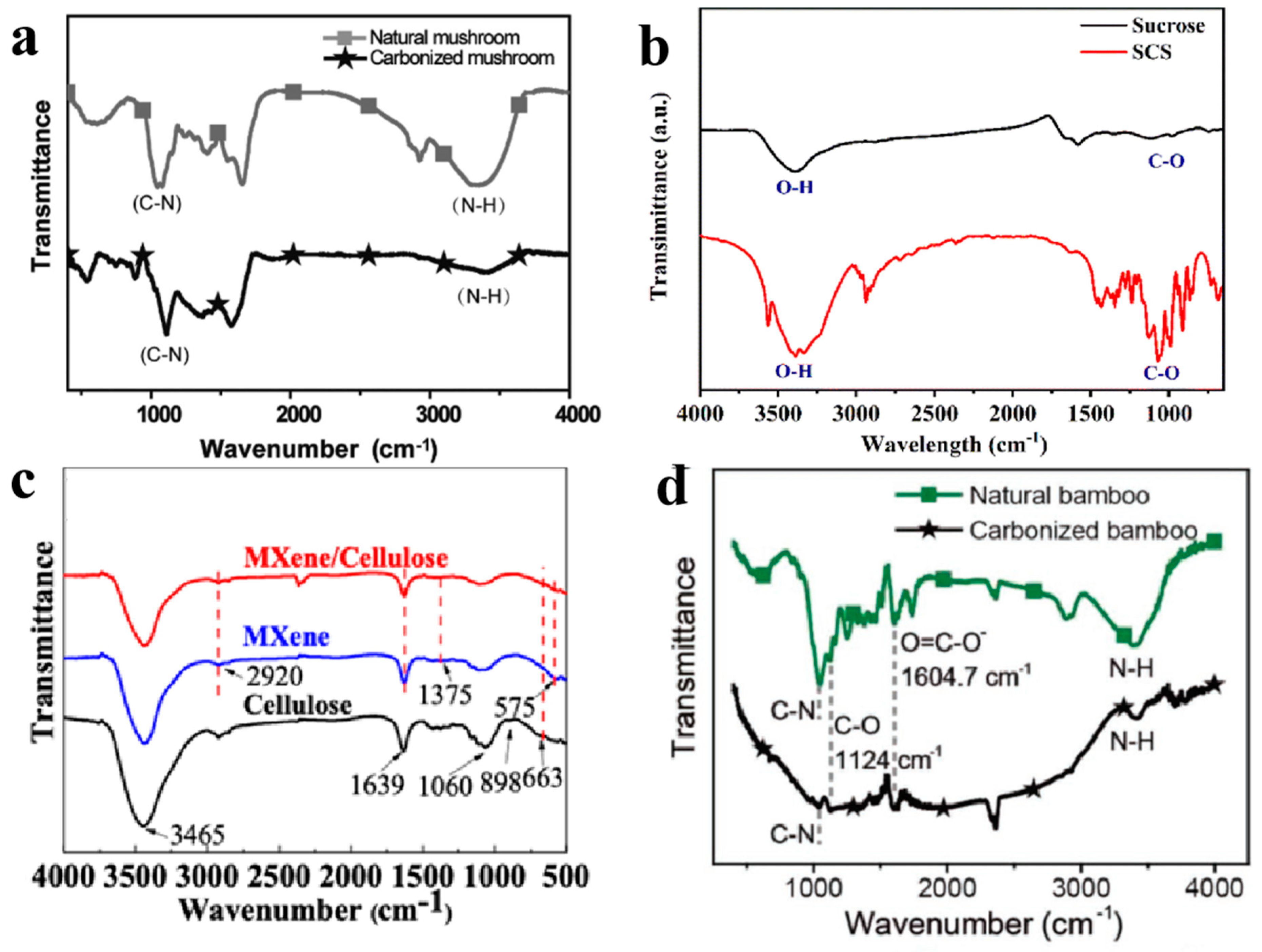
2.2. Fourier Transform Infrared Spectroscopy

Fourier transform infrared spectroscopy (FTIR) offers quantitative and qualitative analysis for organic and inorganic samples. FTIR identifies chemical bonds in a molecule by producing an infrared absorption spectrum. The spectra produce a profile of the sample, a distinctive molecular fingerprint that can be used to screen and scan samples for many different components. FTIR is an effective analytical instrument for detecting functional groups and characterizing covalent bonding information.
Xu et al. [9] described carbonized mushrooms as effective solar steam generators. They used FTIR to confirm the presence of C–N/N–H in the wetting process of original and carbonized mushrooms at 3346 and 1078 cm−1, 3390 and 1111 cm−1, respectively (Figure 2a). Nie et al. [10] proposed a cheap and sustainable 3D porous dome array evaporator that achieves broadband spectrum absorption, thermal insulation, and high hydrophilicity for highly efficient water evaporation and self-desalination. They used FTIR to show that the surface functional groups of carbonized sucrose mainly included C–O, O–H, and C–C, and the content of hydrophilic groups decreased significantly, especially the O–H, indicating that the deoxygenation reaction had happened (Figure 2b). Zha et al. [11] described a simple method for making flexible anti-biofouling fiber photothermal membranes by coating cellulose membranes with MXene for extremely effective solar evaporation. The FTIR spectrum showed C–F and –OH groups on the surface of MXene as two distinctive peaks at 1375 and 575 cm−1, respectively. The typical MXene/cellulose membrane absorption bands at 663 cm−1 (–OH out-of-plane bending), 1639 cm−1 (–OH bending), and 2920 cm−1 (C–H stretching) were observed after dip-coating treatment (Figure 2c). Bian et al. [12] observed an extraordinarily high water vapor production rate in a floating carbonized bamboo sample. The characteristic cellulose structure was visible in the FTIR spectrum. The carboxylate groups in both the original and carbonized bamboos caused peaks at 1124 and 1605 cm−1, which indicated that they both had a large amount of oxygen-containing functional groups. The original and carbonized bamboo had peaks at 3402 and 3415 cm−1, respectively, corresponding to N–H, and peaks at 1047 and 1041 cm−1, respectively, corresponding to C–N (Figure 2d).

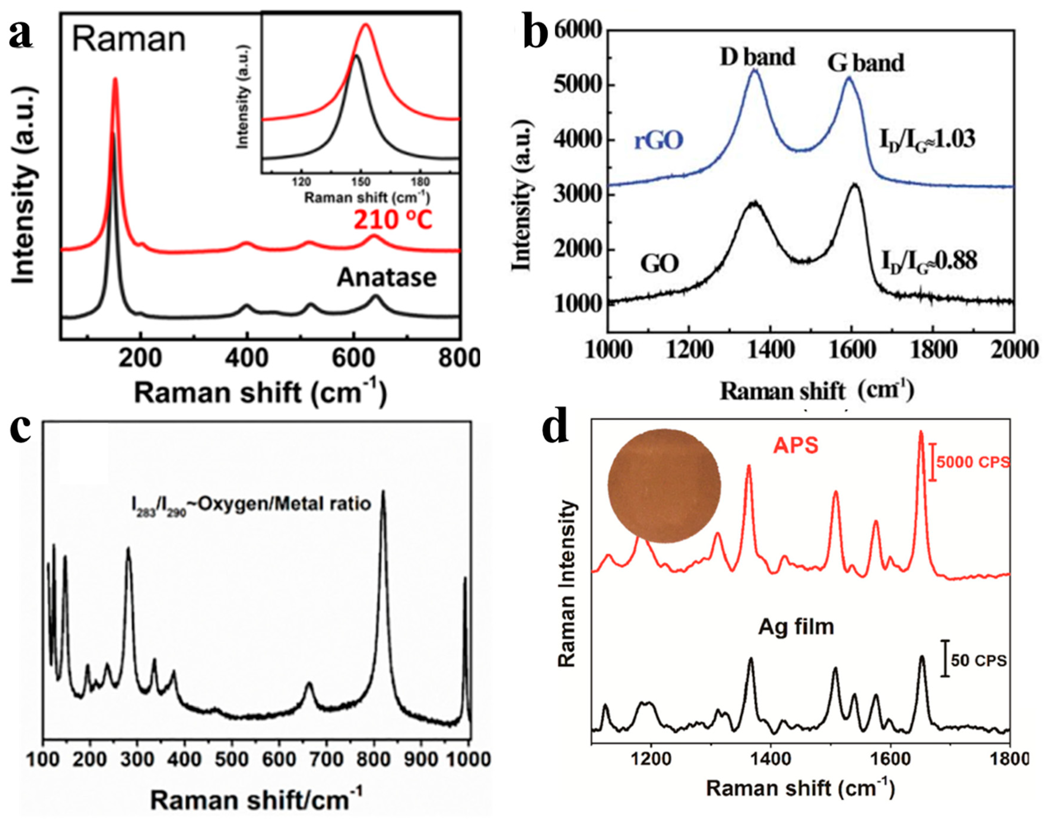
2.3. Raman Spectroscopy

Raman spectroscopy is a nondestructive and highly sensitive technique for micro-area analysis that can be used to identify the structural, optical, and electronic properties of materials. Once the wavelength of incoming light is less than the absorption edge, a semiconductor absorbs optically. The Raman spectrum is a graph that can be used to quantify and visualize the quantity and frequency of photons. Typically, visible or near-infrared (NIR) wavelengths are used to detect Raman waves. Thus, Raman spectroscopy is more advantageous than infrared (IR) spectroscopy. Similar to FTIR, Raman spectroscopy may be used to examine the chemical composition of a material.
Zhu et al. [13] described for the first time black titania with a distinct nanocage structure for solar desalination. They used Raman spectroscopy to clarify the nature of the structural alterations following reduction. Previous research had ascribed the four Raman active modes of anatase to Raman peaks at 136, 390, 509, and 633 cm−1. A comparison between the black TiO2 nanocages and pure anatase nanoparticles revealed a clear widening and blue shift for the peak centered at 136 cm−1. Raman scattering is known to be sensitive to crystallinity and structure, so the shift indicated that the reduction disrupted the original symmetry of the titania lattice (Figure 3a). Shi et al. [14] created a bi-layered photothermal membrane with polystyrene foam on the bottom and a porous film of reduced graphene oxide (rGO) on top. The Raman spectra indicated that GO was successfully reduced to rGO during HI treatment. Two distinct peaks were detected at 1359 and 1609 cm−1 for GO, which matched the well-known G and D bands. The G and D bands remained after reduction, but their intensity ratio (ID/IG) increased noticeably from 0.88 in GO to 1.03 in rGO owing to the more isolated sp2 domains that are present in the latter (Figure 3b). Huang et al. [15] presented MoO3−x, which is a multifunctional material that may be used for both wastewater purification and solar-driven saltwater desalination. Compared to M550Air, the intensity ratios of the two wagging modes of the terminal Mo=O1 group at 283 cm−1 (B2g) and 290 cm−1 (B3g) for M160 were significantly reduced and practically vanished. This drop in the intensity ratio supports earlier research and indicates a break in symmetry caused by OVS (Oxygen Vacancies), which implies a larger concentration of OVs in M160 than in M550Air (Figure 3c). Chen et al. [16] presented tightly packed silver nanoparticles that self-assemble into porous templates, which they used to establish an asymmetric plasmonic structure with the dual purposes of water purification and pollution monitoring. They assessed the effectiveness of chemical detection using the glossy side of APS by using Rhodamine 6 G (R6G), which is the most widely used molecule in Raman spectroscopy. In comparison to a rough Ag film, the glossy side of the asymmetric plasmonic structure detected RG6 at a Raman intensity more than two orders of magnitude greater (Figure 3d).

2.4. X-ray Photoelectron Spectroscopy

When a sample is illuminated by an X-ray beam with energy greater than its work function, some incident X-ray photons are absorbed by the electrons of the component atoms, which is known as the photoelectric effect, and it results in the generation of core holes and emission of photoelectrons. For X-ray photoelectron spectroscopy (XPS), the electron binding energy can be determined by measuring the kinetic energy of expelled photoelectrons and knowing the energy of the incoming X-rays, which is easily determined from their wavelength. The X-ray photoelectron spectrum plots the intensity of the emitted photoelectrons versus the binding energy, and it can be used to determine the oxidation states of the elements in addition to the elemental composition of a specimen by a straightforward peak matching procedure because the electrons of the atom in a particular chemical state have their unique binding energies.
Jiang et al. [17] developed a bi-layered hybrid biofoam made of bacterial nanocellulose and RGO to generate steam by interfacial solar evaporation. They used XPS to establish that GO had undergone chemical changes during the base wash. They separated three peaks in the C 1s spectrum that corresponded to the sp2 domains (C=C with a binding energy of 284 eV) and oxidized sp3 domains (C–O with a binding energy of 286 eV and C=O with a binding energy of 288 eV). They calculated a C/O ratio of 1.74 from the area below the peaks, which indicated that around 57% of the as-synthesized GO surface was oxidized. The C/O ratio rose to 4.95 after a thorough base wash, which indicates that only 20% of the GO surface was oxidized (Figure 4a,b). Li et al. [8] used one-step easy surface oxidation to transform SnSe nanomaterials into a new photothermal–photocatalytic dual-functional core-shell composite. Upon solar irradiation from a single sun, the resulting SnSe@SnO2 exhibited good capabilities for synchronous pollutant destruction and water evaporation. They used XPS to clarify the elemental composition of SnSe@SnO2 and identified Se, Sn, and O-related peaks. The presence of C was attributed to the substrate used to prepare the XPS samples. High-resolution XPS spectra of Sn 3d provided further access to the chemical states of Sn and showed that the Sn 3d5/2 and 3d3/2 peaks broke into two components. The peaks centered at 495.0 and 486.6 eV were assigned to Sn-O bonding, and the peaks at 493.5 and 485.1 eV were attributed to SnSe. The XPS results supported the presence of SnO2 with SnSe (Figure 4c,d).

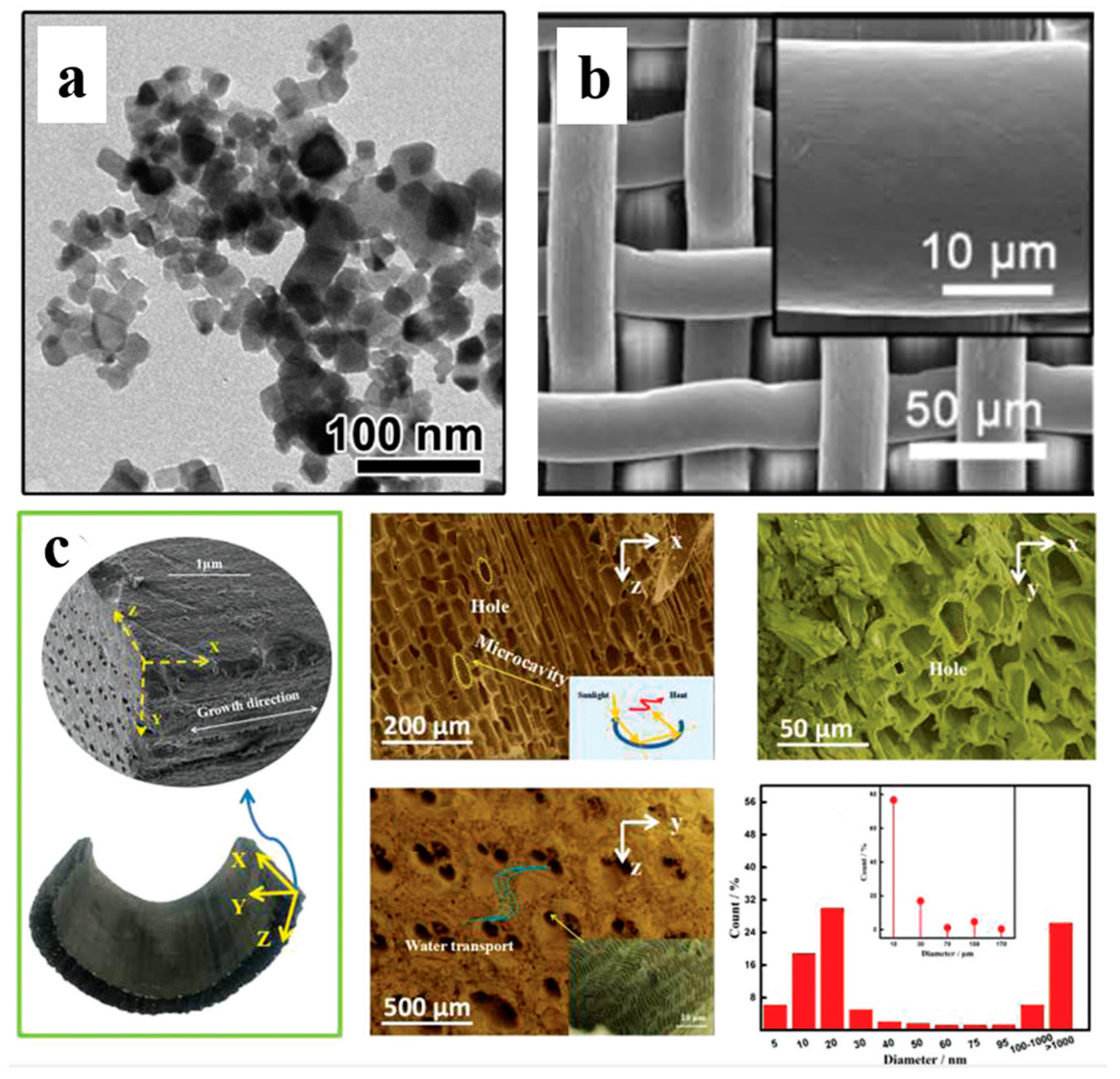
2.5. Scanning Electron Microscopy

Scanning electron microscopy (SEM) is often used to study the morphology and size of nanomaterials. A very thin electron beam is generated and accelerated in the electronic gun and then focused on the sample to excite secondary electrons, Auger electrons, backscattered electrons, characteristic X-rays, etc., on the sample surface. The number of secondary electrons is sensitive to the surface structure of the sample. The secondary electrons are collected by the detector and converted into optical signals, which are then converted into electrical signals by the electro-multiplier tube and amplifier to display scanning images. The surface information of the samples, including the morphology and crystallinity can be characterized by SEM images clearly.
Zhang et al. [18] claimed that a novel photothermal membrane based on polypyrrole (PPy)-coated stainless steel (SS) mesh may self-heal because of its hydrophobicity. They created the membrane by first depositing PPy, which is a polymeric photothermal material, onto an SS mesh substrate. They then modified the PPy coating by using fluoroalkyl silane to produce the desired hydrophobicity. They used SEM to clarify the original unaltered SS mesh and mesh coated with PPy via electropolymerization for 1 h. The original mesh had a smooth wire surface, an average pore size of 26.8 m, and a knitting wire diameter of 24.5 m. The PPy-coated mesh demonstrated microscale surface features (Figure 5a). Ye et al. [19] reported on a variety of TiOx nanomaterials in diverse hues (white, gray, blue, and black) for application as a solar–thermal observer for water desalination, which they made by reducing P25 TiO2 with Mg. The as-prepared samples had particle sizes of around 24 nm, which was the same as the purest P25 TiO2 powder. Generating TiOx lowered the O/Ti ratio, but the sizes and morphologies did not change much, which suggests that the Mg reduction procedure and subsequent HCl wash did not modify the TiO2 structure (Figure 5b). Li et al. [20] showed that bamboo charcoal (BC) is a very effective photothermal material for interfacial solar evaporation thanks to a number of distinctive features. SEM showed that the porous quality of bamboo was carried over to BC, which had numerous irregular pores and grooves. There were several microcavities and a few tiny pores on the upper surface, which facilitated light absorption and steam overflow by redirecting and reflecting sunlight several times. Figure 5c shows that the x–y and y–z directions had polygonal holes as well as regularly combined holes (three-hole). All sections of the vertical growth direction (x–y direction) and part of the growth direction (y–z direction) were submerged in water during the experiment. Capillary forces may have moved water from the BC’s bottom to the top surface through micropores and nanopores, which would ensure enough water is available for solar evaporation.

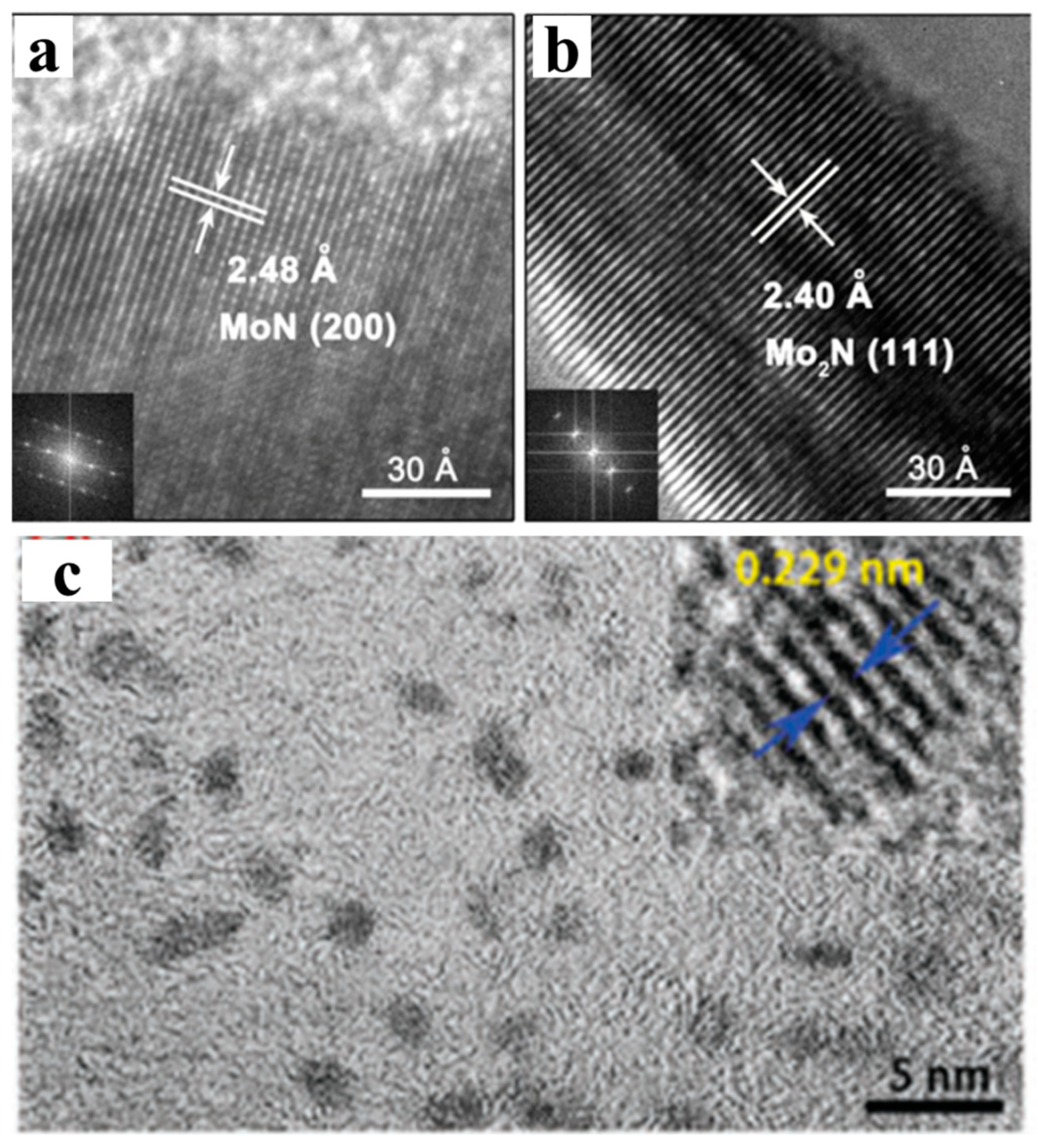
2.6. Transmission Electron Microscopy

The fundamental idea behind transmission electron microscopy (TEM) is that solid-angle scattering occurs when an electron beam that has been accelerated, focused, and projected onto a very thin sample collides with a sample’s atoms. Images can be created from the scattering angle because of its correlation to the sample density and thickness. High-resolution TEM (HRTEM) can be used to determine a material’s crystallinity. Lattice planes (fringes) can be found in the HRTEM image of a crystalline material, and the interplanar distance (d) can be assessed by using a fast Fourier transform (FFT). The crystal phase and Miller indices of these planes may then be determined by contrasting the obtained d with that of a reference sample in the literature or PDFs. Selected-area electron diffraction (SAED) patterns display the distribution of electrons dispersed by a material, and they are frequently examined in combination with HRTEM pictures. The SAED pattern of a crystalline sample should show spots or rings except an intense spot created by directly transmitted electrons in the middle. The crystalline composition of a particular region of a material is determined using the separation between a pair of diffraction spots and their angle with respect to the center spot. Compared to routine XRD patterns, which take minutes or even hours to record (although the time required may be greatly reduced with the use of synchrotron X-ray sources), SAED patterns can be produced in less than 1 s, which makes SAED a valuable additional technique.
Zhu et al. [21] announced the first use of dual-phase MoN/Mo2N to facilitate interfacial solar evaporation under a single source. TEM images showed that the MoN particles had a rambutan-like shape and comprised nanograins with high crystallinity and that were somewhat curved with widths and lengths of 10–15 and 30–50 nm, respectively. HRTEM images were used to identify the dual phases in the MoNx sample. Both cubic Mo2N and hexagonal MoN were found as indicated by the (111) plane of 2.40 and the (200) plane of 2.48. The FFT images of the diffraction pattern showed that both the hexagonal MoN and cubic Mo2N had monocrystalline structures (Figure 6a,b). Ding et al. [22] presented molybdenum oxide quantum dots (MoO3−x QDs) with an absorption spectrum that matched that of solar radiation in both the visible and near-infrared ranges as a proof-of-concept for interfacial solar evaporation. Interestingly, adding more than 1 g of chitosan allowed for the production of uniform and monodispersed tiny quantum dots. Clear lattice fringes were seen in HRTEM pictures, and a lattice parameter of 0.229 nm was determined (Figure 6c).

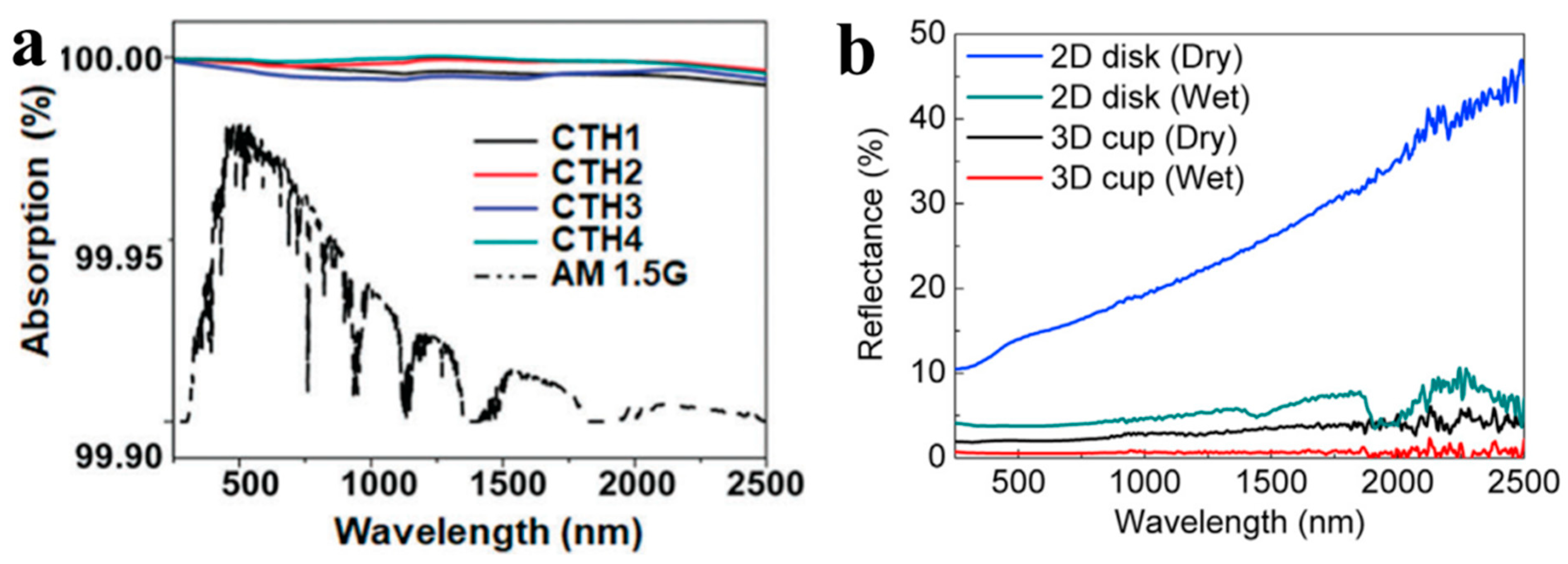
2.7. Diffuse Reflectance Spectroscopy

In diffuse reflectance spectroscopy (DRS), a light beam is used to illuminate a solid sample and then absorbed, reflected, and transmitted. Specular or diffuse reflections of incident light are also considered scattering. A collecting sphere can be used to gather both reflections. However, the incident light beam is frequently perpendicular to the sample surface, which results in specular reflections leaving the integrating sphere and not being picked up. DRS samples are normally prepared with sufficient thickness to prevent transmission. Therefore, only absorption and scattering need to be considered when examining how light interacts with a substance. The reflectance (R) is calculated as the ratio of the reflected incoming fluxes and is measured by a UV-vis spectrophotometer. The reflectance of an enough thick sample and the absorption coefficient-to-scattering coefficient ratio (K/S) are correlated by the Kubelka–Munk function:
F ( R ) = K S = ( 1 R ) 2 2 R
A solid sample is normally ground substantially to make the average grain size greater than the wavelength of the incident light. Using this method allows the scattering coefficient (S) to be considered independent of the wavelength. Therefore, the apparent absorbance, which is defined as log R, is proportional to the absorption coefficient and can be computed to visualize the absorption spectrum. This technique has been used to investigate the ability of various photothermal materials to absorb light.
Zhou et al. [23] demonstrated a hydrogel-based solar evaporator that can produce vapor at a high rate. They used a UV-vis-NIR spectrophotometer to measure the light absorption of CTHs and to clarify their capacity to absorb solar energy. The CTHs exhibited outstanding light absorption through the entire solar spectrum with very little optical loss (Figure 7a). Shi et al. [24] presented three-dimensional (3D) cylindrical cup-shaped structures of mixed metal oxide that demonstrated an energy efficiency of close to 100% under the irradiation of the Sun due to the cup wall’s ability to recover diffuse reflectance and thermal radiation heat loss from the two dimensional (2D) cup bottom. The reflectance spectra of the 3D cup shape in both its dry and wet states were evaluated. The dry 2D disk had a solar absorptance of 0.821, which indicates that 17.9% of solar energy was lost through diffuse reflection. In contrast, the corresponding 3D structure had a solar absorptance of 0.975, which indicates that only 2.5% of energy was lost. Thus, the wall of the 3D cup recovered most of the light reflected from the bottom 2D section because the 3D and 2D structures were made from the same material. In the wet state, the 3D structure had a much greater solar absorptance (0.994) than the 2D sample (0.955) because it had a significantly lower reflectance. These findings demonstrate that the cup wall recovered about 86% of the diffuse reflected light (Figure 7b).

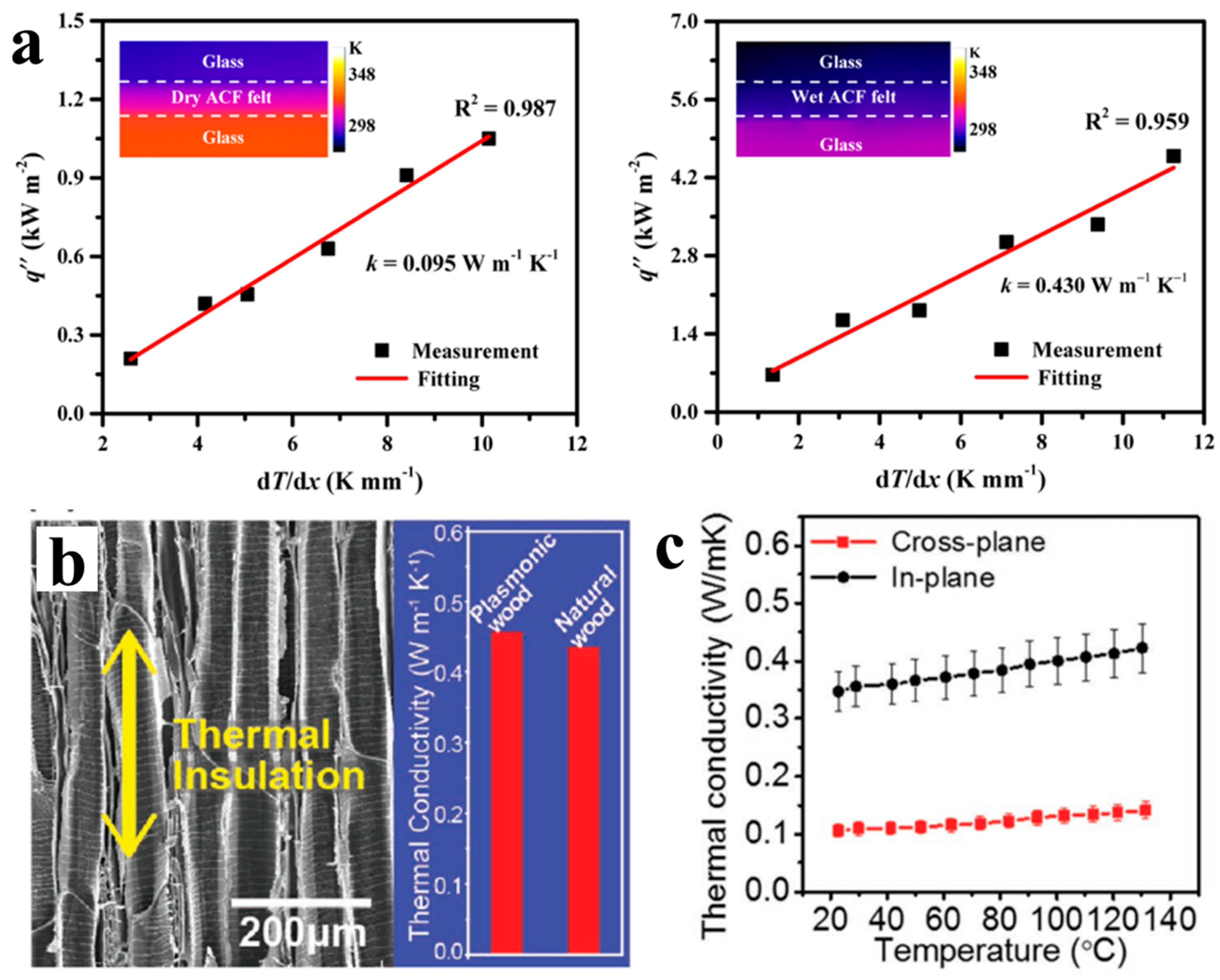
2.8. Thermal Conductivity

According to Fourier’s law, the thermal conductivity k gauges the capacity of a substance to move heat through a medium. While conventionally used for solids, Fourier’s law includes liquids and gases because it is true for all phases of matter.
Li et al. [25] reported that a commercially available activated carbon fiber (ACF) felt had high evaporation efficiency under the irradiation of one Sun. They created a laboratory setup to measure the thermal conductivity, which was 0.095 W m−1 K−1 in the dry state. This suggests that the ACF felt can help with minimizing heat loss. When soaked, the thermal conductivity increased to 0.430 W m−1 K−1, which was less than the thermal conductivity of pure water (0.609 W m−1 K−1 at room temperature). This confirmed that floating ACF could minimize heat loss through bulk water that has not evaporated (Figure 8a). Zhu et al. [26] incorporated tiny metal nanoparticles into the 3D mesoporous matrix of natural wood to create a plasmonic material that they called plasmonic wood. Owing to the low thermal conductivity in the microchannel direction, exposing the plasmonic wood to solar irradiation confined heat on the material surface, which resulted in efficient steam generation. In the wet state, the plasmonic wood exhibited low thermal conductivity in the microchannel direction. When compared to natural wood, the thermal conductivity of plasmonic wood did not increase significantly (Figure 8b). Li et al. [27] constructed a solar steam generation device with nanoscale channels in wood to decouple cross-plane water transport from the preferred thermal transport direction and minimize conductive heat loss. The low thermal conductivity along the cross-plane direction (i.e., perpendicular to the lumen alignment) prevented heat from escaping into the environment, whereas the significantly higher thermal conductivity along the in-plane surface redirected the absorbed heat (Figure 8c).

2.9. Contact Angle

The contact angle is the angle that a liquid or vapor interface forms with a solid surface, and it serves as a quantitative indicator of the wettability of the surface. The interactions between the three interfaces determine the specific angle. Typically, a liquid drop on a surface is used to demonstrate the contact angle. The Young–Laplace equation, which includes the contact angle as a boundary condition, determines the shape of the drop. The contact angle is typically measured by a goniometer, and it is considered a material property because it is independent of geometry.
Zhang et al. [18] presented a proof-of-concept photothermal membrane for interfacial solar evaporation based on a polypyrrole (PPy)-coated stainless steel (SS) mesh with the capacity to self-heal due to its hydrophobicity. They created the membrane by depositing PPy onto an SS mesh substrate. The PPy coating was then modified by using fluoroalkylsilane to give it the desired hydrophobicity. The mesh had a high-water contact angle following the hydrophobic alteration, which rapidly increased with the electropolymerization time throughout the PPy deposition stage. The contact angle converged to 140° when the PPy deposition duration was longer than 30 min. In addition, the mesh showed strong adherence to water droplets while being extremely hydrophobic. The water droplets remained firmly adhered to the surface of the PPy-coated mesh after 1 h of electropolymerization and fluoroalkyl silane modification, even when the mesh was perpendicular or upside down. Water was still able to permeate through the substrate’s rough surface structures despite the substrate’s high contact angle and displace the air pockets that would normally be present under Cassie’s wetting. Wenzel’s wetting behavior is defined as wettability with a high-water contact angle and strong adhesion of water droplets to hydrophobic meshes. Wenzel’s wetting behavior of the mesh can be attributed to the combination of the hydrophobic alteration and micro-sized surface structures. Wenzel’s soaking state allows for close and maximum water-to-solid substrate surface contact, which is advantageous for heat transmission from the mesh to the water, as air is often considered a poor heat conductor (Figure 9a). Long et al. [28] created ethanol-treated carrot biochar as an all-in-one self-floating photothermal device for high-efficiency solar evaporation. The ethanol-treated carrot biochar was shown to have a hydrophilic surface owing to the calculated water contact angle of 44.83°, which is much smaller than the critical angle of 90° (Figure 9b).

3. Performance Evaluation

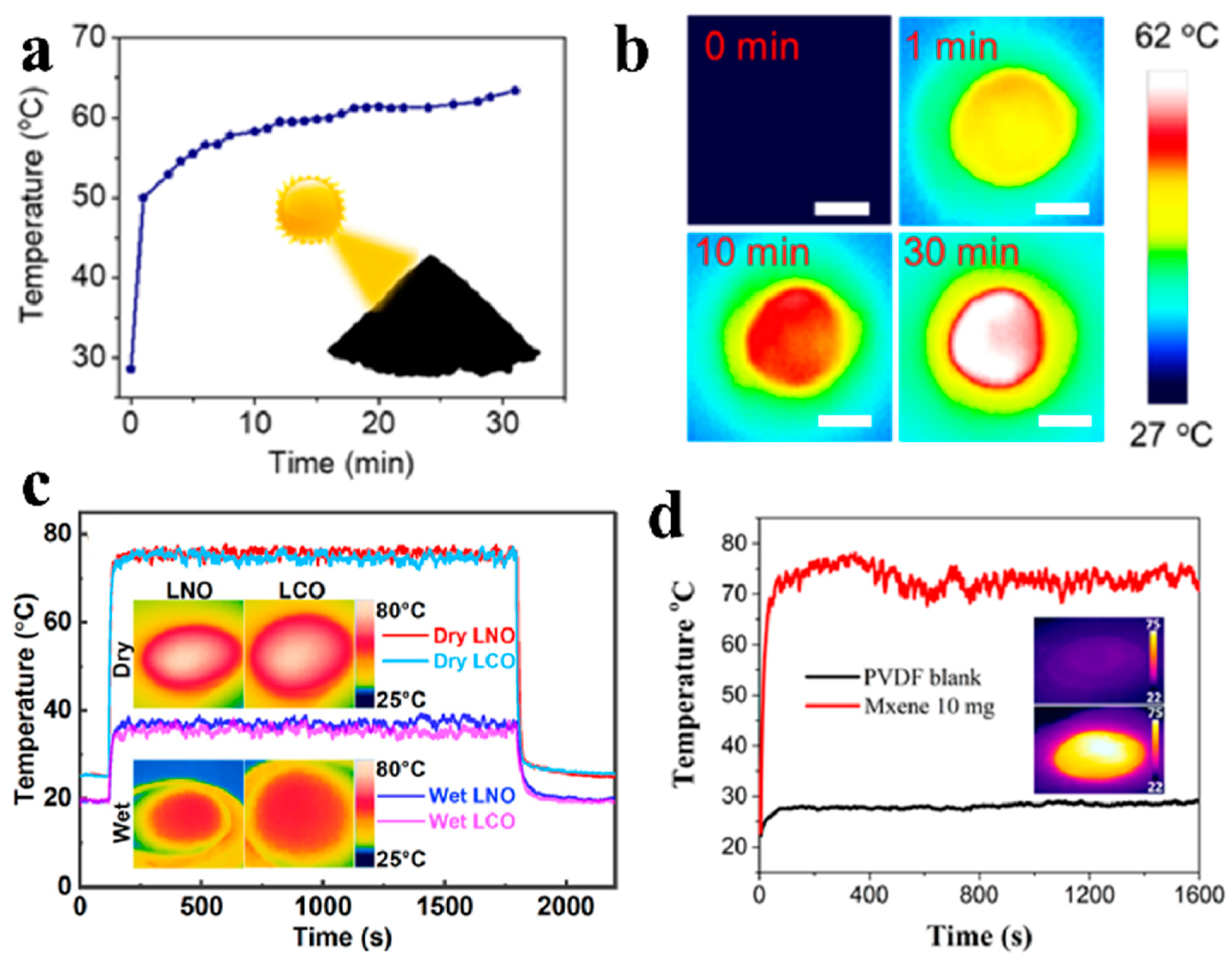
3.1. Surface Temperature

Above a temperature of absolute zero, objects emit infrared radiation depending on the amount of heat they contain, which can be detected by thermal or infrared sensors and transformed into an electrical signal that is then processed to create a picture. Infrared cameras can measure thermal energy with a great degree of accuracy, so they can be employed to evaluate the performance of photothermal materials and the severity of heat-related issues.
Zhao et al. [29] used the self-assembly approach to stack lignin molecules into lignin nanoparticles (L-NPs) at room temperature and studied the photothermal characteristics by using an Xe lamp to simulate solar radiation. Over a period of 30 min, the surface temperature of the L-NPs increased from 28 °C to 63 °C under irradiation of 100 mW cm−2, which suggests good photothermal conversion (Figure 10a,b). Wani et al. [30] created interfacial solar steam generators by using nanostructured metallic LaNiO3 (LNO) and semiconducting LaCoO3 (LCO). They used a thermal infrared camera to track variations in the surface temperature of 2D LCO and LNO under irradiation. The surface temperatures of the dry LCO and LNO samples increased quickly and saturated at greater than 75 °C and 76 °C, respectively, after irradiation. The saturation temperatures of wet LCO and LNO were 36 °C and 37 °C, respectively, which indicated a high heat-to-vapor conversion capacity (Figure 10b). Li et al. [31] presented an aqueous-droplet light heating system and a meticulous mathematical process for accurate assessment of the internal photothermal conversion efficiency of a range of nanomaterials. The MXene-PVDF membrane attained an equilibrium temperature of approximately 75 °C, which was in stark contrast to the equilibrium temperature of approximately 30 °C for the PVDF substrate (Figure 10c).

3.2. Evaporation Rate

The evaporation rate is the rate at which a water mass evaporates, standardized by the evaporation area. Typically, a steady state or equilibrium can be reached in 2000 s under irradiation from one Sun. Using the slope of the mass loss curve as determined by linear fitting in the steady state makes the evaporation rate easier to calculate. Photothermal materials are generally exposed to sunlight vertically and from the top. The projection area is typically used to calculate the evaporation rate and efficiency. It is typically equivalent to the evaporation area, with the exception of 3D systems, where the evaporation area of the absorber expands.

3.3. Conversion Efficiency

Long et al. [28] derived the following equation to precisely calculate the photothermal conversion efficiency η, which they used to evaluate the effectiveness of their ECB-based solar steam generation:
η = m hLV/Copt qin,
where Copt is the optical concentration, qin is the solar irradiance on the surface, m is the net rate of steam production, and hLV is the liquid-vapor phase transition enthalpy. This may be calculated as
hLV = λLV + Cp ∆T,
where Cp is the specific heat capacity of bulk water (4.2 kJ kg−1 K−1), λLV is the latent heat of water evaporation at standard atmospheric pressure (2.257 MJ kg−1), and ∆T is the temperature change throughout the evaporation process.
Chen et al. [32] found that black Ag nanostructures transform solar energy into heat quite well. Ag assemblages had a steam production efficiency of up to 95.2% compared to just 7.3% for the substrate (i.e., filter paper). Chen et al. [33] combined tetrakis (4-carboxyphenyl) porphyrin (TCPP) and Co2+ to form porphyrin paddlewheel frameworks-3 (PPF-3) with an improved photothermal effect. The PPF-3 membrane had a photothermal conversion efficiency of 70.3%, which was a 420% increase over that of the TCPP membrane. Zhu et al. [34] found that carbonized daikon had a highly developed honeycomb cellular structure with many interconnecting channels. It absorbed more than 95.5% of solar energy in the visible spectrum and more than 93.5% in the near-infrared spectrum thanks to the close-packed pores acting as photon traps. Gong et al. [35] presented an easy, scalable, and inexpensive method of generating a high-efficiency solar steam generator based on one-step calcination of commercial melamine sponges (MS) in air. The in-air calcinated MS (AMS) with a thermal insulator achieved a high photothermal efficiency of 92% when irradiated by 1 kW m−2.
In general, the heat loss caused by an evaporator has three components: radiation, convection, and conduction. The calculations of these three components are presented below.
Radiation: The radiation loss is calculated by using the Stefan–Boltzmann law:
Φ = ε A σ (T14 − T24),
where Φ is the heat flux, ε is the emissivity, A is the evaporation surface area, σ is the Stefan-Boltzmann constant, T1 is the average temperature of the absorber after irradiation, and T2 is the initial temperature.
Convection: The convection loss is calculated by using Newton’s law of cooling:
Q = h A ∆T,
where Q is the heat, h is the convection heat transfer coefficient, A is the evaporation surface area, and ∆T is the temperature difference between the absorber after irradiation and the initial temperature. The convection heat transfer coefficient is about 5 W m2 K.
Conduction: The conduction loss is calculated as follows:
Q = C m ∆T,
where Q is the heat, C is the specific heat capacity of water (4.2 J g−1 °C−1), t is the irradiation time, m is the water mass, and ∆T is the temperature variation of water within 60 min.
Xu et al. [9] reported that carbonized mushrooms are efficient solar steam generation devices with radiation, convection, and conduction losses of 6%, 5%, and 1%, respectively (Figure 11a). Yang et al. [36] presented an exceedingly straightforward and independent solar energy converter made entirely of a 3D crosslinked honeycomb graphene foam material that is already commercially available. The radiation, convection, and conduction losses were 4.4%, 2.9%, and 2.7%, respectively (Figure 11b).

3.4. Inductively Coupled Plasma Mass Spectrometry

Inductively coupled plasma mass spectrometry (ICP-MS) is a technique based on ionizing samples with inductively coupled plasma and then detecting the resulting atomic ions. It is typically utilized for measuring metals and non-metals in liquid samples at low detectable limits. It is used in isotopic labeling because it can identify various isotopes of the same element. A nebulizer is used to turn a liquid into an aerosol, which is then transported to the plasma to be ionized. Ions produced in the argon plasma are then sent from the detector through the mass analyzer.
Tang et al. [37] created a wood-based 3D solar evaporator by controlling the hydrophilicity of a charred wood surface and altering the height of the wood above the water surface. They used ICP-MS to determine the amounts of principal ions Na+, Mg2+, K+, and Ca2+ in the original seawater before desalination and condensed water after purification. They found that desalination drastically decreased all four major ion concentrations. The Na+ content was far below the limit for drinking water established by the World Health Organization and the US Environmental Protection Agency (Figure 12a). Wu et al. [38] created an evaporator made of carbon extracted from bamboo leaves and used ICP-MS to show that the quantities of four principal ions (Na+, Mg2+, K+, and Ca2+) in the treated water drastically reduced by nearly three orders of magnitude relative to the original seawater after desalination under irradiation of one Sun (Figure 12b).

4. Conclusions and Outlook

This review presents the most significant techniques for characterizing photothermal materials in two main categories: Intrinsic properties (including chemical and physical properties) and performance evaluation. The information presented here should help readers determine the better technique in their own research as well as become more comfortable with applying multiple techniques to increase the accuracy and reliability of their results. The review would help researchers in selecting appropriate techniques for characterizing interfacial solar evaporation, especially young researchers and students with an interest in photothermal materials and other relevant areas. In addition, the photothermal materials and techniques in large-scale applications will be realized in the near future. And this review should provide the references for the engineers.

Author Contributions

Investigation, X.W., Y.H., S.H. (Shucheng He); writing—original draft preparation, H.Y.; correction, J.C., L.W.; writing—review and editing, R.F., S.H. (Shaolong Huang). All authors have read and agreed to the published version of the manuscript.

Funding

This research was funded by Fundamental Research Funds for the Central Universities, grant number 3122020055.

Institutional Review Board Statement

Not applicable.

Informed Consent Statement

Not applicable.

Data Availability Statement

Not applicable.

Conflicts of Interest

The authors declare no conflicts of interest.

References

  1. Zhang, C.; Shi, Y.; Wang, W.; Li, H.; Li, R.; Hong, S.; Wang, P. Distinct stage-wise environmental energy harvesting behaviors within solar-driven interfacial water evaporation coupled with convective airflow. Nano Energy 2023, 107, 108142. [Google Scholar] [CrossRef]
  2. Yu, H.; Wang, D.; Jin, H.; Wu, P.; Wu, X.; Chu, D.; Lu, Y.; Yang, X.; Xu, H. 2D MoN1.2-rGO Stacked Heterostructures Enabled Water State Modification for Highly Efficient Interfacial Solar Evaporation. Adv. Funct. Mater. 2023, 33, 2214828. [Google Scholar] [CrossRef]
  3. Tao, P.; Ni, G.; Song, C.; Shang, W.; Wu, J.; Zhu, J.; Chen, G.; Deng, T. Solar-driven interfacial evaporation. Nat. Energy 2018, 3, 1031–1041. [Google Scholar] [CrossRef]
  4. Zhang, L.; Ran, J.; Qiao, S.-Z.; Jaroniec, M. Characterization of semiconductor photocatalysts. Chem. Soc. Rev. 2019, 48, 5184–5206. [Google Scholar] [CrossRef]
  5. Lin, H.; Wang, X.; Yu, L.; Chen, Y.; Shi, J. Two-Dimensional Ultrathin MXene Ceramic Nanosheets for Photothermal Conversion. Nano Lett. 2017, 17, 384–391. [Google Scholar] [CrossRef]
  6. Sun, L.; Li, Z.; Su, R.; Wang, Y.; Li, Z.; Du, B.; Sun, Y.; Guan, P.; Besenbacher, F.; Yu, M. Phase-Transition Induced Conversion into a Photothermal Material: Quasi-Metallic WO(2.9) Nanorods for Solar Water Evaporation and Anticancer Photothermal Therapy. Angew. Chem. Int. Ed. Engl. 2018, 57, 10666–10671. [Google Scholar] [CrossRef]
  7. Ma, Q.; Yin, P.; Zhao, M.; Luo, Z.; Huang, Y.; He, Q.; Yu, Y.; Liu, Z.; Hu, Z.; Chen, B.; et al. MOF-Based Hierarchical Structures for Solar-Thermal Clean Water Production. Adv. Mater. 2019, 31, 1808249. [Google Scholar] [CrossRef]
  8. Li, Z.; Sun, L.; Liu, Y.; Zhu, L.; Yu, D.; Wang, Y.; Sun, Y.; Yu, M. SnSe@SnO2 core-shell nanocomposite for synchronous photothermal–photocatalytic production of clean water. Environ. Sci.-Nano 2019, 6, 1507–1515. [Google Scholar] [CrossRef]
  9. Xu, N.; Hu, X.; Xu, W.; Li, X.; Zhou, L.; Zhu, S.; Zhu, J. Mushrooms as Efficient Solar Steam-Generation Devices. Adv. Mater. 2017, 29, 1606762. [Google Scholar] [CrossRef]
  10. Nie, B.; Zhang, W.; Dou, X.; Meng, Y.; Zhao, X.; Wu, Y.; Li, H. A porous dome array evaporator for high-performance photothermal water evaporation and thermoelectric power generation. J. Mater. Chem. A 2024, 12, 293–302. [Google Scholar] [CrossRef]
  11. Zha, X.-J.; Zhao, X.; Pu, J.-H.; Tang, L.-S.; Ke, K.; Bao, R.-Y.; Bai, L.; Liu, Z.-Y.; Yang, M.-B.; Yang, W. Flexible Anti-Biofouling MXene/Cellulose Fibrous Membrane for Sustainable Solar-Driven Water Purification. ACS Appl. Mater. Interfaces 2019, 11, 36589–36597. [Google Scholar] [CrossRef]
  12. Bian, Y.; Du, Q.; Tang, K.; Shen, Y.; Hao, L.; Zhou, D.; Wang, X.; Xu, Z.; Zhang, H.; Zhao, L.; et al. Carbonized Bamboos as Excellent 3D Solar Vapor-Generation Devices. Adv. Mater. Technol. 2019, 4, 1800593. [Google Scholar] [CrossRef]
  13. Zhu, G.; Xu, J.; Zhao, W.; Huang, F. Constructing Black Titania with Unique Nanocage Structure for Solar Desalination. ACS Appl. Mater. Interfaces 2016, 8, 31716–31721. [Google Scholar] [CrossRef]
  14. Shi, L.; Wang, Y.; Zhang, L.; Wang, P. Rational design of a bi-layered reduced graphene oxide film on polystyrene foam for solar-driven interfacial water evaporation. J. Mater. Chem. A 2017, 5, 16212–16219. [Google Scholar] [CrossRef]
  15. Huang, S.; Long, Y.; Yi, H.; Yang, Z.; Pang, L.; Jin, Z.; Liao, Q.; Zhang, L.; Zhang, Y.; Chen, Y.; et al. Multifunctional molybdenum oxide for solar-driven water evaporation and charged dyes adsorption. Appl. Surf. Sci. 2019, 491, 328–334. [Google Scholar] [CrossRef]
  16. Chen, C.; Zhou, L.; Yu, J.; Wang, Y.; Nie, S.; Zhu, S.; Zhu, J. Dual functional asymmetric plasmonic structures for solar water purification and pollution detection. Nano Energy 2018, 51, 451–456. [Google Scholar] [CrossRef]
  17. Jiang, Q.; Tian, L.; Liu, K.-K.; Tadepalli, S.; Raliya, R.; Biswas, P.; Naik, R.R.; Singamaneni, S. Bilayered Biofoam for Highly Efficient Solar Steam Generation, Adv. Mater. 2016, 28, 9400–9407. [Google Scholar]
  18. Zhang, L.; Tang, B.; Wu, J.; Li, R.; Wang, P. Hydrophobic Light-to-Heat Conversion Membranes with Self-Healing Ability for Interfacial Solar Heating, Adv. Mater. 2015, 27, 4889–4894. [Google Scholar]
  19. Ye, M.; Jia, J.; Wu, Z.; Qian, C.; Chen, R.; O’Brien, P.G.; Sun, W.; Dong, Y.; Ozin, G.A. Synthesis of Black TiOx Nanoparticles by Mg Reduction of TiO2 Nanocrystals and their Application for Solar Water Evaporation. Adv. Energy Mater. 2017, 7, 1601811. [Google Scholar] [CrossRef]
  20. Li, Z.; Wang, C.; Lei, T.; Ma, H.; Su, J.; Ling, S.; Wang, W. Arched Bamboo Charcoal as Interfacial Solar Steam Generation Integrative Device with Enhanced Water Purification Capacity. Adv. Sustain. Syst. 2019, 3, 1800144. [Google Scholar] [CrossRef]
  21. Zhu, L.; Sun, L.; Zhang, H.; Yu, D.; Aslan, H.; Zhao, J.; Li, Z.; Yu, M.; Besenbacher, F.; Sun, Y. Dual-phase molybdenum nitride nanorambutans for solar steam generation under one sun illumination. Nano Energy 2019, 57, 842–850. [Google Scholar] [CrossRef]
  22. Ding, D.; Huang, W.; Song, C.; Yan, M.; Guo, C.; Liu, S. Non-stoichiometric MoO3−x quantum dots as a light-harvesting material for interfacial water evaporation. Chem. Commun. 2017, 53, 6744–6747. [Google Scholar] [CrossRef] [PubMed]
  23. Zhou, X.; Zhao, F.; Guo, Y.; Zhang, Y.; Yu, G. A hydrogel-based antifouling solar evaporator for highly efficient water desalination. Energy Environ. Sci. 2018, 11, 1985–1992. [Google Scholar] [CrossRef]
  24. Shi, Y.; Li, R.; Jin, Y.; Zhuo, S.; Shi, L.; Chang, J.; Hong, S.; Ng, K.-C.; Wang, P. A 3D Photothermal Structure toward Improved Energy Efficiency in Solar Steam Generation. Joule 2018, 2, 1171–1186. [Google Scholar] [CrossRef]
  25. Li, H.; He, Y.; Hu, Y.; Wang, X. Commercially Available Activated Carbon Fiber Felt Enables Efficient Solar Steam Generation. ACS Appl. Mater. Interfaces 2018, 10, 9362–9368. [Google Scholar] [CrossRef]
  26. Zhu, M.; Li, Y.; Chen, F.; Zhu, X.; Dai, J.; Li, Y.; Yang, Z.; Yan, X.; Song, J.; Wang, Y.; et al. Plasmonic Wood for High-Efficiency Solar Steam Generation. Adv. Energy Mater. 2018, 8, 1701028. [Google Scholar] [CrossRef]
  27. Li, T.; Liu, H.; Zhao, X.; Chen, G.; Dai, J.; Pastel, G.; Jia, C.; Chen, C.; Hitz, E.; Siddhartha, D.; et al. Scalable and Highly Efficient Mesoporous Wood-Based Solar Steam Generation Device: Localized Heat, Rapid Water Transport. Adv. Funct. Mater. 2018, 28, 1707134. [Google Scholar] [CrossRef]
  28. Long, Y.; Huang, S.; Yi, H.; Chen, J.; Wu, J.; Liao, Q.; Liang, H.; Cui, H.; Ruan, S.; Zeng, Y.-J. Carrot-inspired solar thermal evaporator. J. Mater. Chem. A 2019, 7, 26911–26916. [Google Scholar] [CrossRef]
  29. Zhao, X.; Huang, C.; Xiao, D.; Wang, P.; Luo, X.; Liu, W.; Liu, S.; Li, J.; Li, S.; Chen, Z. Melanin-Inspired Design: Preparing Sustainable Photothermal Materials from Lignin for Energy Generation. ACS Appl. Mater. Interfaces 2021, 13, 7600–7607. [Google Scholar] [CrossRef]
  30. Ahmad Wani, T.; Garg, P.; Bera, S.; Bhattacharya, S.; Dutta, S.; Kumar, H.; Bera, A. Narrow-Bandgap LaMO3 (M=Ni, Co) nanomaterials for efficient interfacial solar steam generation. J. Colloid Interface Sci. 2022, 612, 203–212. [Google Scholar] [CrossRef]
  31. Li, R.; Zhang, L.; Shi, L.; Wang, P. MXene Ti3C2: An Effective 2D Light-to-Heat Conversion Material. ACS Nano 2017, 11, 3752–3759. [Google Scholar] [CrossRef] [PubMed]
  32. Chen, J.; Feng, J.; Li, Z.; Xu, P.; Wang, X.; Yin, W.; Wang, M.; Ge, X.; Yin, Y. Space-Confined Seeded Growth of Black Silver Nanostructures for Solar Steam Generation. Nano Lett. 2019, 19, 400–407. [Google Scholar] [CrossRef]
  33. Chen, L.; Li, D.; Wang, Y.; Duan, C. Highly efficient solar steam generation of supported metal–organic framework membranes by a photoinduced electron transfer process. Nanoscale 2019, 11, 11121–11127. [Google Scholar] [CrossRef]
  34. Zhu, M.; Yu, J.; Ma, C.; Zhang, C.; Wu, D.; Zhu, H. Carbonized daikon for high efficient solar steam generation. Sol. Energy Mater. Sol. C 2019, 191, 83–90. [Google Scholar] [CrossRef]
  35. Gong, F.; Li, H.; Wang, W.; Huang, J.; Xia, D.; Liao, J.; Wu, M.; Papavassiliou, D.V. Scalable, eco-friendly and ultrafast solar steam generators based on one-step melamine-derived carbon sponges toward water purification. Nano Energy 2019, 58, 322–330. [Google Scholar] [CrossRef]
  36. Yang, Y.; Zhao, R.; Zhang, T.; Zhao, K.; Xiao, P.; Ma, Y.; Ajayan, P.M.; Shi, G.; Chen, Y. Graphene-Based Standalone Solar Energy Converter for Water Desalination and Purification. ACS Nano 2018, 12, 829–835. [Google Scholar] [CrossRef] [PubMed]
  37. Tang, J.; Zheng, T.; Song, Z.; Shao, Y.; Li, N.; Jia, K.; Tian, Y.; Song, Q.; Liu, H.; Xue, G. Realization of Low Latent Heat of a Solar Evaporator via Regulating the Water State in Wood Channels. ACS Appl. Mater. Interfaces 2020, 12, 18504–18511. [Google Scholar] [CrossRef]
  38. Wu, Y.; Kong, R.; Ma, C.; Li, L.; Zheng, Y.; Lu, Y.; Liang, L.; Pang, Y.; Wu, Q.; Shen, Z.; et al. Simulation-Guided Design of Bamboo Leaf-Derived Carbon-Based High-Efficiency Evaporator for Solar-Driven Interface Water Evaporation. Energy Environ. Mater. 2022, 5, 1323–1331. [Google Scholar] [CrossRef]
Figure 1. X-ray diffraction patterns. (a) The freeze-dried Ti3C2 MXene. Reproduced with permission from ref. [5]. Copyright 2017, American Chemical Society. (b) WO3 and WO2.9 nanorods. Reproduced with permission from ref. [6]. Copyright 2018, Wiley-VCH. (c) MHS. Reproduced with permission from ref. [7], Copyright 2018, Wiley-VCH. (d) SnSe@SnO2 structure. Reproduced with permission from ref. [8], Copyright 2018, The Royal Society of Chemistry.
Applsci 14 07523 g001
Figure 2. FTIR spectra. (a) Carbonized mushrooms. Reproduced with permission from ref. [9], Copyright 2017, Wiley-VCH. (b) The porous dome array evaporator. Reproduced with permission from ref. [10], Copyright 2024, The Royal Society of Chemistry. (c) The flexible anti-biofouling fiber photothermal membranes. Reproduced with permission from ref. [11], Copyright 2019, American Chemical Society. (d) The carbonized bamboo. Reproduced with permission from ref. [12], Copyright 2019, Wiley-VCH.
Applsci 14 07523 g002
Figure 3. Raman spectra. (a) Black titania. Reproduced with permission from ref. [13], Copyright 2016, American Chemical Society. (b) The bi-layered photothermal membrane. Reproduced with permission from ref. [14], Copyright 2017, The Royal Society of Chemistry. (c) MoO3−x nanoflowers. Reproduced with permission from ref. [15], Copyright 2019, Elsevier. (d) The asymmetric plasmonic structure. Reproduced with permission from ref. [16], Copyright 2018, Elsevier.
Applsci 14 07523 g003
Figure 4. XPS measurements. (a,b) The bi-layered hybrid biofoam. Reproduced with permission from ref. [17], Copyright 2019, Wiley-VCH. (c,d) SnSe@SnO2 structure. Reproduced with permission from ref. [8], Copyright 2018, The Royal Society of Chemistry.
Applsci 14 07523 g004
Figure 5. SEM. (a) PPy-coated-SS photothermal membrane. Reproduced with permission from ref. [18], Copyright 2015, Wiley-VCH. (b) TiOx nanomaterials. Reproduced with permission from ref. [19], Copyright 2017, Wiley-VCH. (c) The bamboo charcoal. Reproduced with permission from ref. [20], Copyright 2019, Wiley-VCH.
Applsci 14 07523 g005
Figure 6. TEM pictures. (a,b) The dual-phase MoN/Mo2N. Reproduced with permission from ref. [21], Copyright 2018, Elsevier. (c) MoO3−x QDs. Reproduced with permission from ref. [22], Copyright 2017, The Royal Society of Chemistry.
Applsci 14 07523 g006
Figure 7. Diffuse Reflectance Spectroscopy. (a) The hydrogel-based solar evaporator. Reproduced with permission from ref. [23], Copyright 2018, The Royal Society of Chemistry. (b) 3D cylindrical cup-shaped structures. Reproduced with permission from ref. [24], Copyright 2018, Elsevier.
Applsci 14 07523 g007
Figure 8. Thermal conductivity. (a) The activated carbon fiber. Reproduced with permission from ref. [25], Copyright 2018, American Chemical Society. (b) The plasmonic wood. Reproduced with permission from ref. [26], Copyright 2018, Wiley-VCH. (c) The solar steam generation device with nanoscale channels in wood. Reproduced with permission from ref. [27], Copyright 2018, Wiley-VCH.
Applsci 14 07523 g008
Figure 9. Contact angle. (a) PPy-coated-SS photothermal membrane. Reproduced with permission from ref. [18], Copyright 2015, Wiley-VCH. (b) The ethanol-treated carrot biochar. Reproduced with permission from ref. [28], Copyright 2019, The Royal Society of Chemistry.
Applsci 14 07523 g009
Figure 10. Surface temperature. (a,b) L-NPs. Reproduced with permission from ref. [29], Copyright 2021, American Chemical Society. (c) The interfacial solar steam generators by using nanostructured metallic LNO and semiconducting LCO. Reproduced with permission from ref. [30], Copyright 2022, Elsevier. (d) The MXene-PVDF membrane. Reproduced with permission from ref. [31], Copyright 2017, American Chemical Society.
Applsci 14 07523 g010
Figure 11. Heat loss. (a) Carbonized mushrooms. Reproduced with permission from ref. [9], Copyright 2017, Wiley-VCH. (b) 3D crosslinked honeycomb graphene foam material. Reproduced with permission from ref. [36], Copyright 2018, American Chemical Society.
Applsci 14 07523 g011
Figure 12. Ion concentration of collected water generated by (a) wood-based 3D solar evaporator. Reproduced with permission from ref. [37], Copyright 2020, American Chemical Society. (b) The evaporator is made of carbon extracted from bamboo leaves. Reproduced with permission from ref. [38], Copyright 2022, Wiley-VCH.
Applsci 14 07523 g012
Disclaimer/Publisher’s Note: The statements, opinions and data contained in all publications are solely those of the individual author(s) and contributor(s) and not of MDPI and/or the editor(s). MDPI and/or the editor(s) disclaim responsibility for any injury to people or property resulting from any ideas, methods, instructions or products referred to in the content.

